Embed Size (px)
Citation preview
1041Hussain S, et al. Arch Dis Child 2020;105:1041–1048. doi:10.1136/archdischild-2020-319203
Global child health
Original research
Reduction of cardiac iron overload by optimising iron chelation therapy in transfusion dependent thalassaemia using cardiac T2* MRI: a quality improvement project from PakistanShabneez Hussain ,1 Zahra Hoodbhoy,2 Fatima Ali,2 Erum Hasan,3 Najveen Alvi,2 Aijaz Hussain,1 Khajista Ishrat,1 Zia Ur Rahman,1 Azizuddin Qamruddin,1 Azra Parvin,1 Babar S Hasan2
To cite: Hussain S, Hoodbhoy Z, Ali F, et al. Arch Dis Child 2020;105:1041–1048.
1Fatimid Foundation, Karachi, Sindh, Pakistan2Department of Pediatrics and Child Health, The Aga Khan University and Hospital, Karachi, Sindh, Pakistan3Kashif Iqbal Thalassaemia Care Centre, Karachi, Sindh, Pakistan
Correspondence toDr Shabneez Hussain, Fatimid Foundation, Karachi, Sindh, Pakistan; shabneez. haem@ gmail. comPublished Online First 29 September 2020
Global child health
© Author(s) (or their employer(s)) 2020. No commercial re- use. See rights and permissions. Published by BMJ.
ABSTRACTObjectives Cardiac T2* MRI (T2*CMR), for accurate estimation of myocardial siderosis, was introduced as part of a QI collaborative to optimise chelation therapy in order to improve cardiac morbidity in transfusion dependent thalassaemia (TDT) patients. We report the impact of this QI initiative from two thalassaemia centres from this collaborative.Design and setting A key driver based quality initiative was implemented to improve chelation in TDT patients registered at these two centres in Karachi, Pakistan. Protocol optimisation and compliance to treatment through training, communication and feedback were used as the drivers for QI intervention. Preintervention variables (demographics, chelation history, T2*CMR, echocardiography and holters) were collected from January 2015 to December 2016) and compared with variables in the post implementation phase (January to December 2019). A standardised adverse event severity for chelators and its management was devised for safe drug therapy as well as ensuring compliance to the regimen. Preintervention and postintervention variables were compared using non- parametric test. P value<0.05 was statistically significant.Results 100 patients with TDT, median age 17 (9–34) years, were included. An increase or stabilisation of T2*CMR was documented in 82% patients in the postintervention phase especially in patients with severe myocardial iron overload (5.5 vs 5.3 ms, p <0.01). Significantly fewer patients had abnormal echocardiographic findings (3.5% vs 26%, p <0.05) in the postintervention versus preintervention period.Conclusion This QI initiative improved the chelation therapy leading to improved cardiac status in TDT patients at the participating centres.
INTRODUCTIONRecurrent transfusions are the mainstay of therapy in transfusion dependent thalassaemia (TDT) but leads to significant hepatic, endocrine and cardiac iron overload over time if chelation is inadequate. Cardiac iron overload in inadequately chelated patients with TDT develops as early as 5.5 years,1 2 whereas detectable cardiac iron overload has been reported in 24%–36% of well chelated patients at 9.5–18 years of age.3 Cardiac iron overload may
be reversible through early detection and optimal intensive chelation.4 Several factors like access to care,5 unavailability of accurate cardiac iron measurements (such as cardiac T2* magnetic reso-nance imaging (T2*CMR)) and chelation agents,5 poor chelation compliance due to patient and physician discomfort with drug related adverse events (AE),6 7 and lack of standardised treatment protocols8 contribute towards poor chelation of patients with TDT in low middle income countries (LMICs) like Pakistan. A preliminary analysis from Pakistan demonstrated that 50% of patients being treated at a tertiary care centre using ferritin as a marker for iron load and a way to direct chelation management, had severe myocardial iron load on
What is already known on this topic?
► Transfusion dependent thalassaemia (TDT) patients in resource constrained settings are known to have severe cardiac disease on cardiac T2* MRI.
► QI based collaborative using a key driver diagram approach is shown to improve outcomes in complex diseases.
► A quality improvement collaborative has been established in Pakistan to improve morbidity and mortality in TDT patients.
What this study adds?
► Demonstrates effectiveness of a QI initiative focused on process re- engineering using existing resources to manage transfusion dependent thalassaemia (TDT) in a low resource setting.
► Devised and assessed the clinical applicability of a novel chelation drug related adverse events severity level definition and its standard management in this population to ensure improved compliance to the prescribed chelation regimens.
► Demonstrate the impact of standardised care in improving patient management and their compliance in chronic diseases.
on February 4, 2022 by guest. P
rotected by copyright.http://adc.bm
j.com/
Arch D
is Child: first published as 10.1136/archdischild-2020-319203 on 29 S
eptember 2020. D
ownloaded from
on F
ebruary 4, 2022 by guest. Protected by copyright.
http://adc.bmj.com
/A
rch Dis C
hild: first published as 10.1136/archdischild-2020-319203 on 29 Septem
ber 2020. Dow
nloaded from
1042 Hussain S, et al. Arch Dis Child 2020;105:1041–1048. doi:10.1136/archdischild-2020-319203
Original research
Glo
bal
chil
d he
alth
T2*CMR (<10 ms), while 70% of the patients were in New York Heart Association (NYHA) functional class II or worse which correlated with decreasing T2*CMR value.9
In Pakistan, patients with TDT are treated by various non- governmental organisations or thalassaemia treatment centres and thus there is significant practice variation in assessment and management of myocardial iron load. The impact of quality improvement (QI) initiative using a key driver diagram (KDD) approach, done individually or as part of a collaborative, in improving clinical outcomes is very well established especially in congenital heart disease.10–12 A continuous QI process coupled with Plan, Do, Study, Act (PDSA) cycle makes such an inter-vention highly effective.12 13 A similar QI collaborative for care of patients with TDT was established in Pakistan with the goal to reduce mortality and morbidity in TDT.14 A main KDD was previously created with the aim to reduce cardiac and endo-crine organ iron overload related mortality and morbidity, by specific interventions (both cardiac and endocrine) in patients with TDT.14 15 This report focuses on a QI initiative to specifi-cally decrease cardiac iron siderosis and subsequently its related morbidity at two out of the four centres which were part of this collaborative.
MATERIALS AND METHODSAs part of the collaborative,14 a QI initiative specifically focused on reducing cardiac siderosis and its related morbidity, was designed at two of the four collaborating not- for- profit
thalassaemia treatment centres (Fatimid Foundation, Karachi (FFK) and Kashif Iqbal Thalassaemia Care Centre (KITCC)). Employing a PDSA cycle methodology and KDD approach, the QI initiative was executed from January 2015 to December 2019. Preintervention data was collected from January 2015 to December 2016 and compared with postintervention data from January 2019 to December 2019 for both process and outcome metric (as described in the section on “outcome and process metrics”). Interventions were implemented with 6- monthly informal process evaluations from January 2017 to December 2018. Comparison of data and final decision of the PDSA cycle to repeat i.e. continue or change the strategies were made in January 2020. Decision to collect the data and analyse both the process and outcome metric (first PDSA cycle ie, after 2 years of beginning of the initiative) was due to the limitation of the T2*CMR centre (The Aga Khan University and Hospital (AKUH)) of performing only 10 T2*CMRs/month. Additionally, significant behaviour change strategies needed to be employed (education, reinforcement etc) to bring about standardisation of chelation, medication compliance and understanding and running a PDSA cycle (see online supplemental material 1 for additional details).
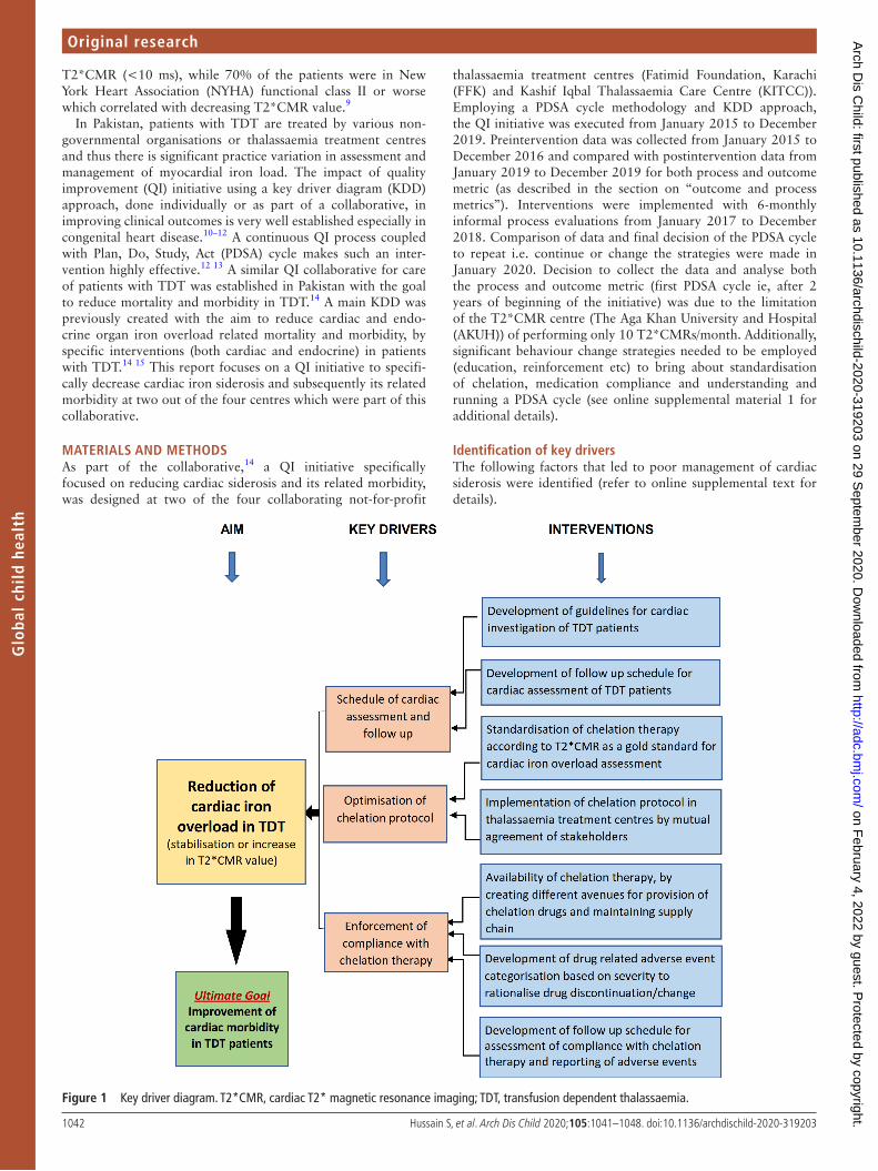
Identification of key driversThe following factors that led to poor management of cardiac siderosis were identified (refer to online supplemental text for details).
Figure 1 Key driver diagram. T2*CMR, cardiac T2* magnetic resonance imaging; TDT, transfusion dependent thalassaemia.
on February 4, 2022 by guest. P
rotected by copyright.http://adc.bm
j.com/
Arch D
is Child: first published as 10.1136/archdischild-2020-319203 on 29 S
eptember 2020. D
ownloaded from
1043Hussain S, et al. Arch Dis Child 2020;105:1041–1048. doi:10.1136/archdischild-2020-319203
Original research
Global child health
1. Assessment: There was lack of standardisation in cardiac as-sessment of patients with TDT.
2. Management: Chelation therapy was guided by serum ferri-tin level (a poor marker of myocardial iron load) rather than the gold standard T2*CMR value.
3. AE: Discontinuation of drugs was common even due to mi-nor drug related AE or poor patient counselling by physi-cians, leading to inconsistent chelation.
Three key drivers and their respective interventions were identified to address these issues (figure 1 and table 1).
Outcome and process metricsTwo metrics were used to assess the performance of the QI initia-tive that is, outcome and process metrics. Outcome metrics for this initiative were defined as a decrease in cardiac morbidity
measured by percentage of patients with an increase or stabili-sation of T2*CMR, increase in median T2*CMR of the cumu-lative cohort and percentage of patients with improvement in cardiac dysfunction (as evaluated by echocardiogram) in the postintervention phase (table 1). Process metrics were used to assess the effectiveness of the interventions aligned with the key drivers. Process was evaluated every 6 months from the begin-ning of the intervention and continuous reinforcement of physi-cian and patients to standardise evaluation and management of cardiac siderosis was done at the same time. For this initiative process metrics were defined as compliance with cardiac assess-ment and its follow- up, standardisation of chelation therapy, compliance with chelation therapy, reporting of AE severity level (table 2), percentage of patients with inappropriate discontinu-ation/continuation of chelation therapy after AE reporting and
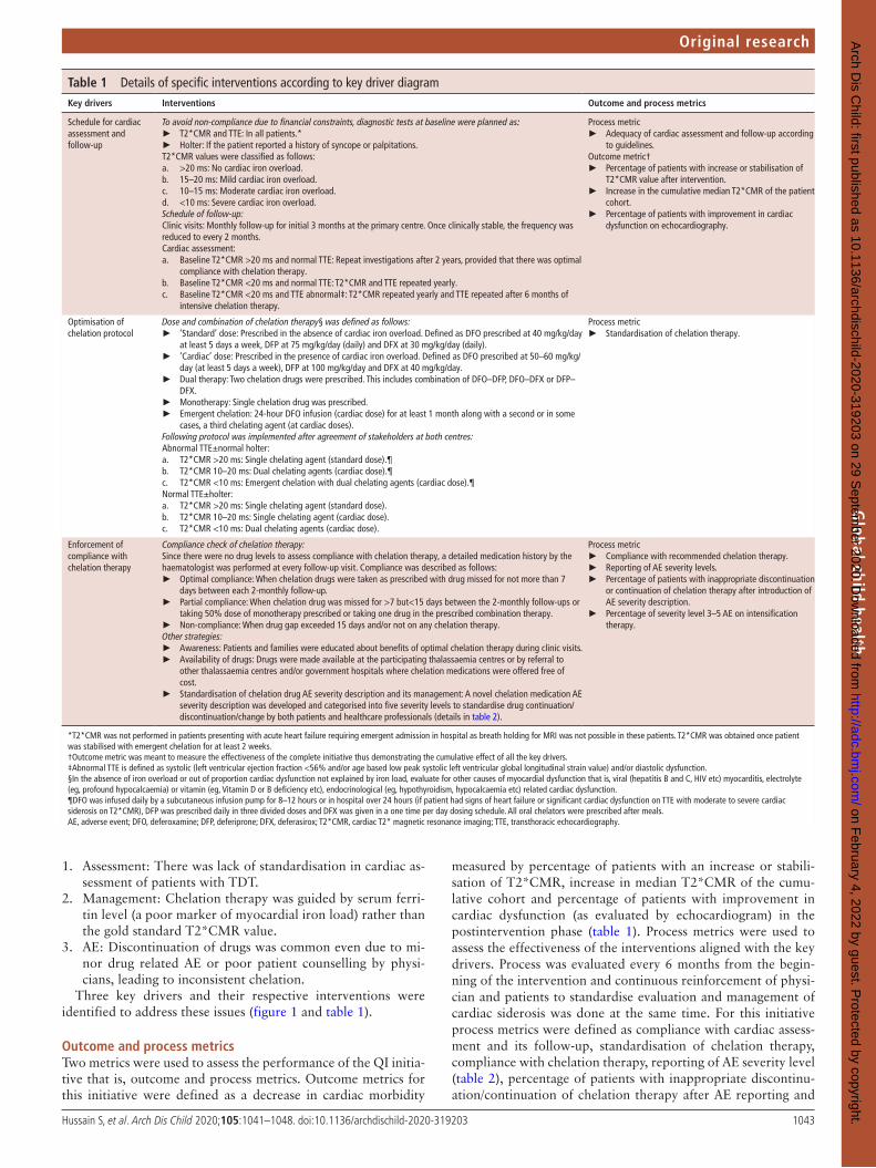
Table 1 Details of specific interventions according to key driver diagramKey drivers Interventions Outcome and process metrics
Schedule for cardiac assessment and follow- up
To avoid non- compliance due to financial constraints, diagnostic tests at baseline were planned as: ► T2*CMR and TTE: In all patients.* ► Holter: If the patient reported a history of syncope or palpitations.
T2*CMR values were classified as follows:a. >20 ms: No cardiac iron overload.b. 15–20 ms: Mild cardiac iron overload.c. 10–15 ms: Moderate cardiac iron overload.d. <10 ms: Severe cardiac iron overload.Schedule of follow- up:Clinic visits: Monthly follow- up for initial 3 months at the primary centre. Once clinically stable, the frequency was reduced to every 2 months.Cardiac assessment:a. Baseline T2*CMR >20 ms and normal TTE: Repeat investigations after 2 years, provided that there was optimal
compliance with chelation therapy.b. Baseline T2*CMR <20 ms and normal TTE: T2*CMR and TTE repeated yearly.c. Baseline T2*CMR <20 ms and TTE abnormal‡: T2*CMR repeated yearly and TTE repeated after 6 months of
intensive chelation therapy.
Process metric ► Adequacy of cardiac assessment and follow- up according
to guidelines.Outcome metric†
► Percentage of patients with increase or stabilisation of T2*CMR value after intervention.
► Increase in the cumulative median T2*CMR of the patient cohort.
► Percentage of patients with improvement in cardiac dysfunction on echocardiography.
Optimisation of chelation protocol
Dose and combination of chelation therapy§ was defined as follows: ► ‘Standard’ dose: Prescribed in the absence of cardiac iron overload. Defined as DFO prescribed at 40 mg/kg/day
at least 5 days a week, DFP at 75 mg/kg/day (daily) and DFX at 30 mg/kg/day (daily). ► ‘Cardiac’ dose: Prescribed in the presence of cardiac iron overload. Defined as DFO prescribed at 50–60 mg/kg/
day (at least 5 days a week), DFP at 100 mg/kg/day and DFX at 40 mg/kg/day. ► Dual therapy: Two chelation drugs were prescribed. This includes combination of DFO–DFP, DFO–DFX or DFP–
DFX. ► Monotherapy: Single chelation drug was prescribed. ► Emergent chelation: 24- hour DFO infusion (cardiac dose) for at least 1 month along with a second or in some
cases, a third chelating agent (at cardiac doses).Following protocol was implemented after agreement of stakeholders at both centres:Abnormal TTE±normal holter:a. T2*CMR >20 ms: Single chelating agent (standard dose).¶b. T2*CMR 10–20 ms: Dual chelating agents (cardiac dose).¶c. T2*CMR <10 ms: Emergent chelation with dual chelating agents (cardiac dose).¶Normal TTE±holter:a. T2*CMR >20 ms: Single chelating agent (standard dose).b. T2*CMR 10–20 ms: Single chelating agent (cardiac dose).c. T2*CMR <10 ms: Dual chelating agents (cardiac dose).
Process metric ► Standardisation of chelation therapy.
Enforcement of compliance with chelation therapy
Compliance check of chelation therapy:Since there were no drug levels to assess compliance with chelation therapy, a detailed medication history by the haematologist was performed at every follow- up visit. Compliance was described as follows:
► Optimal compliance: When chelation drugs were taken as prescribed with drug missed for not more than 7 days between each 2- monthly follow- up.
► Partial compliance: When chelation drug was missed for >7 but<15 days between the 2- monthly follow- ups or taking 50% dose of monotherapy prescribed or taking one drug in the prescribed combination therapy.
► Non- compliance: When drug gap exceeded 15 days and/or not on any chelation therapy.Other strategies:
► Awareness: Patients and families were educated about benefits of optimal chelation therapy during clinic visits. ► Availability of drugs: Drugs were made available at the participating thalassaemia centres or by referral to
other thalassaemia centres and/or government hospitals where chelation medications were offered free of cost.
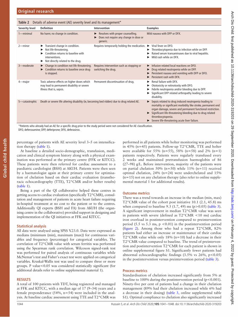
► Standardisation of chelation drug AE severity description and its management: A novel chelation medication AE severity description was developed and categorised into five severity levels to standardise drug continuation/discontinuation/change by both patients and healthcare professionals (details in table 2).
Process metric ► Compliance with recommended chelation therapy. ► Reporting of AE severity levels. ► Percentage of patients with inappropriate discontinuation
or continuation of chelation therapy after introduction of AE severity description.
► Percentage of severity level 3–5 AE on intensification therapy.
*T2*CMR was not performed in patients presenting with acute heart failure requiring emergent admission in hospital as breath holding for MRI was not possible in these patients. T2*CMR was obtained once patient was stabilised with emergent chelation for at least 2 weeks.†Outcome metric was meant to measure the effectiveness of the complete initiative thus demonstrating the cumulative effect of all the key drivers.‡Abnormal TTE is defined as systolic (left ventricular ejection fraction <56% and/or age based low peak systolic left ventricular global longitudinal strain value) and/or diastolic dysfunction.§In the absence of iron overload or out of proportion cardiac dysfunction not explained by iron load, evaluate for other causes of myocardial dysfunction that is, viral (hepatitis B and C, HIV etc) myocarditis, electrolyte (eg, profound hypocalcaemia) or vitamin (eg, Vitamin D or B deficiency etc), endocrinological (eg, hypothyroidism, hypocalcaemia etc) related cardiac dysfunction.¶DFO was infused daily by a subcutaneous infusion pump for 8–12 hours or in hospital over 24 hours (if patient had signs of heart failure or significant cardiac dysfunction on TTE with moderate to severe cardiac siderosis on T2*CMR), DFP was prescribed daily in three divided doses and DFX was given in a one time per day dosing schedule. All oral chelators were prescribed after meals.AE, adverse event; DFO, deferoxamine; DFP, deferiprone; DFX, deferasirox; T2*CMR, cardiac T2* magnetic resonance imaging; TTE, transthoracic echocardiography.
on February 4, 2022 by guest. P
rotected by copyright.http://adc.bm
j.com/
Arch D
is Child: first published as 10.1136/archdischild-2020-319203 on 29 S
eptember 2020. D
ownloaded from
1044 Hussain S, et al. Arch Dis Child 2020;105:1041–1048. doi:10.1136/archdischild-2020-319203
Original research
Glo
bal
chil
d he
alth percentage of patients with AE severity level 3–5 on intensifica-
tion therapy (table 1).At baseline a detailed socio- demographic, transfusion, medi-
cation and clinical symptom history along with a physical exam-ination was performed at the primary centre (FFK or KITCC). These patients were then referred for cardiac assessment to a paediatric cardiologist (BSH) at AKUH. Patients were then seen by a haematologist again at their primary centre for optimisa-tion of chelation based on their cardiac evaluation (transtho-racic echocardiography (TTE), T2*CMR and/or holter results) (table 1).
Being a part of the QI collaborative helped these centres in getting access to cardiac evaluation (specifically T2*CMR), consul-tation and management of patients in acute heart failure requiring in- hospital treatment at no cost to the patient or to the centres. Additionally QI experts (BSH and FA) from AKUH (the organ-ising centre in the collaborative) provided support in designing and implementation of the QI initiatives at FFK and KITCC.
Statistical analysisAll data were analysed using SPSS V.23.0. Data were expressed as medians (minimum (min), maximum (max)) for continuous vari-ables and frequency (percentage) for categorical variables. The correlation of T2*CMR value with serum ferritin was performed using the Spearman rank correlation. Wilcoxon signed- rank test was performed for paired analysis of continuous variables while McNemar’s test and Fisher’s exact test were applied on categorical variables. Kruskal- Wallis test was used to compare three or more groups. P value<0.05 was considered statistically significant (for additional details refer to online supplemental material 1).
RESULTSA total of 100 patients with TDT, being registered and managed at FFK and KITCC, with a median age of 17 (9–34) years and a female preponderance (54%, n=54) were included in the anal-ysis. At baseline cardiac assessment using TTE and T2*CMR was
performed in all patients while holter monitoring was performed in 45% (n=45) patients. Follow- up T2*CMR, TTE and holter were available for 55% (n=55), 58% (n=58) and 2% (n=1) patients respectively. Patients were regularly transfused every 2 weeks and maintained pretransfusion haemoglobin of 86 (27–98) g/L. Before intervention, majority of the patients were on partial chelation 50% (n=50) while 11% (n=11) received optimal chelation, 24% (n=24) were underchelated and 15% (n=15) not on any chelation therapy (also refer to online supple-mental material 1 for additional results).
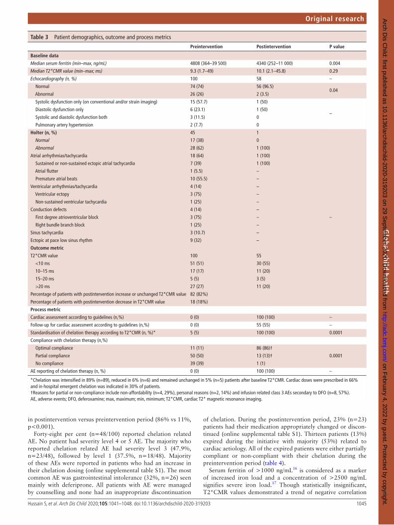
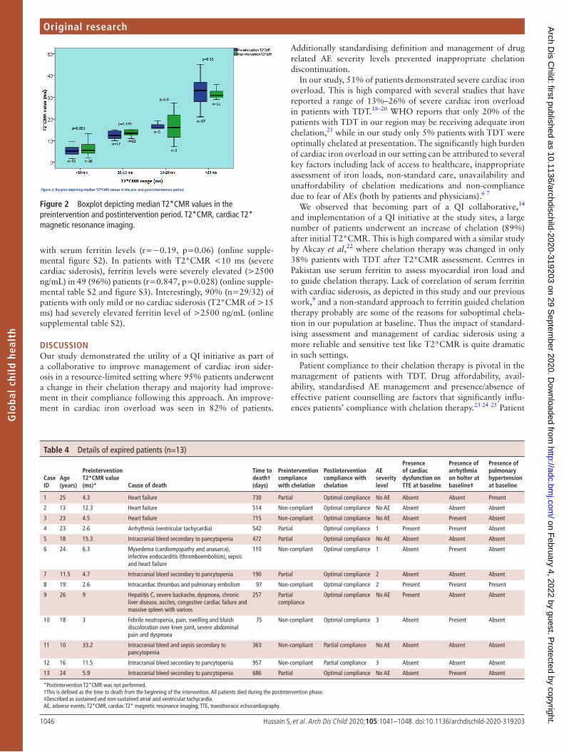
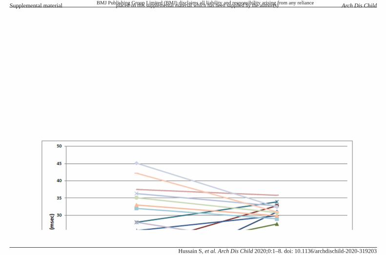
Outcome metricsThere was a trend towards an increase in the median (min, max) T2*CMR value of the cohort post initiative 10.1 (2.1, 45.8) ms when compared to baseline 9.3 (1.7, 49) ms (p>0.05) (table 3). A significant improvement in median T2*CMR value was seen in patients with severe (defined as T2*CMR <10 ms) cardiac iron overload in postintervention compared to preintervention period (5.5 vs 5.3 ms, p <0.01) in the postintervention period (figure 2). Among those who had a repeat T2*CMR, 82% patients had either an increase or maintenance of their cardiac T2*CMR value while only 18% (n=10) had a decrease in their T2*CMR value compared to baseline. The trend of preinterven-tion and postintervention T2*CMR for each patient is shown in online supplemental figure S1. Significantly fewer patients had abnormal echocardiographic findings (3.5% vs 26%, p<0.05) in the postintervention versus preintervention period (table 3).
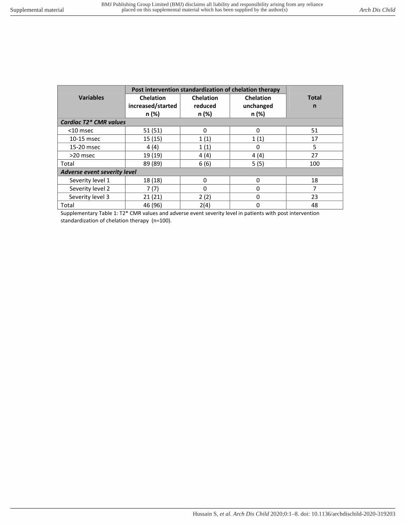
Process metricsStandardisation of chelation increased significantly from 5% at baseline to 100% during the postintervention period (p<0.001). Ninety- five per cent of patients had a change in their chelation management (89% had their chelation increased while 6% had a decrease in their dosing) (table 3, online supplemental table S1). Optimal compliance to chelation also significantly increased
Table 2 Details of adverse event (AE) severity level and its management*
Severity level Definition Intervention Examples
1—minimal No harm; no change in condition. ► Resolves with proper counselling. ► Does not require any change in dose or
generic.
Mild nausea with DFP or DFX.
2—minor ► Transient change in condition. ► Not life- threatening. ► Condition returns to baseline with
interventions. ► Not directly related to the drug.
Requires temporarily holding the medication. ► Viral fever on DFO. ► Thrombocytopenia due to infection while on DFP. ► Raised serum transaminases due to viral hepatitis. ► Mild rash while on DFX.
3—moderate ► Change in condition not life threatening. ► Condition returns to baseline once drug
is stopped.
Requires intervention such as stopping or switching the drug.
► Infusion related local reactions on DFO. ► Drug related neutropenia while on DFP. ► Persistent nausea and vomiting with DFP or DFO. ► Persistent rash with DFX.
4—major Toxic adverse effects on higher doses which may lead to permanent disability or severe illness that is, sepsis.
Permanent discontinuation of drug. ► Renal failure with DFX. ► Ototoxicity or retinotoxity with DFO. ► Febrile neutropenia and/or bleeding due to DFP. ► Significant DFP related arthropathy leading to severe
disability.
5—catastrophic Death or severe life altering disability (ie, becoming bed ridden) due to drug related AE. ► Sepsis related to drug induced neutropenia leading to mortality or significant morbidity like stroke, permanent end organ damage, severe and permanent functional restriction.
► Significant life- threatening bleeding due to drug related thrombocytopenia.
► Severe life- threatening acute liver failure.
*Patients who already had an AE for a specific drug prior to the study period were not considered for AE classification.DFO, deferoxamine; DFP, deferiprone; DFX, deferasirox.
on February 4, 2022 by guest. P
rotected by copyright.http://adc.bm
j.com/
Arch D
is Child: first published as 10.1136/archdischild-2020-319203 on 29 S
eptember 2020. D
ownloaded from
1045Hussain S, et al. Arch Dis Child 2020;105:1041–1048. doi:10.1136/archdischild-2020-319203
Original research
Global child health
in postintervention versus preintervention period (86% vs 11%, p<0.001).
Forty- eight per cent (n=48/100) reported chelation related AE. No patient had severity level 4 or 5 AE. The majority who reported chelation related AE had severity level 3 (47.9%, n=23/48), followed by level 1 (37.5%, n=18/48). Majority of these AEs were reported in patients who had an increase in their chelation dosing (online supplemental table S1). The most common AE was gastrointestinal intolerance (32%, n=26) seen mainly with deferiprone. All patients with AE were managed by counselling and none had an inappropriate discontinuation
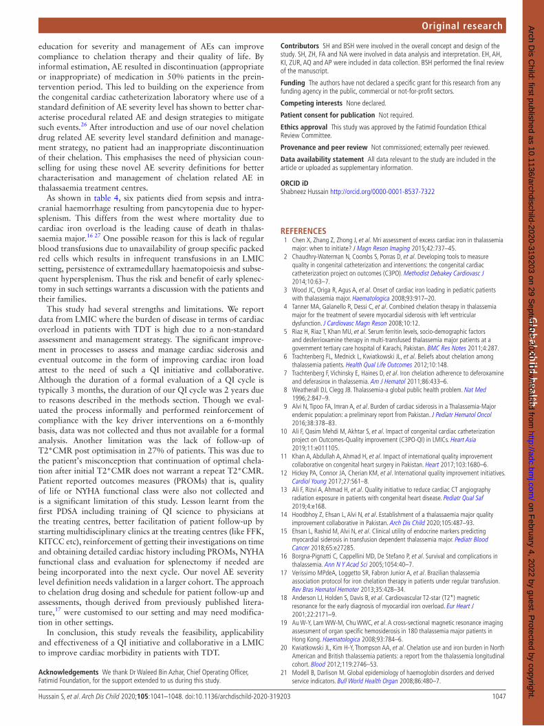
of chelation. During the postintervention period, 23% (n=23) patients had their medication appropriately changed or discon-tinued (online supplemental table S1). Thirteen patients (13%) expired during the initiative with majority (53%) related to cardiac aetiology. All of the expired patients were either partially compliant or non- compliant with their chelation during the preintervention period (table 4).
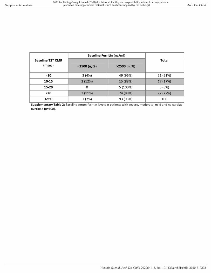
Serum ferritin of >1000 ng/mL16 is considered as a marker of increased iron load and a concentration of >2500 ng/mL signifies severe iron load.17 Though statistically insignificant, T2*CMR values demonstrated a trend of negative correlation
Table 3 Patient demographics, outcome and process metrics
Preintervention Postintervention P value
Baseline data
Median serum ferritin (min–max, ng/mL) 4808 (364–39 500) 4340 (252–11 000) 0.004
Median T2*CMR value (min–max; ms) 9.3 (1.7–49) 10.1 (2.1–45.8) 0.29
Echocardiography (n, %) 100 58 –
Normal 74 (74) 56 (96.5)0.04
Abnormal 26 (26) 2 (3.5)
Systolic dysfunction only (on conventional and/or strain imaging) 15 (57.7) 1 (50)
– Diastolic dysfunction only 6 (23.1) 1 (50)
Systolic and diastolic dysfunction both 3 (11.5) 0
Pulmonary artery hypertension 2 (7.7) 0
Holter (n, %) 45 1
–
Normal 17 (38) 0
Abnormal 28 (62) 1 (100)
Atrial arrhythmias/tachycardia 18 (64) 1 (100)
Sustained or non- sustained ectopic atrial tachycardia 7 (39) 1 (100)
Atrial flutter 1 (5.5) –
Premature atrial beats 10 (55.5) –
Ventricular arrhythmias/tachycardia 4 (14) –
Ventricular ectopy 3 (75) –
Non- sustained ventricular tachycardia 1 (25) –
Conduction defects 4 (14) –
First degree atrioventricular block 3 (75) –
Right bundle branch block 1 (25) –
Sinus tachycardia 3 (10.7) –
Ectopic at pace low sinus rhythm 9 (32) –
Outcome metric
T2*CMR value 100 55
<10 ms 51 (51) 30 (55)
10–15 ms 17 (17) 11 (20)
15–20 ms 5 (5) 3 (5)
>20 ms 27 (27) 11 (20)
Percentage of patients with postintervention increase or unchanged T2*CMR value 82 (82%)
Percentage of patients with postintervention decrease in T2*CMR value 18 (18%)
Process metric
Cardiac assessment according to guidelines (n,%) 0 (0) 100 (100) –
Follow- up for cardiac assessment according to guidelines (n,%) 0 (0) 55 (55) –
Standardisation of chelation therapy according to T2*CMR (n, %)* 5 (5) 100 (100) 0.0001
Compliance with chelation therapy (n,%)
Optimal compliance 11 (11) 86 (86)†
Partial compliance 50 (50) 13 (13)† 0.0001
No compliance 39 (39) 1 (1)
AE reporting of chelation therapy (n, %) 0 (0) 100 (100) –
*Chelation was intensified in 89% (n=89), reduced in 6% (n=6) and remained unchanged in 5% (n=5) patients after baseline T2*CMR. Cardiac doses were prescribed in 66% and in- hospital emergent chelation was indicated in 30% of patients.†Reasons for partial or non- compliance include non- affordability (n=4, 29%), personal reasons (n=2, 14%) and infusion related class 3 AEs secondary to DFO (n=8, 57%).AE, adverse events; DFO, deferoxamine; max, maximum; min, minimum; T2*CMR, cardiac T2* magnetic resonance imaging.
on February 4, 2022 by guest. P
rotected by copyright.http://adc.bm
j.com/
Arch D
is Child: first published as 10.1136/archdischild-2020-319203 on 29 S
eptember 2020. D
ownloaded from
1046 Hussain S, et al. Arch Dis Child 2020;105:1041–1048. doi:10.1136/archdischild-2020-319203
Original research
Glo
bal
chil
d he
alth
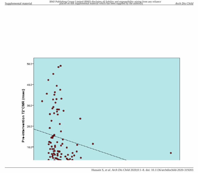
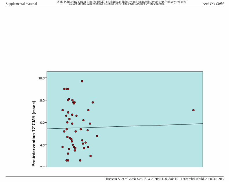
with serum ferritin levels (r=−0.19, p=0.06) (online supple-mental figure S2). In patients with T2*CMR <10 ms (severe cardiac siderosis), ferritin levels were severely elevated (>2500 ng/mL) in 49 (96%) patients (r=0.847, p=0.028) (online supple-mental table S2 and figure S3). Interestingly, 90% (n=29/32) of patients with only mild or no cardiac siderosis (T2*CMR of >15 ms) had severely elevated ferritin level of >2500 ng/mL (online supplemental table S2).
DISCUSSIONOur study demonstrated the utility of a QI initiative as part of a collaborative to improve management of cardiac iron sider-osis in a resource- limited setting where 95% patients underwent a change in their chelation therapy and majority had improve-ment in their compliance following this approach. An improve-ment in cardiac iron overload was seen in 82% of patients.
Additionally standardising definition and management of drug related AE severity levels prevented inappropriate chelation discontinuation.
In our study, 51% of patients demonstrated severe cardiac iron overload. This is high compared with several studies that have reported a range of 13%–26% of severe cardiac iron overload in patients with TDT.18–20 WHO reports that only 20% of the patients with TDT in our region may be receiving adequate iron chelation,21 while in our study only 5% patients with TDT were optimally chelated at presentation. The significantly high burden of cardiac iron overload in our setting can be attributed to several key factors including lack of access to healthcare, inappropriate assessment of iron loads, non- standard care, unavailability and unaffordability of chelation medications and non- compliance due to fear of AEs (both by patients and physicians).6 7
We observed that becoming part of a QI collaborative,14 and implementation of a QI initiative at the study sites, a large number of patients underwent an increase of chelation (89%) after initial T2*CMR. This is high compared with a similar study by Akcay et al,22 where chelation therapy was changed in only 38% patients with TDT after T2*CMR assessment. Centres in Pakistan use serum ferritin to assess myocardial iron load and to guide chelation therapy. Lack of correlation of serum ferritin with cardiac siderosis, as depicted in this study and our previous work,9 and a non- standard approach to ferritin guided chelation therapy probably are some of the reasons for suboptimal chela-tion in our population at baseline. Thus the impact of standard-ising assessment and management of cardiac siderosis using a more reliable and sensitive test like T2*CMR is quite dramatic in such settings.
Patient compliance to their chelation therapy is pivotal in the management of patients with TDT. Drug affordability, avail-ability, standardised AE management and presence/absence of effective patient counselling are factors that significantly influ-ences patients’ compliance with chelation therapy.23 24 25 Patient
Figure 2 Boxplot depicting median T2*CMR values in the preintervention and postintervention period. T2*CMR, cardiac T2* magnetic resonance imaging.
Table 4 Details of expired patients (n=13)
Case ID
Age (years)
Preintervention T2*CMR value (ms)* Cause of death
Time to death†(days)
Preintervention compliance with chelation
Postintervention compliance with chelation
AE severity level
Presence of cardiac dysfunction on TTE at baseline
Presence of arrhythmia on holter at baseline‡
Presence of pulmonary hypertension at baseline
1 25 4.3 Heart failure 730 Partial Optimal compliance No AE Absent Absent Present
2 13 12.3 Heart failure 514 Non- compliant Optimal compliance No AE Absent Absent Absent
3 23 4.5 Heart failure 715 Non- compliant Optimal compliance No AE Absent Present Absent
4 23 2.6 Arrhythmia (ventricular tachycardia) 542 Partial Optimal compliance 1 Present Present Absent
5 18 15.3 Intracranial bleed secondary to pancytopenia 472 Partial Optimal compliance No AE Absent Absent Absent
6 24 6.3 Myxedema (cardiomyopathy and anasarca), infective endocarditis (thromboembolism), sepsis and heart failure
110 Non- compliant Optimal compliance 1 Absent Present Absent
7 11.5 4.7 Intracranial bleed secondary to pancytopenia 190 Partial Optimal compliance 2 Absent Absent Absent
8 19 2.6 Intracardiac thrombus and pulmonary embolism 97 Non- compliant Optimal compliance 2 Present Present Present
9 26 9 Hepatitis C, severe backache, dyspnoea, chronic liver disease, ascites, congestive cardiac failure and massive spleen with varices
257 Partial compliance
Optimal compliance No AE Present Absent Absent
10 18 3 Febrile neutropenia, pain, swelling and bluish discoloration over knee joint, severe abdominal pain and dyspnoea
75 Non- compliant Optimal compliance 3 Absent Present Absent
11 10 33.2 Intracranial bleed and sepsis secondary to pancytopenia
363 Non- compliant Partial compliance No AE Absent Absent Absent
12 16 11.5 Intracranial bleed secondary to pancytopenia 957 Non- compliant Partial compliance 3 Absent Absent Absent
13 24 5.9 Intracranial bleed secondary to pancytopenia 686 Partial Optimal compliance No AE Absent Present Absent
*Postintervention T2*CMR was not performed.†This is defined as the time to death from the beginning of the intervention. All patients died during the postintervention phase.‡Described as sustained and non- sustained atrial and ventricular tachycardia.AE, adverse events; T2*CMR, cardiac T2* magnetic resonance imaging; TTE, transthoracic echocardiography.
on February 4, 2022 by guest. P
rotected by copyright.http://adc.bm
j.com/
Arch D
is Child: first published as 10.1136/archdischild-2020-319203 on 29 S
eptember 2020. D
ownloaded from
1047Hussain S, et al. Arch Dis Child 2020;105:1041–1048. doi:10.1136/archdischild-2020-319203
Original research
Global child health
education for severity and management of AEs can improve compliance to chelation therapy and their quality of life. By informal estimation, AE resulted in discontinuation (appropriate or inappropriate) of medication in 50% patients in the prein-tervention period. This led to building on the experience from the congenital cardiac catheterization laboratory where use of a standard definition of AE severity level has shown to better char-acterise procedural related AE and design strategies to mitigate such events.26 After introduction and use of our novel chelation drug related AE severity level standard definition and manage-ment strategy, no patient had an inappropriate discontinuation of their chelation. This emphasises the need of physician coun-selling for using these novel AE severity definitions for better characterisation and management of chelation related AE in thalassaemia treatment centres.
As shown in table 4, six patients died from sepsis and intra-cranial haemorrhage resulting from pancytopenia due to hyper-splenism. This differs from the west where mortality due to cardiac iron overload is the leading cause of death in thalas-saemia major.16 27 One possible reason for this is lack of regular blood transfusions due to unavailability of group specific packed red cells which results in infrequent transfusions in an LMIC setting, persistence of extramedullary haematopoiesis and subse-quent hypersplenism. Thus the risk and benefit of early splenec-tomy in such settings warrants a discussion with the patients and their families.
This study had several strengths and limitations. We report data from LMIC where the burden of disease in terms of cardiac overload in patients with TDT is high due to a non- standard assessment and management strategy. The significant improve-ment in processes to assess and manage cardiac siderosis and eventual outcome in the form of improving cardiac iron load attest to the need of such a QI initiative and collaborative. Although the duration of a formal evaluation of a QI cycle is typically 3 months, the duration of our QI cycle was 2 years due to reasons described in the methods section. Though we eval-uated the process informally and performed reinforcement of compliance with the key driver interventions on a 6- monthly basis, data was not collected and thus not available for a formal analysis. Another limitation was the lack of follow- up of T2*CMR post optimisation in 27% of patients. This was due to the patient’s misconception that continuation of optimal chela-tion after initial T2*CMR does not warrant a repeat T2*CMR. Patient reported outcomes measures (PROMs) that is, quality of life or NYHA functional class were also not collected and is a significant limitation of this study. Lesson learnt from the first PDSA including training of QI science to physicians at the treating centres, better facilitation of patient follow- up by starting multidisciplinary clinics at the treating centres (like FFK, KITCC etc), reinforcement of getting their investigations on time and obtaining detailed cardiac history including PROMs, NYHA functional class and evaluation for splenectomy if needed are being incorporated into the next cycle. Our novel AE severity level definition needs validation in a larger cohort. The approach to chelation drug dosing and schedule for patient follow- up and assessments, though derived from previously published litera-ture,17 were customised to our setting and may need modifica-tion in other settings.
In conclusion, this study reveals the feasibility, applicability and effectiveness of a QI initiative and collaborative in a LMIC to improve cardiac morbidity in patients with TDT.
Acknowledgements We thank Dr Waleed Bin Azhar, Chief Operating Officer, Fatimid Foundation, for the support extended to us during this study.
Contributors SH and BSH were involved in the overall concept and design of the study. SH, ZH, FA and NA were involved in data analysis and interpretation. EH, AH, KI, ZUR, AQ and AP were included in data collection. BSH performed the final review of the manuscript.
Funding The authors have not declared a specific grant for this research from any funding agency in the public, commercial or not- for- profit sectors.
Competing interests None declared.
Patient consent for publication Not required.
Ethics approval This study was approved by the Fatimid Foundation Ethical Review Committee.
Provenance and peer review Not commissioned; externally peer reviewed.
Data availability statement All data relevant to the study are included in the article or uploaded as supplementary information.
ORCID iDShabneez Hussain http:// orcid. org/ 0000- 0001- 8537- 7322
REFERENCES 1 Chen X, Zhang Z, Zhong J, et al. Mri assessment of excess cardiac iron in thalassemia
major: when to initiate? J Magn Reson Imaging 2015;42:737–45. 2 Chaudhry- Waterman N, Coombs S, Porras D, et al. Developing tools to measure
quality in congenital catheterization and interventions: the congenital cardiac catheterization project on outcomes (C3PO). Methodist Debakey Cardiovasc J 2014;10:63–7.
3 Wood JC, Origa R, Agus A, et al. Onset of cardiac iron loading in pediatric patients with thalassemia major. Haematologica 2008;93:917–20.
4 Tanner MA, Galanello R, Dessi C, et al. Combined chelation therapy in thalassemia major for the treatment of severe myocardial siderosis with left ventricular dysfunction. J Cardiovasc Magn Reson 2008;10:12.
5 Riaz H, Riaz T, Khan MU, et al. Serum ferritin levels, socio- demographic factors and desferrioxamine therapy in multi- transfused thalassemia major patients at a government tertiary care hospital of Karachi, Pakistan. BMC Res Notes 2011;4:287.
6 Trachtenberg FL, Mednick L, Kwiatkowski JL, et al. Beliefs about chelation among thalassemia patients. Health Qual Life Outcomes 2012;10:148.
7 Trachtenberg F, Vichinsky E, Haines D, et al. Iron chelation adherence to deferoxamine and deferasirox in thalassemia. Am J Hematol 2011;86:433–6.
8 Weatherall DJ, Clegg JB. Thalassemia- a global public health problem. Nat Med 1996;2:847–9.
9 Alvi N, Tipoo FA, Imran A, et al. Burden of cardiac siderosis in a Thalassemia- Major endemic population: a preliminary report from Pakistan. J Pediatr Hematol Oncol 2016;38:378–83.
10 Ali F, Qasim Mehdi M, Akhtar S, et al. Impact of congenital cardiac catheterization project on Outcomes- Quality improvement (C3PO- QI) in LMICs. Heart Asia 2019;11:e011105.
11 Khan A, Abdullah A, Ahmad H, et al. Impact of international quality improvement collaborative on congenital heart surgery in Pakistan. Heart 2017;103:1680–6.
12 Hickey PA, Connor JA, Cherian KM, et al. International quality improvement initiatives. Cardiol Young 2017;27:S61–8.
13 Ali F, Rizvi A, Ahmad H, et al. Quality initiative to reduce cardiac CT angiography radiation exposure in patients with congenital heart disease. Pediatr Qual Saf 2019;4:e168.
14 Hoodbhoy Z, Ehsan L, Alvi N, et al. Establishment of a thalassaemia major quality improvement collaborative in Pakistan. Arch Dis Child 2020;105:487–93.
15 Ehsan L, Rashid M, Alvi N, et al. Clinical utility of endocrine markers predicting myocardial siderosis in transfusion dependent thalassemia major. Pediatr Blood Cancer 2018;65:e27285.
16 Borgna- Pignatti C, Cappellini MD, De Stefano P, et al. Survival and complications in thalassemia. Ann N Y Acad Sci 2005;1054:40–7.
17 Veríssimo MPdeA, Loggetto SR, Fabron Junior A, et al. Brazilian thalassemia association protocol for iron chelation therapy in patients under regular transfusion. Rev Bras Hematol Hemoter 2013;35:428–34.
18 Anderson LJ, Holden S, Davis B, et al. Cardiovascular T2- star (T2*) magnetic resonance for the early diagnosis of myocardial iron overload. Eur Heart J 2001;22:2171–9.
19 Au W- Y, Lam WW- M, Chu WWC, et al. A cross- sectional magnetic resonance imaging assessment of organ specific hemosiderosis in 180 thalassemia major patients in Hong Kong. Haematologica 2008;93:784–6.
20 Kwiatkowski JL, Kim H- Y, Thompson AA, et al. Chelation use and iron burden in North American and British thalassemia patients: a report from the thalassemia longitudinal cohort. Blood 2012;119:2746–53.
21 Modell B, Darlison M. Global epidemiology of haemoglobin disorders and derived service indicators. Bull World Health Organ 2008;86:480–7.
on February 4, 2022 by guest. P
rotected by copyright.http://adc.bm
j.com/
Arch D
is Child: first published as 10.1136/archdischild-2020-319203 on 29 S
eptember 2020. D
ownloaded from
1048 Hussain S, et al. Arch Dis Child 2020;105:1041–1048. doi:10.1136/archdischild-2020-319203
Original research
Glo
bal
chil
d he
alth
22 Akcay A, Salcioglu Z, Oztarhan K, et al. Cardiac T2* MRI assessment in patients with thalassaemia major and its effect on the preference of chelation therapy. Int J Hematol 2014;99:706–13.
23 Viprakasit V, Gattermann N, Lee JW, et al. Geographical variations in current clinical practice on transfusions and iron chelation therapy across various transfusion- dependent anaemias. Blood Transfus 2013;11:108–22.
24 Nadeem Ikram KH, Younas M, Amanat S. Ferritin levels in patients of beta thalassaemia major. International Journal of Pathology 2004;2:71–4.
25 Mednick L, Yu S, Trachtenberg F, et al. Symptoms of depression and anxiety in patients with thalassemia: prevalence and correlates in the thalassemia longitudinal cohort. Am J Hematol 2010;85:802–5.
26 Bergersen L, Gauvreau K, Foerster SR, et al. Catheterization for congenital heart disease adjustment for risk method (CHARM). JACC Cardiovasc Interv 2011;4:1037–46.
27 Chouliaras G, Yiannoutsos CT, Berdoukas V, et al. Cardiac related death in thalassaemia major: time trend and risk factors in a large Greek unit. Eur J Haematol 2009;82:381–7.
on February 4, 2022 by guest. P
rotected by copyright.http://adc.bm
j.com/
Arch D
is Child: first published as 10.1136/archdischild-2020-319203 on 29 S
eptember 2020. D
ownloaded from
Supplemental material
Details of the 2 thalassemia centers:
FFK and KITC have approximately 800 and 600 registered TDT patients respectively
providing an average of 50 blood transfusions per day (25 transfusion chairs in each
center).
Factors associated with poor management of myocardial siderosis:
A QI team that included a haematologist (SH), pediatric cardiologists (BSH and FA), QI
experts (BSH and FA) and medical officers, was formed. Continuing medical education
(CMEs), morbidity and mortality meetings (M & M), group discussions and previously
published studies were used to identify factors associated with poor management of
myocardial iron siderosis.
Assessment: Screening of TDT patients for cardiac dysfunction was not performed on a
yearly basis. Diagnostic test such as transthoracic echocardiogram (TTE) was the only
investigation advised and only when the patient presented with cardiac symptoms. Result
of these investigations were not tracked or used to alter management.
Management: Follow up of TDT patients to assess compliance to chelation therapy was
not structured.
Adverse events (AE): There was also a lack of compliance with chelation therapy,
commonly reported due to financial constraints or variable management of chelation drug
related AE.
Statistical Analysis:
Process and outcome metrics were analyzed cumulatively from both the centers (FFK and
KITC). Since the QI initiative was introduced in a standardized manner in both the
centers by the same team, there was no difference in implementation of intervention in
the two and thus no comparative analysis was done between these centers.
Results:
Outcome metric: There was no difference (p= 0.24) in the magnitude of change in T2*
CMR value for the different age categories (10-15 years, 15-20 years, 20-25 years, 25-30
years & 30 to 35 years).
Adverse events: Other DFP induced AEs included arthralgia (19.4%, n=16), neutropenia
(8.4%, n=7) and thrombocytopenia (13.4%, n=11). Two patients (2.4%) developed rash
secondary to DFX while DFO caused infusion related local AE in 17% (n=14) patients.
Nonspecific AEs occurred in 7.4% (n=6) patients such as weakness and back pain
thought to be secondary to DFO (n=4) and DFP (n=2).
BMJ Publishing Group Limited (BMJ) disclaims all liability and responsibility arising from any relianceSupplemental material placed on this supplemental material which has been supplied by the author(s) Arch Dis Child
doi: 10.1136/archdischild-2020-319203–8.:10 2020;Arch Dis Child, et al. Hussain S
BMJ Publishing Group Limited (BMJ) disclaims all liability and responsibility arising from any relianceSupplemental material placed on this supplemental material which has been supplied by the author(s) Arch Dis Child
doi: 10.1136/archdischild-2020-319203–8.:10 2020;Arch Dis Child, et al. Hussain S
Variables
Post intervention standardization of chelation therapy
Total
n
Chelation
increased/started
n (%)
Chelation
reduced
n (%)
Chelation
unchanged
n (%)
Cardiac T2* CMR values
<10 msec 51 (51) 0 0 51
10-15 msec 15 (15) 1 (1) 1 (1) 17
15-20 msec 4 (4) 1 (1) 0 5
>20 msec 19 (19) 4 (4) 4 (4) 27
Total 89 (89) 6 (6) 5 (5) 100
Adverse event severity level
Severity level 1 18 (18) 0 0 18
Severity level 2 7 (7) 0 0 7
Severity level 3 21 (21) 2 (2) 0 23
Total 46 (96) 2(4) 0 48
Supplementary Table 1: T2* CMR values and adverse event severity level in patients with post intervention
standardization of chelation therapy (n=100).
BMJ Publishing Group Limited (BMJ) disclaims all liability and responsibility arising from any relianceSupplemental material placed on this supplemental material which has been supplied by the author(s) Arch Dis Child
doi: 10.1136/archdischild-2020-319203–8.:10 2020;Arch Dis Child, et al. Hussain S
Baseline T2* CMR
(msec)
Baseline Ferritin (ng/ml)
Total
<2500 (n, %)
>2500 (n, %)
<10 2 (4%) 49 (96%) 51 (51%)
10-15 2 (12%) 15 (88%) 17 (17%)
15-20 0 5 (100%) 5 (5%)
>20 3 (11%) 24 (89%) 27 (27%)
Total 7 (7%) 93 (93%) 100
Supplementary Table 2: Baseline serum ferritin levels in patients with severe, moderate, mild and no cardiac
overload (n=100).
BMJ Publishing Group Limited (BMJ) disclaims all liability and responsibility arising from any relianceSupplemental material placed on this supplemental material which has been supplied by the author(s) Arch Dis Child
doi: 10.1136/archdischild-2020-319203–8.:10 2020;Arch Dis Child, et al. Hussain S
BMJ Publishing Group Limited (BMJ) disclaims all liability and responsibility arising from any relianceSupplemental material placed on this supplemental material which has been supplied by the author(s) Arch Dis Child
doi: 10.1136/archdischild-2020-319203–8.:10 2020;Arch Dis Child, et al. Hussain S
BMJ Publishing Group Limited (BMJ) disclaims all liability and responsibility arising from any relianceSupplemental material placed on this supplemental material which has been supplied by the author(s) Arch Dis Child
doi: 10.1136/archdischild-2020-319203–8.:10 2020;Arch Dis Child, et al. Hussain S
BMJ Publishing Group Limited (BMJ) disclaims all liability and responsibility arising from any relianceSupplemental material placed on this supplemental material which has been supplied by the author(s) Arch Dis Child
doi: 10.1136/archdischild-2020-319203–8.:10 2020;Arch Dis Child, et al. Hussain S