Embed Size (px)
Citation preview
How Early Can Myocardial Iron Overload Occur in BetaThalassemia Major?Gaohui Yang1., Rongrong Liu1., Peng Peng2, Liling Long2, Xinhua Zhang3, Weijia Yang4,
Shaohong Tan5, Hongfei Pan6, Xingjiang Long7, Taigang He8,9, Lisa Anderson8, Yongrong Lai1*
1 Department of Hematology, the First Affiliated Hospital of Guangxi Medical University, Nanning, China, 2 Department of Radiology, the First Affiliated Hospital of
Guangxi Medical University, Nanning, China, 3 Department of Hematology, 303rd Hospital of People’s Liberation Army, Nanning, China, 4 Department of Pediatrics,
Women and Children Hospital of Guilin, Guilin, China, 5 Department of Pediatrics, Women and Children Hospital of Yulin, Yulin, China, 6 Department of Pediatrics,
Affiliated Hospital of Youjiang Medical University for Nationality, Baise, China, 7 Department of Pediatrics, The People’s Hospital of Liuzhou, Liuzhou, China,
8 Cardiovascular Sciences Research Centre, St George’s University of London, London, United Kingdom, 9 Biomedical Research Unit, Royal Brompton Hospital, London,
United Kingdom
Abstract
Background: Myocardial siderosis is the most common cause of death in patients with beta thalassemia major(TM). Thisstudy aimed at investigating the occurrence, prevalence and severity of cardiac iron overload in a young Chinese populationwith beta TM.
Methods and Results: We analyzed T2* cardiac magnetic resonance (CMR), left ventricular ejection fraction (LVEF) andserum ferritin (SF) in 201 beta TM patients. The median age was 9 years old. Patients received an average of 13 units ofblood per year. The median SF level was 4536 ng/ml and 165 patients (82.1%) had SF.2500 ng/ml. Myocardial ironoverload was detected in 68 patients (33.8%) and severe myocardial iron overload was detected in 26 patients (12.6%).Twenty-two patients #10 years old had myocardial iron overload, three of whom were only 6 years old. No myocardial ironoverload was detected under the age of 6 years. Median LVEF was 64% (measured by CMR in 175 patients). Five of 6patients with a LVEF,56% and 8 of 10 patients with cardiac disease had myocardial iron overload.
Conclusions: The TM patients under follow-up at this regional centre in China patients are younger than other reportedcohorts, more poorly-chelated, and have a high burden of iron overload. Myocardial siderosis occurred in patients youngerthan previously reported, and was strongly associated with impaired LVEF and cardiac disease. For such poorly-chelated TMpatients, our data shows that the first assessment of cardiac T2* should be performed as early as 6 years old.
Citation: Yang G, Liu R, Peng P, Long L, Zhang X, et al. (2014) How Early Can Myocardial Iron Overload Occur in Beta Thalassemia Major? PLoS ONE 9(1): e85379.doi:10.1371/journal.pone.0085379
Editor: Xiaolei Xu, Mayo Clinic, United States of America
Received September 5, 2013; Accepted November 25, 2013; Published January 22, 2014
Copyright: � 2014 Yang et al. This is an open-access article distributed under the terms of the Creative Commons Attribution License, which permitsunrestricted use, distribution, and reproduction in any medium, provided the original author and source are credited.
Funding: This work was supported by research grants from National Natural Science Foundations of China (Grant No. 30860307; No. 81160175) and GuangxiNatural Science Foundations (Grant No. 2011GXNSFD018033). Dr.. He receives support from Wellcome Trust, and Higher Education Funding for England (HEFCE)and British Heart Foundation (BHF) (FS/08/26225). The funders had no role in study design, data collection and analysis, decision to publish, or preparation of themanuscript.
Competing Interests: The authors have declared that no competing interests exist.
* E-mail: [email protected]
. These authors contributed equally to this work.
Introduction
Myocardial siderosis remains the most common cause of death
in patients with beta thalassemia major (TM) [1]. In affluent
countries such as the UK, there has been a dramatic improvement
in survival since the introduction of cardiovascular magnetic
resonance(CMR) for assessment of myocardial iron [2], but iron
induced cardiomyopathy still accounts for the majority of all
deaths [3]. Early detection of myocardial iron overload is
important because survival is only 50% once overt heart failure
is manifest [4] and patients remain asymptomatic until late in the
course of development of iron overload cardiomyopathy. Usefully,
noninvasive CMR can quantify iron overload in different organs
such as the heart and liver even before the development of
symptoms [5,6]. In this regard, CMR relaxometry T2* is being
increasingly used worldwide for monitoring transfusion-dependent
TM [7,8].
The guidelines for the clinical management of thalassemia
edited by Thalassemia international federation (TIF) recom-
mended that the first assessment of cardiac T2* would be done
at puberty for the well-chelated patients who received chelation
therapy early and regularly [9], but there is not a specific guideline
for poorly-chelated patients. Most TM patients in developing
countries including China are poorly-chelated and many of them
die in childhood or early adolescence due to the lack of access to
specialist care [10,11]. For poorly-chelated patients, there is
limited data on when the myocardial siderosis occurs and when
the CMR T2* screening should be initiated. To answer these
questions, we designed this study to determine the age when
cardiac iron overload occurs and the prevalence of abnormal
PLOS ONE | www.plosone.org 1 January 2014 | Volume 9 | Issue 1 | e85379
brought to you by COREView metadata, citation and similar papers at core.ac.uk
provided by St George's Online Research Archive
cardiac MRI T2* and myocardial function on a large young TM
population in mainland China.
Materials and Methods
Study PopulationWe studied 201 TM patients (192 children and 9 adults) from
November 2010 to January 2013. The median age was 9 (4–25)
years old. All patients were transfusion-dependent and required
transfusions from early childhood. Thirty-five patients (17.4%)
received splenectomy. Five patients (2.5%) were well-chelated and
196 patients (97.5%) were poorly-chelated. Patients were consid-
ered to be well-chelated if the following criteria were met [12,13]:
(i) initiate treatment after first 10–20 transfusions or serum ferritin
(SF) level above 1000 ng/ml; (ii) chelators dose: Deferoxamine
(DFO) is 20–40 mg/kg/day in children and 50–60 mg/kg/day in
adults, at least 5 days a week. Deferiprone (DFP) is 75 mg/kg/day.
Deferasirox (DFX) is 20–40 mg/kg/day; (iii) good compliance
with chelation therapy, defined as a$90% adherence to the
instructions given by the hematologists. The study was performed
at the first affiliated hospital of Guangxi Medical University.
Restrospective review of medical records, coded exchange of
clinical data, SF levels and MRI examininations were authorized
by the local institute. All of patients provided their written
informed consent to participate in this study. Adults ($18) finished
the informed consent by themselves. And for minors (,18), the
informed consents were obtained from the guardians on the behalf
of the minors participants involved in this study. The study was
approved by the Medical Ethics Committee of First Affiliated
Hospital of Guangxi Medical University (the approval number:
NO. 2008(KY-002).
Serum FerritinLaboratory blood tests were performed 2 weeks after the last
blood transfusion, followed by MRI evaluations with a 1-month
interval in all patients. Measurements were carried out by an
Electrochemiluminescence immunoassay (COBASE E 601, Roch,
USA).
MRI ProtocolsPatients were scanned with a 1.5T MRI scanner (Siemens
Avanto, Siemens Medical Systems, Erlangen, Germany) with the
combination of body-matrix and spine-matrix surface coils. To
ensure quality of the work, the local staff involved in this study
(technicians, radiologists, and hematologists) received intensive
onsite training from CMR specialists of Royal Brompton Hospital
(London, UK). Each scan included the measurement of heart T2*
and left ventricular ejection fraction (LVEF) using previously
published techniques [14,15,16]. For T2* imaging, a single short
axis mid-ventricular slice was acquired at eight echo times (2.97–
21.68 ms) using a gradient-echo sequence within a single breath-
hold. For myocardial T2* measurement, a homogeneous full
thickness region of interest was chosen in the septum and signal
intensities at lengthening echo times analysed using a truncation
model [17]. LVEF were determined from steady-state free
precession cines using CMR tools (Cardiovascular Imaging
Solutions, London, UK).
Statistical AnalysisData were expressed as mean6 one standard deviation, or
median (range) as appropriate. Mann Whitney U test or t test were
used for comparison of continuous variables between groups. The
significance of the correlation between parameters was assessed
using Spearman rank as the data was not normally distributed.
Categorical data were compared using the chi-square test.
Multivariate analysis used multiple linear regression analyses.
Statistical analyses were carried out using SPSS Statistics 13.0
(SPSS Inc., USA). A p value less than 0.05 was considered
statistically significant, and all p values were two sided.
Results
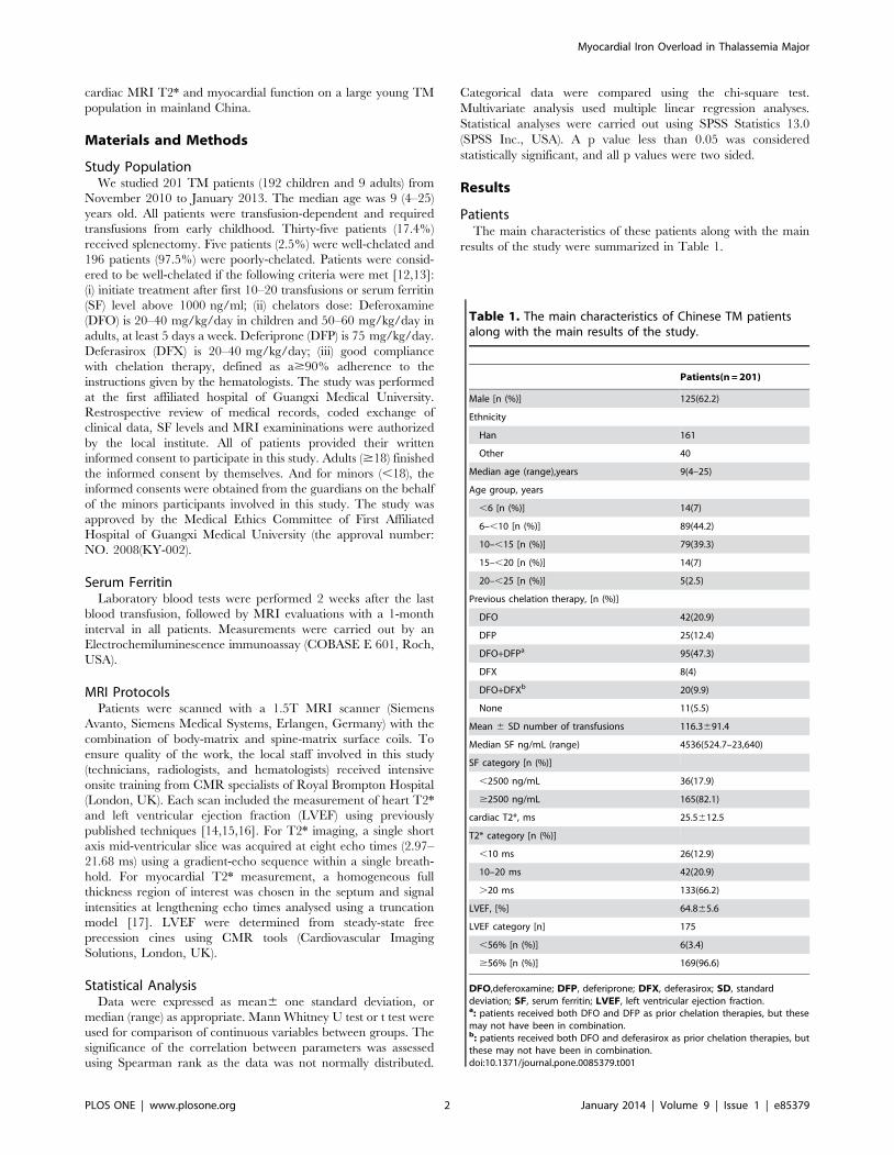
PatientsThe main characteristics of these patients along with the main
results of the study were summarized in Table 1.
Table 1. The main characteristics of Chinese TM patientsalong with the main results of the study.
Patients(n = 201)
Male [n (%)] 125(62.2)
Ethnicity
Han 161
Other 40
Median age (range),years 9(4–25)
Age group, years
,6 [n (%)] 14(7)
6–,10 [n (%)] 89(44.2)
10–,15 [n (%)] 79(39.3)
15–,20 [n (%)] 14(7)
20–,25 [n (%)] 5(2.5)
Previous chelation therapy, [n (%)]
DFO 42(20.9)
DFP 25(12.4)
DFO+DFPa 95(47.3)
DFX 8(4)
DFO+DFXb 20(9.9)
None 11(5.5)
Mean 6 SD number of transfusions 116.3691.4
Median SF ng/mL (range) 4536(524.7–23,640)
SF category [n (%)]
,2500 ng/mL 36(17.9)
$2500 ng/mL 165(82.1)
cardiac T2*, ms 25.5612.5
T2* category [n (%)]
,10 ms 26(12.9)
10–20 ms 42(20.9)
.20 ms 133(66.2)
LVEF, [%] 64.865.6
LVEF category [n] 175
,56% [n (%)] 6(3.4)
$56% [n (%)] 169(96.6)
DFO,deferoxamine; DFP, deferiprone; DFX, deferasirox; SD, standarddeviation; SF, serum ferritin; LVEF, left ventricular ejection fraction.a: patients received both DFO and DFP as prior chelation therapies, but thesemay not have been in combination.b: patients received both DFO and deferasirox as prior chelation therapies, butthese may not have been in combination.doi:10.1371/journal.pone.0085379.t001
Myocardial Iron Overload in Thalassemia Major
PLOS ONE | www.plosone.org 2 January 2014 | Volume 9 | Issue 1 | e85379
Serum FerritinThe median SF level of the patients was 4536 (525–23,640) ng/
ml. The SF was .2500 ng/ml in 165 patients (82.1%). There was
a significant correlation between SF and the total transfusion units
(r = 0.175, p = 0.013). A similar result was found in the correlation
between SF and age (r = 0.144, p = 0.042).
Cardiac T2*The mean cardiac T2* value was 25.5612.5 ms (4.5–58.7 ms)
for all 201 patients. Cardiac T2*#20 ms were detected in 68
patients (33.8%), and cardiac T2*,10 ms were detected in 26
patients (12.6%). For patients #10 years old (n = 113), 32 patients
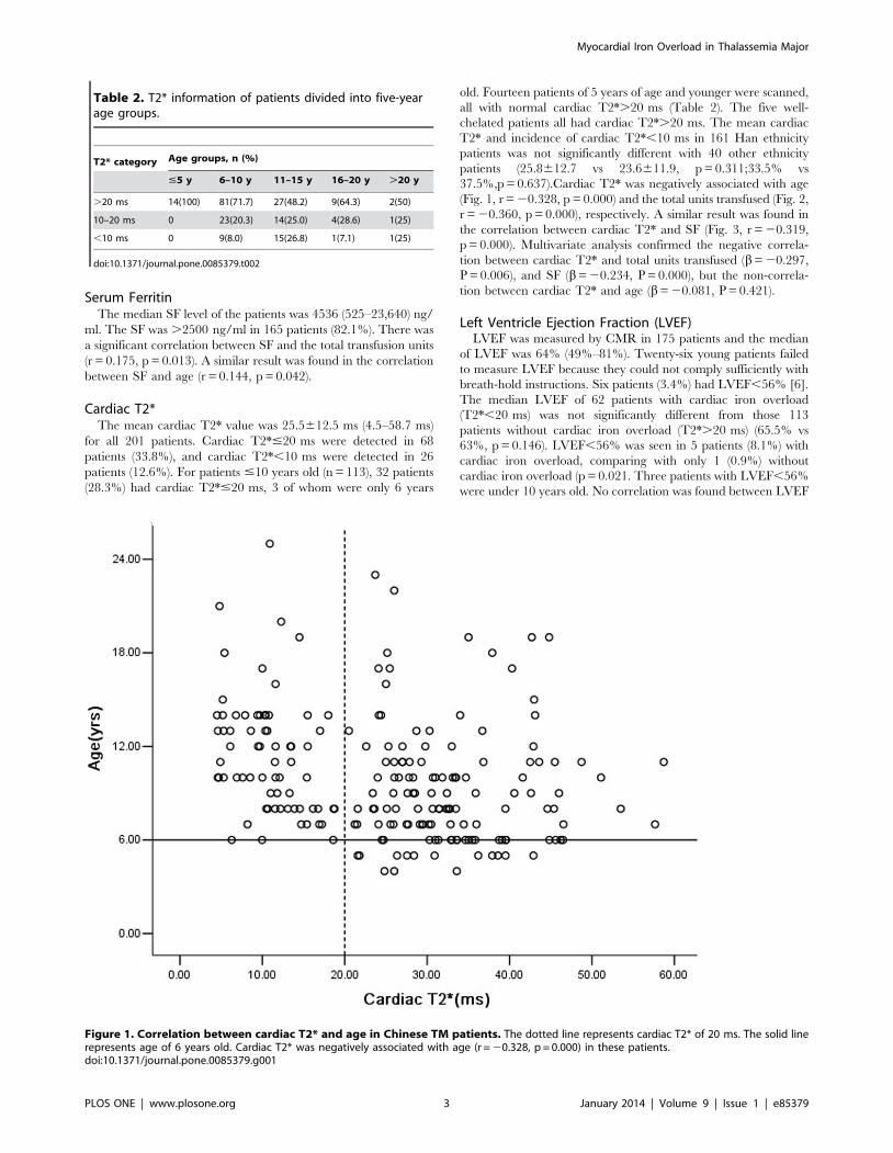
(28.3%) had cardiac T2*#20 ms, 3 of whom were only 6 years
old. Fourteen patients of 5 years of age and younger were scanned,
all with normal cardiac T2*.20 ms (Table 2). The five well-
chelated patients all had cardiac T2*.20 ms. The mean cardiac
T2* and incidence of cardiac T2*,10 ms in 161 Han ethnicity
patients was not significantly different with 40 other ethnicity
patients (25.8612.7 vs 23.6611.9, p = 0.311;33.5% vs
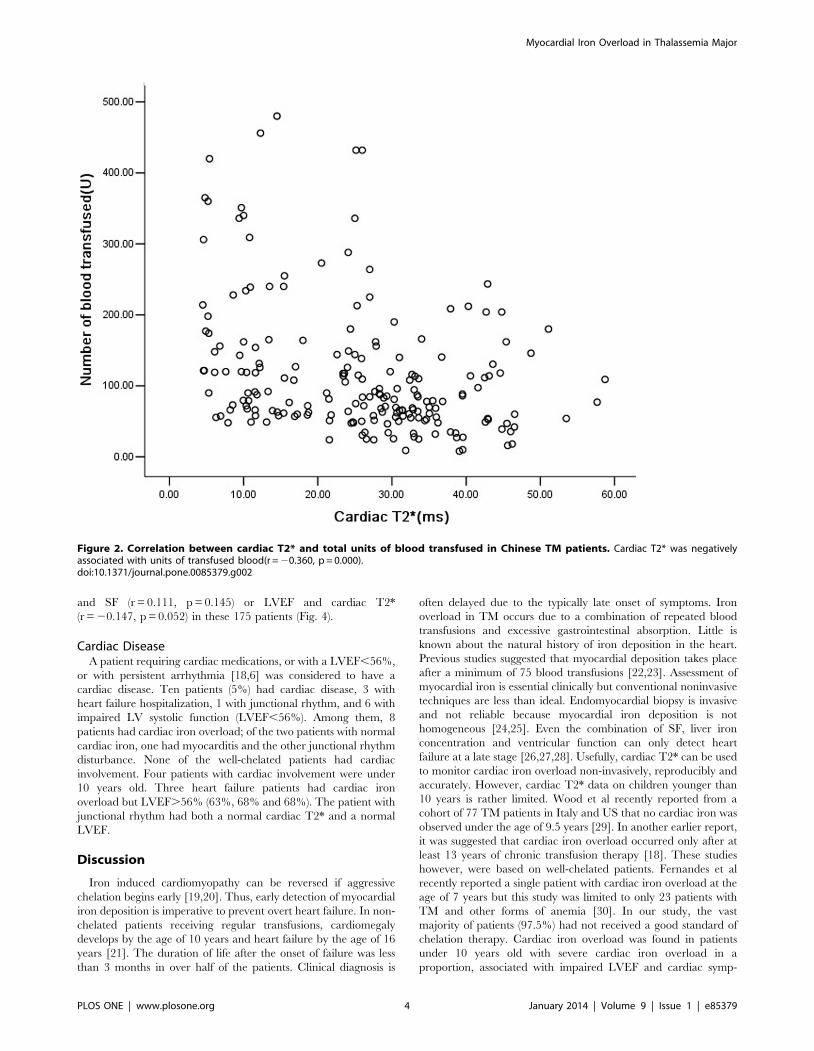
37.5%,p = 0.637).Cardiac T2* was negatively associated with age
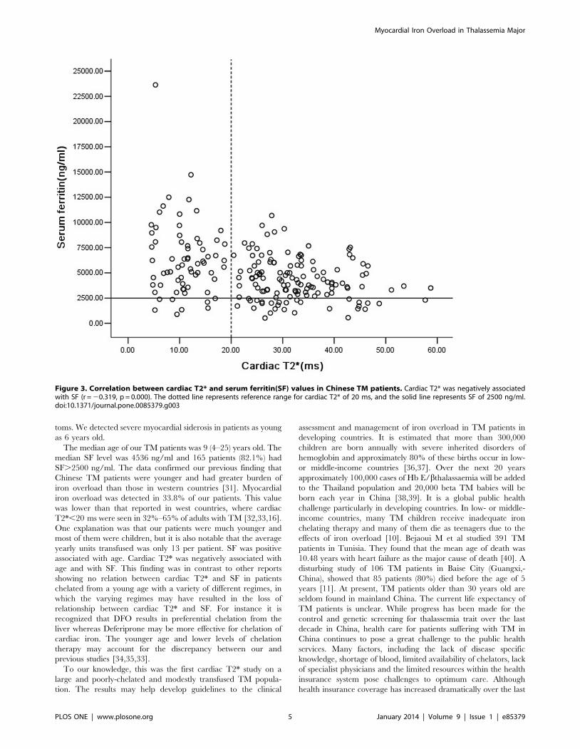
(Fig. 1, r = 20.328, p = 0.000) and the total units transfused (Fig. 2,
r = 20.360, p = 0.000), respectively. A similar result was found in
the correlation between cardiac T2* and SF (Fig. 3, r = 20.319,
p = 0.000). Multivariate analysis confirmed the negative correla-
tion between cardiac T2* and total units transfused (b= 20.297,
P = 0.006), and SF (b= 20.234, P = 0.000), but the non-correla-
tion between cardiac T2* and age (b= 20.081, P = 0.421).
Left Ventricle Ejection Fraction (LVEF)LVEF was measured by CMR in 175 patients and the median
of LVEF was 64% (49%–81%). Twenty-six young patients failed
to measure LVEF because they could not comply sufficiently with
breath-hold instructions. Six patients (3.4%) had LVEF,56% [6].
The median LVEF of 62 patients with cardiac iron overload
(T2*,20 ms) was not significantly different from those 113
patients without cardiac iron overload (T2*.20 ms) (65.5% vs
63%, p = 0.146). LVEF,56% was seen in 5 patients (8.1%) with
cardiac iron overload, comparing with only 1 (0.9%) without
cardiac iron overload (p = 0.021. Three patients with LVEF,56%
were under 10 years old. No correlation was found between LVEF
Figure 1. Correlation between cardiac T2* and age in Chinese TM patients. The dotted line represents cardiac T2* of 20 ms. The solid linerepresents age of 6 years old. Cardiac T2* was negatively associated with age (r = 20.328, p = 0.000) in these patients.doi:10.1371/journal.pone.0085379.g001
Table 2. T2* information of patients divided into five-yearage groups.
T2* category Age groups, n (%)
#5 y 6–10 y 11–15 y 16–20 y .20 y
.20 ms 14(100) 81(71.7) 27(48.2) 9(64.3) 2(50)
10–20 ms 0 23(20.3) 14(25.0) 4(28.6) 1(25)
,10 ms 0 9(8.0) 15(26.8) 1(7.1) 1(25)
doi:10.1371/journal.pone.0085379.t002
Myocardial Iron Overload in Thalassemia Major
PLOS ONE | www.plosone.org 3 January 2014 | Volume 9 | Issue 1 | e85379
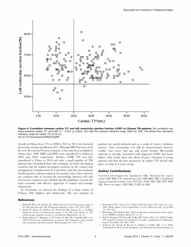
and SF (r = 0.111, p = 0.145) or LVEF and cardiac T2*
(r = 20.147, p = 0.052) in these 175 patients (Fig. 4).
Cardiac DiseaseA patient requiring cardiac medications, or with a LVEF,56%,
or with persistent arrhythmia [18,6] was considered to have a
cardiac disease. Ten patients (5%) had cardiac disease, 3 with
heart failure hospitalization, 1 with junctional rhythm, and 6 with
impaired LV systolic function (LVEF,56%). Among them, 8
patients had cardiac iron overload; of the two patients with normal
cardiac iron, one had myocarditis and the other junctional rhythm
disturbance. None of the well-chelated patients had cardiac
involvement. Four patients with cardiac involvement were under
10 years old. Three heart failure patients had cardiac iron
overload but LVEF.56% (63%, 68% and 68%). The patient with
junctional rhythm had both a normal cardiac T2* and a normal
LVEF.
Discussion
Iron induced cardiomyopathy can be reversed if aggressive
chelation begins early [19,20]. Thus, early detection of myocardial
iron deposition is imperative to prevent overt heart failure. In non-
chelated patients receiving regular transfusions, cardiomegaly
develops by the age of 10 years and heart failure by the age of 16
years [21]. The duration of life after the onset of failure was less
than 3 months in over half of the patients. Clinical diagnosis is
often delayed due to the typically late onset of symptoms. Iron
overload in TM occurs due to a combination of repeated blood
transfusions and excessive gastrointestinal absorption. Little is
known about the natural history of iron deposition in the heart.
Previous studies suggested that myocardial deposition takes place
after a minimum of 75 blood transfusions [22,23]. Assessment of
myocardial iron is essential clinically but conventional noninvasive
techniques are less than ideal. Endomyocardial biopsy is invasive
and not reliable because myocardial iron deposition is not
homogeneous [24,25]. Even the combination of SF, liver iron
concentration and ventricular function can only detect heart
failure at a late stage [26,27,28]. Usefully, cardiac T2* can be used
to monitor cardiac iron overload non-invasively, reproducibly and
accurately. However, cardiac T2* data on children younger than
10 years is rather limited. Wood et al recently reported from a
cohort of 77 TM patients in Italy and US that no cardiac iron was
observed under the age of 9.5 years [29]. In another earlier report,
it was suggested that cardiac iron overload occurred only after at
least 13 years of chronic transfusion therapy [18]. These studies
however, were based on well-chelated patients. Fernandes et al
recently reported a single patient with cardiac iron overload at the
age of 7 years but this study was limited to only 23 patients with
TM and other forms of anemia [30]. In our study, the vast
majority of patients (97.5%) had not received a good standard of
chelation therapy. Cardiac iron overload was found in patients
under 10 years old with severe cardiac iron overload in a
proportion, associated with impaired LVEF and cardiac symp-
Figure 2. Correlation between cardiac T2* and total units of blood transfused in Chinese TM patients. Cardiac T2* was negativelyassociated with units of transfused blood(r = 20.360, p = 0.000).doi:10.1371/journal.pone.0085379.g002
Myocardial Iron Overload in Thalassemia Major
PLOS ONE | www.plosone.org 4 January 2014 | Volume 9 | Issue 1 | e85379
toms. We detected severe myocardial siderosis in patients as young
as 6 years old.
The median age of our TM patients was 9 (4–25) years old. The
median SF level was 4536 ng/ml and 165 patients (82.1%) had
SF.2500 ng/ml. The data confirmed our previous finding that
Chinese TM patients were younger and had greater burden of
iron overload than those in western countries [31]. Myocardial
iron overload was detected in 33.8% of our patients. This value
was lower than that reported in west countries, where cardiac
T2*,20 ms were seen in 32%–65% of adults with TM [32,33,16].
One explanation was that our patients were much younger and
most of them were children, but it is also notable that the average
yearly units transfused was only 13 per patient. SF was positive
associated with age. Cardiac T2* was negatively associated with
age and with SF. This finding was in contrast to other reports
showing no relation between cardiac T2* and SF in patients
chelated from a young age with a variety of different regimes, in
which the varying regimes may have resulted in the loss of
relationship between cardiac T2* and SF. For instance it is
recognized that DFO results in preferential chelation from the
liver whereas Deferiprone may be more effective for chelation of
cardiac iron. The younger age and lower levels of chelation
therapy may account for the discrepancy between our and
previous studies [34,35,33].
To our knowledge, this was the first cardiac T2* study on a
large and poorly-chelated and modestly transfused TM popula-
tion. The results may help develop guidelines to the clinical
assessment and management of iron overload in TM patients in
developing countries. It is estimated that more than 300,000
children are born annually with severe inherited disorders of
hemoglobin and approximately 80% of these births occur in low-
or middle-income countries [36,37]. Over the next 20 years
approximately 100,000 cases of Hb E/bthalassaemia will be added
to the Thailand population and 20,000 beta TM babies will be
born each year in China [38,39]. It is a global public health
challenge particularly in developing countries. In low- or middle-
income countries, many TM children receive inadequate iron
chelating therapy and many of them die as teenagers due to the
effects of iron overload [10]. Bejaoui M et al studied 391 TM
patients in Tunisia. They found that the mean age of death was
10.48 years with heart failure as the major cause of death [40]. A
disturbing study of 106 TM patients in Baise City (Guangxi,-
China), showed that 85 patients (80%) died before the age of 5
years [11]. At present, TM patients older than 30 years old are
seldom found in mainland China. The current life expectancy of
TM patients is unclear. While progress has been made for the
control and genetic screening for thalassemia trait over the last
decade in China, health care for patients suffering with TM in
China continues to pose a great challenge to the public health
services. Many factors, including the lack of disease specific
knowledge, shortage of blood, limited availability of chelators, lack
of specialist physicians and the limited resources within the health
insurance system pose challenges to optimum care. Although
health insurance coverage has increased dramatically over the last
Figure 3. Correlation between cardiac T2* and serum ferritin(SF) values in Chinese TM patients. Cardiac T2* was negatively associatedwith SF (r = 20.319, p = 0.000). The dotted line represents reference range for cardiac T2* of 20 ms, and the solid line represents SF of 2500 ng/ml.doi:10.1371/journal.pone.0085379.g003
Myocardial Iron Overload in Thalassemia Major
PLOS ONE | www.plosone.org 5 January 2014 | Volume 9 | Issue 1 | e85379
decade in China, from 15% in 2000 to 96% in 2011, the financial
protection remains insufficient [41]. Although DFO has been used
for over 40 years in Western countries, it has only been available in
China since 1998. DEP and DFX were introduced to China in
2003 and 2010, respectively. Further, CMR T2* was first
introduced to China in 2010 and only a small number of TM
patients have benefitted from this technique. In brief, developing
countries lag far behind developed countries in the control and
management of thalassemia. It is therefore vital that international
health agencies and governments of countries where these diseases
are common start to develop the partnerships between rich and
low-income countries and establish specific guidelines towards the
most economic and effective approach to control and manage
thalassemia.
In conclusion, we present the findings of a large cohort of
Chinese TM children and adolescents. The vast majority of
patients are poorly-chelated and as a result of relative chelation
naivety, clear associations can still be demonstrated between
cardiac iron stores and age and serum ferritin. Myocardial
siderosis is strongly associated with impaired LVEF and heart
failure. Our results show the effects of poor chelation of young
patients and that the first assessment of cardiac T2* should take
place as early as 6 years of age.
Author Contributions
Conceived and designed the experiments: YRL. Performed the experi-
ments: GHY RRL P-P. Analyzed the data: GHY RRL YRL. Contributed
reagents/materials/analysis tools: P-P LLL TGH XHZ WJY SHT HFP
XJL. Wrote the paper: GHY RRL TGH LA YRL.
References
1. Modell B, Khan M, Darlison M. (2000) Survival in beta-thalassaemia major in
the UK: data from the UK Thalassaemia Register. Lancet 355: 2051–2052.
2. Modell B, Khan M, Darlison M, Westwood MA, Ingram D, et al.(2008)
Improved survival of thalassaemia major in the UK and relation to T2*
cardiovascular magnetic resonance. J Cardiovasc Magn Reson 10: 42.
3. Borgna-Pignatti C, Rugolotto S, De Stefano P, Zhao H, Cappellini MD, et al.
(2004) Survival and complications in patients with thalassemia major treated
with transfusion and deferoxamine. Haematologica 89: 1187–1193.
4. Kremastinos DT, Tsetsos GA, Tsiapras DP, Karavolias GK, Ladis VA, et al.
(2001) Heart failure in beta thalassemia: a 5-year follow-up study. Am J Med
111: 349–354.
5. Brittenham GM, Badman DG. (2003) Noninvasive measurement of iron: report
of an NIDDK workshop. Blood 101: 15–19.
6. Kirk P, Roughton M, Porter JB, Walker JM, Tanner MA, et al. (2009) Cardiac
T2* magnetic resonance for prediction of cardiac complications in thalassemia
major. Circulation 120: 1961–1968.
7. Anderson LJ, Wonke B, Prescott E, Holden S, Walker JM, et al. (2002)
Comparison of effects of oral deferiprone and subcutaneous desferrioxamine on
Figure 4. Correlation between cardiac T2* and left ventricular ejection fraction (LVEF) in Chinese TM patients. No correlation wasfound between cardiac T2* and LVEF (r = 20.147, p = 0.052). The solid line presents reference range (56%) for LVEF. The dotted line representsreference range for cardiac T2* of 20 ms.doi:10.1371/journal.pone.0085379.g004
Myocardial Iron Overload in Thalassemia Major
PLOS ONE | www.plosone.org 6 January 2014 | Volume 9 | Issue 1 | e85379
myocardial iron concentrations and ventricular function in beta-thalassaemia.
Lancet 360: 516–520.8. Tanner MA, Galanello R, Dessi C, Smith GC, Westwood MA, et al. (2007) A
randomized, placebo-controlled, double-blind trial of the effect of combined
therapy with deferoxamine and deferiprone on myocardial iron in thalassemiamajor using cardiovascular magnetic resonance. Circulation 115: 1876–1884.
9. Guidelines for clinical management of thalassaemia 2ND Revised Edition (2008)pp. 86.
10. Angastiniotis M, Modell B (1998) Global epidemiology of hemoglobin disorders.
Ann N Y Acad Sci 850: 251–269.11. Pan HF, Long GF, Li Q, Feng YN, Lei ZY, et al. (2007) Current status of
thalassemia in minority populations in Guangxi, China. Clin Genet 71: 419–426.
12. Aessopos A, Farmakis D, Hatziliami A, Fragodimitri C, Karabatsos F, et al.(2004) Cardiac status in well-treated patients with thalassemia major.
Eur J Haematol 73: 359–366.
13. Guidelines for clinical management of thalassaemia 2ND Revised Edition (2008)pp. 61–63.
14. Westwood M, Anderson LJ, Firmin DN, Gatehouse PD, Charrier CC, et al.(2003) A single breath-hold multiecho T2* cardiovascular magnetic resonance
technique for diagnosis of myocardial iron overload. J Magn Reson Imaging 18:
33–39.15. Maceira AM, Prasad SK, Khan M, Pennell DJ (2006) Normalized left
ventricular systolic and diastolic function by steady state free precessioncardiovascular magnetic resonance. J Cardiovasc Magn Reson 8: 417–426.
16. Alpendurada F, Carpenter JP, Deac M, Kirk P, Walker JM, et al. (2010)Relation of myocardial T2* to right ventricular function in thalassaemia major.
Eur Heart J 31: 1648–1654.
17. He T, Zhang J, Carpenter JP, Feng Y, Smith GC, et al. (2013) Automatedtruncation method for myocardial T2* measurement in thalassemia. J Magn
Reson Imaging 37: 479–483.18. Wood JC, Tyszka JM, Carson S, Nelson MD, Coates TD. (2004) Myocardial
iron loading in transfusion-dependent thalassemia and sickle cell disease. Blood
103: 1934–1936.19. Aldouri MA, Wonke B, Hoffbrand AV, Flynn DM, Ward SE, et al. (1990) High
incidence of cardiomyopathy in beta-thalassaemia patients receiving regulartransfusion and iron chelation: reversal by intensified chelation. Acta Haematol
84: 113–117.20. Tanner MA, Galanello R, Dessi C, Smith GC, Westwood MA, et al. (2008)
Combined chelation therapy in thalassemia major for the treatment of severe
myocardial siderosis with left ventricular dysfunction. J Cardiovasc Magn Reson10: 12.
21. Engle MA EM, Ch S. (1964) Late cardiac complictions of chronic, severe,refractory anemia with hemochromatosis. Circulation 30: 698–705.
22. Wood JC. (2008) Cardiac iron across different transfusion-dependent diseases.
Blood Rev 22 Suppl 2: S14–21.23. Tziomalos K, Perifanis V. (2010) Liver iron content determination by magnetic
resonance imaging. World J Gastroenterol 16: 1587–1597.24. Fitchett DH, Coltart DJ, Littler WA, Leyland MJ, Trueman T, et al. (1980)
Cardiac involvement in secondary haemochromatosis: a catheter biopsy studyand analysis of myocardium. Cardiovasc Res 14: 719–724.
25. Barosi G, Arbustini E, Gavazzi A, Grasso M, Pucci A. (1989) Myocardial iron
grading by endomyocardial biopsy. A clinico-pathologic study on iron
overloaded patients. Eur J Haematol 42: 382–388.
26. Brittenham GM, Griffith PM, Nienhuis AW, McLaren CE, Young NS, et al.
(1994) Efficacy of deferoxamine in preventing complications of iron overload in
patients with thalassemia major. N Engl J Med 331: 567–573.
27. Olivieri NF, Nathan DG, MacMillan JH, Wayne AS, Liu PP, et al. (1994)
Survival in medically treated patients with homozygous beta-thalassemia.
N Engl J Med 331: 574–578.
28. Davis BA, O’Sullivan C, Jarritt PH, Porter JB. (2004) Value of sequential
monitoring of left ventricular ejection fraction in the management of thalassemia
major. Blood 104: 263–269.
29. Wood JC, Origa R, Agus A, Matta G, Coates TD, Galanello R. (2008) Onset of
cardiac iron loading in pediatric patients with thalassemia major. Haematologica
93: 917–920.
30. Fernandes JL, Fabron A Jr, Verissimo M. (2009) Early cardiac iron overload in
children with transfusion-dependent anemias. Haematologica 94: 1776–1777.
31. Yong-rong Lai, Chun-fu Li, Shao-liang Huang, Qi-Li, Zhi-xiang Shen, et al.
(2013) Efficacy of deferasirox for the treatment of iron overload in Chinese
thalassemia major patients: results from a prospective, open-label, multicenter
clinical trial. Transfus Med 2013 Dec;23(6):389–96. doi: 10.1111/tme.12077.
Epub 2013 Oct 23.
32. Tanner MA, Galanello R, Dessi C, Westwood MA, Smith GC, et al. (2006)
Myocardial iron loading in patients with thalassemia major on deferoxamine
chelation. J Cardiovasc Magn Reson 8: 543–547.
33. Positano V, Pepe A, Santarelli MF, Ramazzotti A, Meloni A, et al. (2009)
Multislice multiecho T2* cardiac magnetic resonance for the detection of
heterogeneous myocardial iron distribution in thalassaemia patients. NMR
Biomed 22: 707–715.
34. Anderson LJ, Holden S, Davis B, Prescott E, Charrier CC, et al. (2001)
Cardiovascular T2-star (T2*) magnetic resonance for the early diagnosis of
myocardial iron overload. Eur Heart J 22: 2171–2179.
35. Leung AW, Chu WC, Lam WW, Lee V, Li CK. (2009) Magnetic resonance
imaging assessment of cardiac and liver iron load in transfusion dependent
patients. Pediatr Blood Cancer 53: 1054–1059.
36. Christianson A HCP, Modell B (2006) March of Dimes Global Report on Birth
Defects. New York, NY: March of Dimes Birth Defects Foundation.
37. Modell B, Darlison M. (2008) Global epidemiology of haemoglobin disorders
and derived service indicators. Bull World Health Organ 86: 480–487.
38. Weatherall DJ, Clegg JB. (1996) Thalassemia–a global public health problem.
Nat Med 2: 847–849.
39. Weatherall DJ, Clegg JB (2001) The thalassaemia syndromes. Oxford, Blackwell
Science.
40. Bejaoui M, Guirat N. (2013) Beta thalassemia major in a developing country:
epidemiological, clinical and evolutionary aspects. Mediterr J Hematol Infect Dis
5: e2013002.
41. Meng Q, Xu L, Zhang Y, Qian J, Cai M, et al. (2012) Trends in access to health
services and financial protection in China between 2003 and 2011: a cross-
sectional study. Lancet 379: 805–814.
Myocardial Iron Overload in Thalassemia Major
PLOS ONE | www.plosone.org 7 January 2014 | Volume 9 | Issue 1 | e85379