Embed Size (px)
Citation preview
Folia Biologica (Praha) 62, 95-102 (2016)
Original Article
Iron Overload Causes Alterations of E-Cadherin in the Liver(ironoverload/E-cadherin/liver/adherensjunctions)
Y. FUJIKURA1,3, J. KRIJT1, C. POVÝŠIL2,Z.MĚLKOVÁ3,P.PŘIKRYL1, M. VOKURKA1,E.NEČAS1
1Institute of Pathophysiology, 2Institute of Pathology, 3Institute of Immunology and Microbiology, First Faculty of Medicine, Charles University in Prague, Czech Republic
Abstract. Iron overload causes tissue damage in the liver, but its initial effects at the molecular and cellu-lar level are not well understood. Epithelial cadherin (E-cad) is a major adhesion protein in adherens junctions and is associated with several signal trans-duction pathways. Dysfunction of E-cad causes in-stability of adherens junctions, which leads to cell in-vasion, cell migration, and carcinogenesis. We found in liver samples from iron-overloaded mice that the apparent molecular mass of E-cad was reduced from 125 to 115 kDa in sodium dodecyl sulphate poly-acrylamide gel electrophoresis under reducing con-ditions and immunoblotting, and that the cellular expression of E-cad was decreased in immunohisto-chemistry. The mRNA level of E-cad, however, did not change significantly, suggesting that the altera-tions are posttranslational. Interestingly, incubation of control liver extracts with Fe2+ alone also produced the same mobility shift. Neither an oxidant nor an antioxidant influenced this shift in vitro, suggesting that reactive oxygen species, which are generated by iron and known to cause damage to macromolecules, are not involved. Treatment of the 115 kDa E-cad with deferoxamine, an iron chelator, thus removing Fe2+, shifted the molecular mass back to 125 kDa, demonstrating that the shift is reversible. The obser-vation also implies that the alteration that causes the mobility shift is not due to transcriptional control,
ReceivedJune25,2015.AcceptedMarch23,2016.
ThisworkwassupportedbythegrantsPRVOUKP24/LF1/3andSVV2014-260033fromCharlesUniversityinPrague,CzechRe-public.
Corresponding author:YuzoFujikura, Institute of Immunologyand Microbiology, First Faculty of Medicine, Charles University in Prague, Studničkova 7, 12800 Prague 2, Czech RepublicPhone: (+420)224968460;Fax: (+420)224968588; e-mail:[email protected]
Abbreviations:AJs–adherensjunctions,DFO–deferoxamine,E-cad – epithelial cadherin, Hjv+/+ – haemojuvelin wild type, Hjv–/– –haemojuvelinknockout,LB–lysisbuffer,ROS–reactiveoxy-gen species.
deglycosylation, and proteolysis. This reversible mo-bility shift of E-cad has not been previously known. The alteration of E-cad that causes the mobility shift might be an initial step to liver diseases by iron over-load.
IntroductionPrimary iron overload is a genetic disorder of iron
metabolism causing inappropriately increased iron ab-sorption due to a defect in the hepcidin signalling path-wayasseeninhereditaryhaemochromatosis;secondary(acquired)ironoverloadmostoftendevelopsasacom-plication of repeated transfusions and is associated with cardiomyopathy and chronic liver diseases (Pietrangelo, 2003;SebastianiandPantopoulos,2011).Ironitself isnot carcinogenic. However, iron overload is associated with tissue damage and carcinogenesis. Primarily iron overload causes fibrosis, most frequently in the liver,heart,andpancreas.Theliverfibrosisduetoironexcessevolves into cirrhosis and may eventually lead to the de-velopment of hepatocellular carcinoma. In pathological iron overload, iron gradually saturates the iron-binding capacity of transferrin, which under physiological con-ditionsmaintains iron soluble and nontoxic (Ponka etal., 1998), and non-transferrin-bound iron eventuallygets internalized into cells. Iron’s toxicity is largelybasedonitsabilitytogeneratereactiveoxygenspecies(ROS)(HalliwellandGutteridge,1990),anincreaseofwhichbeyondtheantioxidantcapacityoftheorganismcausesoxidativestress(PapanikolaouandPantopoulos,2005).Althoughiron-inducedchronicoxidativestressisknown to cause damage to macromolecules, the mecha-nisms of initial tissue damage triggered by iron overload are not well understood.
Cell adhesion proteins cadherins are single-pass trans-membrane glycoproteins that are built on the contacts betweenadjacentcells.Extracellulardomainsofcadher-ins on the opposing membranes mediate Ca2+-dependent homophilic interaction and cytoplasmic domains form a complexwithcatenins(α-,β-,γ-,andp120),whichin-teractwithactinfilamentsandmicrotubules(MengandTakeichi, 2009). In this way cadherins, together with
96 Vol.62
several adaptor proteins, functionally link the cytoskele-tonofadjacentcells(Taguchietal.,2011)andcontributeto cell-cell adhesion to form mature adherens junctions (AJs). E-cad is the major cadherin in liver epithelialcellsandisdirectlyboundbyβ-andp120catenin.Thebinding of the catenins masks sequences containing the motif of the E-cad degradation pathway (Huber et al., 2001;MiyashitaandOzawa,2007;Nanesetal.,2012).Some of cell surface E-cad is constitutively endocytosed to regulate cadherin-based adhesion, through which E-cad isdegradedby lysosomeand/orproteasome,orrecycledbacktotheplasmamembrane(Leetal.,1999;Davisetal.,2003;Xiaoetal.,2003).
Inactivation of the E-cad cell adhesion system causes organfibrosisduetothereducedcell-celladhesiveness(Hirohashi, 1998;Guarino et al., 2009).A correlationbetween the cellular expression of the E-cadherin/catenincomplexandthedevelopmentandprogressionof hepatocellular carcinoma has been reported (Zhai et al.,2008).E-cad-mediatedcelladhesionisknowntobeinactivated by genetic alterations and transcriptional re-pression(Berxetal.,1998;Alvesetal.,2009).However,it has been suggested that E-cad dysfunction cannot be explainedatthegenetic/epigeneticlevelaloneandthatother mechanisms may operate at the posttranslational level(Pinhoetal.,2011)inresponsetochangesinexter-nal/internalsignals.
While investigating membrane proteins in the liver, we noticed that the intensity and mobility of cadherin bands were altered when the iron content of samples was high. Since the direct effect of iron on E-cad in the liver has not been fully elucidated, we investigated E-cad in the liver from iron-overloaded mice, in order to detect possible initial alterations of E-cad by iron overload. Liver samples were analysed at the molecular and cel-lular level by immunoblotting and immunohistochemis-try, respecti vely. The detected alterations may provide better understanding of the initiation of liver diseases by iron overload.
Material and Methods
Animals
Haemojuvelin wild type (Hjv+/+)andknockout(Hjv–/–, a model of juvenile haemochromatosis) mice were agenerous gift from Prof. Silvia Arber, Basel, Switzerland (Niederkofleretal.,2005).Iron-overloadedmiceoftheC57BL/6strainwerepreparedbyinjectionofirondex-tran(Sigma-Aldrich,StLouis,MO)at200mgiron/kg,and after three days liver samples were obtained. All animalexperimentswereapprovedbytheEthicsCom-mittee of the First Faculty of Medicine, Charles Uni ver-sity in Prague.
Protein extraction Liverproteinwasextracted in lysisbuffer (LB)de-
scribedbyReynoldsetal.(1994):20mMTris-HClpH7.4 containing 0.5% NP-40, 150 mM NaCl, 1 mM
EDTA, Protease Inhibitor Cocktail (Roche, Basel, Swit-zerland), andPhosphatase InhibitorCocktails 2 and3(Sigma-Aldrich). Extracts were obtained by homoge-nizing20–50mgoftissuein250μlofLBandsubse-quentcentrifugationat14,000gfor15minat4°C.Forfractionation,proteinwasextractedin20mMTris-HClpH6.8containing0.28Msucrose,50mMNaCl,2mMEDTA, and the protease and phosphatase inhibitor cock-tails.The homogenatewas centrifuged at 6,000g for 15mintoremovelargemembranefragments(P1),andthesupernatantwascentrifugedat16,000gfor45min.The pellet that contains membrane proteins was saved (P2)andthesupernatantwassubjectedtoultracentrifu-gationat80,000g for 55 min. The pellet that contains endosomes (Hoshino et al., 2005)was saved (P3).P2andP3werewashedonceanddissolvedinLB.Forthedose response tests, LB extracts of the liver fromC57BL/6micesubjectedtodifferentdosesofiron,200,350,and700mg/kg,wereprepared.Proteinconcentra-tionwasmeasuredbyBradfordreagent(Sigma-Aldrich)with BSA as the standard. Other chemicals were ob-tained from Sigma-Aldrich.
ImmunoblottingProteins,50or60μgofeachsample,wereseparated
in6or8%SDS-PAGEunderreducingconditionsandtransferred to PVDF membrane (GE Healthcare, Little Chalfont,UK)byelectroblotting.The standardproce-dureusingTBScontaining0.1%Tween-20(TBS/T)forwashing,5%(w/v)non-fatmilkinTBS/Tforblocking,16hincubationat4°Cwiththerabbitprimaryantibody(E-cad: 1 : 3000,CellSignalingTechnology, #3195,Danvers,MA;actin:1 : 8000,Sigma-Aldrich)inTBS/Tand2hincubationatroomtemperaturewiththeperoxi-dase-conjugatedanti-rabbitsecondaryantibody(1 : 1000,JacksonImmunoResearch,WestGrove,PA)inTBS/Twas employed. Positive signals were detected by the chemiluminescence method (Cell Signaling Techno lo-gy).BenchMarkProteinLadder(Invitrogen,Carlsbad,CA)wasusedtoestimatethemolecularmassofproteinbands. The immunoblots are representatives of at least twoindependentexperiments.
mRNA measurementLiver RNA was isolated using a Qiagen RNeasy Plus
MiniKit (QiagenGmbH,Stockach,Germany).cDNAwas synthesized using a Fermentas RevertAid First StrandcDNASynthesisKit(ThermoFisherScientific,Waltham, MA). For real-time PCR determination ofE-cad gene (Cdh1)expression,aRocheLightCyclerin-strument in combination with the Fast Start SYBR Green protocol (Roche Diagnostics GmBH, Mannheim, Germany)wasused.TargetmRNAcontentwascalculat-ed relative to GAPDH mRNA content. Primers (forward andreverse)wereasfollows:Gapdh, CGGTGTGAA-CGGATTTGC and GCAGTGATGGCATGGACTGT, and Cdh1, CCATGTGTGTGACTGTGAAGG and CAGCTGGCTCAAATCAA AGTC.
Y. Fujikura et al.
Vol.62 97
Fe2 + in vitro assays
Extractscontaining50μgofproteininLBweresub-jectedtothetreatmentin50μMHepespH7.5contain-ing100mMNaCl,2mMDTT,andtheproteaseinhibi-torcocktailatthefinalvolumeof20μl.Fordetectionofthe effects of Fe2+, extractswereincubatedwithFeSO4 at 1mM for 30min at 30 °C. For the dose responsecheck,controlextractsweretreatedwithdifferentcon-centrations of FeSO4. For H2O2 assay,controlextractswere incubated with different concentrations of H2O2 for30minat30°C.ForN-acetylcysteine,glutathione,anddithiothreitolassays,controlextractswerefirstin-cubated with the agent for 5 min, and then FeSO4 was addedat1mM,followedby30minincubationat30°C.Deferoxamine (DFO) was added to the extracts after30minincubationwithFeSO4at1mMat30°C,andtheextracts were further incubated for 30 min. ExtractsfromC57BL/6iron-overloadmicegeneratedbyinjec-tionofirondextran(700mg/kgbodyweight)werealsotested for the DFO treatment. Since phosphorylation maybeinvolvedintheshift,LBextractsthatcontainedphosphatase inhibitors were diluted more than ten times intheassayssothattheinfluenceoftheinhibitorswasnegligible. All the reagents were obtained from Sigma-Aldrich.
ImmunohistochemistryLiversampleswerefixedinformalinandembedded
inparaffin,fromwhichhistologicalsectionswerecol-lected. For protein detection, sections were dewaxedandincubatedwithmethanolcontaining30%H2O2 for 20min to block endogenous peroxidase activity. Sec-tions were then immersed in 10 mM citrate buffer pH 6.0andheatedinamicrowaveovenat100°Cfor20minto enhance antigen retrieval, washed three times with distilledwater,andblockedwith1%BSAfor30min.Subsequently,sectionswereincubatedovernightat4°Cwithantibody,firstwithantiE-cad(1 : 400,CellSigna-lingandTechnology), thenwith theperoxidase-conju-gatedsecondaryantibody(1 : 500,JacksonImmunoRe-search) for 30 min at 37 °C. Finally, sections werewashed three times with PBS buffer and the colour was displayed with DAB. Nuclei were lightly counterstained withhaematoxylin.Perl’s iron stainmethodwas usedfor detection of iron deposition.
Results
Mobility shift of E-cadherin in the liver of iron-overloaded mice
First, we compared the livers from Hjv+/+ mice (liver ironcontentlessthan100μg/gwetweight)withthosefrom Hjv–/– mice (liver iron content approximately2000μg/g wet weight), a murine model of juvenilehaemochromatosis(Niederkofleretal.,2005).Liverex-tracts were prepared in LB and analysed by immuno-blotting. In the control sample, the E-cad band was de-
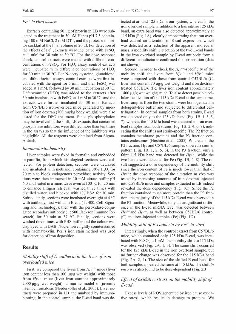
tectedataround125kDainoursystem,whereasintheironoverloadsample,inadditiontoalessintense125kDaband,anextrabandwasalsodetectedapproximatelyat115kDa(Fig.1A),clearlydemonstratingthatironover-load caused an alteration of E-cad expression, whichwas detected as a reduction of the apparent molecular mass, a mobility shift. Detection of the two E-cad bands in the iron overload sample by E-cad antibody from a differentmanufacturerconfirmedtheobservation(datanotshown).
Second, in order to check the Hjv–/–-specificityofthemobility shift, the livers from Hjv+/+ and Hjv–/– mice were comparedwith those from controlC57BL/6 (C,liverironcontent70μg/gwetweight)andirondextran-treatedC57BL/6(Fe, liver ironcontentapproximately1400μg/gwetweight)mice.Toalsodetectpossiblecel-lular localization of the 115 kDa E-cad at the same time, liver samples from the two strains were homogenized in detergent-free buffer and subjected to differential cen-trifugation. In control samples from both strains, E-cad wasdetectedonlyasthe125kDaband(Fig.1B.1,3,5,7),whereasthe115kDabandwasdetectedinironover-loadsamplesfrombothstrains(Fig.1B.2,4,6,8),indi-catingthattheshiftisnotstrain-specific.TheP2fractioncontainsmembrane proteins and the P3 fraction con-tainsendosomes(Hoshinoetal.,2005).WhereasintheP2fraction,HjvandC57BL/6samplesshowedasimilarpattern(Fig.1B.1,2,5,6), in theP3fraction,onlyafaint 115 kDa band was detected for Hjv–/–, while the twobandsweredetectedforFe(Fig.1B,4,8).There-sult suggested a dose dependency of the mobility shift since the iron content of Fe is much lower than that of Hjv–/–; the dose response of the alteration in vivo was tested by increasing amounts of iron dextran injectedintoC57BL/6miceandsamplesextractedinLBindeedrevealed thedosedependency (Fig.1C).Since theP2fractioncontainedmuchmoreproteinthantheP3frac-tion, the majority of the 115 kDa E-cad was observed in theP2fraction.Meanwhile,onlyaninsignificantdiffer-ence in the E-cad mRNA level was detected between Hjv+/+and Hjv–/–, as well as between C57BL/6 control(C)andiron-injectedsamples(Fe)(Fig.1D).
Mobility shift of E-cadherin by Fe2+ in vitroInterestingly,whenthecontrolextractfromC57BL/6
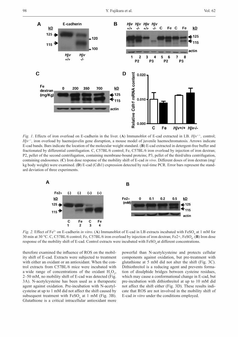
mice,whichcontainedonly125kDaE-cad,wasincu-bated with FeSO4 at 1 mM, the mobility shift to 115 kDa wasobserved(Fig.2A.1,3).Thesameshiftoccurredforthe125kDaE-cadintheironoverloadsample,butno further change was observed for the 115 kDa band (Fig.2A.2,4).Thesizeof theshiftedE-cadbandforboth samples appeared the same at 115 kDa. The shift in vitrowasalsofoundtobedose-dependent(Fig.2B).
Effect of oxidative stress on the mobility shift of E-cad
ExcesslevelsofROSgeneratedbyironcauseoxida-tive stress, which results in damage to proteins. We
Effects of Iron Overload on E-Cadherin
98 Vol.62
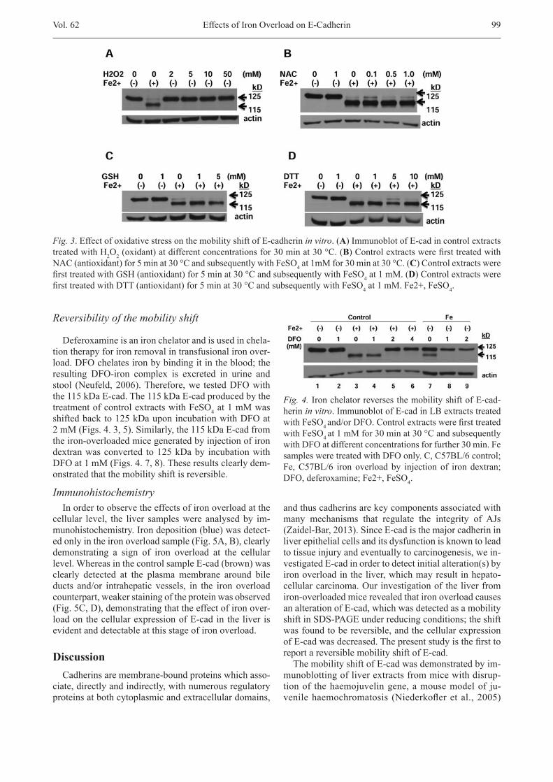
thereforeexaminedtheinfluenceofROSonthemobil-ityshiftofE-cad.Extractsweresubjectedtotreatmentwitheitheranoxidantoranantioxidant.Whenthecon-trol extracts fromC57BL/6micewere incubatedwithawide range of concentrations of the oxidant H2O2, 2–50mM,nomobilityshiftofE-cadwasdetected(Fig.3A). N-acetylcysteine has been used as a therapeuticagentagainstoxidation.Pre-incubationwithN-acetyl-cysteine at up to 1 mM did not affect the shift caused by subsequent treatment with FeSO4 at 1mM (Fig. 3B).Glutathione is a critical intracellular antioxidantmore
powerful than N-acetylcysteine and protects cellular components against oxidation, but pre-treatment withglutathione at 5mMdid not alter the shift (Fig. 3C).Dithiothreitol is a reducing agent and prevents forma-tion of disulphide bridges between cysteine residues, which may cause a conformational change in E-cad, but pre-incubation with dithiothreitol at up to 10 mM did notaffecttheshifteither(Fig.3D).Theseresultsindi-cate that ROS are not involved in the mobility shift of E-cad in vitro under the conditions employed.
Fig. 1. Effects of iron overload on E-cadherin in the liver. (A)ImmunoblotofE-cadextractedinLB.Hjv+/+,control;Hjv–/–, iron overload by haemojuvelin gene disruption, a mouse model of juvenile haemochromatosis. Arrows indicate E-cad bands. Bars indicate the location of the molecular weight standard. (B)E-cadextractedindetergent-freebufferandfractionatedbydifferentialcentrifugation.C,C57BL/6control;Fe,C57BL/6ironoverloadbyinjectionofirondextran;P2,pelletofthesecondcentrifugation,containingmembrane-boundproteins;P3,pelletofthethird/ultracentrifugation,containing endosomes. (C)IrondoseresponseofthemobilityshiftofE-cadin vivo.Differentdosesofirondextran(mg/kgbodyweight)wereexamined.(D)E-cad(Cdh1)expressiondetectedbyreal-timePCR.Errorbarsrepresentthestand-arddeviationofthreeexperiments.
Fig. 2. Effect of Fe2+ on E-cadherin in vitro. (A)ImmunoblotofE-cadinLBextractsincubatedwithFeSO4 at 1 mM for 30minat30°C.C,C57BL/6control;Fe,C57BL/6ironoverloadbyinjectionofirondextran;Fe2+,FeSO4. (B)IrondoseresponseofthemobilityshiftofE-cad.ControlextractswereincubatedwithFeSO4at different concentrations.
Y. Fujikura et al.
Vol.62 99
Reversibility of the mobility shift
Deferoxamineisanironchelatorandisusedinchela-tion therapy for iron removal in transfusional iron over-load.DFOchelatesironbybindingitintheblood;theresulting DFO-iron complex is excreted in urine andstool (Neufeld,2006).Therefore,we testedDFOwiththe 115 kDa E-cad. The 115 kDa E-cad produced by the treatmentofcontrolextractswithFeSO4 at 1 mM was shiftedbackto125kDauponincubationwithDFOat2mM(Figs.4.3,5).Similarly,the115kDaE-cadfromthe iron-overloaded mice generated by injection of iron dextranwasconverted to125kDaby incubationwithDFOat1mM(Figs.4.7,8).Theseresultsclearlydem-onstrated that the mobility shift is reversible.
ImmunohistochemistryIn order to observe the effects of iron overload at the
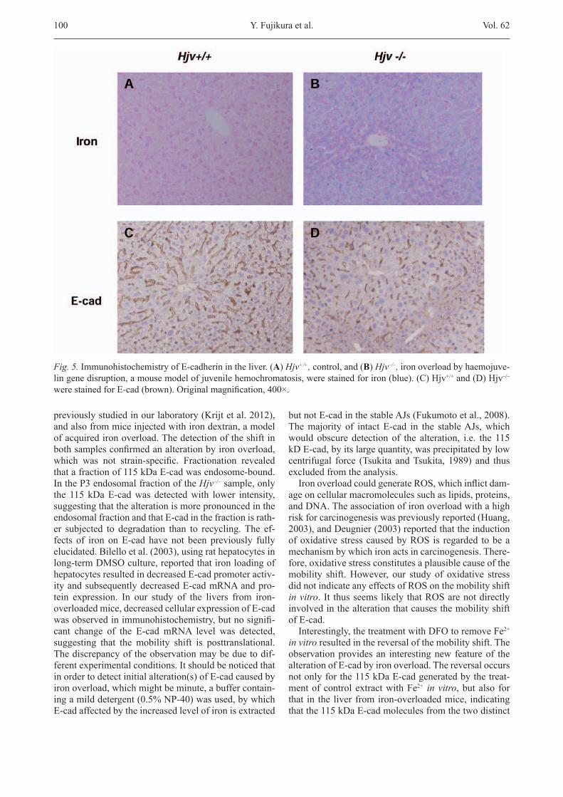
cellular level, the liver samples were analysed by im-munohistochemistry.Irondeposition(blue)wasdetect-edonlyintheironoverloadsample(Fig.5A,B),clearlydemonstrating a sign of iron overload at the cellular level.WhereasinthecontrolsampleE-cad(brown)wasclearly detected at the plasma membrane around bile ducts and/or intrahepatic vessels, in the iron overloadcounterpart, weaker staining of the protein was observed (Fig.5C,D),demonstratingthattheeffectofironover-loadonthecellularexpressionofE-cadintheliver isevident and detectable at this stage of iron overload.
DiscussionCadherins are membrane-bound proteins which asso-
ciate, directly and indirectly, with numerous regulatory proteinsatbothcytoplasmicandextracellulardomains,
and thus cadherins are key components associated with many mechanisms that regulate the integrity of AJs (Zaidel-Bar,2013).SinceE-cadisthemajorcadherininliver epithelial cells and its dysfunction is known to lead to tissue injury and eventually to carcinogenesis, we in-vestigatedE-cadinordertodetectinitialalteration(s)byiron overload in the liver, which may result in hepato-cellular carcinoma. Our investigation of the liver from iron-overloaded mice revealed that iron overload causes an alteration of E-cad, which was detected as a mobility shiftinSDS-PAGEunderreducingconditions;theshiftwasfoundtobereversible,andthecellularexpressionofE-cadwasdecreased.Thepresentstudyisthefirsttoreport a reversible mobility shift of E-cad.
The mobility shift of E-cad was demonstrated by im-munoblottingofliverextractsfrommicewithdisrup-tion of the haemojuvelin gene, a mouse model of ju-venilehaemochromatosis (Niederkofleretal.,2005)
Fig. 3.EffectofoxidativestressonthemobilityshiftofE-cadherinin vitro. (A)ImmunoblotofE-cadincontrolextractstreated with H2O2(oxidant)atdifferentconcentrationsfor30minat30°C.(B)ControlextractswerefirsttreatedwithNAC(antioxidant)for5minat30°CandsubsequentlywithFeSO4at1mMfor30minat30°C.(C)ControlextractswerefirsttreatedwithGSH(antioxidant)for5minat30°CandsubsequentlywithFeSO4 at 1 mM. (D)ControlextractswerefirsttreatedwithDTT(antioxidant)for5minat30°CandsubsequentlywithFeSO4at1mM.Fe2+,FeSO4.
Fig. 4. Iron chelator reverses the mobility shift of E-cad-herin in vitro.ImmunoblotofE-cadinLBextractstreatedwith FeSO4and/orDFO.Controlextractswerefirsttreatedwith FeSO4at1mMfor30minat30°CandsubsequentlywithDFOatdifferentconcentrationsforfurther30min.FesamplesweretreatedwithDFOonly.C,C57BL/6control;Fe,C57BL/6 iron overload by injection of iron dextran;DFO,deferoxamine;Fe2+,FeSO4.
Effects of Iron Overload on E-Cadherin
100 Vol.62
previouslystudiedinourlaboratory(Krijtetal.2012),andalsofrommiceinjectedwithirondextran,amodelof acquired iron overload. The detection of the shift in bothsamplesconfirmedanalterationbyironoverload,which was not strain-specific. Fractionation revealedthat a fraction of 115 kDa E-cad was endosome-bound. IntheP3endosomalfractionoftheHjv–/– sample, only the 115 kDa E-cad was detected with lower intensity, suggesting that the alteration is more pronounced in the endosomal fraction and that E-cad in the fraction is rath-er subjected to degradation than to recycling. The ef-fects of iron on E-cad have not been previously fully elucidated.Bilelloetal.(2003),usingrathepatocytesinlong-term DMSO culture, reported that iron loading of hepatocytes resulted in decreased E-cad promoter activ-ity and subsequently decreased E-cad mRNA and pro-tein expression. In our study of the livers from iron-overloadedmice,decreasedcellularexpressionofE-cadwasobservedinimmunohistochemistry,butnosignifi-cant change of the E-cad mRNA level was detected, suggesting that the mobility shift is posttranslational. The discrepancy of the observation may be due to dif-ferentexperimentalconditions.Itshouldbenoticedthatinordertodetectinitialalteration(s)ofE-cadcausedbyiron overload, which might be minute, a buffer contain-ingamilddetergent(0.5%NP-40)wasused,bywhichE-cadaffectedbytheincreasedlevelofironisextracted
butnotE-cadinthestableAJs(Fukumotoetal.,2008).The majority of intact E-cad in the stable AJs, which would obscure detection of the alteration, i.e. the 115 kD E-cad, by its large quantity, was precipitated by low centrifugal force (Tsukita andTsukita, 1989) and thusexcludedfromtheanalysis.IronoverloadcouldgenerateROS,whichinflictdam-
age on cellular macromolecules such as lipids, proteins, and DNA. The association of iron overload with a high risk for carcinogenesis was previously reported (Huang, 2003),andDeugnier(2003)reportedthattheinductionofoxidativestresscausedbyROSis regarded tobeamechanism by which iron acts in carcinogenesis. There-fore,oxidativestressconstitutesaplausiblecauseofthemobility shift.However, our study of oxidative stressdid not indicate any effects of ROS on the mobility shift in vitro. It thus seems likely that ROS are not directly involved in the alteration that causes the mobility shift of E-cad.
Interestingly, the treatment with DFO to remove Fe2+ in vitro resulted in the reversal of the mobility shift. The observation provides an interesting new feature of the alteration of E-cad by iron overload. The reversal occurs not only for the 115 kDa E-cad generated by the treat-mentofcontrolextractwithFe2+ in vitro, but also for that in the liver from iron-overloaded mice, indicating that the 115 kDa E-cad molecules from the two distinct
Fig. 5. Immunohistochemistry of E-cadherin in the liver. (A)Hjv+/+, control, and (B)Hjv–/–, iron overload by haemojuve-lingenedisruption,amousemodelofjuvenilehemochromatosis,werestainedforiron(blue).(C)Hjv+/+and(D)Hjv–/– werestainedforE-cad(brown).Originalmagnification,400×.
Y. Fujikura et al.
Vol.62 101
samples represent the same polypeptide. Thus, it may also imply that the alteration by iron overload in vivo is reversible. Also, the observation eliminates possible causes for the shift such as transcriptional control, pro-teolysis, and deglycosylation. The reversibility also confirmsnoparticipationofROSintheshift.Therefore,it points toward a conformational change as a possible cause of the shift, and also possible direct involvement of Fe2+inthechange.Toxicmetalcadmiumwasreportedto disrupt cadherin function in the kidney by displacing Ca2+fromitsbindingsitesat theextracellulardomain,possibly causing a conformational change (Pro zialeck et al., 2003). However, displacement of Ca2+ by Fe2+ in cadherin is not known. Meanwhile, it is known that the negativelychargedphosphategrouphashighaffinitytometalions(Machidaetal.,2007).SinceE-cadisphos-phorylated at the normal state and there are several po-tential phosphorylation sites at the C-terminus in the cytoplasmicdomain(StappertandKemler,1994),itispossible that an interaction between the Fe ion and phosphate groups takes place, which could result in a conformational change, and thus the mobility shift. Phosphorylation is known to cause the mobility shift of catenins due to a possible conformational change (Calauttietal.,1998;Fukumotoetal.,2008),andalsotoregulate the function of the cadherin/catenin complex(Zaidel-Bar, 2013), and therefore investigation of thephosphorylationsitesinE-cadwouldbethenextsteptoelucidating the mobility shift.
Immunohistochemistry detected decreased cellular expression of E-cad. Since the mobility shift is notcaused by proteolysis, the antibody should equally de-tectbothformsofE-cad,115and125kDa.Thediffer-ence between the control and the iron overload sample in immunohistochemistry may therefore be due to that either some fraction of the 115 kDa E-cad is degraded, or the epitope is less accessible to the antibody in the 115 kDa E-cad, perhaps due to a conformational change, or both. The vulnerability of the 115 kD E-cad was de-tectedintheP3fraction.Interestingly,theepitopeoftheemployed antibody is located near the potential multiple phosphorylation site at the C-terminus. Thus, the lower accessibility to the antibody cannot be excluded.Theiron dose dependency of the mobility shift, in vitro as well as in vivo, and the decreased cellular expressiondetected by immunohistochemistry suggest that increas-ing amounts of iron would result in more pervasive al-teration of E-cad, and thus more progressed cellular damage.Ourobservationfitswellwiththefindingthatthe intensity of the cadherin signal gradually weakens from non-tumorous tissue to tumour regions in hepato-cellularcarcinomas(Zhaietal.,2008).
The mobility shift is caused by an alteration of E-cad induced by iron overload, which would result in weak-ened cell adhesions in AJs and could eventually lead to the onset of carcinogenesis. Further studies of the altera-tion may therefore provide a new insight into the roles of E-cad in AJs and also contribute to better understand-
ing the pathology caused by iron overload, which could lead to the development of therapeutic methods.
AcknowledgementThe authors are grateful to Dr. Christine Bartels for
critical reading of the manuscript.
ReferencesAlves,C.C.,Carneiro,F.,Hoefler,H.,Becker,K.F.(2009)
Role of the epithelial-mesenchymal transition regulator Slug in primary human cancers. Front. Biosci. 14, 3035-3050.
Berx,G.,Becker,K.F.,Hofler,H.,vanRoy,F.(1998)Muta-tionsofthehumanE-cadherin(CDH1)gene.Hum. Mutat. 12,226-237.
Bilello,J.P.,Cable,E.E.,Isom,H.C.(2003)ExpressionofE-cadherin and other paracellular junction gene is decreased in iron-loaded hepatocytes. Am. J. Pathol. 162, 1323-1338.
Calautti, E., Cabodi, S., Stein, P. L., Hatzfeld, M., Kedersha, N.,Dotto,G.P.(1998)TyrosinephosphorylationandSrcfamily kinases control keratinocyte cell-cell adhesion. J. Cell Biol. 141,1449-1465.
Davis,M.A., Ireton,R.C.,Reynolds,A.B. (2003)A corefunctionforp120-cateninincadherinturnover.J. Cell Biol. 163, 525-534.
Deugnier,Y. (2003) Ironand livercancer.Alcohol 30, 145-150.
Fukumoto, Y., Shintani, Y., Reynolds, A. B., Johnson, K. R., Wheelock,M.J.(2008)Theregulatoryorphosphorylationdomainofp120catenincontrolsE-cadherindynamicsatthe plasma membrane. Exp. Cell Res. 314, 52-67.
Guarino,M.,Tosoni,A.,Nebuloni,M.(2009)Directcontribu-tionofepitheliumtoorganfibrosis:epithelial-mesenchy-mal transition. Hum. Pathol. 40,1365-1376.
Halliwell, B., Gutteridge, J.M. C. (1990) The role of freeradicals and catalytic metal ions in human diseases. Meth-ods Enzymol. 186,1-85.
Hirohashi,S.(1998)InactivationoftheE-cadherin-mediatedcell adhesion system in human cancers. Am. J. Pathol. 153, 333-339.
Hoshino, T., Sakisaka, T., Baba, T., Yamada, T., Kimura, T., Takai,Y.(2005)RegulationofE-cadherinendocytosisbynectin through afadin, rap1, and p120ctn. J. Biol. Chem. 280,24095-24103.
Huang,X.(2003)Ironoverloadanditsassociationwithcan-cer risk in humans: evidence for iron as a carcinogenicmetal. Mutat. Res. 533,153-171.
Huber, A. H., Stewart, D. B., Laurents, D. V., Nelson, W. J., Weis,W.I.(2001)Thecadherincytoplasmicdomainisun-structuredintheabsenceofβ-catenin.J. Biol. Chem. 276, 12301-12309.
Krijt,J.,Frýdlová,J.,Kukačková,L.,Fujikura,Y.,Přikryl,P.,Vokurka,M.,Nečas,E.(2012) Effect of iron overload and irondeficiencyonliverhemojuvelin protein. PLoS One 7, e37391.
Le,T.L.,Yap,A.S.,Stow,J.L.(1999)RecyclingofE-cadher-in:apotentialmechanismforregulatingcadherindynam-ics. J. Cell Biol. 146,219-232.
Machida, M., Kosako, H., Shirakabe, K., Kobayashi, M., Ush-iyama, M., Inagawa, J., Hirano, J., Nakano, T., Bando, Y.,
Effects of Iron Overload on E-Cadherin
102 Vol.62
Nishida,E.,Hattori,S.(2007)Purificationofphosphopro-teinsbyimmobilizedmetalaffinitychromatographyanditsapplication to phosphoproteome analysis. FEBS J. 274, 1576-1587.
Meng,W.,Takeichi,M.(2009).Adherensjunction:moleculararchitecture and regulation. Cold Spring Harb. Perspect. Biol. 1,a002899.
Miyashita,Y.,M.Ozawa.(2007)Increasedinternalizationofp120-uncoupledE-cadherinandarequirementforadileu-cine motif in the cytoplasmic domain for endocytosis of the protein. J. Biol. Chem. 282,11540-11548.
Nanes, B. A., Chiasson-MacKenzie, C., Lowery, A. M., Ishiy-ama, N., Faundez, V., Ikura, M., Vincent, P. A., Kowalczyk, A.P.(2012)p120-cateninbindingmasksanendocyticsig-nal conserved in classical cadherins. J. Cell Biol.199,365-380.
Neufeld, E. J. (2006) Oral chelators deferasirox and defer-iprone for transfusional iron overload in thalassemia ma-jor:newdata,newquestions.Blood 107, 3436-3441.
Niederkofler,V., Salie,R.,Arber, S. (2005)Hemojuvelin isessential for dietary iron sensing, and its mutation leads to severe iron overload. J. Clin. Invest. 115, 2180-2186.
Papanikolaou, G., Pantopoulos, K. (2005) Iron metabolismandtoxicity.Toxicol. Appl. Pharmacol. 202, 199-211.
Pietrangelo,A.(2003)Haemochromatosis.Gut 52,23-30.Pinho, S. S., Seruca, R., Gärtner, F., Yamaguchi, Y., Gu, J.,
Taniguch,N.,Reis,C.A.(2011)ModulationofE-cadherinfunction and dysfunction by N-glycosylation. Cell Mol. Life Sci. 68,1011-1020.
Ponka,P.,Beaumont,C.,Richardson,D.R.(1998)Functionand regulation of transferrin and ferritin. Semin. Hematol. 35,35-54.
Prozialeck,W.C.,Lamar,P.C.,Lynch,S.M.(2003)Cadmi-um alters the localization of N-cadherin, E-cadherin, and β-cateninintheproximaltubuleepithelium.Toxicol. Appl. Pharmacol. 189,180-195.
Reynolds, A. B., Daniel, J., McCrea, P. D., Wheelock, M. J., Wu,J.,Zhang,Z.(1994)Identificationofanewcatenin:the tyrosine kinase substrate p120cas associates with E-cadherincomplexes.Mol. Cell Biol. 14,8333-8342.
Sebastiani,G.,Pantopoulos,K. (2011)Disorders associatedwithsystemicorlocalironoverload:frompathophysiolo-gy to clinical practice. Metallomics 3,971-986.
Stappert,J.,Kemler,R.(1994)AshortcoreregionofE-cad-herin is essential for catenin binding and is highly phos-phorylated. Cell Adhes. Commun. 2,319-327.
Taguchi,K.,Ishiuchi,T.,Takeichi,M.(2011)Mechanosensi-tive EPLIN-dependent remodeling of adherens junctions regulates epithelial reshaping. J. Cell Biol. 194,643-656.
Tsukita,S.,Tsukita,S.(1989)Isolationofcell-to-celladher-ens junctions from rat liver. J. Cell Biol. 108,31-41.
Xiao,K.,Allison,D.F.,Buckley,K.M.,Kottke,M.D.,Vin-cent,P.A.,Faundez,V.,Kowalczyk,A.P.(2003)Cellularlevelsofp120cateninfunctionasasetpointforcadherinexpressionlevelsinmicrovascularendothelialcells.J. Cell Biol. 163,535-545.
Zaidel-Bar,R.(2013)Cadherinadhesomeataglance.J. Cell Sci. 126,373-378.
Zhai,B.,Yan,H.X.,Liu,S.Q.,Chen,L,Wu,M.C.,Wang,H.Y.(2008)ReducedexpressionofE-cadherin/catenincom-plexinhepatocellularcarcinomas.World J. Gastroenterol. 14,5665-5673.
Y. Fujikura et al.


![Original Article Differences in Vancomycin Clearance ... · therapy for critically ill patients causes pharmacokinetic alterations due to the hydrophilicity of vancomycin [5]. The](https://img.pdfslide.us/doc/110x75/604a7ed7a84ba66f5a2e7480/original-article-differences-in-vancomycin-clearance-therapy-for-critically.jpg)