Embed Size (px)
Citation preview

RECTAL CANCER
Patient Handbook KAISER PERMANENTE – EAST BAY AREA OAKLAND, RICHMOND, PINOLE, AND ALAMEDA
2

2

Table of contents
Introduction 2
Diagnosing and Staging Rectal Cancer 3
Stages of Rectal Cancer 4
Chemotherapy and Radiation Therapy 6
Surgical Treatment 6
Enterostomal Therapy 7
Treatment Flow Sheet 8
What to expect after Surgery • Activity 9 • Driving 9 • Medication 9 • Diet 9 • Adding fiber to your diet 10
Some common problems • Diarrhea 10 • Constipation 10 • Gas 10
Wound Care 10
Contacting the Surgery Department 11
Important Phone Numbers 11
Patient Handbook – East Bay Area | 1
Introduction Being diagnosed with cancer can be very overwhelming. At times you may feel fearful, and have more questions than answers. We understand that moving through the various stages of treatment can be very difficult. Here at Kaiser Permanente we believe in a team approach to guiding you along this journey. We want you to know that you are not alone, and we will do what we can to support you through this process.
General Information
The information provided in this brochure is intended to be used as a general guide on the diagnosis and treatment of rectal cancer. It is not intended to be used in place of a detailed discussion with your care provider. Your treatment plan will be determined by you and your provider based on the staging and clinical evaluation.
Rectal Cancer
Rectal cancer is a disease in which malignant (cancer) cells form in the tissues of the rectum.
The rectum is part of the body’s digestive system. The digestive system is made up of the esophagus, stomach, and the small and large intestines. The first 6 feet of the large intestine are called the large bowel or colon. The last 6 inches are the rectum and the anal canal. The anal canal ends at the anus (the opening of the large intestine to the outside of the body).
Age and family history can affect the risk of rectal cancer.
Anything that increases your chance of getting a disease is called a risk factor. Having a risk factor does not mean that you will get cancer; not having risk factors doesn’t mean that you will not get cancer. The following are possible risk factors for rectal cancer:
• Being aged 40 or older.
• Having certain hereditary conditions, such as familial adenomatous polyposis (FAP) and hereditary non polyposis colon cancer (HNPCC or Lynch syndrome).
• Having a personal history of any of the following: Colorectal cancer, Polyps (small growths) in the colon or rectum, or cancer of the ovary, endometrium or breast.
• Having Inflammatory Bowel Disease (IBD) such as Crohn’s Disease or Ulcerative Colitis.
• Having a parent, brother, sister, or child with a history of colorectal cancer or polyps.
2 | Rectal Cancer – East Bay Area

DIAGNOSING AND STAGING RECTAL CANCER
Diagnosing and staging rectal cancer Diagnosing Rectal Cancer
How rectal cancer is diagnosed:
• Physical exam and history: Your doctor will perform an examination of your body to check general signs of health, and look for signs or symptoms of disease, such as lumps or anything else that seems unusual. A history of your health habits, past illnesses and treatments will also be taken.
• Family history: You will be asked if any relatives have ever been diagnosed with having cancer and what type of cancer they were diagnosed with.
• Digital rectal exam (DRE): You may have an exam of the rectum. The doctor inserts a lubricated, gloved finger into the lower part of the rectum to feel for lumps or anything else that seems unusual. In women, the vagina may also be examined.
• Flexible Sigmoidoscopy and Colonoscopy: This is a procedure to look inside the rectum and colon for polyps (a small growth or lump), or tissue that appears abnormal. The instrument is a thin, flexible tube with a light and a lens for viewing. It may also have a tool to remove polyps or tissue samples, which are checked under a microscope for signs of cancer.
• Carcinoembryonic antigen (CEA) assay: A test that measures the level of CEA in the blood. CEA is released into the bloodstream from both cancer cells and normal cells. When found in higher than normal amounts, it can be a sign of cancer
Staging Rectal Cancer
The process used to find out whether cancer has spread within the rectum or to other parts of the body is called staging. The information gathered from the staging process determines the stage of the disease and how it is treated. The following tests and procedures may be used in the staging process:
• Chest x-ray: An x-ray of the organs and bones inside the chest. An x-ray is a type of energy beam that can go through the body and onto film, making a picture of areas inside the body.
• CT scan (CAT scan): A procedure that makes a series of detailed pictures of areas inside the body, such as the abdomen, pelvis, or chest, taken from different angles. The pictures are made by a computer linked to an x-ray machine. Fluid may be injected into a vein or swallowed to help the organs or tissues show up more clearly. This procedure is also called computed tomography, computerized tomography, or computerized axial tomography.
• MRI (magnetic resonance imaging): A procedure that uses a magnet, radio waves, and a computer to make a series of detailed pictures of areas inside the body. This procedure is also called nuclear magnetic resonance imaging (NMRI).
• PET scan (positron emission tomography scan): A procedure to find malignant tumor cells in the body. A small amount of radioactive glucose (sugar) is injected into a vein. The PET scanner rotates around the body and makes a picture of where glucose is being used in the body. Malignant tumor cells show up brighter in the picture because they are more active and take up more glucose than normal cells do.
• Endorectal ultrasound: A procedure used to examine the rectum and nearby organs. An ultrasound transducer (probe) is inserted into the rectum and used to bounce high-energy sound waves (ultrasound) off internal tissues or organs and make echoes. The echoes form a picture of body tissues called a sonogram. The doctor can identify tumors by looking at the sonogram. This procedure is also called transrectal ultrasound.
Patient Handbook – East Bay Area | 3
STAGES OF RECTAL CANCER
Stages of rectal cancer AJCC (TNM) Staging System
The most commonly used staging system for colorectal cancer was devised by the American Joint Committee on Cancer (AJCC). It is also known as the TNM system. The purpose of this system is to provide a standardized way in which health care professionals can describe the degree that cancer cells have spread to healthy tissues. This is also a helpful determining factor when planning treatment.
The TNM system refers to three important characteristics of cancer:
• T refers to how far the primary tumor has grown into or through the wall of the rectum
• N describes the location and number of lymph nodes that have cancer cells
• M refers to whether the tumor cells have spread to other organs in the body
Numbers are used after each letter to give more information about the various aspects of the tumor.
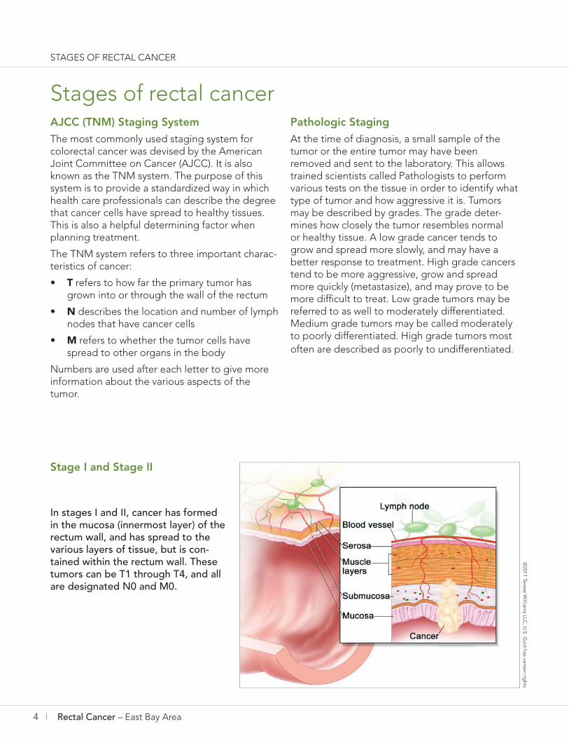
Stage I and Stage II
Pathologic Staging
At the time of diagnosis, a small sample of the tumor or the entire tumor may have been removed and sent to the laboratory. This allows trained scientists called Pathologists to perform various tests on the tissue in order to identify what type of tumor and how aggressive it is. Tumors may be described by grades. The grade determines how closely the tumor resembles normal or healthy tissue. A low grade cancer tends to grow and spread more slowly, and may have a better response to treatment. High grade cancers tend to be more aggressive, grow and spread more quickly (metastasize), and may prove to be more difficult to treat. Low grade tumors may be referred to as well to moderately differentiated. Medium grade tumors may be called moderately to poorly differentiated. High grade tumors most often are described as poorly to undifferentiated.
In stages I and II, cancer has formed in the mucosa (innermost layer) of the rectum wall, and has spread to the various layers of tissue, but is contained within the rectum wall. These tumors can be T1 through T4, and all are designated N0 and M0.
©2011 Terese W
illiams LLC
, U.S. G
ovt has certain rig
hts
4 | Rectal Cancer – East Bay Area
STAGES OF RECTAL CANCER
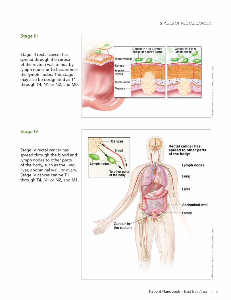
Stage III
Stage III rectal cancer has spread through the serosa of the rectum wall to nearby lymph nodes or to tissues near the lymph nodes. This stage may also be designated as T1 through T4, N1 or N2, and M0.
©2011 Terese W
illiams LLC
, U.S. G
ovt has certain rig
hts
Stage IV
Stage IV rectal cancer has spread through the blood and lymph nodes to other parts of the body, such as the lung, liver, abdominal wall, or ovary. Stage IV cancer can be T1 through T4, N1 or N2, and M1.
©2011 Terese W
illiams LLC
, U.S. G
ovt has certain rig
hts
Patient Handbook – East Bay Area | 5

TREAMENT OF RECTAL CANCER
Treatment of rectal cancer Chemotherapy and Radiation Therapy
Chemotherapy is a cancer treatment that uses drugs to stop the growth of cancer cells, either by killing the cells or by stopping the cells from dividing. The way the chemotherapy is given depends on the type and stage of the cancer being treated.
Radiation therapy is a cancer treatment that uses high-energy x-rays or other types of radiation to kill cancer cells. The way the radiation therapy is given depends on the type and stage of the cancer being treated tumor.
Surgical Treatment
Surgery is the most common treatment for all stages of rectal cancer. The cancer is removed using one of the following types of surgery:
• Polypectomy: If the cancer is found in a polyp (a small growth or piece of tissue), the polyp is often removed during a procedure called a colonoscopy.
• Local excision: If the cancer is found on the inside surface of the rectum and has not spread into the wall of the rectum, the cancer and a
small amount of surrounding healthy tissue is removed.
• Resection: If the cancer has spread into the wall of the rectum, the section of the rectum with cancer and nearby healthy tissue is removed. Sometimes the tissue between the rectum and the abdominal wall is also removed. The lymph nodes near the rectum are removed and checked under a microscope for signs of cancer. An opening (stoma) may be made to allow stool to empty into a bag or pouch. The opening may be temporary in order to facilitate healing from the surgery, or in some cases it can be permanent.
After the cancer is removed, the surgeon can do an anastomosis (sew the healthy parts of the colon together, sew the remaining rectum to the colon, or sew the colon to the anus); or make a stoma (an opening) from the rectum to the outside of the body for waste to pass through. A bag or pouch is placed around the stoma to collect the waste. Sometimes the stoma is needed only until the rectum has healed, and then it can be reversed. If the entire rectum is removed, however, the ostomy may be permanent.
Ressection of the rectum with anastomosis
Resection of the rectum with anastomosis. The rectum and part of the colon are removed, and then the colon and anus are joined
Radiation therapy or chemotherapy may be given before surgery to shrink the tumor, make it easier to remove the cancer, and lessen problems with bowel control after surgery. Treatment given before surgery is called neoadjuvant therapy. Even if all the cancer that can be seen at the time of the operation is removed, some patients may be given radiation therapy or chemotherapy after surgery to kill any cancer cells that may be left. Treatment given after the surgery, to lower the risk that the cancer will come back, is called adjuvant therapy.
6 | Rectal Cancer – East Bay Area
©2011 Terese W
illiams LLC
, U.S. G
ovt has certain rig
hts
TREAMENT OF RECTAL CANCER
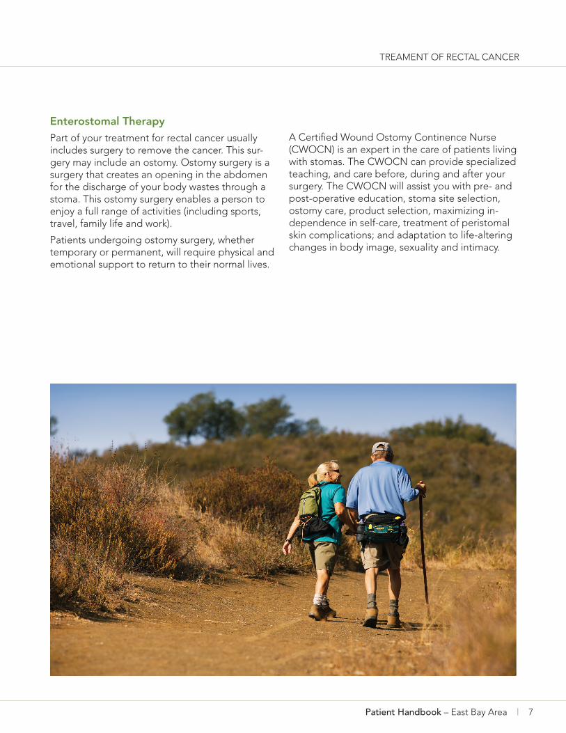
Enterostomal Therapy
Part of your treatment for rectal cancer usually includes surgery to remove the cancer. This surgery may include an ostomy. Ostomy surgery is a surgery that creates an opening in the abdomen for the discharge of your body wastes through a stoma. This ostomy surgery enables a person to enjoy a full range of activities (including sports, travel, family life and work).
Patients undergoing ostomy surgery, whether temporary or permanent, will require physical and emotional support to return to their normal lives.
A Certified Wound Ostomy Continence Nurse (CWOCN) is an expert in the care of patients living with stomas. The CWOCN can provide specialized teaching, and care before, during and after your surgery. The CWOCN will assist you with pre- and post-operative education, stoma site selection, ostomy care, product selection, maximizing independence in self-care, treatment of peristomal skin complications; and adaptation to life-altering changes in body image, sexuality and intimacy.
Patient Handbook – East Bay Area | 7
8 | Rectal Cancer – East Bay Area
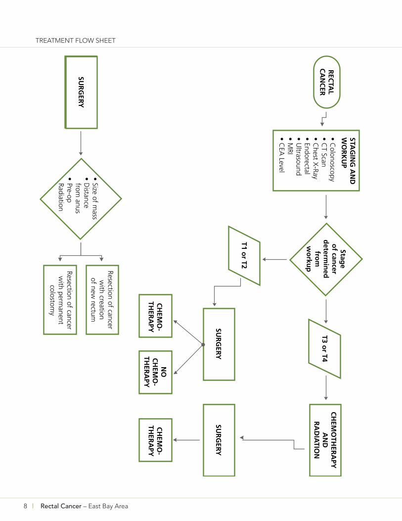
TREATMENT FLOW SHEET
WHAT TO EXPECT AFTER SURGERY
What to expect after surgery Activity
You will be encouraged to sit and walk every 2 hours as early as the day of surgery. Walking has been shown to reduce recovery time and decrease complications after surgery. Walking helps to protect you from developing blood clots and breathing problems. It also helps to get your bowels functioning again after anesthesia and helps with gas pains that most people experience after this surgery. A breathing exercise device (incentive spirometer) will be given to you in the hospital and you will be instructed on how to use it properly. It is important to use it regularly and as instructed in order to prevent respiratory complications after surgery. Avoid heavy lifting (more than about 10 pounds), or strenuous activity for about 6 weeks, or as advised by your surgeon. You will be given specific instructions upon discharge from the hospital.
Driving
You should not drive while you are taking narcotic medication. Driving under the influence of narcotic medication carries the same risks as driving under the influence of alcohol. We also do not recommend that you drive if you are unable to comfortably sit in traffic for extended periods of time, or quickly move your foot from the gas pedal to the brake pedal.
Medication
You should expect some discomfort after surgery, but it is important to try to manage the pain so that it does not get out of control. Keeping pain under control helps patients to recover and heal more quickly. While you are in the hospital, you will be given medication around the clock. This may be given via an intravenous (IV) line into your vein, or as an oral medication to take by mouth, or you may be given a combination of both. When you return home, we advise that you continue to take your pain medication on a schedule for the first 2-3 days. This will help to keep the level of pain manageable. Gradually, you can space
out the interval of time between doses until you are taking on an “as needed basis”. You may be advised to take an over the counter (OTC) non-steroidal, anti-inflammatory medication (NSAID) in addition to your narcotic medication. Please note that these are two different types of medication, so it will be okay to use as directed. These medications work well together, and provide added pain relief. You can discuss this further with your surgeon’s nurse.
Diet
Your physician may recommend a soft or low fiber diet for you following surgery. A soft diet is useful when your body is ready for more than liquids but still unable to handle a regular solid diet. Soft food is easier to digest while you are recovering from your surgery. The diet consists of a variety of normal foods, cooked or prepared in such a way that they have a soft texture.
Some examples of foods included in a soft diet are:
• all beverages • cooked or canned fruits • fresh banana or avocado • soft-cooked or canned vegetables • moist, tender meats or fish • eggs • low-fat milk products, yogurt • mild-flavored cheese • potatoes (mashed, baked, boiled) • refined cooked cereal • refined white, wheat, or rye bread • plain white rice • pasta • ice cream, frozen yogurt; sherbet custards,
pudding
Some foods to avoid are those that are high in fiber, such as raw fruits and vegetables or meats that are tough. Avoiding high-fiber foods like whole-grain breads and cereals immediately after surgery, fried or greasy foods, and spicy foods is also recommended.
Patient Handbook – East Bay Area | 9
WHAT TO EXPECT AFTER SURGERY
Adding fiber to your diet
Dietary fiber is the indigestible part of whole grains, vegetables and fruits. Fiber can also be found in beans and legumes, and some dietary supplements such as Metamucil, Citrucel and their generic equivalents. An adequate fiber intake is an important part of keeping your colon healthy; however, your surgeon may advise you to limit your consumption of high fiber foods immediately after surgery. Your colon will need time to heal, and fiber intake should be increased slowly. Adequate fluid intake is very important, particularly when increasing your fiber intake. Strive to drink 8 to 10 glasses of fluid per day unless your medical doctor has restricted your fluid intake.
Some common problems
Diarrhea Foods that can help resolve loose stools or diarrhea
• Applesauce • Bananas • Cream of wheat or rice • Peanut butter • Rice • Tapioca • Weak tea
Contact the Surgery Advice Nurse if you are having diarrhea accompanied by abdominal cramps, nausea and vomiting, or more than 6 loose stools in a day.
Constipation • Increase your fluids, especially water • Increase exercise, even if it means just a little
extra walking • Eat more fiber (add slowly and as directed
by your surgeon) such as: – Bran or whole grain cereals – Fresh fruit – Vegetables – Whole grain bread
Do not increase your intake of high fiber foods if your surgeon has advised against this. Contact the Surgery Advice Nurse if you do not have a bowel movement within 4 days.
Gas Gas production is a normal part of intestinal function. However, excessive gas can cause bloating, nausea and discomfort. What you eat can affect the amount of gas that your body produces. You may already be aware of which foods cause excessive gas production for you, and you should try to avoid them. Over the counter (OTC) medications that contain Simethicone such as GasX or Mylicon are recommended to decrease gas. Maalox, Mylanta, Tums or their generic equivalents may help as well. Drinking warm fluids like tea, soup or broth may help also.
Wound care
If you have a laparoscopic surgery, you may go home with 3-5 small bandages on your abdomen. The bandages cover small incisions that are usually closed with dissolving sutures and steri strips (thin strips of white tape), or skin glue. If you have an open surgery, you may go home with one medium incision on your abdomen. The incision is usually closed with dissolving sutures and steri strips, or skin staples may be used. If staples are used, they will be removed in 1-2 weeks.
You may have a tube outside of your skin that drains into a small container. You will be instructed on how to take care of this before you leave the hospital. The tube and the container are usually removed in the clinic at your first postoperative appointment. It will be very important for you to keep a written record of the amount of fluid that drains into the bulb in a 24 hour period of time.
Your discharge instructions will give you specific information on when you can get the incision area wet, when you can shower, and when you can take a tub bath.
10 | Rectal Cancer – East Bay Area

Contacting the Surgery Department Contact the Surgery Department for any of these symptoms:
• Temperature over 101 degrees Fahrenheit
• Abdomen is uncomfortably distended
• No bowel movement within 4 days after discharge
• Bowel movements stop abruptly
• Separation of wound edges
• Green, yellow or brown drainage from the wound
• Foul smelling drainage from the wound
• Increased redness, swelling, warmth or pain
• Severe nausea and/or vomiting
• Increased abdominal pain that is not relieved by pain medication
An Advice Nurse is available in the Surgery Department Monday through Friday from 8:30 am to 5:30 pm. After hours or on the weekends, you can call the Kaiser Permanente Medical Advice Line at (510) 752-1190. If you think you have an emergency, do not wait to speak with an Advice Nurse. Call 911, or come to the nearest emergency room. If your surgeon is here in Oakland, we advise you to come to the Kaiser Permanente Oakland Emergency Department if you can safely do so.
Important Phone Numbers
Kaiser Permanente Oakland Surgery Department Colo-rectal Advice Nurse: (510) 752-2711 Monday through Friday, 9:30 am to 5:30 pm
General Surgery Advice Nurse: (510) 752-1105 Monday through Friday, 8:30 am to 5:00 pm
General Surgery Appointments: (510) 752-2079 or (510) 752-2375 Monday through Friday, 8:30 am to 5:00 pm
Kaiser Permanente Medical Advice Staff: (510) 752-1190* Available 24 hours a day, seven days a week
*there may be a different number to call is you live outside of the East Bay service area
Patient Handbook – East Bay Area | 11

NOTES
12 | Rectal Cancer – East Bay Area


2