Embed Size (px)
Citation preview
Indian J Plast Surg 2007 Vol 40 SupplementS35
This P
DF is av
ailab
le for
free d
ownlo
ad fro
m
a site
hoste
d by M
edkn
ow P
ublic
ation
s
(www.m
edkn
ow.co
m).
CMYK35
Maxillary reconstruction
James S. BrownRegional Maxillofacial Service, Aintree Hospital, Liverpool, UK
Address for correspondence: Mr. James Brown, Merseyside Regional Head and Neck Cancer Centre University Hospital Aintree, Lower
Lane, Liverpool, L9 7al 0151 529 5283. E-mail: [email protected]
ABSTRACT
This article aims to discuss the various defects that occur with maxillectomy with a full review of the literature and discussion of the advantages and disadvantages of the various techniques described. Reconstruction of the maxilla can be relatively simple for the standard low maxillectomy that does not involve the orbital ß oor (Class 2). In this situation the structure of the face is less damaged and the there are multiple reconstructive options for the restoration of the maxilla and dental alveolus. If the maxillectomy includes the orbit (Class 4) then problems involving the eye (enopthalmos, orbital dystopia, ectropion and diplopia) are avoided which simpliÞ es the reconstruction. Most controversy is associated with the maxillectomy that involves the orbital ß oor and dental alveolus (Class 3). A case is made for the use of the iliac crest with internal oblique as an ideal option but there are other methods, which may provide a similar result. A multidisciplinary approach to these patients is emphasised which should include a prosthodontist with a special expertise for these defects.
KEY WORDS
Head and neck cancers, maxillary reconstruction, maxillectomy
Free full text on www.ijps.org
Maxillary reconstruction still remains an area of controversy in Head and Neck Surgery. In the past the main method of rehabilitation involved
prosthesis with an obturator which often produced very acceptable results considering the extent of the ablative surgery. The operation was simpler and could be accomplished in relatively shorter time and also the defect could be inspected to detect the presence of recurrence at an earlier stage. Very little research had been done on the quality of life outcomes in this group and it is still uncertain how patients cope with this form of prosthetic rehabilitation, especially for larger defects.
Tumours arising in the paranasal sinuses and the maxillary alveolus form the majority of maxillectomy cases and the incidence of squamous cell carcinoma arising in these sites is relatively uncommon. As a result there are few studies reporting on the management of these cases. Most papers tend to report a single method of reconstruction and few studies have been reported to
compare the outcome of using different rehabilitative or reconstructive methods.
ANATOMY AND FORM
In most cases of mid-face ablation the facial skin is spared and the main loss of structure is part of the maxilla. The maxilla provides a skeletal base for the form of the face and supports the orbit and the alar part of the nose. It also provides the alveolar bone, which supports the teeth of the upper jaw. The majority of this tissue is bone and its replacement should be the main aim of the reconstruction. If the facial skin does not require resection then the soft tissue structure that is lost with a maxillectomy is mucoperiosteum, which lines the nasal cavity and the palate. The most natural replacement of this tissue is in fact muscle which when used to line bone forms a pseudo-periosteum that becomes epithelialised and firmly attaches to the bone. The structure is immobile and is the passive partner of the lower jaw. Mobility of
M
Indian J Plast Surg 2007 Vol 40 Supplement S36
This P
DF is av
ailab
le for
free d
ownlo
ad fro
m
a site
hoste
d by M
edkn
ow P
ublic
ation
s
(www.m
edkn
ow.co
m).
36 CMYK
the soft tissue reconstruction is not an advantage as in the floor of the mouth and tongue.
CLASSIFICATION AND CONCEPTS IN RECONSTRUCTION
There are a number of working classifications of the maxillary defect but it is important that any classification is simple to remember and clearly indicates the increasing complexity of the defect. We[1] have described a system, which includes the dental loss as well as the facial or surgical defect [Figure 1].
SURGICAL COMPONENT (VERTICAL)
Class 1 Minimal loss of the alveolar bone without an oro-antral fistula. Loss of the hard palate only. Partial maxillectomy with no breach of the oral cavity or loss of the alveolus
Class 2 Including the alveolus and antral walls, but not extending to the orbital rim or adnexae
Class 3 As in Class II but including the orbital floor or medial wall
Class 4Maxillectomy with orbital exenteration
DENTAL COMPONENT (HORIZONTAL)
a) Less than or equal to half the dental alveolusb) More than half the dental alveolus or crossing the
midlinec) The entire maxillary alveolus
The principle of this classification is that the need for composite reconstruction increases as the Class of the
defect increases. There is evidence that patients with larger defects had a decreased quality of life.
A Class 1 defect can be simply treated with obturation or a soft tissue flap often preferred at the junction of the hard and soft palate. Partial maxillectomies involving the lateral nasal wall for an inverted papilloma can be left without obturation or reconstruction with excellent results.
In the classic hemi-maxillectomy defect (Class 2) it is the extent of the loss of the dental alveolus (Class 2a-c), which complicates both the obturation and the reconstruction. In my experience this is very important if there has been a high resection anteriorly with loss of the septum and support of the alar base.
The Class 3 defect is the most complex problem for either the reconstructive surgeon or the prosthodontist. The problem with such a defect is the primary need to reconstruct the orbital floor and medial wall, provide support for the cheek skin and a reliable base for an implant-retained dental prosthesis. By retaining the eye the problems of facial disfigurement due to enopthalmos, ectropion and the functional problem of epiphora need to be addressed.
In the Class 4 defect it is necessary to enucleate or exenterate the eye, which results in a significant facial deformity but this surgery necessitates both an orbital and dental prosthesis. The orbital prosthesis can be used to disguise poor support of the cheek if the orbital rim has not been restored and the problems of enopthalmos and ectropion are avoided. The use of a composite graft with a reasonable height of bone is required if dental rehabilitation is to be achieved.
The principle of maxillary reconstruction is now accepted although concern remains regarding the inability to directly examine the defect. Postoperative scans and regular endoscopy can play a more important role in clinical surveillance, but there is no evidence that filling the defect is associated with a compromised outcome in terms of survival. The role of cavity inspection may be more useful for lower grade disease such as an ameloblastoma or low-grade salivary tumours but the prognosis for patients with recurrent squamous cell carcinoma remains poor, whatever the method of rehabilitation.
Review of the literature clearly shows the lack of consensus on the management of this defect. Table 1 shows the Figure 1: ClassiÞ cation of the maxillectomy defect (23)
Brown
Class 1 Class 2 Class 3 Class 4
a b c
Indian J Plast Surg 2007 Vol 40 SupplementS37
This P
DF is av
ailab
le for
free d
ownlo
ad fro
m
a site
hoste
d by M
edkn
ow P
ublic
ation
s
(www.m
edkn
ow.co
m).
CMYK37
Maxillary reconstruction
articles published since 2000 describing different free flap options on maxillary reconstruction and Table 2 lists larger series and the choice of reconstructive options. Teaching and guidance as to the best technique, therefore, is based more on authority than clinical evidence.
Multidisciplinary decision-making in the manage-ment of the maxillectomy defectIn the reconstructive practice the role of the Restorative Dentist is essential in the management of any patient with head and neck cancer and this is most important for patients with a planned maxillectomy as part of their treatment. Each case requires discussion in terms of the disease, age, medical fitness and most important the patient’s aspirations. Table 3 shows the essential differences between obturation and reconstruction which
need to be considered while making the decision. The team should be able to offer the full range of prosthetic and reconstructive options and devise a treatment plan to restore the form and function lost as a result of the ablative surgery.
Although the evidence in the literature is weak there is a trend to suggest that patients are more satisfied following reconstruction when compared to obturation. We published a study involving 28 patients, 10 managed with obturation and 18 with a free flap and although the groups were not matched the results favoured the reconstructed group although these defects were larger and a higher percentage required radiotherapy [Table 4]. While comparing the groups the oral pain and soreness was significantly less and overall satisfaction with function was greater in the reconstructed group. The facial appearance is generally not compromised with a Class 2 defect, which comprised the majority of the comparative group.
Eric Genden from Mark Urken’s group compared matched patients with Class 2a defects with obturation and reconstruction (one scapula, three iliac crest with internal oblique) but they only had four in each group with four controls.[27] The results favoured the reconstructed group and the donor site did not seem a significant problem in them.
For the purposes of this article I will concentrate on the reconstruction of the Class 2-4 defects as Class 1 cases often need no reconstruction or a soft tissue flap is all that is required since there is no loss of the functioning dental alveolus.
RECONSTRUCTION OF CLASS 2, 3 AND 4 MAXILLARY DEFECTS
Class 2aThere are a multitude of techniques that have been described which can give good results in the standard
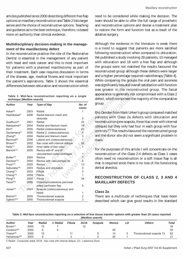
Table 1: Mid-face reconstruction reporting on a single technique (Medline search)
Author Year Type of fl ap No. of casesAskar[2] 2003 Radial 1Hashikawa[3] 2006 Radial titanium mesh and obdurate 5Guelfucci[4] 2001 Radial 23Nakayama[5] 2004 Radial and titanium mesh 18Kim[6] 2005 Radial (osteocutaneous) 23Santamaria[7] 2000 Radial 2, (osteocutaneous) 4Zhang[8] 2004 Radial and titanium mesh 3Barnouti[9] 2006 Lateral arm (osteocutaneous) 1Brown[10] 2002 Iliac crest with internal oblique 24Kelly[11] 2004 Inner table of iliac crest 1Bianchi[12] 2005 Rectus with 8th and 9th vascularised costal cartilages 1Butler[13] 2004 Rectus 7Davison[14] 2004 Rectus with vascularised rib 6Pryor[15] 2005 Rectus 13Sakuraba[16] 2003 Rectus and obturation 5Chang[17] 2003 Fibula 3Chang[18] 2004 Fibula 1Peng[19] 2005 Fibula 34Yazar[20] 2006 Osteomyocutaneous peroneal artery perforator ß ap 6Jones[21] 2003 Scapula (osteocutaneous) and radial 1Bidros[22] 2005 Thoracodorsal scapula 2Uglesic[23] 2000 Thoracodorsal scapula 3
Table 2: Mid-face reconstruction reporting on a selection of free tissue transfer options with greater than 25 cases reported (Medline search)
Author Year Radial C Radial Fibula DCIA Scapula Rectus LD Others TotalBrown[1] 2000 15 7 12 34Cordeiro[24] 2000 6 4 45 55Triana[25] 2000 10 11 9 14 5 Thoracodorsal scapula 13 62Yamamoto[26] 2004 6 - 2 - 2 20 1 31C Radial - Composite radial, DCIA - Iliac crest with internal oblique, LD - Latissimus Dorsi
Indian J Plast Surg 2007 Vol 40 Supplement S38
This P
DF is av
ailab
le for
free d
ownlo
ad fro
m
a site
hoste
d by M
edkn
ow P
ublic
ation
s
(www.m
edkn
ow.co
m).
38 CMYK
low maxillectomy that does not cross the midline. In my opinion this is the only defect that can be reconstructed without a composite free flap and hence the role of pedicled flaps is useful and often adequate.
Pedicled fl apsThe most commonly described pedicled flap is temporalis but this flap is limited to an arc of rotation that reaches the ipsilateral canine. If the flap is to be used for a defect involving the ipsilateral central incisor then its reach may be a problem and dehiscence an expected complication. Tideman[28] described this flap in conjunction with non-vascularised bone from the iliac crest maintained in a titanium mesh structure. The buccal fat pad is a useful source of tissue but can usually only reach more posterior defects.[29] The use of the temporoparietal fascia and outer calvarium based on the superficial temporal artery has also been described although the source of bone is limited.[30] Although the use of the deltopectoral flap is not preferred
there is a large series that describes good results as long as the flap is delayed prior to transfer.[31] More recently, the submental island flap[27] has been developed in many areas of facial and oral reconstruction as reverse-flow can be utilised to increase the length of the pedicle to reach the orbit. This is perhaps the simplest and most reliable reconstruction when bone is not required for defects posterior to the canine in the dentate patient. It is more preferable to use a muscle-based flap such as temporalis for the edentulous patient as the epithelialised muscle base is more suitable to bear a denture.
Free tissue transferIf the reconstruction is for a dentate patient with a posterior defect in which the ipsilateral canine can be saved then a soft tissue flap is all that is required as there is sufficient bone to support the face and a partial denture need only hold the premolar teeth.
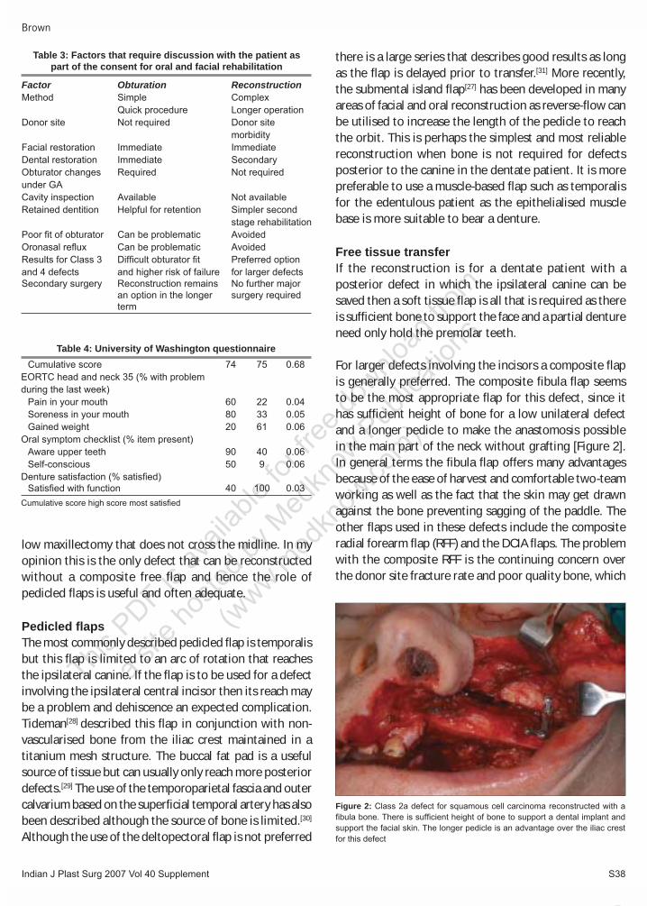
For larger defects involving the incisors a composite flap is generally preferred. The composite fibula flap seems to be the most appropriate flap for this defect, since it has sufficient height of bone for a low unilateral defect and a longer pedicle to make the anastomosis possible in the main part of the neck without grafting [Figure 2]. In general terms the fibula flap offers many advantages because of the ease of harvest and comfortable two-team working as well as the fact that the skin may get drawn against the bone preventing sagging of the paddle. The other flaps used in these defects include the composite radial forearm flap (RFF) and the DCIA flaps. The problem with the composite RFF is the continuing concern over the donor site fracture rate and poor quality bone, which
Table 3: Factors that require discussion with the patient as part of the consent for oral and facial rehabilitation
Factor Obturation ReconstructionMethod Simple Complex Quick procedure Longer operationDonor site Not required Donor site morbidityFacial restoration Immediate ImmediateDental restoration Immediate SecondaryObturator changes Required Not requiredunder GACavity inspection Available Not availableRetained dentition Helpful for retention Simpler second stage rehabilitationPoor Þ t of obturator Can be problematic AvoidedOronasal reß ux Can be problematic AvoidedResults for Class 3 DifÞ cult obturator Þ t Preferred optionand 4 defects and higher risk of failure for larger defectsSecondary surgery Reconstruction remains No further major an option in the longer surgery required term
Table 4: University of Washington questionnaireCumulative score 74 75 0.68
EORTC head and neck 35 (% with problem during the last week)
Pain in your mouth 60 22 0.04Soreness in your mouth 80 33 0.05Gained weight 20 61 0.06
Oral symptom checklist (% item present)Aware upper teeth 90 40 0.06Self-conscious 50 9 0.06
Denture satisfaction (% satisÞ ed) SatisÞ ed with function 40 100 0.03
Cumulative score high score most satisÞ ed
Figure 2: Class 2a defect for squamous cell carcinoma reconstructed with a Þ bula bone. There is sufÞ cient height of bone to support a dental implant and support the facial skin. The longer pedicle is an advantage over the iliac crest for this defect
Brown
Indian J Plast Surg 2007 Vol 40 SupplementS39
This P
DF is av
ailab
le for
free d
ownlo
ad fro
m
a site
hoste
d by M
edkn
ow P
ublic
ation
s
(www.m
edkn
ow.co
m).
CMYK39
Maxillary reconstruction
is insufficient for an implant-retained denture.
Class 2b-cOnce the low maxillectomy (Class 2) defect crosses the midline (Class 2b) or involves the whole of the functioning dental alveolus (Class 2c), then a composite flap is essential to restore the loss of bone which includes the important anterior alveolus, support the alar region and nasal columella and provide an adequate bony base for an implant-retained prosthesis. The choice of flap for this more complex defect depends on the height of bone that is lost in the anterior maxilla and nasal septum. If the loss of bone only includes the dental alveolus then a fibula flap is the ideal choice. If the height of bone loss includes a significant part of the nasal piriforms, the nasal septum and extending towards the nasal bones (>2 cm) then a vertically
placed iliac crest with internal oblique provides the ideal flap. In the original paper first describing the use of iliac crest with internal oblique I suggested the use of the horizontal placement of the bone for the lateral defect using part of the bone to fill the palatal defect also. But it has been found unnecessary to provide bony reconstruction of the palate and also the use of the internal oblique for soft tissue.
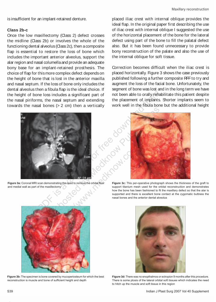
Correction becomes difficult when the iliac crest is placed horizontally. Figure 3 shows the case previously published following a further composite RFF to try and augment the loss of the facial bone. Unfortunately, the segment of bone was lost and in the long term we have not been able to orally rehabilitate this patient despite the placement of implants. Shorter implants seem to work well in the fibula bone but the additional height
Figure 3c: This per-operative photograph shows the thickness of the graft to support titanium mesh used for the orbital reconstruction and demonstrates how the bone has been fashioned to Þ t the maxillary defect so that the alar is supported and there is excellent bone contact at the zygomatic buttress the nasal bones and the anterior dental alveolus
Figure 3a: Coronal MRI scan demonstrating the need to remove the orbital ß oor and medial wall as part of the maxillectomy
Figure 3b: The specimen is bone covered by mucoperiosteum for which the best reconstruction is muscle and bone of sufÞ cient height and depth
Figure 3d: There was no enopthalmos or ectropion 9 months after this procedure. There is some ptosis of the lateral orbital soft tissues which indicates the need to hitch up the muscle and soft tissue in this region
Indian J Plast Surg 2007 Vol 40 Supplement S40
This P
DF is av
ailab
le for
free d
ownlo
ad fro
m
a site
hoste
d by M
edkn
ow P
ublic
ation
s
(www.m
edkn
ow.co
m).
40 CMYK
of the iliac crest is an advantage to ensure successful osseointegration.
Class 3a-cIn terms of the maxillectomy or mid-face defect the loss of the maxilla and orbital floor presents the most complex reconstructive problem. The aim of reconstruction is to close the oronasal fistula, restore the functioning dental alveolus, provide support for the facial skin and most importantly support the orbit and eyelids. The iliac crest with internal oblique is an ideal flap to fulfil these requirements. It is the only bone source that will be able to provide sufficient bone for an implant-retained dental prosthesis, support of the face and a platform for the reconstruction of the orbital floor with either titanium mesh or free bone from the hip or the calvarium. The muscle will obturate the oral defect and provide an epithelialised lining for the lateral nose.
The relationships of the hard and soft tissue components of the iliac crest with internal oblique are ideal. The criticism of the use of the iliac crest with internal oblique relates to the poor quality of the soft tissue, the short length of the pedicle and the problems with the donor site.[32] There is persisting concern regarding the iliac crest donor site but in a retrospective study comparing 19 iliac crests with 16 fibula cases there was no difference in patient tolerance and if anything the iliac crest caused less major problems for this patient group.[33] We have also carried out a study asking patients the preference for the site of a scar in free tissue transfer and the fibula site was not favoured compared to an abdominal scar (in press PRS). The problem of the shorter pedicle is an important factor and considerable skill is required to overcome this problem. It is possible to site the bone harvest posterior to the anterior superior iliac spine and I use the facial vessels overlying the body of the mandible for the anastomosis. Others including Mark Urken’s group now use vein grafts for both the artery and vein and bring the pedicle into the main part of the neck. In my experience it is virtually always possible to use the facial or retromandibular vein but a graft may be required if there is insufficient run-off from the facial artery.
The scapula donor site offers a reasonable height of bone but this may be too thin in the dental alveolus to accommodate implants. Uglesic[23] has described the use of the angle of the scapula supplied by the angular branch of the thoracodorsal artery in combination with the muscle and/or skin of the latissimus dorsi, which I have termed the thoracodorsal scapula [Table 1]. This has the
advantage of the longer pedicle, which can easily reach the neck and the use of epithelialised muscle in the oral cavity, which results in a more natural denture-bearing surface. The bone stock that can be safely transferred is limited and this part of the scapula is thinner making an implant-retained prosthesis unreliable. In my opinion this may be a better alternative than the fibula flap, which has a limited bone height and relies on skin coverage in the nasal cavity and the mouth. It is possible to carry out two osteotomies of the fibula bone to reconstruct the orbital rim, the lateral nasal wall and the dental alveolus but this is not possible for Class 3b defects and results in a short pedicle as most of the bone length is utilised. It is also not possible to bring the dental alveolar section as far as the zygomatic buttress to complete the bony reconstruction. In the longer term there is a risk of a facial deformity as there is no direct support to the facial skin. I have not seen a reported case of a double-barrelled fibula flap for a Class 3 defect but in this situation the orientation of the skin island would be difficult and the pedicle shortened. There would be little capacity to shape the bone graft to fit the contours of the face.
The key to the reconstruction of the maxilla is the provision of a reliable bony structure to support the soft tissue components of the orbit and face and provide a basis for the wearing of a tissue-borne or implant-retained dental prosthesis. In my opinion neither the scapula nor the fibula donor sites can make this provision for the Class 3 defect and my preference is the iliac crest with the internal oblique for the reasons stated above.
Class 4aOnce the decision has been taken to exenterate the orbit in the management of mid-face malignancy then the problems for the reconstructive surgeon are reduced. The complications of diplopia, enopthalmos and ectropion are obviated by the removal of the eye and the provision of a prosthetic eye can mask some of the deformity if vascularised bone has not been transferred as part of the reconstruction.
My first choice reconstruction for a Class 4 defect is still the iliac crest with internal oblique.[1;10,34] It is possible to achieve an excellent result with full long-term support of the facial skin and a base for an implant or tissue-borne dental prosthesis. A close working relationship with both the maxillofacial technician and the restorative dentist is an essential part of achieving this sort of result. As in the Class 3 defect neither the scapula nor the fibula flap
Brown
Indian J Plast Surg 2007 Vol 40 SupplementS41
This P
DF is av
ailab
le for
free d
ownlo
ad fro
m
a site
hoste
d by M
edkn
ow P
ublic
ation
s
(www.m
edkn
ow.co
m).
CMYK41
Table 5: Summary of free fl ap reconstructive options in maxillary reconstruction
Class 1 Class 2 Class 3 Class 4Summary of defect Minimal defect only Low maxillectomy not High maxillectomy involving Radical maxillectomy with requiring reconstruction involving the orbit the orbital adnexae orbital exenteration at the junction of the hard and soft palate Ideal reconstructive 1. Radial forearm ß ap 1. Fibula ß ap 1. Iliac crest IO 1. Iliac crest IOoption in order of 2. ALT 2. Iliac crest IO 2. Scapula 2. Rectus, preference 3. Scapula Latissimusdorsi, 4. Radial ALTIO - Internal oblique, ALT - Anterolateral thigh ß ap
can provide sufficient bone for the support of the orbital prosthesis, the facial skin and the dental rehabilitation. The best compromise reconstruction is a large soft tissue flap with abundant muscle to obturate the whole of the defect from the roof of the orbit to the dental
alveolus. This provides sufficient bulk for good healing and although there will inevitably be some facial collapse due to the ptosis of the flap with gravity and no chance of a successful dental rehabilitation, some quality of life can be maintained.
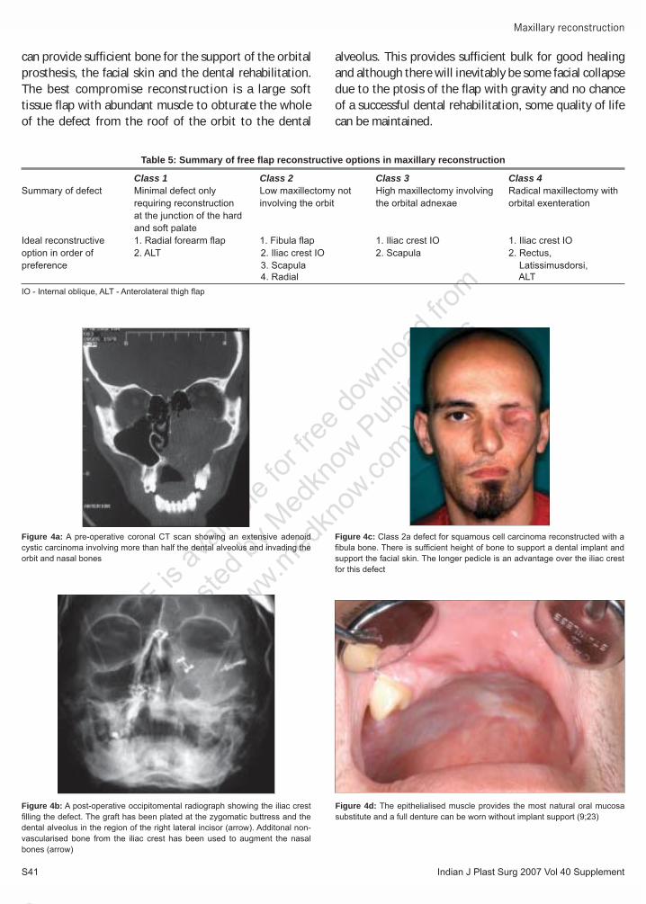
Figure 4a: A pre-operative coronal CT scan showing an extensive adenoid cystic carcinoma involving more than half the dental alveolus and invading the orbit and nasal bones
Figure 4b: A post-operative occipitomental radiograph showing the iliac crest Þ lling the defect. The graft has been plated at the zygomatic buttress and the dental alveolus in the region of the right lateral incisor (arrow). Additonal non-vascularised bone from the iliac crest has been used to augment the nasal bones (arrow)
Figure 4c: Class 2a defect for squamous cell carcinoma reconstructed with a Þ bula bone. There is sufÞ cient height of bone to support a dental implant and support the facial skin. The longer pedicle is an advantage over the iliac crest for this defect
Figure 4d: The epithelialised muscle provides the most natural oral mucosa substitute and a full denture can be worn without implant support (9;23)
Maxillary reconstruction
Indian J Plast Surg 2007 Vol 40 Supplement S42
This P
DF is av
ailab
le for
free d
ownlo
ad fro
m
a site
hoste
d by M
edkn
ow P
ublic
ation
s
(www.m
edkn
ow.co
m).
42 CMYK
Class 4b-cIf this defect crosses the midline or involves the nasal bones then an iliac crest with internal oblique is the only flap that can provide sufficient bone to support the facial and nasal skin as well as providing a choice for dental rehabilitation [Figure 4]. There is an option of using cranial bone or non-vascularised iliac crest covered with a vascularised soft tissue myocutaneous flap but the use of these is likely to result in loss of the bone graft and inevitable facial collapse. The patient in Figure 4 also demonstrates the advantage of using muscle to obturate the exenterated orbit. In this situation it is possible to maintain the eyelids, which are sutured together using the internal oblique muscle to support this suture line and prevent dehiscence. The contraction of the muscle allows the orbital space to reform providing the ideal cavity for the provision of an implant-retained orbital prosthesis. Although in this patient there are no completion photographs I have included the case because it shows the provision of a bony base for both oral and orbital rehabilitation following a Class 4b defect with partial loss of the nasal bones. Completed cases have been previously published.[1,10]
CONCLUSIONS
As the level of evidence in the literature is provided by retrospective case series any article on the best methods for mid-face reconstruction must be opinion-based. The main tissue loss in the maxillectomy defect without the loss of facial or nasal skin is bone, which we can reconstruct in terms of from and true function. Any algorithm must therefore be based on the restoration of the bony skeleton as the most important part of the proposed reconstruction. Although the lower level Class 2 defect can be restored by bone from the fibula and scapula donor site, the iliac crest must be the first choice for defects requiring restoration of bone to support the orbit and facial skin and provide a basis for successful dental and orbital rehabilitation [Table 5].
REFERENCES
1. Brown JS, Rogers SN, Mcnally DN, Boyle M. A modified classiÞ cation for the maxillectomy defect. Head Neck 2000;22:17-26.
2. Askar I, Oktay MF, Kilinc N. Use of radial forearm free ß ap with palmaris longus tendon in reconstruction of total maxillectomy with sparing of orbital contents. J Craniofac Surg 2003;14:220-7.
3. Hashikawa K, Tahara S, Ishida H, Yokoo S, Sanno T, Terashi H, et al. Simple reconstruction with titanium mesh and radial forearm ß ap after globe-sparing total maxillectomy: A 5-year follow-up
study. Plast Reconstr Surg 2006;117:963-7.4. GuelÞ B, Bizeau A, Gras R, Giovanni A, Casanova D, Zanaret M.
Reconstruction of the palate vault by free forearm cutaneous ß ap in oncology. [Article in French]. Ann Otolaryngol Chir Cervicofac 2001;118:233-7.
5. Nakayama B, Hasegawa Y, Hyodo I, Ogawa T, Fujimoto Y, Kitano H, et al. Reconstruction using a three-dimensional orbitozygomatic skeletal model of titanium mesh plate and soft-tissue free ß ap transfer following total maxillectomy. Plast Reconstr Surg 2004;114:631-9.
6. Kim JH, Rosenthal EL, Ellis T, Wax MK. Radial forearm osteocutaneous free ß ap in maxillofacial and oromandibular reconstructions. Laryngoscope 2005;115:1697-701.
7. Santamaria E, Granados M, Barrera-Franco JL. Radial forearm free tissue transfer for head and neck reconstruction: Versatility and reliability of a single donor site. Microsurgery 2000;20:195-201.
8. Zhang S, Meng Z, Dong Z. Repair of maxillary defects by free forearm ß ap and titanium mesh. [Article in Chinese]. Zhongguo Xiu Fu Chong Jian Wai Ke Za Zhi 2004;18:459-61.
9. Barnouti L, Caminer D, Barnouti L, Caminer D. Maxillary tumours and bilateral reconstruction of the maxilla. ANZ J Surg 2006;76:267-9.
10. Brown JS, Jones DC, Summerwill A, Rogers SN, Howell RA, Cawood JI, et al. Vascularized Iliac crest with internal oblique muscle for immediate reconstruction after maxillectomy. Br J Oral Maxillofac Surg 2002;40:183-90.
11. Kelly CP, Moreira-Gonzalez A, Ali MA, Topf J, Persiani RJ, Jackson IT, et al. Vascular Iliac crest with inner table of the ilium as an option in maxillary reconstruction. J Craniofac Surg 2004;15:23-8.
12. Bianchi B, Bertolini F, Ferrari S, Tullio A. The rectus abdominis myocutaneous ß ap combined with vascularized costal cartilages for orbito-malar facial reconstruction. J Oral Maxillofac Surg 2005;63:1026-9.
13. Butler CE, Lewin JS. Reconstruction of large composite oromandibulomaxillary defects with free vertical rectus abdominis myocutaneous ß aps. Plast Reconstr Surg 2004;113:499-507.
14. Davison SP, Boehmler JH, Ganz JC, Davidson B. Vascularized rib for facial reconstruction. Plast Reconstr Surg 2004;114:15-20.
15. Pryor SG, Moore EJ, Kasperbauer JL. Orbital exenteration reconstruction with rectus abdominis microvascular free ß ap. Laryngoscope 2005;115:1912-6.
16. Sakuraba M, Kimata Y, Ota Y, Uchiyama K, Kishimoto S, Harii K, et al. Simple maxillary reconstruction using free tissue transfer and prostheses. Plast Reconstr Surg 2003;111:594-600.
17. Chang DW, Langstein HN. Use of the free Þ bula ß ap for restoration of orbital support and midfacial projection following maxillectomy. J Reconstr Microsurg 2003;19:147-52.
18. Chang YM, CoskunÞ rat OK, Wei FC, Tsai CY, Lin HN. Maxillary reconstruction with a Þ bula osteoseptocutaneous free ß ap and simultaneous insertion of osseointegrated dental implants. Plast Reconstr Surg 2004;113:1140-5.
19. Peng X, Mao C, Yu GY, Guo CB, Huang MX, Zhang Y. Maxillary reconstruction with the free Þ bula ß ap. Plast Reconstr Surg 2005;115:1562-9.
20. Yazar S, Cheng MH, Wei FC, Hao SP, Chang KP. Osteomyocutaneous peroneal artery perforator flap for reconstruction of composite maxillary defects. Head Neck 2006;28:297-304.
21. Jones JW. Reconstruction of a complex hemifacial deformity with multiple simultaneous free-ß ap transfers: Case report. J Reconstr Microsurg 2003;19:73-8.
22. Bidros RS, Metzinger SE, Guerra AB. The thoracodorsal artery perforator-scapular osteocutaneous (TDAP-SOC) Flap for reconstruction of palatal and maxillary defects. Ann Plast Surg
Brown
Indian J Plast Surg 2007 Vol 40 SupplementS43
This P
DF is av
ailab
le for
free d
ownlo
ad fro
m
a site
hoste
d by M
edkn
ow P
ublic
ation
s
(www.m
edkn
ow.co
m).
CMYK43
Maxillary reconstruction
Source of Support: Nil, Confl ict of Interest: None declared.
2005;54:59-65.23. Uglesic V, Virag M, Varga S, Knezevic P, Milenovic A.
Reconstruction following radical maxillectomy with ß aps supplied by the subscapular artery. J Craniomaxillofac Surg 2000;28:153-60.
24. Cordeiro PG, Santamaria E. A ClassiÞ cation system and algorithm for reconstruction of maxillectomy and midfacial defects. Plast Reconstr Surg 2000;105:2331-46.
25. Triana RJ Jr, Uglesic V, Virag M, Varga SG, Knezevic P, Milenovic A, et al. Microvascular free ß ap reconstructive options in patients with partial and total maxillectomy defects. Arch Fac Plast Surg 2000;2:91-101.
26. Yamamoto Y, Kawashima K, Sugihara T, Nohira K, Furuta Y, Fukuda S. Surgical management of maxillectomy defects based on the concept of buttress reconstruction. Head Neck 2004;26:247-56.
27. Genden EM, Buchbinder D, Urken ML. The submental island ß ap for palatal reconstruction: A novel technique. J Oral Maxillofac Surg 2004;62:387-90.
28. Tideman H, Samman N, Cheung LK. Immediate reconstruction following maxillectomy: A new method. Int J Oral Maxillofac Surg 1993;22:221-5.
29. Amin MA, Bailey BM, Swinson B, Witherow H. Use of the buccal fat pad in the reconstruction and prosthetic rehabilitation
of oncological maxillary defects. Br J Oral Maxillofac Surg 2005;43:148-54.
30. Choung PH, Nam IW, Kim KS. Vascularized cranial bone grafts for mandibular and maxillary reconstruction the parietal osteofascial ß ap. J Craniomaxillofac Surg 1991;19:235-42.
31. Konno A, Togawa K, Iizuka K. Primary reconstruction after total or extended total maxillectomy for maxillary cancer. Plast Reconstr Surg 1981;67:440-8.
32. Futran ND. Primary reconstruction of the maxilla following maxillectomy with or without sacriÞ ce of the orbit. J Oral Maxillofac Surg 2005;63:1765-9.
33. Rogers SN, Lakshmiah SR, Narayan B, Lowe D, Brownson P, Brown JS, et al. A comparison of the long-term morbidity following deep circumflex iliac and fibula free flaps for reconstruction following head and neck cancer. Plast Reconstr Surg 2003;112:1517-27.
34. Brown JS. Deep circumß ex iliac artery free ß ap with internal oblique muscle as a new method of immediate reconstruction of maxillectomy defect. Head Neck 1996;18:412-21.
Author Help: Choosing an appropriate category of article for faster publication
The manuscript system (www.journalonweb.com) allows the authors to check a likely publication date for a newly submitted article. Based on number of articles in review, number of accepted articles and acceptance rate, the system estimates the likely publication date for an article submitted on a given date.
If there are too many articles in a category e.g., case report, a newly submitted case report if accepted may have to wait for a long period before publication. Hence, the author can check other categories e.g. letter to editor or images, for such paper and submit to another category of articles.