Embed Size (px)
Citation preview

Proc. Natl. Acad. Sci. USAVol. 93, pp. 5419-5424, May 1996Genetics
Recombinational repair of gaps in DNA is asymmetric in Ustilagomaydis and can be explained by a migrating D-loop modelDAVID 0. FERGUSON AND WILLIAM K. HOLLOMAN*Hearst Microbiology Research Center, Department of Microbiology, Cornell University Medical College, New York, NY 10021
Communicated by Charles M. Radding, Yale University, New Haven, CT, February 9, 1996 (received for review October 6, 1995)
ABSTRACT Recombinational repair of double-strandedDNA gaps was investigated in Ustilago maydis. The experi-mental system was designed for analysis of repair of anautonomously replicating plasmid containing a cloned genedisabled by an internal deletion. It was discovered thatcrossing over rarely accompanied gap repair. The strong biasagainst crossing over was observed in three different genesregardless of gap size. These results indicate that gap repairin U. maydis is unlikely to proceed by the mechanism envi-sioned in the double-stranded break repair model of recom-bination, which was developed to account for recombination inSaccharomyces cerevisiae. Experiments aimed at exploringprocessing of DNA ends were performed to gain understand-ing of the mechanism responsible for the observed bias. Aheterologous insert placed within a gap in the coding sequenceof two different marker genes strongly inhibited repair if theDNA was cleaved at the promoter-proximal junction joiningthe insert and coding sequence but had little effect on repairif the DNA was cleaved at the promoter-distal junction. Geneconversion of plasmid restriction fragment length polymor-phism markers engineered in sequences flanking both sides ofa gap accompanied repair but was directionally biased. Theseresults are interpreted to mean that the DNA ends flanking agap are subject to different types of processing. A modelfeaturing a single migrating D-loop is proposed to explain thebias in gap repair outcome based on the observed asymmetryin processing the DNA ends.
Genetic analysis of meiotic recombination has revealed a closeconnection between gene conversion, the nonreciprocal trans-fer of information, and crossing over (for a review, see ref. 1).Insight into the mechanistic basis for this connection has comefrom investigation of DNA double-stranded break and gaprepair, and models accounting for the association have beenproposed based on the homolog interaction of one or bothDNA ends resulting from duplex DNA breakage (2, 3). As atest of the double-strand break repair model in Saccharomycescerevisiae, the repair of a gap in plasmid DNA was measuredafter transformation of mitotic cells (4, 5). The observedassociation of plasmid integration with repair of a gap in 50%of the events was in precise agreement with the predicteddistribution hypothesized in the model and has reinforced theview of universality of the double-strand break repair pathway.
Nevertheless, studies on double-stranded break or gap re-pair in other systems have not generally revealed a closeassociation with crossing over. For instance, mating typeinterconversion in S. cerevisiae takes place by gene conversionfollowing introduction of a double-stranded DNA break at theMAT locus, but no crossing over is found to be associated (6).Similarly, the double-stranded break resulting from P elementmobilization in Drosophila melanogaster was found to berepaired through recombination with a homologous sequence,but no crossing over was observed to accompany the repair (7).
In a recent study (8) of transformation of S. cerevisiae withplasmid DNA cut to the linear form by introduction of adouble-stranded break that was quite similar in experimentaldesign to the prototype system used by Orr-Weaver andSzostak (4), the overwhelming majority of double-strandedbreak repair events was found to occur without associatedcrossing over. No mechanistic explanation has been presentedto reconcile the contradictory findings. It is possible thatalternative pathways are in operation for repair of double-stranded breaks, which are not universal (9), or that recom-bination at particular genetic loci is strongly influenced by thelocal chromatin structure, which might not be uniform (10). Itseems clear that a mechanism responsible for the strong biasagainst crossing over noted in examples above is operational inmitotic cells.We have been investigating the genetic and molecular basis of
recombination in Ustilago maydis and have initiated studies usingplasmid DNA substrates. Experimental systems were designed tocategorize the types of recombination events taking place be-tween nonreplicating plasmids and chromosomal sequences (11)and to examine extrachromosomal recombination between rep-licating plasmids (12). Our studies indicated that recombinationwas strongly stimulated by introduction of double-strandedbreaks and that a potent end-joining activity was present thatcould repair double-stranded breaks in the absence of homologinteraction (12). These observations plus the differences noted inthe degree of association between double-stranded break repairand crossing over in various systems have led us to investigate themechanism of double-stranded break repair in U. maydis.
In the present work, we designed a system for studying therecombinational repair of double-stranded gaps in DNA. Inparticular, we were interested in determining (i) if gap repairin U. maydis is accompanied by crossing over and (ii) whetheror not processing of the DNA ends might provide clues to themechanism of recombination.
MATERIALS AND METHODSPlasmids. The U. maydis LEU1 gene used in these studies
was contained on a 3.0-kbp Hindlll-EcoRI genomic DNAfragment that fully complements the leul-1 mutation (13, 14).pCM216 (12) is pBluescript II SKI (Stratagene) containingthis 3.0-kbp fragment and the 383-bp U. maydis ARS thatconfers autonomous replication (15). An essential 710-bpNcoIfragment extending from nucleotides +656 to + 1366 relativeto the initiating ATG of the LEUI gene was removed fromwithin the coding region of the LEU1 gene in pCM216 to yieldpCM291. pCM524 was constructed from pCM216 by replace-ment of 195 bp residing between the unique SphI and MluI siteswith 785 bp of heterologous DNA inserted in by a tripleligation involving the digested plasmid, a 337-bp SphI-EcoRVfragment from the tetracycline resistance gene of pBR322, and
Abbreviation: RFLP, restriction fragment length polymorphisms.*To whom reprint requests should be addressed at: Department ofMicrobiology, Box 62, Cornell University Medical College, 1300 YorkAvenue, New York, NY 10021. e-mail: [email protected].
5419
The publication costs of this article were defrayed in part by page chargepayment. This article must therefore be hereby marked "advertisement" inaccordance with 18 U.S.C. §1734 solely to indicate this fact.
5420 Genetics: Ferguson and Holloman
a 448-bp EcoRV-MluI fragment from the lacIq gene ofpETllc(Novagen). pCM521 was derived from pCM216 by an inversepolymerase chain reaction procedure such that a 35-bp blockwas deleted from the LEUJ sequence and a SmaI site wasconcomitantly created after closure of the sequences flankingthe deleted stretch. Two oligonucleotides complementary toLEU1 coding and noncoding sequences were designed asprimers to direct PCR DNA synthesis on pCM216 templateaway from each other. The 5' ends were positioned 35 bp apartso that blunt-end ligation of PCR product would effectivelycreate a 35-bp gap. The 5' termini were located at nucleotides+1155 and +1190. Primer location was such that blunt-endrejoining generated the SmaI site. Each primer was alsodesigned to contain a single-base change that would result ina restriction site polymorphism in the LEUJ gene, but thesewere silent mutations that would not alter the amino acidsequence. Oligonucleotides were 5'-GGGCACGAGCATA-CAGTACAGTCCA-3' and 5'-GGGGCTCGACAAAATCT-TCCAGGCG-3' with the restriction site mutations 13 residuesfrom the 5' ends in both cases. These mutations destroyed SphI(GCATGC) and BglII sites (AGATCT), as underlined, in therespective oligonucleotides. The PCR reaction was performedwith pCM216 DNA as template using Vent DNA polymerase(New England Biolabs). DNA sequence analysis confirmed thesite-directed mutations and the 35-bp gap. The PYR6 gene wascontained on a 4.3-kbp PstI fragment that was subcloned fromthe original 8-kbp isolate (16). pCM242 contained this PstIfragment inserted into the multiple cloning site of pUC12 andthe 383-bp U. maydis ARS inserted at the SspI site. pCM549 ispCM242 with 90 base pairs deleted from the PYR6 gene byremoval of an internal NruI fragment. This gap ranged fromnucleotides 592-682 relative to the putative translational startcodon. pCM556 is pCM242 with a 206-bp fragment removedfrom PYR6 between unique EcoRV and SphI sites, located atnucleotides 293 and 499, respectively. The removed fragmentwas replaced with a 337-bp EcoRV-Sphl fragment from thetetracycline resistance gene of pBR322. The ADEI gene wascontained on a 3.3-kbp fragment subcloned from the originalisolate (17). pCM369 is pBluescript II SKI containing this3.3-kbp ADE1 gene fragment inserted between the uniqueXbaI and ApaI sites in the polylinker and the 383-bp ARSinserted in place of a nonessential 127-bp SspI fragment in theplasmid. pCM523 is pCM369, with a 90-bp NcoI fragmentremoved from within the ADE] gene. All plasmids wereamplified in Escherichia coli strain XL-1 Blue (Stratagene)endAlI hsdR17supE44 thil A- recAl gyrA96 relAl lac- [F' proABlacIq lacZ DM15 TnlO etR].
U. maydis Procedures. Preparation of media, procedures forgrowth and transformation of U. maydis, and methods forDNA preparation and Southern analysis have been describedpreviously (11, 17). U. maydis strains used in this study includeUCM5 (leul-] adel-l a2b2) and UCM163 (pyr6-7 inosl-3nicl-2 a2b2) in which leu, ade, pyr, inos, and nic indicaterequirements for leucine, adenine, uracil, inositol, and nico-tinic acid, respectively, and a2b2 indicates the genotype at themating type loci. Protoplasts used in individual transforma-tions were prepared from 107 cells. DNA used in analysis ofrepair of gaps was prepared by cleavage of the appropriateplasmid followed by thermal inactivation of the added restric-tion endonuclease(s). Recombination frequencies were stan-dardized for a particular batch of protoplasts by comparisonwith the transformation frequency obtained with the appro-priate autonomously replicating plasmid containing the intactgene of interest (17). The plasmids used were pCM216 forstandardization at LEU1, pCM242 for PYR6, and pCM369 forADEJ. The recombination frequency is the number of pro-totrophs arising from the gapped plasmid divided by thenumber of transformants per microgram of appropriate trans-formation control. The specific recombination frequency is thenumber of prototrophs arising from the gapped plasmid per
microgram divided by the number of transformants per mi-crogram of the appropriate autonomously replicating stan-dard. In general, transformations with the control plasmidstandards were carried out using 30 ng of DNA.
RESULTSGap Repair Strategy. Recombinational repair of double-
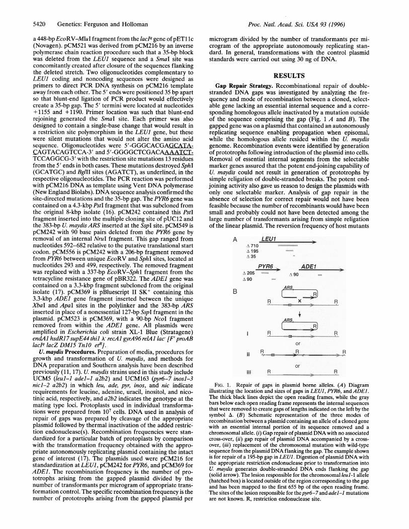
stranded DNA gaps was investigated by analyzing the fre-quency and mode of recombination between a cloned, select-able gene lacking an essential internal sequence and a corre-sponding homologous allele inactivated by a mutation outsideof the sequence comprising the gap (Fig. 1 A and B). Thegapped gene was on a plasmid that contained an autonomouslyreplicating sequence enabling propagation when episomal,while the homologous allele resided within the U. maydisgenome. Recombination events were identified by generationof prototrophs following introduction of the plasmid into cells.Removal of essential internal segments from the selectablemarker genes assured that the potent end-joining capability ofU. maydis could not result in generation of prototrophs bysimple religation of double-stranded breaks. The potent end-joining activity also gave us reason to design the plasmids withonly one selectable marker. Analysis of gap repair in theabsence of selection for correct repair would not have beenfeasible because the number of recombinants would have beensmall and probably could not have been detected among thelarge number of transformants arising from simple religationof the linear plasmid. The reversion frequency of host mutants
A LEU1iA 710A 195A 35
-PYR6 +-
ADE1lA 205A 90
B
ii
iii
A 90
ARS.
R x R.J__J..
.ARS _
R Rj > _ j
orR~~RR
orR R
FIG. 1. Repair of gaps in plasmid borne alleles. (A) Diagramillustrating the location and sizes of gaps in LEUI, PYR6, and ADEL.The thick black lines depict the open reading frames, while the graybars below each open reading frame represents the internal sequencesthat were removed to create gaps of lengths indicated on the left by thesymbol A. (B) Schematic representation of the three modes ofrecombination between a plasmid containing an allele of a cloned genewith an essential internal portion of its sequence removed and achromosomal allele. (i) Gap repair of plasmid DNA with no associatedcross-over, (ii) gap repair of plasmid DNA accompanied by a cross-over, (iii) replacement of the chromosomal mutation with wild-typesequence from the plasmid DNA flanking the gap. The example shownis for repair of a 195-bp gap in LEUI. Digestion of plasmid DNA withthe appropriate restriction endonuclease prior to transformation intoU. maydis generates double-stranded DNA ends flanking the gap(solid arrow). The lesion responsible for the chromosomal leul-1 allele(hatched box) is located outside of the region corresponding to the gapand has been mapped to the first 655 bp of the open reading frame.The sites of the lesion responsible for thepyr6-7 and adel-1 mutationsare not known. R, restriction endonuclease site.
Proc. Natl. Acad. Sci. USA 93 (1996)
A rnie
Proc. Natl. Acad. Sci. USA 93 (1996) 5421
utilized in this study was <10-7, which was so low that norevertants appeared during the course of transformation.Therefore generation of prototrophy under the conditions ofthese experiments was a true measure of recombination.Three modes of recombination were possible (Fig. 1B).
These were (i) gap repair of the missing information in theplasmid allele using the sequence of the chromosomal allele astemplate, but no integration of the plasmid; (ii) gap repair ofthe plasmid allele accompanied by integration of the plasmidinto the genome; and (iii) replacement of the chromosomalmutation by the corresponding wild-type sequence present onthe plasmid, without gap repair. Recombinants in this thirdmode could arise by gene conversion or by a double cross-overspanning the region but will be referred to as gene replace-ments for simplicity. The three modes of recombination were
easily distinguishable by Southern hybridization analysis. Thiswas accomplished by digestion of genomic DNA with a re-
striction enzyme that cut the transforming plasmid DNA at a
unique site within the vector sequence, but not within thesequence of the cloned marker, and followed by hybridizationwith a probe corresponding to the DNA sequence within thegap. Gap repair without associated crossing over was recog-nized as the appearance of a plasmid-length band of multicopyintensity along with a band representing the endogenous allele.Gap repair accompanied by crossing over was recognized bythe absence of the band representing the endogenous allele,but the appearance of two new bands whose sizes were the sumof the endogenous band plus the plasmid. Gene replacementswere recognized by a single band representing the endogenousallele.
Crossing Over Accompanies Gap Repair Infrequently. Re-combinants were readily obtained when plasmid DNA with a
gap was introduced into the appropriate host strain. Trans-formation of leul-1 with plasmid DNA containing a gap inLEU1 of 35 or 710 bp (Fig. 1A) resulted in an approximatelylinear response in Leu+ recombinants with increasing trans-forming DNA up to an input of 10 ,tg (data not shown),yielding several hundred colonies per microgram of inputDNA. To control for variability in cellular competence andDNA uptake, parallel transformations were conducted usingpCM216, an autonomously replicating plasmid containing theintact LEU1 gene. Transformation in this case does not rely onrecombination to confer leucine prototrophy, and therefore isa measure of transformation competence. Furthermore, sincelinear pCM216 containing the entire LEUI gene confers thesame transformation frequency as does the circular form (12),it can be concluded that conformation of DNA makes no
difference in uptake. The frequency of transformation wasabout 2-fold higher when plasmid DNAwith the 35-bp gap wasutilized compared with the plasmids with the 710- or 195-bpgap (Fig. 1 and Table 1).
Sets of recombinants obtained with plasmids containing gapsin LEU1 of 35, 195, and 710 bp were examined by Southernhybridization to determine the mode of recombination as de-scribed above. The majority of recombinants in each set arosethrough gap repair, with gene replacement comprising only 10%of the total (Table 1). Examination of gap repair events revealeda pronounced asymmetry in outcome indicated by a bias againstplasmid crossing over. Regardless of the gap size, plasmids wererepaired without crossing over several times more frequently thanwith crossing over. X: contingency tests confirmed that there wasindeed a bias against crossing over associated with gap repair (P< 0.01) but did not support the notion that the strength of the biaswas related to the size of the gap (0.05 < P < 0.1).
Recombinants obtained after transformation with plasmidscontaining gaps in two other genes were also examined (Table1). Gaps of 90 and 206 bp were generated in the PYR6 gene,and a gap of 90 bp was generated in theADE1 gene. The twogaps in the case ofPYR6 were not overlapping, unlike those inLEUI in which the largest gap spans the smaller two (Fig. 1).Gap repair of PYR6 occurred almost exclusively without associ-ated crossing over. Furthermore, none of the recombinantsobtained in the analysis of PYR6 was due to gene replacement.Repair of the 90-bp gap in theADEl gene also occurred with littleassociated crossing over, although the majority of Adel recom-binants was found to have arisen from gene replacement. Theseresults indicate that recombinational repair of gaps in plasmidDNA takes place with a strong bias against crossing over. It is ofinterest to note that the frequency of recombination atpyr6 andat adel was considerably higher than that observed at leul. Thereason for these differences is unclear, but the occurrence ofmarker specific effects in recombination is well known.
Heterologous DNA Blocks Gap Repair Only from One End.To gain insight into the mechanism responsible for the biasagainst crossing over, we investigated recombination of gappedgenes under conditions in which homologous sequence was
present at only one side of the gap. For these experiments, a
heterologous DNA sequence was inserted into the gap. Trans-formation was then performed using plasmid DNA cut on oneside of the heterologous block or the other, thereby generatinga gap, but with a heterologous stretch at the side eitherproximal or distal to the promoter (Fig. 2). When recombi-nation was measured at LEU1 and PYR6, the results indicatedthat there was no appreciable effect on recombination fre-
Table 1. Analysis of recombinants
Southern analysis
Gap repair(Crossing
Specific recombination Total Gene over)Gap size frequency, 10-2* analyzed replacement - +
LEUI710bp 3.0 49 5 41 3195 bp 2.2 36 5 28 235 bpt 7.1 54 4 39 11
PYR690 bp 24 35 0 34 1206 bp 29 35 0 34 1
ADEI90 bp 20 75 50 22 3
*Frequencies calculated from 2-4 independent transformations, each of which yielded 200-800colonies. Southern analysis was performed on a subset of each transformation.tpCM521 was used which contains RFLP point mutation markers flanking the gap sequence.
Genetics: Ferguson and Holloman

5422 Genetics: Ferguson and Holloman
ARS
t5' 3'
MIul Sphlor
EcoRV
ARS ARS ARSB ( : :+ (_ j, (----
No block promoter distal promoter proximalend blocked end blocked
~I ILEU1 2.2
PYR6 29
0.016
2.7
1.5
14
FIG. 2. Repair of gaps blocked with heterologous DNA. (A) Gaprepair was examined at LEUJ and PYR6 (solid arrow) in which gapswere filled with heterologous DNA (gray bar). By cutting the plasmidwith the appropriate restriction endonuclease prior to transformation,one of the two homologous DNA ends adjacent to the gap wasexposed, while the other remained blocked by the heterologous insert.Digestion with both restriction endonucleases whose sites flank theheterologous insert allowed both ends to be exposed prior to trans-formation. Gap repair of LEU1 was performed using pCM524, whichcontains 825 bp of heterologous DNA in place of an internal 195-bpMluI-SphI fragment, while gap repair of PYR6 was performed usingpCM556, which contains 337 bp of heterologous DNA in place of aninternal 206-bp EcoRV-SphI fragment. (B) Recombination frequen-cies obtained at leul-1 and pyr6-7 after transformation with theappropriate plasmid. The left column shows the frequencies observedwhen plasmid DNA with both ends exposed was transformed. Themiddle column shows frequencies obtained after leaving the promoterdistal side blocked, while the third column shows frequencies obtainedafter leaving the promoter proximal side blocked. The numbers aboveare the average of three independent transformations of 1 gg of inputDNA. Recombination frequencies were calculated as described.
quency when the promoter-proximal end was blocked. Incontrast, there was a precipitous drop in recombination at bothloci when the promoter-distal end was blocked. This overallinhibition of recombination by heterologous DNA blockingone side of a gap but not the other is indicative of asymmetryin the processing mechanism leading to gap repair. However,based upon these findings alone, no definitive conclusion canbe drawn as to whether the heterologous sequence blocks astep in initiation or a later step in resolution.Gene Conversion of Restriction Fragment Length Polymor-
phisms (RFLPs) in Sequences Flanking a Gap. A secondapproach addressing the mechanism of biased gap repair wasto examine gene conversion ofRFLP markers in the sequencesadjacent to a gap. To this end, a 35-bp gap was engineered inLEU1 with point mutations at sites 13 bp from either side ofthe gap. The mutations were silent in terms of codon usage, buteach eliminated a restriction enzyme site. The SphI siteupstream of the gap and proximal to the 5' end of the LEU1gene was altered, and the BglII site downstream of the gap, ordistal, was likewise altered. The disposition of these restrictionenzyme sites was determined in recombinants that arosethrough gap repair unassociated with crossing over. Analysis ofintegrated plasmids was not undertaken due to the smallsample size obtained and increased difficulty in interpretingthe nature of the events by restriction analysis.
Analysis of DNA from 24 recombinants containing gap-repaired but unintegrated plasmids indicated that in all 24cases there was gene conversion directed by the chromosomalleul-1 allele of at least one of the plasmid RFLP markersflanking the gap (Fig. 3). In 19 instances, all of the plasmid
DNA present in a particular recombinant was sensitive tocleavage by SphI, indicating that the mutated SphI site hadbeen converted to the recognition sequence (two-strandedconversion). These events could have resulted either fromenlargement of the gap beyond the engineered restriction sitemutation or from mismatch repair of heteroduplex DNAimmediately flanking the gap. In the remaining five samples,there was a mixed population of gap-repaired plasmids inwhich about half of the DNA was resistant to cleavage by SphIand half was sensitive, indicating that both the mutated SphIsequence and the normal sequence were present. These latterevents presumably arose from postdivision segregation ofheteroduplex DNA formed on the gap-repaired plasmid (one-strand conversion). While no example was found of a gap-repaired recombinant plasmid without gene conversion at theproximal RFLP marker (SphI site), in a significant fraction ofthe gap repair events there was no conversion of the distalRFLP marker (BglII site). In eight recombinants, the mutatedBglII site remained unconverted, while in the other 16 therewas an even distribution of one- and two-strand conversions.The apparent predominance of conversion events on theproximal side of the gap could indicate a polarity or endpreference in processing. These results could mean that thegap is enlarged more readily toward the proximal side or thatthe mismatch repair system operates more efficiently on thatside. Not a single example was found in which there wasconversion of the chromosomal RFLP markers directed by themutated sequences on the plasmid, implying that if the ob-served RFLP conversions arose by mismatch repair, the mech-anism would have to be strongly biased in favor of correctionto the sequence of the invaded duplex (18).
DISCUSSIONThere are two principal findings in this study. First, double-stranded DNA gaps in a cloned gene on a plasmid can berepaired through recombinational transfer of informationfrom homologous sequences within the genome but with little
Aleul alleleon plasmidleul-1 alleleon chromosome
13bp 35bp 13bp
SphlIBglll1
2 strands 19
50
Conversion 1 strandof RFLP
none
----
8
88
FIG. 3. Gene conversion of RFLP markers flanking a gap. (A)pCM521 was digested with SmaI to expose a 35-bp gap in the LEUIgene. The solid black arrow represents the portion of the leul allele inthe proximity of the gap; the gray arrow represents the correspondingchromosomal leul-1 sequence. The black Xs on the plasmid sequencerepresent RFLP markers 13 base pairs from the DNA ends thateliminate the SphI and BglII restriction sites. These sites are presenton the chromosomal allele and are represented by the gray circles.After transformation, the mode of recombination was determined bySouthern hybridization analysis following digestion withBamHI. Geneconversion of the point mutations flanking the gap was analyzed byRFLP analysis. Two categories of gene conversion were defined.Two-strand conversion regenerates the restriction endonuclease targetsite. One-strand conversion results from heteroduplex formationspanning the RFLP site followed by postreplication segregation ofdaughter sequences, one ofwhich contains the mutated restriction siteand one of which contains the normal sequence. The results of thisanalysis are shown aligned with the appropriate RFLP marker for 24recombinants resulting from gap repair without crossing over.
A Blocked gapin LEU1 or PYR6
Proc. Natl. Acad. Sci. USA 93 (1996)

Proc. Natl. Acad. Sci. USA 93 (1996) 5423
associated crossing over. Second, there is unequal processingof the DNA ends flanking the gap.Gap repair as envisioned by the double-strand break repair
model features the formation of an intermediate with Hollidayjunctions flanking both sides of the gap (3). The proposedmeans for resolution of the intermediate to yield cross-over ornoncross-over products invoked symmetrical cleavage of theHolliday structures and provided a theoretical mechanism inaccordance with the close association between gene conver-sion and crossing over of genetic markers that has beenobserved during meiosis and plasmid transformation in yeast.By contrast, in U. maydis little crossing over with the genomicsequence was found to accompany recombinational repair ofgapped plasmids. Examples of gap repair unassociated withcrossing over have been noted in other systems, but are oftenconsidered to be special situations. These include P element-induced gap repair in D. melanogaster (7) and mating typeswitching at the MAT locus in S. cerevisiae (6), both of whichare even more extreme in terms of infrequent crossing over.The bias against crossing over observed here, however, isconsistent with studies on heteroallelic recombination in U.maydis in which the association of crossing over with geneconversion is weak (19), and seems sensible biologically.Frequent crossing over in mitotic cells could lead to chromo-some imbalance as a consequence of pairing through repeatedsequences and could result in disaster for the cell. Thus, itmight be expected that systems are in place to prevent mitoticcrossing over. These findings raise the issue that if gap repairproceeds through an intermediate featuring Holliday struc-tures, as in the double-strand break-repair model, then theremust be some restraint on the mechanism of resolution of thestructures to explain the observed bias. This might arise as aresult of sequence specificity of a Holliday structure resolvingenzyme such as RuvC (20) and/or through constraints inresolution imposed by the tertiary conformation of the Hol-liday structure (21). Alternatively, symmetric Holliday junc-tions could be nullified by the action of a topoisomerase (22),or mechanisms could be in operation that circumvent theHolliday intermediates (22, 23).Two experimental approaches revealed that the DNA ends
adjacent to the gaps were processed asymmetrically. Thestrategy taken in the first approach was to ask if gap repaircould be completed if one DNA terminus contained a heter-ologous block of DNA. In two different genes it was found thatproficient repair of gaps filled with heterologous DNA pro-ceeded, but only when the configuration ofDNA was arrangedso that the junction was cut between the heterologous insertand the marker gene sequence at the promoter-distal end ofthe gap, not at the promoter-proximal end. The strategy takenin the second approach was to follow the fate of restriction sitepolymorphisms in the plasmid LEUI sequence placed 13 basepairs away from a gap, thereby enabling a higher resolutionview of processing of the ends. There was gene conversion ofthe RFLP marker at the proximal end sequence in all of thegap repair events examined, while the RFLP marker at thedistal end escaped gene conversion one-third of the time. Theresults from the two approaches are consistent and could beinterpreted to mean that both DNA strands at the promoter-proximal end of the gap are removed by nucleolytic digestionat an initial step or at a later step in gap repair, while at leastone strand at the distal end is relatively protected throughoutthe process. Asymmetric enlargement of DNA gaps has beennoted in other experimental systems (24, 25).The asymmetry in both processing and outcome observed in
these studies can be accounted for by a recombination modelfeaturing a single migrating D-loop (Fig. 4). A single-stranded3' tail generated by exonucleolytic processing of a brokenDNA end at one side of the gap is imagined to invade thehomologous duplex forming a D-loop and a primer for a repairpolymerase. The D-loop does not become enlarged, but is
B-,,~~
Nicking at A Nicking at B
Gap repair withoutcrossing over _
Gap repair withoutcrossing over
Gap repair withcrossing over
FIG. 4. The migrating D-loop model of recombination. Repairsynthesis at the 3' terminus of the invading strand drives migration ofthe D-loop into the gap and pushes it past the DNA end on theopposite side. The nascent single strand is displaced during D-loopmigration and can thus pair with complementary sequences on theother side of the gap. After second-strand synthesis, an endonucleasecleaves the nascent single strand (arrow A), which results in gap repairwithout crossing over, or the single-strand of the D-loop (arrow B),which leads to formation of a Holliday junction adjacent to therepaired gap. Resolution of the junction by a Holliday endonucleaseleads to crossing over in half of these instances, and thus at most 25%of the total.
instead driven to migrate into the gap in a manner similar towhat was observed for UvsX-dependent DNA synthesis in vitro(26). The newly synthesized strand is continuously extrudedfrom the D-loop and eventually spans the entire gap. It is thenfree to make contact with the distal broken DNA end, andcomplementary base pairing is effected. After second-strandsynthesis, single-stranded endonucleolytic and exonucleolyticdigestion of the newly synthesized strand trailing behind themigrating D-loop will resolve the intermediate resulting inrepair of the gap with no crossing over. Although rare, crossingover was observed in association with gap repair in U. maydis.A cross-over could result from the asymmetric mechanismdescribed above if on occasion the endonuclease cleaved thesingle strand of the D-loop rather than the displaced nascentstrand (Fig. 4) or if the D-loop simply collapsed releasing thenewly synthesized strand. The new free single-stranded endcould then pair with the homolog to yield a Holliday structurein a manner similar to the mechanism proposed in the Me-selson-Radding model (27). The migrating D-loop modeldiffers from the most common depiction of the double-strandbreak-repair model in that only one side of the gap interactswith the homolog and the resulting D-loop is forced to moveinstead of being enlarged. The initiation of gap repair on onlyone side is similar to an early model of double-strand breakrepair proposed by Resnick (2).
It remains an important question in the migrating D-loopmodel as to whether the protected distal side or the unpro-tected proximal side of the gap invades the homolog to initiate
Genetics: Ferguson and Holloman
odb--

5424 Genetics: Ferguson and Holloman
repair. It is possible that in U. maydis the 3' end of the DNAon the promoter distal side of the gap is protected, perhaps bycomponents of the recombination machinery, during thesearch for homology and D-loop formation. It follows then thatthe promoter-proximal end is not as good a substrate for therecombination machinery and is thus susceptible to degrada-tion and possibly also the mismatch repair apparatus. Thedifference in the action of the recombination machinerybetween the two sides could be due to interference by, orinteraction with, local directional processes such as replicationor transcription.
We thank Dr. Sally Leong for providing the cloned PYR6 gene. andDrs. Lorraine Symington, Hannah Klein, and Jim Haber for commentson the manuscript. This work was supported by Grant GM42482 fromthe National Institutes of Health.
1. Petes, T. D., Malone, R. E. & Symington L. S. (1991) in Recom-bination in yeast, eds. Broach, J. R., Pringle, J. R. & Jones, E. W.(Cold Spring Harbor Laboratory Press, Cold Spring Harbor,NY), pp. 407-522.
2. Resnick, M. A. (1976) J. Theor. Biol. 59, 97-106.3. Szostak, J. W., Orr-Weaver, T. L., Rothstein, R. J. & Stahl, F. W.
(1983) Cell 33, 25-35.4. Orr-Weaver, T. L. & Szostak, J. W. (1983) Proc. Natl. Acad. Sci.
USA 80, 4417-4421.5. Nickoloff, J. A., Singer, J. D., Hoekstra, M. F. & Heffron, F.
(1989) J. Mol. Biol. 207, 527-541.6. Strathern, J. N., Klar, A. J., Hicks, J. B., Abraham, J. A., Ivy,
J. M., Nasmyth, K. A. & McGill, C. (1982) Cell 31, 183-192.7. Gloor, G. B., Nassif, N. A., Johnson-Schlitz, D. M., Preston,
C. R. & Engels, W. R. (1991) Science 253, 1110-1117.8. Plessis, A. & Dujon, B. (1993) Gene (Amst.) 134, 41-50.
9. Fishman-Lobell, J., Rudin, N. & Haber, J. E. (1992) Mo. Cell.Biol. 12, 1292-1303.
10. Sugawara, N., Ivanov, E. L., Fishman-Lobell, J., Ray, B. L., Wu,X. & Haber, J. E. (1995) Nature (London) 373, 84-86.
11. Fotheringham, S. & Holloman, W. K. (1990) Genetics 124, 833-843.
12. Fotheringham, S. & Holloman, W. K. (1991) Genetics 129, 1052-1060.
13. Fotheringham, S. & Holloman, W. K. (1989) Mol. Cell. Biol. 9,4052-4055.
14. Rubin, B. P., Li, D. & Holloman, W. K. (1994) Gene (Amst.) 140,131-135.
15. Tsukuda, T., Carleton, S., Fotheringham, S. & Holloman, W. K.(1988) Mol. Cell. Biol. 8, 3703-3709.
16. Kronstad, J. W., Wang, J., Covert, S. F., Holden, D. W., McK-night, G. L. & Leong, S. A. (1989) Gene (Amst.) 79, 97-106.
17. Rubin, B. P., Ferguson, D. 0. & Holloman, W. K. (1994) Mol.Cell Biol. 14, 3863-3875.
18. Ray, B. L., White, C. I. & Haber, J. E. (1991) Mol. Cell. Biol. 11,5372-5380.
19. Holliday, R., Halliwell, R. E., Evans, M. W. & Rowell, V. (1976)Genet. Res. 27, 413-453.
20. Bennett, R. J., Dunderdale, H. J. & West, S. C. (1993) Cell 74,1021-1031.
21. Clegg, R. M., Murchie, A. I., Zechel, A., Carlberg, C., Diekmann,S. & Lilley, D. M. (1992) Biochemistry 31, 4846-4856.
22. Hastings, P. J. (1988) Bioessays 9, 61-64.23. Nassif, N., Penney, J., Pal, S., Engels, W. R. & Gloor, G. B. (1994)
Mol. Cell. Biol. 14, 1613-1625.24. White, C. I. & Haber, J. E. (1990) EMBO J. 9, 663-673.25. Sweetser, D. B., Hough, H., Whelden, J. F., Arbuckle, M. &
Nickoloff, J. A. (1994) Mol. Cell. Bio. 14, 3863-3875.26. Formosa, T. & Alberts, B. M. (1986) Cell 47, 793-806.27. Meselson, M. S. & Radding, C. M. (1975) Proc. Natl. Acad. Sci.
USA 72, 358-361
Proc. Natl. Acad. Sci. USA 93 (1996)