Embed Size (px)
Citation preview
The Bactertal Chromosome Edited by N. Patrick Higgins
2005 ASM Press, Washington, D.C.
Chapter 21
Homologous Recombination by the RecBCD and RecF Pathways
RECOMBINATIONAL REPAIR OF DNA DAMAGE
Homologous, or general, recombination is a cru- cial biological process that involves the paring and transfer of strands between DNA molecules that share a region of significant sequence homology. After its discovery in 1946 by Lederberg and Tatum (48), ho- mologous recombination in bacteria was associated with the sexual process of conjugation and was viewed as an evolutionary mechanism both for shuffling the genome and for spreading favorable alleles. However, more recently, a more immediate function of homol- ogous recombination has been recognized: namely, it is a mechanism for the maintenance of chromosomal integrity that acts to repair DNA lesions, both double- strand DNA breaks and single-strand DNA gaps, gen- erated during the course of DNA replication. The intimate connection between the processes of replica- tion and recombination was initially appreciated in the life cycle of bacteriophage T4 (56) and then later rec- ognized as an important determinant of viability in bacteria (39,45). In T4 phage, recombination is linked to replication to produce a high yield of phage DNA; in Escherichia coli, recombination is linked to repli- cation to permit its completion when interrupted by DNA damage, and also to initiate DNA replication in the absence of origin function. This view of recom- bination as an integral part of efficient chromosome duplication also reconciled the high level of inviability (up to 95%) of recombination-deficient cells (12).
GENERATION OF A DSB IN DNA
A significant fraction of DNA damage affects only one strand of the DNA duplex. Such lesions can often be repaired by one of the repair systems that are
specific for a particular DNA lesion (see reference 28 for a review). These repair systems use the intact com- plementary strand as a template to restore the dam- aged DNkmolecule to its original state. Occasionally both strands of the double-stranded DNA (dsDNA) can be broken opposite to each other, resulting in a double-strand break (DSB). DSBs can be produced, for example, as a direct consequence of ionizing ra- diation (Fig. 1A). However, the bulk of the DSBs in bacteria are generated indirectly as the result of DNA replication through an unrepaired break in just a single strand of DNA (Fig. 1B). Replication of DNA containing a single-strand nick or a gap in the leading strand results in the dissociation of the DNA poly- merase holoenzyme complex and the generation of one blunt DSB (Fig. IB), whereas replication of DNA with a nick in the lagging strand produces a DSB with a 3'-terminated single-stranded DNA (ssDNA) tail.
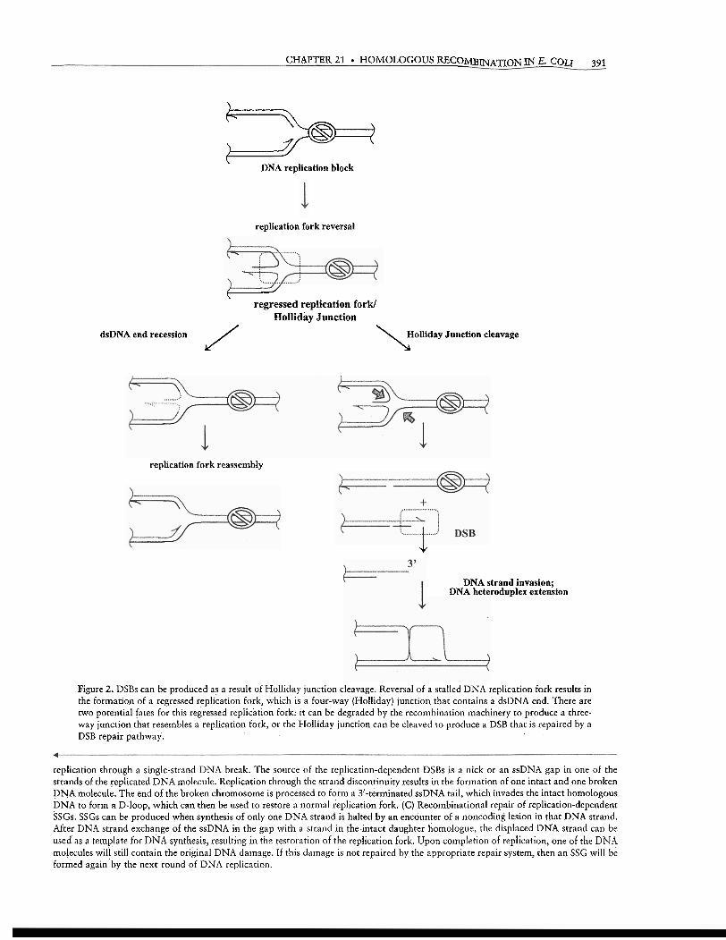
A DSB can also be produced when the DNA replication process is halted by anything that might block the progress of the replisome. Upon dissocia- tion of the replisome, the stalled replication fork can regress to produce a Holliday junction. Such structure contains both a dsDNA end and an intermediate of reconibination and, hence, attracts recombination machinery in a manner similar to that of the simple DSB. In addition, the Holliday junction can be cleaved to provide yet another path for DSB formation (Fig. 2). Left unrepaired, DSBs are lethal, and E. coli can re- pair only a few DSBs per chromosome without dying (43). Thus, DNA replication is a major source of endogenous DSBs that, in turn, are repaired by ho- mologous recombination. This relationship between DNA replication and recombinational repair of DSBs is summarized in Fig. 1.
Recombinational repair, however, requires a homologous DNA molecule to be used as a template from which to restore, by DNA synthesis, the genetic
Maria Spies and Stephen C. Kowalczykowski . Sections of Microbiology and of Molecular and Cellular Biology, Center for Genetics and Development, University of California, Davis, CA 95616-8665.
A)
dire
ctly
indu
ced
DSB
s B
) rep
licat
ion-
depe
nden
t DSB
s C
) rep
licat
ion-
depe
nden
t SS
Gs
nick
or
gap
in t
he
nick
or
gap
in th
e le
adin
g le
adin
g st
rand
tem
plat
e st
rand
tem
plat
e
I I
DN
A st
rand
inva
sion
; he
tero
dupl
ex e
xten
sion
1 re
plic
atio
n re
star
t
Hol
liday
junc
tion
r.01.t
ion
I D
SB
..
..
i
DSB
C
DSB
is r
epai
red;
repl
icat
ion
rest
arts
\
stra
nd in
vasi
on;
hete
rodu
plex
exte
nsio
n
mi
ca
";
/
rest
art
\ nuc
leas
e
reso
lutio
n\
Jhet:,"
d;p1ex
,
. ex
tens
ion
no,n
sodi
ng le
sion
in th
e no
n-co
ding
lesi
on in
the
lead
ing
stra
nd te
mpl
ate
lead
ing
stra
nd te
mpl
ate
DN
A s
tran
d in
vasi
on 1
repl
icat
ion
; 1
Figu
re 1
. Rec
ombi
natio
nal
repa
ir o
f D
NA
dam
age.
The
DN
A s
tran
ds th
at a
re u
sed
as te
mpl
ates
for
the
lead
ing
stra
nd s
ynth
esis
are
sho
wn
in b
lack
, an
d th
e st
rand
s us
ed a
s te
mpl
ates
for
the
lagg
ing
stra
nd s
ynth
esis
are
sho
wn
in g
ray.
Arr
ows
indi
cate
the
dire
ctio
n of
DN
A s
ynth
esis
. (A
) Rec
ombi
natio
nal
repa
ir o
f di
rect
ly in
duce
d D
SBs.
A D
SB, w
hich
occ
urs
in n
ewly
syn
thes
ized
DN
A, c
an b
e re
pair
ed b
y co
mpl
etio
n of
the
fol
low
ing
step
s. F
irst
, the
DSB
is p
roce
ssed
to
prod
uce
3'-t
erm
mat
ed s
sDN
A. T
hen,
one
of
the
ssD
NA
tails
can
inv
ade
the
hom
olog
ous
dsD
NA
dau
ghte
r, d
ispl
acin
g on
e of
the
res
iden
t st
rand
s to
for
m a
D
-loo
p. T
his
stru
ctur
e ca
n be
use
d as
a t
empl
ate
for
DN
A s
ynth
esis
, ulti
mat
ely
resu
lting
in
the
form
atio
n of
a H
ollid
ay j
unct
ion.
Upo
n re
solu
tion
of t
he
Hol
lida
y ju
nctio
n, t
he r
eplic
atio
n fo
rk is
res
tore
d to
its
orig
inal
for
m. W
hen
a D
SB o
ccur
s in
a p
art o
f the
chr
omos
ome
that
is n
ot y
et re
plic
ated
, th
ere
is no
ho
mol
ogou
s D
NA
to s
erve
as
a te
mpl
ate,
and
suc
h a
DSB
can
be
leth
al. (
B) R
ecom
bina
tiona
l rep
air
of re
plic
atio
n-de
pend
ent D
SBs.
DSB
s ca
n be
pro
duce
d by
CHAPTER 21 HOMOLOGOUS RECOMBINATION IN E. C o t 1 391
> DNA replication block
replication fork reversal
w regressed replication fork/
Holliday Junction
dsDNA end recession J Holliday Junction cleavage \
replication fork reassembly
J.
DNA strand invasion; DNA heteroduplex extension
Figure 2. DSBs can be produced as a result of Holliday junction cleavage. Reversal of a stalled DNA replication fork results in the formation of a regressed replication fork, which is a four-way (Holliday) junction that contains a dsDNA end. There are two potential fates for this regressed replication fork: it can be degraded by the recombination machinery to produce a three- way junction that resembles a replication fork, or the Holliday junction can be cleaved to produce a DSB that is repaired by a DSB repair pathway.
replication through a single-strand DNA break. The source of the replication-dependent DSBs is a nick or an ssDNA gap in one of the strands of the replicated DNA molecule. Replication through the strand discontinuity results in the formation of one intact and one broken DNA molecule. The end of the broken cliromosome is processed to form a 3'-terminated ssDNA tail, which invades the intact homologous DNA to form a D-loop, which can then be used to restore a normal replication fork. (C) Recombinational repair of'replication-dependent SSGs. SSGs can be produced when synthesis of only one DNA strand is halted by an encounter of a noncoding lesion in that DNA strand. After DNA strand exchange of the ssDNA in the gap with a strand in theintact daughter homologue, the displaced DNA strand can be used as a template for DNA synthesis, resulting in the restoration of the replication fork. Upon completion of replication, one of the DNA molecules will still contain the original DNA damage. If this damage is not repaired by the appropriate repair system, then an SSG will be formed again by the next round of DNA replication.
392 SPES AND KOWALCZYKOWSKI
content of the damaged DNA molecule. These ho- mologous DNA sequences are generated by DNA replication, and, therefore, the meridiploid character of bacterial chromosomes provides the necessary templates for recombinational repair.
GENERATION OF A SINGLE-STRAND GAP IN DNA
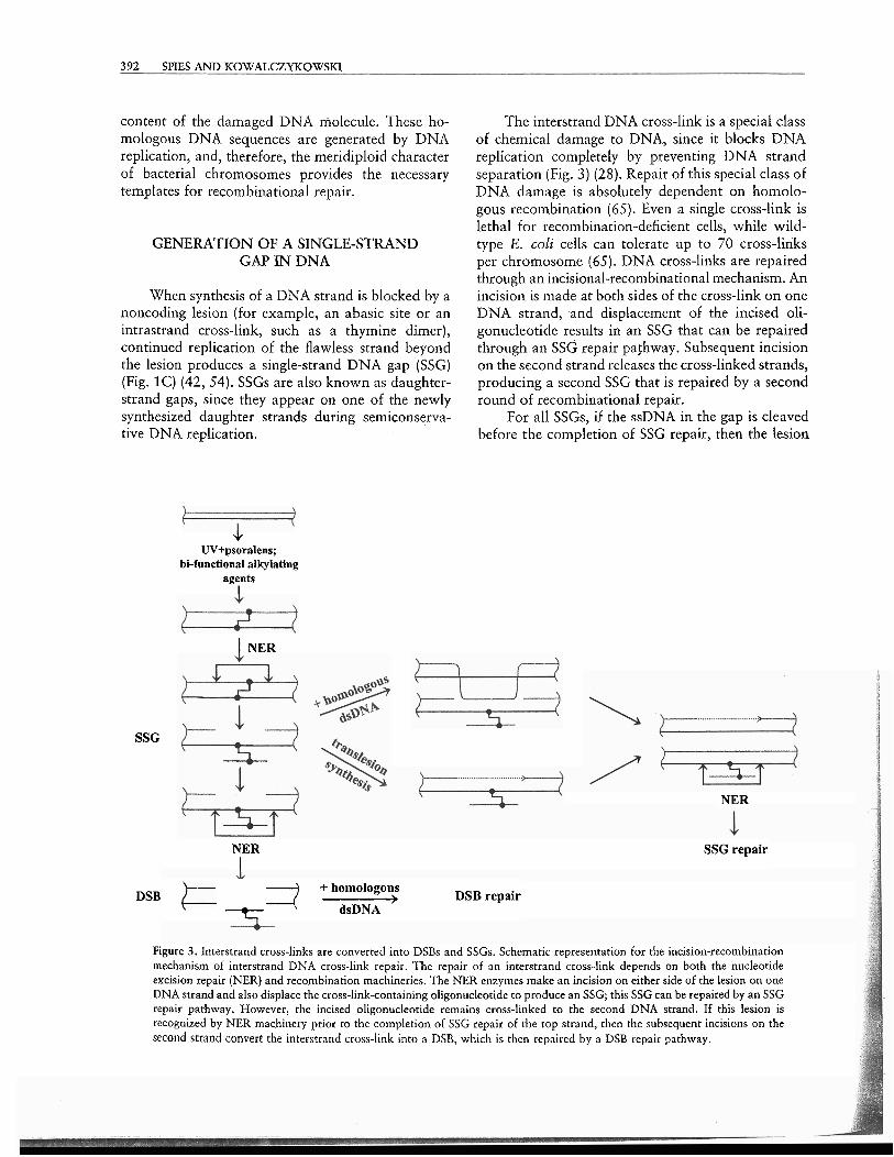
When synthesis of a DNA strand is blocked by a noncoding lesion (for example, an abasic site or an intrastrand cross-link, such as a thymine dimer), continued replication of the flawless strand beyond the lesion produces a single-strand DNA gap (SSG) (Fig. 1C) (42, 54). SSGs are also known as daughter- strand gaps, since they appear on one of the newly synthesized daughter strands during semiconserva- tive DNA replication.
SSG
+ W+psoralens;
bi-functional alkylating agents
I
I NER
The interstrand DNA cross-link is a special class of chemical damage to DNA, since it blocks DNA replication completely by preventing DNA strand separation (Fig. 3) (28). Repair of this special class of DNA damage is absolutely dependent on homolo- gous recombination (65). Even a single cross-link is lethal for recombination-deficient cells, while wild- type E. coli cells can tolerate up to 70 cross-links per chromosome (65). DNA cross-links are repaired through an incisional-recombinational mechanism. An incision is made at both sides of the cross-link on one DNA strand, and displacement of the incised oli- gonucleotide results in an SSG that can be repaired through an SSG repair pathway. Subsequent incision on the second strand releases the cross-linked strands, producing a second SSG that is repaired by a second round of recombinational repair.
For all SSGs, if the ssDNA in the gap is cleaved before the completion of SSG repair, then the lesion
NER
NER SSG repair I
+ homologous DSB &DNA ' DSB repair
r;l
Figure 3. Interstrand cross-links are converted into DSBs and SSGs. Schematic representation for the incision-recombination mechanism of interstrand DNA cross-link repair. The repair of an interstrand cross-link depends on both the nucleotide excision repair (NER) and recombination machineries. The NER enzymes make an incision on either side of the lesion on one DNA strand and also displace the cross-link-containing oligonucleotide to produce an SSG; this SSG can be repaired by an SSG repair pathway. However, the incised oligonucleotide remains cross-linked to the second DNA strand. If this lesion is recognized by NER machinery prior to the completion of SSG repair of the top strand, then the subsequent incisions on the second strand convert the interstrand cross-link into a DSB, which is then repaired by a DSB repair pathway.
CHAPTER 21 HOMOLOGOUS RECOMBINATION IN E. COLI 393
is converted into a DSB. Thus, some fraction of SSGs are converted into DSBs and are repaired through the DSB repair pathway.
CONJUGATION, TRANSDUCTION, AND TRANSFORMATION CREATE
DSBs AND SSGs
DSBs arise riot only as a consequence of DNA damage; in fact, DSBs are formed as a natural step in several normal cellular processes. The molecular feature common to each of these processes is the cellular acquisition of a linear segment of dsDNA. The end of this linear DNA molecule is seen as a DSB, and the recombinational repair of the DSB is initiated. Bacteria can acquire linear DNA by any of three different routes. First, during conjugation, a copy of a chromosome is transferred from one bacte- rium, which possesses a fertility factor (the F' plas- mid), to another. Genetic studies of this process led to isolation of the first recombination-deficient mutants (21). Transformation is a secoild route by which some bacteria take up a segment of DNA from the envi- ronment; unrelated in process, but not in the form of the DNA involved, artificial transformation is widely used as a laboratory technique to introduce foreign dsDNA into cells. Finally, infection by bacteriophage also results in the introduction of linear DNA into bacteria. Note, however, that in addition to DSBs, the processes of conjugation and natural transfor- mation also produce SSGs due to incomplete repli- cation of the DNA intermediates that form during these processes.
HOMOLOGOUS PAIRING AND DNA STRAND EXCHANGE MEDIATED
BY RecA PROTEIN
A step common to all pathways of recombina- tional repair discussed below is the homologous pairing of DNA (Fig. 1). In bacteria, the ubiq~litous RecA protein catalyzes an invasion of ssDNA into homologous duplex DNA and the exchange of DNA strands. Regardless of whether the DNA lesion is a DSB or an SSG, pathway-specific processing of the break produces an extensive region of ssDNA that serves as the substrate for assembly of the RecA nucleoprotein filament. The process of finding DNA sequence homology and exchanging DNA strands occurs in three defined stages: (i) presynapsis, during which the RecA nucleoprotein is assen~bled; (ii) syn-
apsis, during which the homology search and ex-
change of DNA strands occur; and (iii) postsynapsis, during which branch migration can occur. Afterward, resolution of the resulting recombination intermedi- ate produces a recombinant molecule.
In E. coli, recA null mutations reduce conjuga- tional recombination by 100,000-fold (21). The ac- tive form of RecA protein is a nucleoprotein filament formed by the cooperative binding of RecA protein to the ssDNA tails of the processed DSB, or to the ssDNA in the SSG (see reference 6 for a review). Thus, ssDNA is an essential, although transient, in- termediate in the process of homologous recombi- nation. Most of the SSGs, whose average length is minimally 200 nucleotides (78) (but can be as large as 800 nucleotides [37], or half--the average size of an Okazaki fragment [66]), are sufficiently large for RecA nucleoprotein filament assembly; however, DSBs are not the normal direct targets of RecA protein binding. Therefore, DSBs and short SSGs are processed to produce longer regions of ssDNA. This processing requires either a nuclease, a helicase, or both (see below).
However, production of ssDNA is not sufficient for RecA nucleoprotein filament assembly in vivo. Within the cell, ssDNA rapidly forms a complex with the ssDNA-binding (SSB) protein. SSB protein bind- ing has the beneficial consequences of protecting ssDNA from degradation by nucleases and of disrupt- ing inhibitory DNA secondary structures. Unfortu- nately, SSB protein blocks assembly of the RecA nucleoprotein filament by competing directly with RecA protein for ssDNA binding. Thus, the SSB protein-ssDNA complex is a kinetic barrier for the assembly of a RecA-ssDNA nucleoprotein filament, and its formation is inhibitory for the subsequent steps of homologous recombination. However, t o counteract this inhibitory effect of SSB protein, there is a class of the recombination/replieation mediator proteins whose function is to facilitate assembly of RecA protein (and its homologues in other organ- isms) on ssDNA, thereby alleviating the kinetic bar- rier imposed by SSR protein (the role of replication mediator proteins is reviewed in reference 5 ) . Thus, in addition to the production of a single-stranded region within dsDNA, recombination requires the "loading" of RecA protein onto this ssDNA.
In wild-type E. coli, the processing of a bro- ken DNA molecule, and the subsequent delivery of RecA protein to this ssDNA, occurs by either of two pathways: RecBCD or RecF. The pathway names reflect critical and unique enzymes acting in each. The RecBCD pathway is used primarily to initiate re- combinatioti at a DSR, whcrcas the RccF patllrvay is used for recombinational repair at SSGs. The concept
394 SPLES AND KOWALCZYKOWSKI
of recombiriation pathways was initially postulated by Clark (20). In both pathways, the Holliday junc- tions that result are resolved by the RuvABC enzyme complex (see reference 46 for a review) into the re- combinant progeny.
DSB REPAIR BY RECOMBINATION: THE RecBCD PATHWAY
The RecBCD pathway comprises the RecBCD, SSB, RecA, and RuvABC proteins; in addition, a specific DNA locus called x (Chi [crossover hot spot instigator], 5'-GCTGGTGG-3') is required in the dsDNA that is broken.
In wild-type E. coli, RecBCD enzyme is required for approximately 99% of the recombination events associated with conjugation and transduction, i.e., the processes that involve linear DNA and, hence, a DSB. Genetic studies revealed that the products of recB and recC genes are necessary for the repair of DSBs and for conjugational recombination. Deletion of either recB or recC genes reduces the levels of sexual recombination 100- to 1,000-fold and in- creases sensitivity to DNA-damaging agents, such as UV irradiation and mitomycin C (26, 36, 84). This effect, however, is significantly smaller then the 100,000-fold decrease in homologous recombination in the recA mutant cells. In contrast, cells deleted for recD are not only proficient in homologous recom- bination and DSB repair, but they also catalyze ho- mologous recombination at high rates (1).
The RecBCD enzyme is a heterotrimer consisting of three nonidentical polypeptides, RecB (134 kDa), RecC (129 kDa), and RecD (67 kDa) (1, 58). The three subunits compose a complex, multifunctional en- zyme that possesses a number of seemingly disparate catalytic activities, including DNA-dependent ATPase, DNA helicase, ssDNA endo- and exonuclease, and dsDNA exonuclease. In vivo, these activities enable RecBCD enzyme to carry out a highly coordinated set of biochemical reactions resulting in conversion of the broken dsDNA into the active species in homol- ogous recombination: the RecA ~lucleoprotein fila- ment assembled on the ssDNA containing X.
Among DNA helicases, an unusual feature of the RecBCD enzyme is its ability to initiate unwind- ing at a blunt or nearly blunt dsDNA end. RecBCD enzyme binds to blunt DNA ends with a very high affinity ( - 1 nM), forming an initiation complex (30) that has a footprint of about 20 or 21 nucleotides on the 5'-terminated strand and 16 to 17 nucleotides on the ?'-terminated strand.
RecB protein has a modular organization. The N-terminal domain of the RecB sub~init contains
motifs characteristic for the superfamily 1 (SF1) DNA helicases. Moreover, the purified RecB protein is an ssDNA-dependent ATPase and a DNA helicase (10). Its behavior is related to the well-characterized Rep, UvrD, and PcrA helicases. Similar to these en- zymes, RecB protein displays 3' -, 5' helicase activ- ity, requiring a 3'-terminated ssDNA region flanking the duplex DNA that is to be unwound. The C- terminal domain of this same subunit contains a nu- clease motif resembling that of other nucleases, such as FokI and BamHI (77, 85) and h exonuclease (2c). Besides being responsible for the complicated nucle- ase activity of RecBCD enzyme, the C-terminal do- main of the RecB subunit *has another crucial function: it harbors a site for interaction with RecA protein (19).
The amino acid sequence of RecC protein pro- vides no clue as to its role in the holoenzyme. How- ever, the existence of the RecC mutants that enable the holoenzyme to recognize an altered x sequence suggests a significant role in x recognition ( 3 ) . In- teraction with the RecC subunit greatly stimulates the weak nuclease activity of the RecB helicase, and increases its affinity for dsDNA ends (61). The re- sulting RecBC enzyme is a fast, processive helicase that call initiate homologous recombination. How- ever, RecBC enzyme displays negligible nuclease ac- tivity. As a result, homologous recombination in the recD mutant cells strongly depends on the function of RecJ nuclease (51, 51a).
The RecD subunit also contains SF1 helicase motifs. The purified RecD protein is a DNA-dependent ATPase that was recently shown to be a 5' -t 3' helicase (23) similar to the closely related TraI and Dda helicases. Like these other helicases, RecD protein unwinds substrates containing 5'-terminated ssDNA flanking the duplex DNA. Interaction with the RecD subunit further stimulates both the helicase activ- ity and the dsDNA end-binding affinity of RecBC enzyme. Given that the RecB subunit is bound to the 3'-terminated strand, the RecD subunit is bound to the 5'-terminated strand (27), and each motor subunit moves wi-th an opposite polarity, the resulting bipolar RecBCD enzyme translocates with each motor sub- unit moving in the same direction relative to the DSB. In addition, the resulting RecBCD holoenzyme now manifests its vigorous nuclease activity, implying that the RecD subunit also activates the nuclease con- tained within the RecB subunit (41).
The activities described above permit the fol- lowing description of the mechanism of action of the RecBCD helicaselnuclease (Fig. 4). Upon binding to the DNA end (Fig. 4a), the enzymc uses the energy of ATP hydrolysis to translocate along and unwind the dsDNA molecule, consuming approximately two
CHAPTER 21 HOMOLOGOUS RECOMBINATION IN E. COLl 395
396 SPIES AND KOWALCZYKOWSKI I
molecules of ATP per base pair unwound (63) (Fig. 4b). esse~ltial for initiation of hon~ologous recombination: RecBCD enzyme unwinds, on average, 30,000 bp (i) it recesses the DSB to produce an ssDNA-tailed of dsDNA per binding event (62) at a rate of ap- duplex DNA with x at its terminus, and (ii) it cata- proximately 1,000 to 1,300 bpls (at 37°C). DNA un- lyzes formation of the RecA nucleoprotein filament winding by RecBCD enzyme is accompanied by on the ssDNA produced. This RecA nucleoprotein endonucleolytic cleavage of the newly produced filament now can search for homology, promote in- ssDNA. The nuclease activity of RecBCD enzyme is vasion of the homologous recipient, and exchange the asymmetric, with digestion occurring preferentially DNA strands (Fig. 4e). on the 3'-terminated strand relative to the DSB (25). , The exact molecular mechanism by which x
recognition is translated into the observed changes in the activities of the RecBCD enzyme remains un-
THE RECOMBINATlON HOT SPOT, x known. Most models propose either dissociation or inactivation of the RecD subunit (2, 19, 57, 73).
Originally, x was identified as a cis-acting mu- Indeed, as mentioned previously, the RecBC enzyme tation in bacteriophage h that allowed its more effi- (lacking the RecD subunit) is'recombinationally pro- cient growth in E. coli by stimulating the host's ficient both in vivo (13) and in vitro (18). The RecBC recombination system. Stimulation of recombination enzyme is a processive helicase but with little or no is approximately 10-fold (47), and it is confined to nuclease activity (411, which is in contrast to the loci that are downstream of x (70): stimulation is X-activated form of RecBCD enzyme (Fig. 4a'); how- highest at x and then decreases exponentially (17,47). ); however, this distinction is consistent with the re- The x sequence is overrepresented in E. coli: 1,009 x quirement for RecJ nuclease activity in recD mutant sequences are found in the 4.6-hlb genome of the cells in vivo (Fig. 4b') (51). Similar to RecBCD en- hlGl65.S strain (9). Furthermore, over 60% of x se- zyme, RecBC helicase facilitates asymmetric assem- quences are oriented toward replication origin. Such bly of RecA protein only onto ssDNA that is 3' an orientation would facilitate RecBCD-mediated terminal at the enzyme entry site (18) (Fig. 4e'). The reconlbination repair of DSBs created during DNA RecBC-mediated loading of RecA protein is consti- replication (11). tutive and is independent of X, consistent with the
Biochemically, the activities of RecBCD enzyme phenotypic behavior of recD rrlutant cells in vivo. are altered upon recognition of X. Alteration of RecBCD enzyme activity is manifested only when the enzyme approaches X, 5'-GCTGGTGG-3', from its 3' REGULATED HELICASES / NUCLEASES side. In the schematic depiction (Fig. 4), X, which is IN OTHER BACTERIAL SPECIES the sequence in the "top strand" (7), is recognized by an enzyme moving only from right to left. In vivo, For decades, the interaction between RecBCD interaction with x results in the stimulation of ho- enzyme and x was known to exist only in the species mologous recombination downstreal11 of x (69, 71). of enteric bacteria closely related to E. coli. But rel- In vitro, recognition of the x sequence causes RecBCD atively recently, short (5 to 8 bp) sequences, which enzyme to switch the polarity of its nuclease activity protect linear dsDNA from degradation by attenua- (Fig. 4c): upon interaction with X, degradation of the tion of the nuclease activity of RecBCD-like helicasel 3'-terminated strand is downregulated (24,25), while nuclease enzymes, were found in several distantly degradation of the 5'-terminated strand is upregu- related bacteria (8, 16, 67). This finding indicates lated (2a). Consequently, the enzyme produces a that, although apparently not universal, the regula- lengthy ssDNA tail with x at the 3'-terminated end. tion of recombinational helicaseslnucleases by specific As determined from production of the X-specific DNA sequences is widely spread among prokaryotes. ssDNA fragments, the probability of recognizing a While some bacteria possess clear holnologues of single xis about 30 to 40% (3,72). Interaction with x RecBCD enzyme, other species contain its functional also affects the helicase activity of RecBCD enzyme. equivalent, the AddAB enzyme (reviewed in reference Recognition of x causes the enzyme to pause briefly 15). AddAB helicaselnuclease comprises two subunits (typically, a few seconds) at x and to resume trans- encoded by addA and addB genes. The sole motor location after the x site, but at a rate that is reduced subunit of AddAB enzyme, AddA protein, contains an by approximately twofold (68). Another consequence SF1 helicase and a nuclease domain, which display of x modification is that the RecBCD enzymc gains a high degree of similarity to those in RecB pro- the ability to "load" RecA protein onto the newly tein (32). Also similar to RecB protein, AddA is a produced ssDNA (2h) (Pig. 4J). Thus, in rcspolise 3' -+ 5' llelicase, and its 3' + 5' nuclease activity is to X, the RecBCD enzyme acconlplishes both tasks downregulated upon interaction with the cognate
CHAPTER 21 0' HOMOLOGOUS RECOMBINATION IN E. COW 397
recombination hot spot of Bacillus subtilis, X B ~ (5'- AGCGG-3') (14, 16). The AddB subunit has no sub- stantial similarity to either the RecC or the RecD subunit, but it does contain a putative ATPase motif and a second nuclease site similar to that of the AddA protein. The AddB subunit is responsible for the degradation of the 5'-terminated strand. Despite the limited sequence similarity to the RecBCD enzyme, AddAB enzyme is functional in E. coli: its expression overcomes the recombination and repair defects of recBC-deficient cells (40). Similar to RecBCD en- zyme, AddAB enzyme binds to blunt-ended dsDNA, and uses the energy of ATP hydrolysis to translocate along and unwind dsDNA. However, whereas RecBCD enzyme degrades the dsDNA asymmetri- cally, the AddAB enzyme degrades both strands of the DNA duplex equally. Interaction with a correctly oriented X B ~ results in downregulation of only the 3' -t 5' nuclease activity of the translocating AddAR enzyme. The outcome, therefore, is the same as that occurring for the E. coli enzyme, namely, the produc- tion of ssDNA-tailed dsDNAwith x at the 3' terminus. Homologues of AddAB enzyme are found in 12 dif- ferent species of gram-positive bacteria, and x ho- mologues were identified in several bacterial species (reviewed in reference 15).
SINGLE-STRAND GAP REPAIR BY RECOMBINATION: THE RecF PATHWAY
The conjugal recombination deficiency of recB or recC mutants can be overcome by the combined effect of two extragenic suppressor mutations: sbcB and either sbcC or sbcD (suppressor of recBC). The sbcB mutation disables the nuclease activity of exo- nuclease I (44), while sbcC (31) disables one of the two subunits of the SbcCD nuclease, which, in wild-type E. coli, cleaves DNA hairpin and cruci- form structures formed during replication of palin- dromic sequences (22, 49). The combined effect of these mutations is the full activation of an alterna- tive pathway of sexual homologous recombination, referred to as the RecF pathway. Interestingly, the efficiency of conjugational and transductional re- combination by the RecF pathway in the recBC sbcBC cells is similar to that of the RecBCD path- way in wild-type cells, showing that the machinery of this pathway can be as productive as that of the RecBCD pathway. Moreover, some bacterial species whose survival depends on homologous recombina- tion (such as Deinococcus radiodurans) do not pos- sess obvious RecBCD or AddAR cnzynics, implying that a RecF-like pathway is the wild-type pathway in
Homologous recombination in the recBC sbcB sbcC mutant background depends on RecF, RecJ, RecN, RecO, RecQ, RecR, and SSB proteins. The processing of a DSB is likely achieved by the com- bined action of the RecQ helicase and RecJ nuclease. Althougli RecQ helicase is responsible for about 75% of conjugal recombination events occurring in recBC sbcB sbcC mutant cells, the remaining 25% require either UvrD (helicase 11) or HelD (helicase IV) (53). The RecJ protein is an exonuclease that degrades ssDNA in the 5' -+ 3' direction (Fig. 5a to c). RecQ protein is an SF2 DNA helicase with a 3' -+ 5' polarity. Similar to RecBCD enzyme, RecQ helicase can unwind blunt-ended dsDNA (76). The helicase activity of RecQ protein is not limited to blunt dsDNA ends: the enzyme can also unwind an ssDNA-dsDNA junction with a 3'-ssDNA overhang (76), and even internal regions of dsDNA (33). It is hypothesized that DNA unwinding by RecQ helicase is coupled to the degradation of the 5'-terminated strand by RecJ nuclease, resulting in the production of the 3'-terminated ssDNA overhang, which can then be used as a substrate for RecA nucleoprotein assembly (Fig. 5c). In contrast to RecBCD enzyme, RecQ does not facilitate RecA nucleoprotein filament assembly. Therefore, the ssDNA produced by RecQ is bound by SSB protein and must be protected from degradation by nucleases. This explains the require- ment for the sbcB mutation, since the preferred sub- strate for exonuclease I is SSB-complexed ssDNA.
Intriguingly, two major classes of mutations in the structural gene for exonuclease I were found. One class of mutations, referred to as sbcB, restores both recombination and the UV resistance of recBC cells. In contrast, the other class, known as xonA mutants, suppresses only the UV sensitivity but not the recombination deficiency of the recBC mutant bacteria (44). Exonuclease 1 activity is significantly reduced in both classes of mutants; moreover, UV sensitivity directly correlates with the amount of residual activity of the enzyme (60). The sbcB muta- tions are not the same as iiull mutations and, there- fore, are likely gain-of-function mutations. The molecular niechanism distinguishing the two types of mutations still remains to be elucidated, but protec- tion of the 3' end of ssDNA by the sbcB mutations is envls~oned.
The loading of RecA protein lr an essential as- pect of recombination in the RecBCD pathway (4). Not unexpectedly, the RecF pathway provides a RecA-load~ng activlty in the form of the RecFOR complex. The genetlc data had suggested that the lo , ld~ng of RrcA piotcill o~lto SSD ~ o ~ t c c l ~ L D N A depends on the ~oncerted action of RecF, RecO, and
those bacteria. RecR proteins. First, mutant RecA proteins that
398 SPIES AND KOWALCZYKOWSKI
- RecQ
8 RecJ
0 1 RecA 0
a.) RecQ helicase binds to the dsDNA end
b.) RecQ unwinds dsDNA, RecJ nuclease degrades the 5'-terminated strand
c.) Dissociation of RecQ and RecJ proteins
d.) Assembly of RecF, RecO, and RecR proteins at ssDNA-dsDNA junction
e.) RecA protein-loading
- 0 1 homologous dsDNA
f.) Homologous pairing and DNA strand exchange
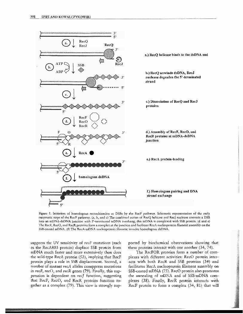
Figure 5. Initiation of homologous recombination at DSBs by the RecF pathway. Schematic representation of the early enzymatic steps of the RecF pathway. (a, b, and c) The combined action of RecQ helicase and RecJ nuclease converts a DSB into an ssDNA-dsDNA junction with 3'-terminated ssDNA overhang; this ssDNA is complexed with SSB protein. (d and e) The RecF, RecO, and RecR proteins form a complex at the junction and facilitate RecA nucleoprotein filament assembly on the SSB-coated ssDNA. (f) The RecA-ssDNA nucleoprotein filament invades homologous dsDNA.
suppress the W sensitivity of recF mutations (such as the RecA803 protein) displace SSB protein from ssDNA much faster and more extensively than does the wild-type RecA protein (52), implying that RecF protein plays a role in SSB displacement. Second, a number of mutant recA alleles cosuppress mutations in recF, recO, and recR genes (79). Finally, this sup- pression is dependent on recJ function, suggesting that RecF, RecO, and RecR proteins function to- gether as a complex (79). This view is strongly sup-
ported by biochemical observations showing -that these proteins interact with one another (34, 74).
The RecFOR proteins form a number of com- plexes with different activities: RecO protein inter- acts with both RecR and SSB proteins (34) and facilitates RecA nucleoprotein filament assembly on SSB-coated ssDNA (75). RecO protein also promotes the annealing of ssDNA and of SSB-ssDNA com- plexes (38). Finally, RecR protein interacts with RecF protein to form a complex (34, 81) that will
CHAPTER 21 HOMOLOGOUS RECOMBINATION IN E. COLI 392
bind to an ssDNA-dsDNA junction (55). Riochemi- cal analysis revealed that RecF protein binds prefer- entially to the ssDNA-dsDNA junction (35j, and that DNA binding by RecF protein is controlled by ATP hydrolysis (80). The RecFOR complex will bind to an ssDNA-dsDNA junction with a base-paired 5' terminus at the junction region, and it will facilitate assembly of RecA protein onto ssDNA adjacent to the junction (55). RecF protein (or RecFR complex) recognizes an ssDNA-dsDNA junction with a 5' end, which is the structure that should be produced by RecQ and RecJ proteins. The RecOR complex (or RecO protein) binds to the DNA-RecF(R) complex, which alters the SSB-ssDNA cornplex nearby and allows RecA protein nucleation; subsequent nucleo- protein filament extension permits assenlbly of RecA protein on the entire ssDNA tail.
Another component of the RecF pathway, RecN protein, has not yet been assigned any biochemical function. However, the slight recombination defi- ciency and mild UV sensitivity of the recJ recN dou- ble mutant, co~nbined with the severe recombination defect (50- to 100-fold reduction) of the recD r.ecJ recN mutant, suggests that RecN protein nright be a functional equivalent of the RecJ nuclease (50).
Despite its apparent complexity, the enzyrnatic machinery of the RecF pathway is as functional in DSB repair as the RecBCD pathway. h/loreover, the components of the RecF pathway have functional homologues or paralogues in all organisms, from bacteriophage to human ( 5 j.
SINGLE-STRAND GAP REPAIR
Because conjugation (and transduction) involves a DSB as the initiating site for recombination, it should not be s~rrprising that the genes that emerged from a screen for mutants defective in sexual recom- bination are essential for the repair of DSBs. How- ever, the consequence of this nearly singular focus on conjugational recombination events led to the erro- neous conclusion that the RecF pathway serves only a minor function in recombination in wild-type cells. Also incorrect was the belief that the discovery of the RecF pathway as the set of genes that permitted re- combination in the absence of the primary RecBCD pathway implied that the RecF pathway was a cryp- tic recombination pathway that could be activated to compensate for the loss of RecBCD enzyme function. Rather, in wild-type cells, the RecF pathway is re- sponsible for the repair of all SSGs. This conclusion emerged from many studies, but was made clearest from recombination assays that did not employ sex- ual events.
A genetic assay system that observes recombi- nation between direct repeats of a chromosomal seg- ment allows following sister chromosome excha~lges required for the repair of DSBs (29). This assay was developed to approximate the function of homolo- gous recombinatio~i in the repair of the DNA lesions produced during replication. The recornbinatiotl events occurring between direct chromosonlal re- peats are detected in the colony-sectoring recombi- nation assay, since the detected recombination events eliminate the joint-point markers located between the repeats. Recombination events detected in this assay are absolutely dependent on RecA protein func- tion. The recF and recJ mutants display a rect phe- notype, recB mutants show only a slight defect, and recB rec] double mutants are capable of supporting duplication segregation. On the other hand, recB recF double mutants are deficient in recombination between chromosomal direct repeats, suggesting that both RecBCD and RecF pathways play major roles in recombination.
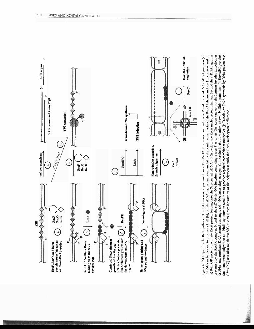
Initiation of homologous recombination on SSGs is presented in Fig. 6. First, the RecFOR com- plex assembles at the ssDNA-dsDNA junction of an SSG containing a base-paired 5' end (Fig. 6a) and fa- cilitates RecA nucleoprotein assembly in the ssDNA region of the gap (55) (Fig. 6e). The RecFR complex can bind to the 3'-containing end of SSG, limiting RecA filament extension into the dsDNA region (82) (Fig. 6f). The resulting RecA nucleoprotein fila- ment formed on the SSG can then invade the ho- mologous dsDNA molecule (Fig. 6g). DNA strand exchange followed by heteroduplex extension re- sults in the formation of two Holliday junctions. To complete the repair, RecA protein must be removed from the junctions, and the Holliday junctions themselves need to be resolved. The processing of a Holliday junctiotl into mature recombinant mole- cules is achieved by the RuvA, RuvB, and RuvC proteins (see references 64 and 83 for reviews). The RuvA tetramer is a four-way junction-specific rec- ognition protein that binds to the Holliday junc- tion and induces the square planar conformation of this junction. The RuvB protein is a specialized translocation protein that can "pump" or move DNA through its circular hexameric active form. To catalyze branch migration, two hexameric rings of RuvB protein bind to opposing arms of the Hol- liday junction that was recognized by the RuvA proteins, and then they translocate the dsDNA out- ward through the center of each ring, resulting in the relative movement of the junction (Fig. 6h). RuvC protein is a four-way junction-specific endonuclr- ase that resolves a Holliday junction by symmetri- cally cleaving the opposite arm of the junctlon to
SSB
un
know
n nu
clea
se
3'
DSB
rep
air
0
> 5'
-
SS
G is
con
vert
ed to
the
DSB
R
ecF,
Rec
O, a
nd R
ecR
R
ecF
0
prot
eins
ass
embl
e at
the
ssD
NA
-dsD
NA
junc
tion
S
SG
exp
ansi
on
R
w
Rec
F 0
Rec
O
Rec
FO
R fa
cili
tate
Rec
A
load
ing
on to
the
SSB
- R
ecR
0
cove
red
gap
v
P
Con
tinu
ed R
ecA
fda
men
t gr
owth
with
in t
he g
ap;
@
Rec
FR
com
plex
pre
vent
s R
ecA
fil
amen
t gro
wth
int
o a 1 Re
cFR
th
e do
wns
trea
m d
sDN
A
trPaaesioll
DN
A s
ynth
esis
re
gion
5' 3'
S
os
iad
~o
n
Hom
olog
ous
pair
ing
and
DN
A s
tran
d ex
chan
ge 0 1 ho
mol
ogou
s ds
DN
A
" .....
......
Het
erod
uple
x ex
tens
ion,
B
ranc
h m
igra
tion
_
__
__
)
0
Rec
A
Ruv
AB
.....
. ..."
C
Hol
liday
Jun
ctio
n R
~~
C
reso
luti
on
Figu
re 6
. SSG
repa
ir b
y th
e R
ecF
path
way
. The
SSG
has
sev
eral
pot
enti
al fa
tes.
The
Rec
FOR
pro
tein
s ca
n bi
nd to
the
5' en
d of
the
ssD
NA
-dsD
NA
junc
tion
(a),
th
e SS
G c
an b
e cl
eave
d to
pro
duce
a D
SB (
b), o
r the
ssD
NA
regi
on c
an b
e ex
pand
ed b
y th
e co
mbi
ned
activ
ities
of
the
Rec
Q h
elic
ase
and
Rec
J nu
clea
se (
c and
d).
(e
) Rec
FOR
pro
tein
s fa
cili
tate
Rec
A p
rote
in l
oadi
ng o
nto
the
SSB
-coa
ted
ssD
NA
. (f)
Gro
wth
of
the
Rec
A n
ucle
opro
tein
fila
men
t bey
ond
the
ssD
NA
regi
on is
pr
even
ted
by t
he R
ecFR
com
plex
bou
nd t
o th
e ss
DN
A-d
sDN
A ju
ncri
on c
onta
inin
g a
free
3' e
nd.
(g) T
he R
ecA
nuc
leop
rote
in f
ilam
ent
inva
des
hom
olog
ous
dsD
NA
and
cat
alyz
es D
NA
str
and
exch
ange
. (h
) DN
A h
eter
odup
lex
expa
nsio
n re
sults
in
the
form
atio
n of
tw
o H
olli
day
junc
tions
. (i
) Ruv
AB
C p
rote
ins
faci
lita
te b
ranc
h m
igra
tion
and
Hol
liday
jun
ctio
n re
solu
tion
to p
rodu
ce r
epai
red
reco
mbi
nant
mol
ecul
es. (
j) T
rans
lesi
on D
NA
syn
thes
is b
y D
NA
pol
ymer
ase
(Um
uD'C
) ca
n al
so r
epai
r th
e SS
G d
ue t
o di
rect
int
erac
tion
of
the
poly
mer
ase
wit
h th
e R
ecA
nuc
leop
rote
in f
ilam
ent.
CHAPTER 21 HOMOLOGOUS RECOMBINATION IN E. C O L ~ 401
produce the recombinant DNA product molecules (Fig. 6i).
Alternatively, an ssDNA endonuclease can con- vert the SSG into a DSB, which can be repaired through a DSB repair mechanism (Fig. 6b). If the re- gion of ssDNA in the gap is too small, the11 it can be expanded by the combined action of RecQ helicase and RecJ nuclease. Similar to the DSB repair situa- tion, RecQ helicase function can be substituted by UvrD helicase or helicase IV (53) (Fig. 6c). The SSG, on which the RecA nucleoprotein filament is assem- bled, is not necessarily repaired only by homologous recombitlation. The error-prone UmuD'C DNA polymerase can be attracted to such a RecA filament assembled on the SSG to catalyze translesion DNA synthesis (59) (Fig. 6j).
CONCLUSION
Homologous recoml~ination can be initiated at either DSBs or SSGs in duplex DNA. Two major pathways are responsible for homologous recombi- nation in wild-type E , coli: the RecBCD and RecF pathways. The RecBCD pathway is specific for the recombinational repair of DSBs, and in the wild-type cells, the RecF pathway is primarily used for recorn- bination that initiates at SSGs. However, with appro- priate suppressor mutations in E. coli, and presumably in bacteria that lack a RecBCD pathway, the RecF pathway can efficiently act at DSBs as well. Despite the different initiating lesions, both pathways have the same subsequent step: conversion of the broken DNA molecule into a central intermediate of recom- bination, which is the RecA protein nucleoprotein filament assembled along the ssDNA. In the RecBCD pathway, this process is carried out by the combined helicase/nuclease activity of RecBCD enzyme, and depends on the presence of the recombination hot spot, X. In the RecF pathway, the conlbined efforts of RecQ helicase and RecJ nuclease are needed in com- bination with the RecFOR complex. In the RecBCD pathway, RecBCD enzyme facilitates assembly of the RecA protein onto SSB-coated ssDNA; in the RecF pathway, this task is accomplished by RecF, RecO, and RecR proteins. The RecA nucleoprotein filament can then initiate invasion of ssDNA into homoiogous dsDNA, progressing into the final stage of homolo- gous recombination, which is resolution by RuvABC proteins.
REFERENCES
1. Amundsen, S. K., A. F. Taylor, A. A I . Chaudhurg, and G. R. Smlth. 1986. reel): the gene for an essential thlrd sub-
unit of exonuclease V. Proc. Natl. Acdd. Sci. USA 83:5558- 5562.
2. Anderson, D. G., J. J. Churchill, and S. C. Kowalczykowski. 1997. Chi-activated RecBCD enzyme possesses 5' -, 3' cleolytic activity, but RecBC enzyme does not: evidence sug- gestlng that the alteration induced by Chi is not silnply ejection of the RecD subunit. Genes Cells 2:117-128.
22. Anderson, D. G., and S. C. Kowalczykowski. 1997. The re- cornbination hot spot x is a regulatory element that switches the polarity of DNA degradation by the RecBCD enzyme. Genes Dew. 11:571-581.
2b. Anderson, D. G., and S . C. Kowalczykowski. 1997. The translocating RecBCD enzyme stimulates recombination by directing RecA protein onto ssDNA in a x regulated manner. Cell 90:77-86.
2c. havind, L., K. S. Makarova, and E. V. Koonin. 2000. Hol- liday junction resolvases and related nucleases: identification of new families, phyletic distribution and evolutionary tra- jectories. Nucleic Acids Res. 28:3417-3432.
3. Arnold, D. A.,P. R. Rianco, and S . C. Kowalczykowski. 1998. The reduced levels of x recognition exhibited by the R ~ ~ B c ~ ~ ~ ~ D enzyme reflect its recon~bination defect in vivo. J. Biol. Chem. 273316476-1 6486.
4. Arnold, D. A., and S. C. Kowalczykowski. 2000. Facilitated loading of RecA protein is essential to recombination by RecBCD enzyme. J. Biol. Chen~. 275:12261-12265.
5. Beernink, H. T., and S. W. Morrical. 1999. RhlPs: recombi- natiodreplication mediator proteins. Trends Biochejn. Set. 24:385-359.
6. Bianco, P. R., and S. C. Kowalczykowski. 1999. RecA protein. In Encj~clopedia oiLife SC~PIICCS. [Online.] http:l/\v\vc-v.els.net. Nature Publishirlg Group, London, England.
7. Bianco, P. R., and S . C. Kowalczykowski. 1997. The re- combination hotspot Chi is recognized by the translocating KecBCD enzyme as the single strand of DNA containing the srquence 5'-GCTGGTGG-3'. Proc. Natl. Acad. Sci. USA 94:6706-6711.
8. Biswas, I., E. Maguin, S. D. Ehrlich, and A. Gruss. 1995. A 7-base-pair sequence protects DNA from exonucleolytic deg- radation in Lactococcus lactis. Proc. Natl. Acud. Sci. USA 92:2244-2248.
9. Blattner, F. R., G. Plunkett 111, C. A. Bloch, N. T. Perna, V. Burland, M. Riley, J. Collado-Vides, J. D. Glasner, C. K. Rode, G. F. Mayhew, J. Gregor, N. W. Davis, H. A. Kirkpatrick, M. A. Goeden, D. J. Rose, B. Mau, and Y. Shao. 1997. The complete genome sequence of Escherichia coli K-12. Science 277:1453-1474.
10. Boehmer, P. E., and P. T. Emmerson. 1992. The IiecB sub- unit of the Escherichia coli RecBCD enzyme couples ATP hydrolysis to DNA unwinding. J. Biol. Chem. 267:4981- 4987.
11. Burland, V., G. Plunkett III, D. L. Daniels, and P. R. Blattner. 1993. DNA sequence and ailalysis of 136 kilobases of the Escherichia coli genome: organizational symmetry around the origin of replication. Genomics 16351-561.
12. Capaldo-Kimball, F., and S. D. Barbour. 1971. I~lvolvernent of recolnbinatio~l genes in growth and viability of Escherichzn coli K-12. J. Bacteuiol. 106:204-212.
13. Chaudhury, A. M., and G. R. Smith. 1984. A new class of Escherichia coli recBC mutants: implications for the role of recBC enzyme in homologous recombination. Proc. Natl. Acad. Sci. USA 81:7850-7854.
14. Chkdin, F., S. D. Ehrlich, and S. C. Kowalczykowski. 2000. T'hc k ~ c i l l u s SLI~LIIIS AclJAK l~eliiasrir~uclrase is regulateJ by its cognate Chi sequence in vitro. J. Mol. Biol. 298: 7-2 0.
402 SPIES AND KOWALCZYKOWSKI
15. Chidin, F., and S. C. Kowalczykowski. 2002. A novel family of regulated helicasesl~lucleases from Grani-positive bacteria: insights into the initiation of DNA recombination. Mol. Microbiol. 43:823-834.
16. Chtdin, F., P. Noirot, V. Biaudet, and S. D. Ehrlich. 1998. 11 five-nucleotide sequence protects DNA from exonucleolytic degradation by AddAB, the RecBCD analogue of Bacillus subtilis. Mol. Microbiol. 29:1369-1377.
17. Cheng, K. C., and G. R. Smith. 1989. Distribution of Chi- stimulated recombinationai exchanges and heteroduplex end- ,
points in phage lambda. Genetics 1232-17. 18. Churchill, J. J., D. G. Anderson, and S. C. Kowalcz~kowski.
1999. The RecBC enzyme loads RecA protein onto ssDNA asymmetrically and independently of Chi, resulting in consti- tutive recombination activation. Genes Deu. 13:901-911.
19. Churchill, J. J., and S. C. Kowalczykowski. 2000. Identifica- tion of the RecA protein-loading domain of RecBCD enzyme. J. Mol. Biol. 297537-542.
20. Clark, A. J. 1973. Recombination deficient mutants of E. coli and other bacteria. Annu. Rev. Genet. 7:67-86.
21. Clark, A. J., and A. D. Margulies. 1965. Isolation and char- acterization of recombination-deficient mutants of Escherichia coli K12. Proc. Natl. Acad. Sci. USA 53:4511159.
22. Connelly, J. C., L. A. Kirkham, and D. R. Leach. 1998. The SbcCD nuclease of Escherichin coli is a structural maintenance of chromosomes (SblC) family protein that cleaves hairpin DNA. Proc. Nntl. Acaci. Sci. USA 95:7969-7974.
23. Dillingham, M. S., M. Spies, and S. C. K~walcz~kowski. 2003. RecBCD enzyme is a bipolar DNA helicase. Nattrre 423:893-897.
24. Dixon, D. A., J. J. Churchill, and S. C. Kowalczykowski. 1994. Reversible inactivation of the Escherichia coli RecBCD enzyme by the recombination hotspot x in vitro: evidence for functional inactivation or loss of the RecD subunit. Proc. Natl. Acad. Sci. USA 91:2980-2984.
25. Dixon, D. A., and S. C. Kowalczykowski. 1993. The recom- bination hotspot x is a regulatory sequence that acts by at- tenuating the nuclease activity of the E. coli RecBCD enzyme. Cell 73:87-96.
26. En~merson, P. T. 1968. Recombination deficient mutants of Escherichia coli K12 that map between thyA and argA. Genetics 60:19-30.
27. Farah, J. A., and G. R. Smith. 1997. The RecBCD enzyme initiation complex for DNA unwinding: enzyme positioning and DNA opening. J. Mol. Biol. 272:699-715.
28. Friedberg, E. C., G. C. Walker, and W. Siede. 1995. DNA Repair and Mutagenesis. ASM Press, Washington, D.C.
29. Galitski, T., and J. R. Roth. 1997. Pathways for homologous recombination between chromoson~al direct repeats in Sal- monella typhimurium. Genetics 146:751-767.
30. Ganesan, S., and G. R. Smith. 1993. Strand-specific binding to duplex DNA ends by the subunits of Escherichia coli recBCD enzyme. J. Mol. Biol. 229:67-78.
31. Gibson, F. P., D. R. Leach, and R. G. Lloyd. 1992. Identifi- cation of sbcD mutations as cosuppressors of recBC that allow propagation of DNA palindromes in Escherichia coli K-12. 1. Bacteriol. 174:1222-1228.
32. Haijema, B. J., G. Venema, and J. Kooistra. 1996. The C terminus of the AddA subunit of the Bacillus stlbtilis ATP- dependent DNase is required for the ATP-dependent exonu- clease activity but not for the helicase activity. J . Bacteriol. 178:5086-5091.
33. Harmon, F. G., and S. C. Kowalczykowski. 2001. Bio- cherl~ical charactcrizotio~l of the DNA hclicasc activity of the Escherichia coli RecQ helicase. J. Bzol. Chem. 276:232- 243.
34. Hegde, S. P., M. H. Qin,X. H. Li, M. A. Atkinson, A. J. Clark, M. Rajagopalan, and M. V. Madiraju. 1996. Interactions of RecF protein with RecO, RecR, and single-stranded DNA binding proteins reveal roles for the RecF-RecO-RecR com- plex in DNA repair and recombination. Proc. Natl. Acnd. Sci. USA 93:14468-14473.
35. Hegde, S. P., M. Rajagopalan, and M. V. V. S. Madiraju. 1996. Preferential binding of Escherichia coli RecF protein to gapped DNA in the presence of adenosine (y-thio) triphos- phate. J. Bacteriol. 178:L84-190.
36. Howard-Flanders, P., and L. Theriot. 1966. Mutants of Escherichia coli K-12 defective in DNA repair and in genetic recombination. Genetirs 53:1137-1150.
37. Iyer, V. N., and W. D. Rupp. 1971. Usefulness of benzoy- lated naphthoylated DEAE-cellulose to distinguish and frac- tionate double-stranded DNA bearing different extents of single-stranded regions. Biochim. Biophys. Acta 228:117- 126.
38. Kantake, N., M. V. Madiraju, T. Sugiyama, and S. C. Ko- walczykowski. 2002. Escherichia coli RecO protein anneals ssDNA complexed with its cognate ssDNA-binding protein: a common step in genetic recombination. Proc. Natl. Acad. Sci. USA 99:15327-15332.
39. Kogoma, T. 1997. Stable DNA replication: interplay between DNA replication, homologous recombination, and transcrip- tion. Microbiol. Mol. Biol. Rev. 61:212-238.
40. Kooistra, J., B. J. Haijema, and G. Venema. 1993. The Ba- cillus subtilis addAB genes are fully functional in Escherichia coli. Mol. Microbiol. 7:9 15-923.
41. Korangy, F., arid D. A. Julin. 1993. Kinetics and process- ivity of ATP hydrolysis and DNA unwinding by the RecBC enzyme from Escherichia coli. Biochemistry 32:4873- 4880.
42. Kowalczykowski, S. C. 2000. Initiation of genetic recombi- nation and recombination-dependent replication. Trends Biochem. Sci. 25:156-165.
43. Krasin, F., and F. Hutchinson. 1977. Repair of DNA double- strand breaks in Escherichia coli, which requires recA function and the presence of a duplicate genome. J. Mol. Biol. 116: 81-98.
44. Kushner, S. R., H. Nagaishi, and A. J. Clark. 1972. Indirect suppression of recB and recC mutations by exonuclease I de- ficiency. Proc. Nntl. Acad. Sci. USA 69:1366-1370.
45. Kuzminov, A. 1995. Collapse and repair of replication forks in Eschericllia coli. Mol. Microbiol. 16:373-384.
46. Kuzminov, A. 1999. Recombinational repair of DNA damage in Escherizhia coli and bacteriophage h. Microbiol. Mol. Biol. Rev. 63:751-813.
47. Lam, S. T., M. M. Stahl, K. D. McMilin, and F. W. Stahl. 1974. Rec-mediated recombinational hot spot activity in bacteriophage lambda. 11. A mutation which causes hot spot activity. Genetics 77:425433.
48. Lederberg, J., and E. L. Tatum. 1953. Sex in bacteria; genetic studies, 1945-1952. Science 118:169-175.
49. Lloyd, R. G., and C. Buckman. 1985. Identification and ge- netic analysis of sbcC mutatioris in conlmonly used recBC sbcB strains of Escherichia coli K-12. J. Bacteriol. 164:836- 844.
50. Lloyd, R. G., and C . Buckman. 1991. Overlapping functions of recD, recJ and recN provide evidence of three epistatic groups of genes in Escherichia coli recombination and DNA repair. Biochimie 73:313-320.
5 1. Lloyd, R. G., M. C. Porton, and C. Buckman. 1988. Effect of rccF, recJ, recN, recO and rw I I I U [ ~ L I ~ I ~ S UI I ~ ~ I t ~ a ~ i o l r t vtr.- viva1 and genetic recombination in a recl) strain of Escherichia coli K12. Mol. Gen. Genet. 212:317-324.
CHAPTER 21 HOMOLOGOUS RECOMBWATION IN E. COLI 403
51a. Lovett, S. T., C. Luisi-DeLuca, and R. D. Kolodner. 1988. The genetic dependence of recombination in recD rnutants of Escherichia coli. Genetics 120:37-45.
52. Madiraju, M. V. V. S., A. Templin, and A. J. Clark. 1988. Properties of a mutant recA-encoded protein reveal a possible role for Escherichia coli recF-encoded protein in genetic recombination. Proc. Natl. Acad. Sci. USA 85:6592-6596.
53. Mendonca, V. M., H. D. Klepin, and S. W. Matson. 1995. DNA helicases in recombination and repair: construction of a AzivrD AhelD ArecQ mutant deficient in recombination and repair. J. Bacteriol. 177:1326-1335.
54. Michel, B., S. D. Ehrlich, and M. Uzest. 1997. DNA double- strand breaks caused by replication arrest. EMBO ]. 16: 430-438.
55. Morimatsu, K., and S. C. Kowalczykowski. 2003. RecFOR proteins load RecA protein onto gapped DNA to accclcrate DNA strand exchange: a universal step of recombinational repair. Mol. Cell 11:1337-1347.
56. Mosig, G. 1987. The essential role of recombination in phage T4 growth. Annu. Rev. Genet. 21:347-371.
57. Myers, R. S., A. Kuzminov, and F. W. Stahl. 1995. The recombination hot spot x activates RecBCD recombination by converting Escheric/~ia coli to a recD mutant phenocopy. Proc. Natl. Acad. Sci. USA 92:6244-6248.
58. Myers, R. S., atid F. W. Stahl. 1994. Chi and the RecBCD enzyme of Escherichia coli. Annu. Rev. Genet. 28:49-70.
59. Pham, P., E. M. Seitz, S. Saveliev, X. Shen, R. Woodgate, M. M. Cox, and M. F. Goodman. 2002. Two dist~~ict modes of RecA action are required for DNA polymerase V-cata- lyzed translesion synthesis. Proc. Natl. Acud. Sci. USA 99:11061-11066.
60. Phillips, G. J., D. C. Prasher, and S. R. Kushner. 1988. Physical and biochelnical characterization of cloned shcB and xonA n~utations from Escherichia coli K-12. J. Bacteriol. 170:2089-2094.
61. Phillips, R. J., D. C. HicMeton, P. E. Boehmer, and P. T. Emmerson. 1997. The RecB protein of Escherichia coli translocates along single-stranded DNA in the 3' to 5' di- rection: a proposed ratchrt mechanism. Mol. Gen. Genet. 254:319-329.
62. Roman, L. J., A. K. Eggleston, and S. C. Kowalczykowski. 1992. Processivity of the DNA helicase activity of Escher- ichia coli recBCD enzyme. J. Biol. Chem. 267:4207-4214.
63. Roman, L. J., and S. C. Kowalczykowski. 1989. Character- ization of the adenosinetriphosphatase activity of the Escherichza coli RecBCD enzyme: relationship of ATP hy- drolysis to the unwinding of duplex DNA. Biochemistry 28:2873-2881.
64. Shinagawa, H., and H. Iwasaki. 1995. Molecular mechan- isms of Holliday junction processing in Escherichia coli. Adv. Biophys. 31:49-65.
65. Sinden, R. R., and R. S. Cole. 1978. Repair of cross-linked DNA and survival of Escherichia coli treated with psoralen and light: effects of mutations influencing genetic recombi- nation and DNA metabolism, J. Bacteriol. 136538-547.
66. Smith, C. L., J. G. Econome, A. Schutt, S. Klco; and C. R. Cantor. 1987. A ~hysical map of the Escherichia coli K12 genome. Science 236:1448-1453.
67. Sourice, S., V. Biaudet, M. El Karoui, S. D. Ehrlich, and A. Gruss. 1998. Identification of the Chi site of Haemophilus influenzae as several sequences related to the Escherichia coli Chi site. hlol. Microbiol. 27:1023-1029.
68. Spies, M., P. R. Bianco, M. S. Dillingham, N. Handa, R. J. Baskin, and S. C, Kowalczykowski. 2003. A molecular throttle: the recombination hotspot, X, controls DNA trans- location by the RecBCD helicase. Cell 114:647-654.
69. Stahl, F. W., J. M. Crasemann, and M. M. Stahl. 1975. Rec- mediated recombinational hot spot activity in bacteriophage lambda. 111. Chi mutations are site-mutations stimulating rec- mediated recombination. 1. 14401. Biol. 94:203-212.
70. Stahl, F. W., K. D. McMilin, M. M. Stahl, J. M. Crasemann, and S. 1,am. 1974. The distribution of crossovers along un- replicated lambda bacteriophage chromosomes. Genetics 77:395-408.
71. Stahl, F. W., and M. M. Stahl. 1975. Rec-mediated recom- binational hot spot activity in bacteriophage lambda. IV. Effect of heterology on Chi-stimulated crossing over. Mol. Gen. Genet. 140:29-37.
72. Taylor, A. F., D. W. Schultz, A. S. Ponticelli, and G. R. Smith. 1985. RecBC enzyme nicking at Chi sites during DNA unwinding: location and orientation-dependence of the cut- ting. Cell 41:153-163.
73. Taylor, A. F., and G. R. Smith. 1999. Regulation of ho- mologous recombination: Chi inactivates RecBCD enzyme by disassembly of the three subunits. Genes Dev. 13:890- 900.
74. Umezu, K., N. W. Chi, and R. D. Kolodner. 1993. Bio- chemical interaction of the Escherichia coli RecF, RecO, and RecR proteins with RecA protein and single-stranded DNA binding protein. Proc. Nntl. Acad. Sci. USA 90:3875- 3879. - -
/>. Umezu, K., and R. D. Kolodner. 1994. Protein interactions in genetic recombination in Escherichia coli. Interactions involving RecO and RecR overcome the inhibition of RecA by single-stranded DNA-binding protein. J. Biol. Chem. 269: 30005-30013.
76. Umezu, K., K. Nakayama, and H. Nakayama. 1990. Es- cherichia coli RecQ protein is a DNA helicase. Proc. Nutl. Acad. Sci. USA 875363-5367.
77. Wang, J., R. Chen, and D. A. Julin. 2000. A single nuclease active site of the Escherichia coli RecBCD enzyme catalyzes single-stranded DNA degradation in both directions. J. Biol. Chern. 27.5507-513.
78. Wang, T. C., and S. H. Chen. 1992. Simllar-sized daughter- strand gaps are produced in the leading and lagging strands of DNA in UV-irradiated E. coli uvrA cells. Biochem. Bio- phys. Res. Commun. 184:1496-1503.
79. Wang, T.-C. V., H. Y. Chang, and J. L. Hung. 1993. Co- suppression of recF, recR and recO mutations by mutant recA alleles in Escherichia coli cells. Mutat. Res. 294:157- 166.
80. Webb, B. L., M. M. Cox, and R. B. Inman. 1999. ATP hy- drolysis and DNA binding by the Escherichia coli RecF protein. J. Biol. Chem. 274:15367-15374.
81. Webb, B. L., M. M. Cox, and R. B. Inman. 1995. An in- teraction between the Escherichia coli RecF and RecR pro- teins dependent on ATP and double-stranded DNA. J. Biol. Chem. 270:31397-31404.
82. Webb, B. L., M. M. Cox, and R. B. Inman. 1997. Recom- binational DNA repair: the RecF and RecR proteins limit the extension of RecA filaments beyond single-strand DNA gaps. Cell 91:347-356.
83. West, S. C. 1996. The RuvABC proteins and I-Iolliday junction processing in Escherichia colt. 1. Bacte~iol. 178:1237-1241.
84. Willetts, N. S., and D. W. Mount. 2969. Genetic analysis of recombination-deficient mutants of Escherichia coli K-12 carrying rec mutations cotransducible with thyA. J . Bacter- iol. 100:923-934.,
85. Yu, M., J. Souaya, and D. A. Julin. 1998. Identification of thc nuclease active site in the multifunctional IiecBCD enzyme by creation of a chimeric enzyme. J. Mol. Biol. 283:797-808.


![RecBCD-dependent promoted the Escherichia RecA andSSBProc. Natl. Acad. Sci. USA88(1991) 3369 140 c.2 ~-120 0 0 80 o E E 40 0 0 20 [recA protein], pM 0 2 4 #J 6 0 2 4 t 6 8 [recBCD](https://img.pdfslide.us/doc/110x75/60fedde5105ceb176675e595/recbcd-dependent-promoted-the-escherichia-reca-andssb-proc-natl-acad-sci-usa881991.jpg)