Embed Size (px)
Citation preview

1
Real-time genomic characterization of advanced pancreatic cancer to enable
precision medicine
Andrew J. Aguirre1,2,3,4*#, Jonathan A. Nowak1,4,5*, Nicholas D. Camarda1,2,6,7*, Richard A. Moffitt8*, Arezou A. Ghazani1,2,6, Mehlika Hazar-Rethinam9, Srivatsan Raghavan1,2,3,4, Jaegil Kim2, Lauren K. Brais1, Dorisanne Ragon1, Marisa W. Welch1, Emma Reilly1, Devin McCabe1,2,6,7, Lori Marini1,5,6, Kristin Anderka2, Karla Helvie1,6, Nelly Oliver1,6, Ana Babic1,4, Annacarolina Da Silva1,4,5, Brandon Nadres9, Emily E. Van Seventer9, Heather A. Shahzade9, Joseph P. St. Pierre1, Kelly P. Burke1,3,4, Thomas Clancy1,4,10, James M. Cleary1,3,4, Leona A. Doyle1,4,5, Kunal Jajoo1,4,11, Nadine J. McCleary1,3,4, Jeffrey A. Meyerhardt1,3,4, Janet E. Murphy9, Kimmie Ng1,3,4, Anuj K. Patel1,3,4, Kimberly Perez1,3,4, Michael H. Rosenthal1,4,12, Douglas A. Rubinson1,3,4, Marvin Ryou1,4,11, Geoffrey I. Shapiro1,3,4, Ewa Sicinska1,4, Stuart G. Silverman1,4,12, Rebecca J. Nagy13, Richard B. Lanman13, Deborah Knoerzer14, Dean J. Welsch14, Matthew B. Yurgelun1,3,4, Charles S. Fuchs1,4,6,7, Levi A. Garraway1,2,3,4,6, Gad Getz2,4,9, Jason L. Hornick1,4,5, Bruce E. Johnson1,2,3,4,6, Matthew H. Kulke1,3,4, Robert J. Mayer1,3,4, Jeffrey W. Miller7, Paul B. Shyn1,4,12, David A. Tuveson15, Nikhil Wagle1,2,3,4,6, Jen Jen Yeh16, William C. Hahn1,2,3,4,
Ryan B. Corcoran4,9, Scott L. Carter1,2,6,7#, Brian M. Wolpin1,3,4#
1 Dana-Farber Cancer Institute, 450 Brookline Avenue, Boston, MA 02215 USA
2 Broad Institute of Harvard and MIT, 415 Main Street, Cambridge, MA 02142 USA
3 Department of Medicine, Brigham and Women’s Hospital, 75 Francis Street, Boston,
MA 02115
4 Harvard Medical School, 25 Shattuck Street, Boston, Massachusetts 02115, USA.
5 Department of Pathology, Brigham and Women’s Hospital, 75 Francis Street, Boston,
MA 02115
6 Joint Center for Cancer Precision Medicine, Dana-Farber Cancer Institute/Brigham
and Women's Hospital, Boston, MA, 02215 USA
7 Harvard T.H. Chan School of Public Health, 677 Huntington Ave, Boston, MA 02115
8 Department of Biomedical Informatics, Department of Pathology, Stony Brook
University, Stony Brook, NY 11794
9 Massachusetts General Hospital Cancer Center, Boston, MA, USA.
10 Department of Surgery, Brigham and Women’s Hospital, 75 Francis Street, Boston,
MA 02115.
11 Department of Gastroenterology, Brigham and Women’s Hospital, 75 Francis Street,
Boston, MA 02115
Research. on August 29, 2018. © 2018 American Association for Cancercancerdiscovery.aacrjournals.org Downloaded from
Author manuscripts have been peer reviewed and accepted for publication but have not yet been edited. Author Manuscript Published OnlineFirst on June 14, 2018; DOI: 10.1158/2159-8290.CD-18-0275

2
12 Department of Radiology, Brigham and Women’s Hospital, 75 Francis Street, Boston,
MA 02115
13 Department of Medical Affairs, Guardant Health, Inc., Redwood City, California, USA
14 BioMed Valley Discoveries, Kansas City, Missouri, USA
15 Cold Spring Harbor Laboratory, Cold Spring Harbor, NY 11724, USA; Lustgarten
Foundation Pancreatic Cancer Research Laboratory, Cold Spring Harbor, NY 11724,
USA
16 Departments of Surgery and Pharmacology, Lineberger Comprehensive Cancer
Center, University of North Carolina, Chapel Hill, North Carolina, USA.
* These authors contributed equally
Co-senior authors
# Correspondence:
Andrew J. Aguirre, Dana-Farber Cancer Institute, 450 Brookline Avenue, Boston MA
02215. Phone: 617-632-5707; E-mail: [email protected]
Scott L. Carter, Dana-Farber Cancer Institute, 450 Brookline Avenue, Boston MA
02215. Phone: 617-632-3038; Email: [email protected]
Brian M. Wolpin, Dana-Farber Cancer Institute, 450 Brookline Avenue, Boston MA
02215. Phone: 617-632-6942; E-mail: [email protected]
Running Title: Genomic precision medicine in advanced pancreatic cancer
Conflict of Interest Disclosure: Matthew B. Yurgelun acknowledges research funding
from Myriad Genetic Laboratories, Inc. James M. Cleary reports research funding from
Merck. Geoffrey I. Shapiro reports research funding from Lilly, Merck, EMD Serono,
Sierra Oncology and Pfizer and is a consultant to Pfizer, G1 Therapeutics, Lilly, Roche
and Merck/EMD Serono. Charles S. Fuchs is a consultant to CytomX, Sanofi, Eli Lilly,
Merck and Entrinsic Health. Levi A. Garraway is an employee of Eli Lilly and Company
and has ownership interest in Tango Therapeutic and Foundation Medicine. Bruce E.
Research. on August 29, 2018. © 2018 American Association for Cancercancerdiscovery.aacrjournals.org Downloaded from
Author manuscripts have been peer reviewed and accepted for publication but have not yet been edited. Author Manuscript Published OnlineFirst on June 14, 2018; DOI: 10.1158/2159-8290.CD-18-0275

3
Johnson reports research funding from Toshiba and Novartis. Nikhil Wagle reports
ownership interest in Foundation Medicine. Ryan B. Corcoran reports research funding
from AstraZeneca and Sanofi, and is a consultant to Astex, Amgen, Avidity Biosciences,
BMS, FOG Pharma, Genentech, LOXO Oncology, Merrimack, N-of-one, Roche,
Roivant, Shire, Symphogen, Taiho and WarpDrive Bio. Richard B. Lanham is an
employee of and has ownership interest in Guardant Health, Inc. Dean J. Welsch is an
employee of BioMed Valley Discoveries. Brian M. Wolpin acknowledges research
funding from Celgene. The remaining authors report no potential conflicts of interest.
Research. on August 29, 2018. © 2018 American Association for Cancercancerdiscovery.aacrjournals.org Downloaded from
Author manuscripts have been peer reviewed and accepted for publication but have not yet been edited. Author Manuscript Published OnlineFirst on June 14, 2018; DOI: 10.1158/2159-8290.CD-18-0275
4
Abstract
Clinically relevant subtypes exist for pancreatic ductal adenocarcinoma (PDAC), but
molecular characterization is not yet standard in clinical care. We implemented a biopsy
protocol to perform time-sensitive whole exome sequencing and RNA-sequencing for
patients with advanced PDAC. Therapeutically relevant genomic alterations were
identified in 48% (34/71) and pathogenic/likely pathogenic germline alterations in 18%
(13/71) of patients. Overall, 30% (21/71) of enrolled patients experienced a change in
clinical management as a result of genomic data. Twenty-six patients had germline
and/or somatic alterations in DNA-damage repair genes, and 5 additional patients had
mutational signatures of homologous recombination deficiency but no identified causal
genomic alteration. Two patients had oncogenic in-frame BRAF deletions, and we
report the first clinical evidence that this alteration confers sensitivity to MAP-kinase
pathway inhibition. Moreover, we identified tumor/stroma gene expression signatures
with clinical relevance. Collectively, these data demonstrate the feasibility and value of
real-time genomic characterization of advanced PDAC.
Statement of Significance
Molecular analyses of metastatic PDAC tumors are challenging due to the
heterogeneous cellular composition of biopsy specimens and rapid progression of the
disease. Using an integrated multi-disciplinary biopsy program, we demonstrate that
real-time genomic characterization of advanced PDAC can identify clinically relevant
alterations that inform management of this difficult disease.
Introduction
Pancreatic ductal adenocarcinoma (PDAC) is the third-leading cause of cancer-related
death in the United States and is projected to become the second leading cause by
2030 (1). Most patients present with advanced disease and die within 12 months of
diagnosis (2,3). Recent genomic studies of primary PDAC resection specimens have
identified recurrent molecular alterations and genomic subtypes of the disease (4-12).
Moreover, RNA analyses of PDAC cohorts have identified gene expression signatures
with prognostic and biological relevance (7,12-14). While these molecular subtypes of
Research. on August 29, 2018. © 2018 American Association for Cancercancerdiscovery.aacrjournals.org Downloaded from
Author manuscripts have been peer reviewed and accepted for publication but have not yet been edited. Author Manuscript Published OnlineFirst on June 14, 2018; DOI: 10.1158/2159-8290.CD-18-0275
5
PDAC may theoretically help guide precision medicine approaches, molecular
characterization of PDAC in patients with advanced disease is not yet standard clinical
practice. Biopsy-driven genomic studies have been challenging due to rapid disease
progression and the small-volume and heterogeneous nature of biopsies that impede
deep molecular characterization. Despite the fact that conventional therapies are often
ineffective, the rate of enrollment of PDAC patients onto clinical trials is extremely low
(15). A proactive, standardized approach to acquire PDAC biopsy tissue and perform
rapid turnaround molecular analysis is required to efficiently generate and utilize
genomic information in PDAC patients.
Results
Biopsy approach and patient cohort
The PancSeq protocol was developed as an institutional review board (IRB)-
approved multi-disciplinary biopsy program to obtain tissue from core needle biopsies or
fine needle aspirates in patients with metastatic or locally advanced PDAC for rapid
turnaround genomic analysis. Between March 2015 and June 2017, 79 patients
underwent biopsy on the PancSeq protocol (Supplementary Table S1, Supplementary
Figure S1). An initial pilot phase (n=10 patients) was conducted in patients having
clinically indicated biopsies to optimize workflow, tissue processing, nucleic acid
extraction, whole exome sequencing (WES) and RNA sequencing (RNA-seq) from
small volume needle biopsies (approximately 15-20 mg of tissue per biopsy).
Subsequently, 69 additional patients underwent biopsy and WES was performed in a
CLIA-certified laboratory, with return of selected somatic and germline variants to
referring clinicians. Patients had a median age of 64 years and most had metastatic
disease (96%) and no prior therapy (70%) (Supplementary Table S1). Most patients had
a diagnosis of PDAC (95%), although several patients with less common histologies
were enrolled. Biopsies were obtained from liver (n=63), pancreas (n=7), peritoneum
(n=6), lymph nodes (n=2) and ovary (n=1) (Supplementary Table S2). Biopsies were
performed percutaneously by interventional radiology (n=72), with endoscopic
ultrasound (n=6), or intraoperatively (n=1), and a median of 5 (range, 1-10) cores or
biopsy specimens were collected per patient. A low rate of serious complications was
Research. on August 29, 2018. © 2018 American Association for Cancercancerdiscovery.aacrjournals.org Downloaded from
Author manuscripts have been peer reviewed and accepted for publication but have not yet been edited. Author Manuscript Published OnlineFirst on June 14, 2018; DOI: 10.1158/2159-8290.CD-18-0275
6
observed, with only one patient having a self-limited hepatic subcapsular hematoma in
the setting of a clinically indicated liver biopsy and therapeutic anticoagulation with an
oral factor Xa inhibitor (rivaroxaban) for prior deep venous thrombosis.
Real-time DNA sequencing and exome analysis
From the FFPE core, tumor content was quantified by histopathology and 92%
(73/79) of cases had sufficient tumor content (≥5% tumor nuclei) to proceed with WES.
ABSOLUTE purity (16) was examined in WES data and successful mutation and copy
number calls could be made for all but two of these samples (Supplementary Table S2,
Supplementary Figure S1).
Consistent with prior genome sequencing studies of primary PDAC samples,
samples from our cohort of advanced PDAC patients displayed low neoplastic
cellularity, with a median cellularity by histologic assessment of 40%, and by WES using
the ABSOLUTE algorithm of 34% (Supplementary Figure S2A-B)(16). Neoplastic
cellularity estimates by histologic assessment and by the ABSOLUTE algorithm on
WES data were significantly correlated (Pearson's product-moment correlation = 0.386,
95% CI (0.195 - 0.548); P = 0.00016; Supplementary Figure S2A). This level of
correlation is consistent with that observed in other studies (12,16) and is impacted by
variability between biopsy specimens as well as differential sensitivity between the two
methods for identification of neoplastic cellularity. For our biopsy samples, we
performed deep WES with a median of mean target coverage of 191X in the tumor and
176X in the normal (Supplementary Table S2). Within the CLIA-certified phase of the
study, analyzed results were available within an average of 39 days (range, 16-67) from
the date of clinically indicated biopsies and 28 days (range, 15-51) for research-only
biopsies (Supplementary Table S2). Additional time for return of results for clinically
indicated biopsies was required, as these specimens were held in the pathology
department until a formal histologic diagnosis was confirmed. A report of clinically
relevant events was returned to the referring clinician detailing somatic mutations, small
insertions/deletions, and copy number alterations (CNAs) as well as pathogenic/likely
pathogenic germline alterations in a curated list of 81 PDAC-relevant genes
(Supplementary Table S3). These genes were chosen based on somatic or germline
Research. on August 29, 2018. © 2018 American Association for Cancercancerdiscovery.aacrjournals.org Downloaded from
Author manuscripts have been peer reviewed and accepted for publication but have not yet been edited. Author Manuscript Published OnlineFirst on June 14, 2018; DOI: 10.1158/2159-8290.CD-18-0275
7
clinical relevance, therapeutic actionability and/or recurrent mutation across published
PDAC genome sequencing cohorts (4-12). Most of these genes (n=69) were associated
with clinical trial or off-label FDA-approved targeted therapies. Comprehensive analysis
of all genes from WES data was simultaneously performed in the research setting.
Landscape of somatic mutations and copy number alterations (CNAs) in
advanced PDAC
Genome sequencing studies have elucidated the molecular landscape of
archived primary PDAC tumors and have demonstrated distinct molecular subtypes of
disease (6-12). In our cohort of 71 advanced PDAC patients, we identified significantly
recurrent mutations in KRAS, TP53, CDKN2A, SMAD4, ARID1A and TGFBR2 – a
collection of genes that were also recurrently mutated in primary PDAC tumors
(7,10,12) (Figure 1). Moreover, we observed frequent mutations in additional tumor
suppressor genes (e.g. RNF43) or oncogenes (e.g. BRAF, GNAS), as well as recurrent
mutations in genes involved in DNA-damage repair (DDR) and chromatin modification
(Figure 1). Recurrent high-level amplifications were observed in several genomic loci,
encompassing genes such as MYC, AKT2 and GATA6 (Figure 1, Supplementary Figure
S3). Deletions were identified at numerous loci, including frequent homozygous
deletions of CDKN2A and SMAD4 (Figure 1, Supplementary Figure S3).
Mutational signature analysis from WES data
Mutational signature analysis was performed using a Bayesian variant of the
non-negative matrix factorization (NMF) approach in a two-stage manner from the set of
single nucleotide variants (SNV) in our dataset, as previously described
(SignatureAnalyzer, Supplementary Experimental Methods, (17-20)). First, we
performed de novo signature discovery and our analytic pipeline identified three primary
signatures: SigA that best resembled COSMIC signature 3 with cosine similarity 0.87
(BRCA mutant signature suggestive of homologous recombination deficiency [HRD]);
SigB that best resembled COSMIC signature 1 with cosine similarity 0.96 (C>T
transitions at CpG dinucleotides, Aging); and SigC that best resembled COSMIC
signature 17 with cosine similarity 0.91 (etiology unknown) (Supplementary Figure S4A-
Research. on August 29, 2018. © 2018 American Association for Cancercancerdiscovery.aacrjournals.org Downloaded from
Author manuscripts have been peer reviewed and accepted for publication but have not yet been edited. Author Manuscript Published OnlineFirst on June 14, 2018; DOI: 10.1158/2159-8290.CD-18-0275
8
B). In addition, we observed a relative elevation of C>G transversions and C>T
transitions at TC[A/T] contexts in SigA corresponding to canonical hotspots of APOBEC
mutagenesis (COMSIC signature 2 and 13), suggesting that APOBEC signature is
possibly operative in this cohort, but not cleanly separable due to a lack of mutations
(17). Based on this de novo analysis, we concluded that four main mutational processes
were likely active in these data (Aging/COSMIC1; BRCA/HRD/COSMIC3;
APOBEC/COSMIC2+13; and COSMIC17). To better evaluate discrete contributions of
these mutational processes in our data and to minimize signature contamination, we
next performed a projection analysis to infer a signature activity across our biopsy
cohort using the signature profiles of the five contributing COSMIC signatures:
COSMIC1, 2, 3, 13, 17 (Figure 2A and Supplemental Experimental Methods).
Consistent with the de novo analysis, the inferred signature activity with these five
COSMIC signatures reveals a clear contribution of the Aging/COSMIC1 signature in
almost all patients and a notable activity of the HRD/COSMIC3 signatures in many
patients (Figure 2A).
To further investigate the HRD/COSMIC3 signature in these data, we integrated
mutation and copy number data from WES and gene expression data from RNA-seq for
a core set of known homologous recombination (HR) genes (BRCA1, BRCA2, PALB2
and RAD51C) that are known to be associated with the HRD/COSMIC3 signature (18)
and categorized samples as “HRD altered” or “WT.” We defined HRD altered samples
based on the presence of damaging germline and somatic mutations (null, truncating,
and splice-site variants), homozygous deletions, or more than 2-fold down-regulation of
mRNA-expression levels. We observed a significant enrichment of HRD/COSMIC3
signature mutations within HRD altered samples (Figure 2B, P < 0.000002 by one-tailed
Wilcoxon rank-sum test). The increased occurrence of large deletions of up to 50 base
pairs with overlapping microhomology is another characteristic mark of HR-deficient
samples, and we indeed observed an increased incidence of such deletions (≥ 9 base
pairs) in our HRD altered samples (Figure 2C, p < 0.00002 by one-tailed Wilcoxon rank-
sum test). Eight out of the top fourteen samples with HRD/COSMIC3 signature activity
harbored deleterious mutations or homozygous deletions in one of the four core HRD
genes. Six out of these eight HRD-altered samples had both germline and somatic
Research. on August 29, 2018. © 2018 American Association for Cancercancerdiscovery.aacrjournals.org Downloaded from
Author manuscripts have been peer reviewed and accepted for publication but have not yet been edited. Author Manuscript Published OnlineFirst on June 14, 2018; DOI: 10.1158/2159-8290.CD-18-0275

9
events or a somatic alteration with co-existing loss-of-heterozygosity (LOH) in BRCA1
or BRCA2, supporting a “two-hit” hypothesis. Another sample (0400253) had both a
p.Q750* nonsense and a p.F1016S missense mutation in PALB2. Furthermore, one
sample harbored homozygous deletion of RAD51C. Two additional samples with
HRD/COSMIC3 signature activity but without genomic alterations in the four core HRD
genes displayed down-regulation of RAD51C expression in the RNA-sequencing data,
an event previously associated with HRD/COSMIC3 signature activity (18). Thus, a total
of 10 of 14 samples with a high HRD/COSMIC3 signature activity could be explained by
genomic alterations or downregulation of gene expression in BRCA1, BRCA2, PALB2
or RAD51C (Figure 2B).
Notably, we also observed 4 samples that did not have clear DNA alterations or
mRNA downregulation of BRCA1, BRCA2, PALB2 or RAD51C but nevertheless had
enrichment of the HRD/COSMIC3 mutational signature at a level equal to or greater
than those samples in the HRD altered class (Figure 2B-C). We conducted a broader
examination of genes involved in DDR, including those responsible for HR, non-
homologous end joining (NHEJ), base excision repair (BER), nucleotide excision repair
(NER) and DDR checkpoint responses. One patient with a high activity of the
HRD/COSMIC3 signature had a ERCC2N238S mutation in a conserved helicase domain
of this key enzyme involved in NER. This ERCC2N238S mutation corresponds to one of
the recurrent mutational hotspots observed in patients with muscle-invasive urothelial
carcinoma who achieved a complete response to neoadjuvant cisplatin-based therapy
(21). ERCC2 mutations have been associated with a separate mutational signature that
has a significant overlap with the HRD/COSMIC3 signature (20); thus, the apparent
enrichment of the HRD/COSMIC3 signature for this PDAC patient with an ERCC2N238S
mutation may be attributed to the presence of an ERCC2 mutational signature. Notably,
this patient experienced a partial response to platinum-based FOLFIRINOX therapy (5-
FU, leucovorin, irinotecan and oxaliplatin), but progressed after 4-5 months of
treatment. For the remaining three samples with unexplained HRD/COSMIC3 signature
enrichment, we did not observe other mutations or copy number events in recognized
DDR genes. Thus, our data suggest that the HRD/COSMIC3 signature analysis in WES
Research. on August 29, 2018. © 2018 American Association for Cancercancerdiscovery.aacrjournals.org Downloaded from
Author manuscripts have been peer reviewed and accepted for publication but have not yet been edited. Author Manuscript Published OnlineFirst on June 14, 2018; DOI: 10.1158/2159-8290.CD-18-0275
10
data may detect additional patients with HRD not identified solely by mutation profiling
of specific genes known to be related to HR.
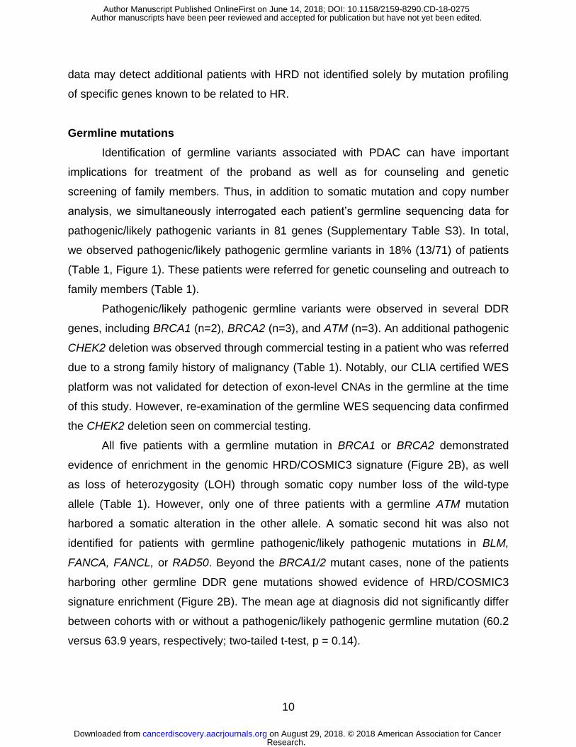
Germline mutations
Identification of germline variants associated with PDAC can have important
implications for treatment of the proband as well as for counseling and genetic
screening of family members. Thus, in addition to somatic mutation and copy number
analysis, we simultaneously interrogated each patient’s germline sequencing data for
pathogenic/likely pathogenic variants in 81 genes (Supplementary Table S3). In total,
we observed pathogenic/likely pathogenic germline variants in 18% (13/71) of patients
(Table 1, Figure 1). These patients were referred for genetic counseling and outreach to
family members (Table 1).
Pathogenic/likely pathogenic germline variants were observed in several DDR
genes, including BRCA1 (n=2), BRCA2 (n=3), and ATM (n=3). An additional pathogenic
CHEK2 deletion was observed through commercial testing in a patient who was referred
due to a strong family history of malignancy (Table 1). Notably, our CLIA certified WES
platform was not validated for detection of exon-level CNAs in the germline at the time
of this study. However, re-examination of the germline WES sequencing data confirmed
the CHEK2 deletion seen on commercial testing.
All five patients with a germline mutation in BRCA1 or BRCA2 demonstrated
evidence of enrichment in the genomic HRD/COSMIC3 signature (Figure 2B), as well
as loss of heterozygosity (LOH) through somatic copy number loss of the wild-type
allele (Table 1). However, only one of three patients with a germline ATM mutation
harbored a somatic alteration in the other allele. A somatic second hit was also not
identified for patients with germline pathogenic/likely pathogenic mutations in BLM,
FANCA, FANCL, or RAD50. Beyond the BRCA1/2 mutant cases, none of the patients
harboring other germline DDR gene mutations showed evidence of HRD/COSMIC3
signature enrichment (Figure 2B). The mean age at diagnosis did not significantly differ
between cohorts with or without a pathogenic/likely pathogenic germline mutation (60.2
versus 63.9 years, respectively; two-tailed t-test, p = 0.14).
Research. on August 29, 2018. © 2018 American Association for Cancercancerdiscovery.aacrjournals.org Downloaded from
Author manuscripts have been peer reviewed and accepted for publication but have not yet been edited. Author Manuscript Published OnlineFirst on June 14, 2018; DOI: 10.1158/2159-8290.CD-18-0275
11
RNA sequencing of PDAC biopsies
Recent gene expression studies have identified subtypes of PDAC with
prognostic and biological relevance (7,12-14). These studies converge on at least two
major neoplastic subtypes of PDAC including a squamous/basal-
like/quasimesenchymal subtype and a classical/pancreatic progenitor subtype. To
investigate these neoplastic PDAC subsets within our metastatic biopsy cohort, we
performed RNA-seq on a separate biopsy specimen from that used for WES. We
achieved successful RNA extraction and sequencing from 80% (63/79) of patients
(Supplementary Figure S1). Consistent with our prior observations in primary PDAC
specimens, we observed clear distinctions at the RNA level of the basal-like and
classical subtypes (14) (Figure 3A). However, samples with low neoplastic cellularity
were more difficult to classify with the basal-like and classical subtype gene sets, and
several of these samples showed a strong association with gene expression from
normal liver tissue (Figure 3A-B, Supplementary Figure S5A), suggesting that the
biopsy may have captured primarily adjacent liver parenchyma rather than the target
tumor lesion. Moreover, integrated analysis of tumor and normal gene expression
enabled clustering of samples by tumor-specific subtype and site of biopsy as well as
identification of outlier samples based on tumor type, atypical genetic lesions or
predominant contribution from adjacent normal or stromal gene expression
(Supplementary Figure S5B).
In addition to neoplastic subtypes of PDAC, two stromal subtypes have been
identified: “normal” and “activated” stromal subtypes (14). To capture a single composite
stroma signature, we generated a merged “stroma score” reflecting the combined total
expression of the top 25 activated and top 25 normal expressed stroma genes. Notably,
biopsies from metastatic liver lesions on average demonstrated lower stroma scores
than those from other sites or compared with primary tumor resection specimens
(Figure 3C). Comparison of ABSOLUTE neoplastic cellularity derived from WES
revealed a trend toward higher neoplastic cellularity for liver lesions (mean 0.39) versus
pancreatic biopsies or resections (mean 0.30; two sample t-test, p = 0.07,
Supplementary Figure S2B). Despite liver metastases showing on average lower
stroma scores, many samples did show elevated scores at a level similar to those seen
Research. on August 29, 2018. © 2018 American Association for Cancercancerdiscovery.aacrjournals.org Downloaded from
Author manuscripts have been peer reviewed and accepted for publication but have not yet been edited. Author Manuscript Published OnlineFirst on June 14, 2018; DOI: 10.1158/2159-8290.CD-18-0275
12
in primary resections or biopsies of other sites. Thus, the stroma score shows important
differences according to site of biopsy but also significant interpatient variability within
each biopsy site.
Clinically relevant genomic events
Genomic analyses of primary specimens have suggested that approximately
40% of patients with PDAC may harbor clinically relevant genomic events that could
potentially impact treatment decisions (12). However, most PDAC patients do not
undergo timely genomic analysis to identify these events for clinical decision making.
Leveraging our rapid turnaround CLIA-certified sequencing program, we performed real
time assessment of genomic data for use in clinical decision making (Supplementary
Figure S6A)(Supplementary Table S4). Excluding common events in KRAS or
CDKN2A, 48% (34/71) of patients within this cohort had cancers with at least one
genomic alteration that could potentially confer eligibility for current clinical trials or
support off-label usage of an agent approved for another indication (Supplementary
Figure S6A). Furthermore, 11% (8/71) of the patients had cancers with two or more
such events, suggesting a potential basis for genotype-driven combination therapy
trials.
KRAS mutations were observed in 90% (64/71) of patients in our cohort.
Although this alteration has been used to enroll patients onto clinical trials of MAPK-
directed therapy, these trials have demonstrated limited efficacy to date (22). However,
in the subset of KRAS wild-type tumors, we observed mutations and CNAs in additional
MAPK pathway activating genes, such as BRAF mutations (see below), a ROS1
translocation and high-level FGFR1 amplification (Figure 1, Supplementary Figure
S6A). Across the entire cohort, we observed multiple other alterations that could warrant
experimental therapies, including RNF43 mutation (e.g. Porcupine inhibitor), AKT2
amplification (e.g. AKT inhibitor), TSC2 mutation (e.g. MTOR inhibitor), MYC
amplification (e.g. bromodomain inhibitor), and CDK4 amplification (e.g. CDK4 inhibitor)
(Supplementary Figure S6A). Moreover, sequencing identified other lesions that may
contraindicate therapy with certain agents, such as RB1 mutations (n=4 patients) and
CDK4/6 inhibition.
Research. on August 29, 2018. © 2018 American Association for Cancercancerdiscovery.aacrjournals.org Downloaded from
Author manuscripts have been peer reviewed and accepted for publication but have not yet been edited. Author Manuscript Published OnlineFirst on June 14, 2018; DOI: 10.1158/2159-8290.CD-18-0275
13
A total of 24% (17/71) of patients enrolled on the PancSeq study were treated
with an experimental agent, either through enrollment onto a clinical trial or through off-
label use of an approved agent (Table 2). In particular, genomic information from WES
dictated the choice of experimental agent in 15% (11/71) of cases. Moreover, 18%
(13/71) of patients had a clinically relevant germline mutation necessitating referral for
genetic counseling (Table 1). Thus, accounting for both new therapeutic options and
genetic counseling indications, a total of 30% (21/71) of patients enrolled on the
PancSeq protocol experienced a change in clinical management as a result of the
obtained genomic data (Tables 1 and 2).
DNA-damage repair mutations and PARP-inhibitor therapy
In 20% of patients, we observed germline and/or somatic alterations in one or
more of the following DDR genes: BRCA1, BRCA2, PALB2, ATM and CHEK2 (Figure 1,
Supplementary Figure S6B). When considering mutations in a wider spectrum of genes
across several DDR classes, as well as specific mutational signatures consistent with
HRD, we identified a total of 44% (31/71) of patients displaying genomic evidence of
potential DDR deficiency (Table 1, Supplementary Figure S6B). DDR gene mutations
may confer increased sensitivity to platinum chemotherapy (9). Moreover, BRCA1 or
BRCA2 gene mutations have been reported to confer sensitivity to poly-ADP
polymerase (PARP) inhibition in preclinical models and early clinical trials of PDAC
(9,23,24). All six patients in the cohort with germline or somatic mutations in the BRCA1
or BRCA2 genes demonstrated evidence of the HRD/COSMIC3 mutational signature
(Supplementary Figure S6B). By contrast, none of the patients with mutations in ATM,
ATR or CHEK2 demonstrated enrichment of the HRD/COSMIC3 signature. All six
patients with BRCA mutations received an oxaliplatin-based chemotherapy regimen
(FOLFIRINOX or FOLFOX) in the first or second line setting and all demonstrated some
degree of radiographic response to these regimens (Figure 1, Supplementary Table
S4). Two of these patients with a germline BRCA1 mutation were subsequently enrolled
onto a randomized trial of the PARP inhibitor olaparib versus placebo for maintenance
therapy after receipt of 4-6 months of FOLFIRINOX (NCT02184195). Two further
patients with a BRCA2 mutation received off-label olaparib, including one patient with a
Research. on August 29, 2018. © 2018 American Association for Cancercancerdiscovery.aacrjournals.org Downloaded from
Author manuscripts have been peer reviewed and accepted for publication but have not yet been edited. Author Manuscript Published OnlineFirst on June 14, 2018; DOI: 10.1158/2159-8290.CD-18-0275
14
germline mutation and one with a somatic mutation. Besides patients with these
BRCA1/2 mutations, two other patients with DDR gene mutations were treated with a
PARP inhibitor (Table 2). Eight of ten patients with ATM, ATR or CHEK2 mutations
were treated with an oxaliplatin-based chemotherapy regimen and five demonstrated
clinical benefit as defined by partial response or stable disease at first radiographic
follow-up scans.
As noted above, two patients with BRCA2 alterations were treated with off-label
olaparib. The patient harboring a somatic BRCA2 mutation was a 45 year-old man who
presented with jaundice and abdominal pain and was diagnosed with metastatic PDAC
involving the liver and lymph nodes, with a serum CA19-9 of 3,592 U/mL on
presentation. He underwent biopsy of a metastatic liver lesion which confirmed a poorly
differentiated PDAC. Genome sequencing on the PancSeq protocol revealed KRAS
(p.G12R), TP53 (p.M246fs), and somatic BRCA2 (p.LS298fs) mutations (Figure 4A). He
received first line therapy with the platinum-containing regimen FOLFIRINOX and
experienced normalization of serum CA19-9 levels (Figure 4B) and a complete
radiographic response to therapy within 5 months of treatment (Figure 4C).
FOLFIRINOX was discontinued after a total of 13 two-week cycles due to transaminitis
and neuropathy. The patient chose to forego further cytotoxic chemotherapy and was
initiated on off-label olaparib 300 mg twice daily as maintenance therapy. He remains
free of radiographically detectable disease 28 months after diagnosis and 20 months
after beginning olaparib (Figure 4C).
BRAF mutant PDAC and response to MAP-kinase inhibition
Within our CLIA-certified cohort, we discovered two patients with in-frame
deletions in the BRAF oncogene (Figure 5A). This class of deletions near the alphaC-
helix region of the kinase domain has recently been shown to occur in KRAS wild-type
PDAC and activates the protein to drive MAPK signaling (12,25,26). Furthermore,
PDAC cell lines grown in vitro or as in vivo xenografts have been shown to be sensitive
to MAPK inhibition, although this approach had not been tested in humans (26). To
further define the frequency of this BRAF alteration in PDAC, we investigated a larger
collection of samples (N=406) profiled with a targeted genome sequencing panel at
Research. on August 29, 2018. © 2018 American Association for Cancercancerdiscovery.aacrjournals.org Downloaded from
Author manuscripts have been peer reviewed and accepted for publication but have not yet been edited. Author Manuscript Published OnlineFirst on June 14, 2018; DOI: 10.1158/2159-8290.CD-18-0275
15
Dana-Farber Cancer Institute (27,28) and found four KRAS wild-type tumors that
harbored the same in-frame deletion (p.N486_P490del) and one additional KRAS wild-
type tumor with a small, likely oncogenic in-frame insertion (p.T599dup). Thus, BRAF in-
frame insertions or deletions occurred in approximately 10% of our patients with KRAS
wild-type PDAC or 1% of all PDAC patients (12,25,26).
In one case, a 66 year-old woman presented with dyspepsia and weight loss and
was diagnosed with PDAC and liver metastases. She underwent first-line therapy with
FOLFIRINOX but experienced progression of disease after only five two-week cycles.
She underwent a biopsy and WES on the PancSeq protocol and then began second-
line therapy with gemcitabine plus nab-paclitaxel that was discontinued after 6 four-
week cycles due to disease progression (Figure 5B). Sequencing revealed a BRAF in-
frame deletion (p.N486-P490del) and TP53 mutation (p.V157G) (Figure 5A-B). Given
that cell lines harboring this mutation were sensitive to MEK inhibitors but resistant to
selective BRAF inhibitors in pre-clinical models (26), she was initiated on off-label
treatment with the MEK1/2 inhibitor trametinib, which is FDA-approved for use in
BRAFV600E mutant melanoma (Figure 5B) (29-31). Within four weeks of initiating
therapy, her serum CA19-9 had fallen from 36,000 to 8,100 U/ml, and the first restaging
scan done 8 weeks after initiation of trametinib showed a partial response to therapy
(Figure 5C-D). Serial plasma samples were collected to measure the fractional
abundance of BRAF and TP53 mutant alleles in circulating cell-free DNA (cfDNA) by
droplet digital PCR (ddPCR) (32,33). cfDNA measurements for BRAF and TP53 alleles
(two clonal alterations in this tumor) revealed a dramatic decline in response to
trametinib therapy that mirrored the radiographic findings (Figure 5E). After 5 months on
trametinib therapy, a rise in the fractional abundance of the BRAF and TP53 mutant
alleles in cfDNA was observed. After six months of therapy, radiographic progression
was identified. Evaluation of a more comprehensive panel of genes within cfDNA was
pursued through a commercial test (Guardant360®) at the time of progression and
demonstrated the emergence of multiple subclonal mutations in the MAP2K2 (MEK2)
gene (Figure 5E). Notably, these mutations are homologous to MEK1 mutations that
have been previously described to confer resistance to MEK inhibition in vitro in
mutagenesis studies in BRAF mutant melanoma (34). Retrospective analysis of the
Research. on August 29, 2018. © 2018 American Association for Cancercancerdiscovery.aacrjournals.org Downloaded from
Author manuscripts have been peer reviewed and accepted for publication but have not yet been edited. Author Manuscript Published OnlineFirst on June 14, 2018; DOI: 10.1158/2159-8290.CD-18-0275
16
MEK2 mutations in serial plasma samples collected through the patient’s treatment
course revealed emergence of these mutant alelles in concert with increases in
fractional abundance of mutant BRAF and TP53 alleles (Figure 5E). Thus, these data
likely reflect the emergence of heterogeneous polyclonal resistance mechanisms that
evolved under the selective pressure of trametinib therapy.
Given the emergence of MAP2K2 resistance mutations while on trametinib
therapy, the patient was subsequently treated with ulixertinib/BVD-523, an inhibitor of
ERK1/2 (which signals downstream of MEK1/2), on a single-patient investigational new
drug application. After treatment with ulixertinib/BVD-523 for 17 days, cfDNA analysis
by ddPCR demonstrated a rapid decline in the fractional abundance of the BRAF and
TP53 mutant alleles (Figure 5E). The MEK2 resistance alleles also rapidly decreased in
absolute frequency and in relative abundance with respect to the overall tumor burden
(as measured by the total MEK2/TP53 ratio) (Figure 5E). Unfortunately, the patient’s
functional status was quickly declining as she initiated therapy with ulixertinib/BVD-523,
and this medication was stopped after 3 weeks of therapy. A last blood collection after
cessation of ulixertinib/BVD-523 demonstrated rebound of the fractional abdundance of
the BRAF and TP53 mutant alleles. The patient expired at home several weeks later
with hospice services. This patient’s clinical course suggests that a BRAF in-frame
deletion may serve as an important biomarker for response to MAPK inhibition in
patients with pancreatic cancer.
Discussion
Many barriers to precision medicine exist for patients with PDAC, including low
neoplastic cellularity of tumors and the aggressive nature of the disease that makes
timely biopsy and genomic analysis difficult. We have established an integrated biopsy
program for advanced PDAC patients that enables CLIA-certified genomic profiling and
rapid turnaround of WES data to referring clinicians. To overcome the challenges of low
neoplastic cellularity, we have performed deep WES on small volume tumor biopsies
and germline DNA and have employed analytic algorithms to accurately quantitate
tumor DNA content and to identify mutations and CNAs. In addition, we have identified
molecular driver alterations and clinically relevant subsets of disease in real time, during
Research. on August 29, 2018. © 2018 American Association for Cancercancerdiscovery.aacrjournals.org Downloaded from
Author manuscripts have been peer reviewed and accepted for publication but have not yet been edited. Author Manuscript Published OnlineFirst on June 14, 2018; DOI: 10.1158/2159-8290.CD-18-0275
17
the course of the patient’s illness. In an initial biopsy cohort of 79 patients, we obtained
high quality genomic information for 71 patients and identified potentially actionable
somatic and germline alterations in 48% of cases. These include 26 patients harboring
tumors with DDR gene mutations and 7 with KRAS wild-type tumors, including two with
BRAF alterations and one with a ROS1 translocation. We have demonstrated how
these data can impact clinical decision making, utilizing molecular information to treat
multiple patients on clinical trials or with use of off-label targeted therapies.
Nevertheless, further study will be necessary to confirm the rate and therapeutic
implications of actionable alterations in a multi-institutional population of patients with
PDAC. Overall, 30% (21/71) of enrolled patients experienced a change in clinical
management as a result of genomic data, including 15% of patients for which genomic
information dictated the choice of an experimental agent and 18% of patients whose
germline data warranted referral for genetic counseling. These numbers far exceed the
average experience for PDAC patients, and these data support the implementation of
genomic evaluation as a standard clinical practice in patients with advanced PDAC.
We identified pathogenic/likely pathogenic germline mutations in 18% of patients
in our cohort of advanced PDAC patients. Analysis of germline variants was performed
within a set of 81 genes included in our CLIA-certified analysis pipeline (Supplementary
Table S3). Thus, this analysis may underestimate the true number of PDAC patients
with a hereditary pancreatic cancer predisposition. However, 18% is higher than the
approximately 4-12% incidence of pathogenic germline variants that has been reported
in recent primary tumor cohorts (12,35,36). This higher rate of germline variants may be
due to the larger number of genes examined, different distribution of Ashkenazi Jewish
patients (who are known to have higher rates of germline variants), the selected patient
population who provided informed consent for research biopsy, and potential
enrichment of germline variants in patients with advanced PDAC. Regarding the latter
point, a similar observation of enrichment for germline mutations in DNA-damage repair
genes has been made in metastatic prostate cancer (37). Nevertheless, the frequency
of germline mutations in patients with advanced PDAC will require further validation.
Annotation of Ashkenazi Jewish descent was incomplete for patients included in the
Research. on August 29, 2018. © 2018 American Association for Cancercancerdiscovery.aacrjournals.org Downloaded from
Author manuscripts have been peer reviewed and accepted for publication but have not yet been edited. Author Manuscript Published OnlineFirst on June 14, 2018; DOI: 10.1158/2159-8290.CD-18-0275
18
current study. However, only 3 of the 13 identified germline alterations were known to
be Ashkenazi Jewish founder mutations.
While all tumors with germline mutant BRCA1 and BRCA2 genes demonstrated
somatic mutation or LOH of the second allele, only 2 of 9 (22%) of the other identified
germline mutant genes demonstrated somatic mutation or LOH. The functional
implications for loss or retention of the wild-type allele remain an important unanswered
question that is worthy of further investigation. Given the prevalence of germline
mutations in PDAC patients, several groups now advocate for universal multigene
germline testing for all patients, irrespective of family history or age at diagnosis (35,38-
40). The most effective approach to implementing universal germline testing as well as
the optimal bioinformatic and functional frameworks for interpreting these data remain
important to define.
As has been suggested in prior studies of primary PDAC resection specimens
(7,9,12), we observed a striking prevalence of germline and somatic mutations in DDR
genes in our cohort of advanced PDAC patients. Beyond the 37% (26/71) of patients
with DDR gene mutations or CNAs, nine of whom had HRD/COSMIC3 signature
enrichment, an additional 7% (5/71) of patients had enrichment of an HRD/COSMIC3
signature but no clear associated HR gene alteration. Two of these cases could be
explained by downregulation of mRNA expression of RAD51C; however, three samples
had HRD/COSMIC3 signature activity but no clear causal genetic or gene expression
feature to explain the signature. While the therapeutic implications of HRD/COSMIC3
signature enrichment without an identifiable mutation need further validation in
preclinical models, this result suggests that more patients may have functional DDR
deficiency than are detectable by DNA mutational profiling alone, thus arguing in favor
of performing more global genomic characterization approaches such as WES in
concert with integrative analyses of gene expression in PDAC patients. Notably, whole
genome sequencing of PDAC has also suggested that chromosomally unstable tumors
with a large number of structural variation events are associated with a BRCA
mutational signature (9). We were unable to similarly evaluate the number of structural
rearrangements in these tumors using WES, and additional studies will be needed to
Research. on August 29, 2018. © 2018 American Association for Cancercancerdiscovery.aacrjournals.org Downloaded from
Author manuscripts have been peer reviewed and accepted for publication but have not yet been edited. Author Manuscript Published OnlineFirst on June 14, 2018; DOI: 10.1158/2159-8290.CD-18-0275
19
understand the association of DDR gene mutations, HRD/COSMIC3 mutational
signature enrichment and structural variation events.
In this study, six patients with DDR gene mutations were treated off-label or
enrolled on clinical trials of PARP inhibitor therapy. Understanding the gene- and allele-
specific differences for DDR genes conferring sensitivity to platinum chemotherapy,
PARP inhibition or other inhibitors of DDR checkpoints will be critical to enabling
effective stratification of patients with PDAC onto efficacious therapies. As noted above,
the functional implications of genomic correlates on therapeutic responsiveness,
including somatic LOH vs. retention of the wild type allele or COSMIC3/HRD signature
activity, also needs further investigation in patient-derived models as well as in human
clinical trials.
We and others have noted the importance of alternative oncogenic driver events
in KRAS wild-type PDAC (10,12,41). Here, we demonstrate multiple oncogenic and
targetable lesions that occur in KRAS wild-type tumors, including alterations in BRAF,
ROS1, FGFR1 and other genes. In particular, we have identified in-frame deletions in
BRAF in two patients in our cohort. These lesions have been reported to result in
oncogenic activation (25,26). We report the first human therapeutic experience with
MAPK inhibition in a patient harboring a BRAF in-frame deletion. We demonstrate
substantial clinical benefit in a patient who had a partial response to the MEK1/2
inhibitor trametinib (Figure 5). Furthermore, by serial monitoring of driver alterations in
plasma cfDNA through multiple lines of treatment, we provide molecular evidence for
disease response and progression, as well as the emergence of heterogeneous MEK2
resistance alleles that correspond to radiographic progression of disease on trametinib.
This patient was subsequently treated with an ERK inhibitor after progression on
trametinib and showed a second decline in mutant BRAF and TP53 alleles within cfDNA
and suppression of MAP2K2 resistance alleles, suggesting further evidence of response
to MAPK inhibition. A second patient with rapidly progressive BRAF mutant disease was
also treated with off-label trametinib but failed to show a response. This heterogeneity of
primary and secondary resistance mechanisms will necessitate effective combination
therapy strategies with MAPK inhibition. These data suggest that a larger multi-center
clinical trial with proper molecular correlates, including cfDNA monitoring of therapeutic
Research. on August 29, 2018. © 2018 American Association for Cancercancerdiscovery.aacrjournals.org Downloaded from
Author manuscripts have been peer reviewed and accepted for publication but have not yet been edited. Author Manuscript Published OnlineFirst on June 14, 2018; DOI: 10.1158/2159-8290.CD-18-0275
20
response and resistance, should be developed to fully investigate the therapeutic
efficacy of MAPK inhibition in patients with activating oncogenic BRAF deletions.
In parallel to WES of DNA alterations, we also performed RNA sequencing and
demonstrated that RNA signatures of neoplastic PDAC subtypes are readily discernible
from small volume metastatic biopsies. The two primary neoplastic subtypes of PDAC
have been shown to have prognostic importance and may correlate with
chemosensitivity (14,42); thus, identifying these subtypes and proactively incorporating
this stratification into clinical trials remains an important translational priority. Detection
of a high stromal signature was consistent across primary tumors and several
metastatic sites but more variable in liver biopsy specimens, suggesting that the stromal
make-up of liver lesions may be distinct from that of primary tumors. This observation
has been made recently using an automated histological approach (43) and will need
rigorous follow-up with histologic and molecular approaches. Stromal fibroblasts have
been proposed to play an important role in promoting PDAC progression and in blunting
chemotherapeutic response (42,44,45); however, attempts to target fibroblasts in
mouse models as well as clinical trials have yielded conflicting results (46-48).
Additional trials of stroma-directed therapies are underway, including vitamin D receptor
agonists to modulate stromal gene expression and improve chemosensitivity (42). Our
data highlight the importance of biopsy and RNA-seq analysis to identify patients and
even specific metastatic lesions that may respond to stroma-directed therapy.
Together with recent work from other PDAC referral centers (41,49), this study
demonstrates the feasibility and value of real-time genomic characterization of
advanced PDAC and provides a path forward for treatment of PDAC patients with
molecularly defined therapy. To harness all potential therapeutic opportunities in this
highly aggressive disease, we propose that biopsy-based genomic analysis early in a
patient’s treament course should become standard of care for all PDAC patients.
Methods
Investigators obtained informed, written consent for each patient enrolled to the
PancSeq protocol (DF/HCC #14-408), and this study was performed in accordance with
standard ethical guidelines approved by the Dana-Farber/Harvard Cancer Center IRB.
Research. on August 29, 2018. © 2018 American Association for Cancercancerdiscovery.aacrjournals.org Downloaded from
Author manuscripts have been peer reviewed and accepted for publication but have not yet been edited. Author Manuscript Published OnlineFirst on June 14, 2018; DOI: 10.1158/2159-8290.CD-18-0275
21
DNA and RNA were extracted from tumor samples (and normal whole blood for
germline DNA control), and whole exome sequencing (WES) was performed in a
laboratory certified by the Clinical Laboratory Improvement Amendments (CLIA,
#22D2055652)(12,50). WES data was processed through the Broad Institute “Picard”
pipeline (http://picard.sourceforge.net/), generating a BAM file for each sample.
Mutation calling was performed using the MuTect algorithm (51). MutSigCV2 was used
to determine significantly mutated genes (52). GISTIC2.0 was used to identify recurrent
deletions and amplifications (53). ABSOLUTE (16) was used to determine purity, ploidy,
and whole genome doubling status using allelic copy number data along with the allelic
fraction of all somatic mutations as input. Annotated WES data were cross-referenced
with a curated list of 81 PDAC-relevant genes and variants in these genes were
reviewed by a certified clinical geneticist. A report of clinically relevant germline and
somatic events was returned to the referring clinician detailing somatic mutations, small
insertions/deletions, and CNAs as well as pathogenic/likely pathogenic germline
alterations. Only germline variants with population frequency of <1% upon comparison
with the ExAC database (http://exac.broadinstitute.org/) were retained for review as
pathogenic/likely pathogenic. Mutational signature analysis was performed on the set of
single nucleotide variants (SNV) in our dataset using SignatureAnalyzer, as previously
described (17-20). RNA sequencing was performed on poly-A selected mRNA at the
Broad Institute, and gene expression signatures were derived from Moffitt et al. (14).
cfDNA analysis was performed by ddPCR as previously described (32,33), or through a
commercial assay (Guardant360®)(54). See also the Supplementary Experimental
Methods for further description of experimental procedures.
Acknowledgements
We dedicate this work to the memory of our patients, including Dr. Andrew Tager, a
beloved colleague, mentor and friend whose courageous battle with pancreatic cancer
continues to inspire our pursuit of improved diagnosis and treatment for this difficult
disease.
Financial Support
Research. on August 29, 2018. © 2018 American Association for Cancercancerdiscovery.aacrjournals.org Downloaded from
Author manuscripts have been peer reviewed and accepted for publication but have not yet been edited. Author Manuscript Published OnlineFirst on June 14, 2018; DOI: 10.1158/2159-8290.CD-18-0275
22
We acknowledge primary research support from Lustgarten Foundation (AJ Aguirre, S
Raghavan, WC Hahn, DA Tuveson, BM Wolpin), and Dana-Farber Cancer Institute
Hale Center for Pancreatic Cancer Research (AJ Aguirre, S Raghavan, WC Hahn, BM
Wolpin), with additional support from Hope Funds for Cancer Research (AJ Aguirre, S
Raghavan), the Doris Duke Charitable Foundation (AJ Aguirre), Conquer Cancer
Foundation of ASCO Young Investigator Award (AJ Aguirre, S Raghavan), Broman
Fund for Pancreatic Cancer Research (K Ng), National Institutes of Health National
Cancer Institute K08 CA218420-01 (AJ Aguirre), P50CA127003 (AJ Aguirre, MB
Yurgelun, WC Hahn, RB Corcoran, BM Wolpin), U01 CA224146 (AJ Aguirre, WC
Hahn), U01 CA210171 (BM Wolpin), R01 CA199064 (JJ Yeh), The Harvard Clinical and
Translational Science Center UL1 TR001102 (AJ Aguirre, S Raghavan), DFCI Medical
Oncology Translational Research Project Award (MB Yurgelun), Pancreatic Cancer
Action Network (AJ Aguirre, BM Wolpin), Noble Effort Fund (BM Wolpin), Peter R.
Leavitt Family Fund (BM Wolpin), Wexler Family Fund (BM Wolpin), and Promises for
Purple (BM Wolpin).
Research. on August 29, 2018. © 2018 American Association for Cancercancerdiscovery.aacrjournals.org Downloaded from
Author manuscripts have been peer reviewed and accepted for publication but have not yet been edited. Author Manuscript Published OnlineFirst on June 14, 2018; DOI: 10.1158/2159-8290.CD-18-0275
23
References
1. Rahib L, Smith BD, Aizenberg R, Rosenzweig AB, Fleshman JM, Matrisian LM.
Projecting cancer incidence and deaths to 2030: the unexpected burden of
thyroid, liver, and pancreas cancers in the United States. Cancer research
2014;74(11):2913-21 doi 10.1158/0008-5472.CAN-14-0155.
2. Wolfgang CL, Herman JM, Laheru DA, Klein AP, Erdek MA, Fishman EK, et al.
Recent progress in pancreatic cancer. CA Cancer J Clin 2013;63(5):318-48 doi
10.3322/caac.21190.
3. Ryan DP, Hong TS, Bardeesy N. Pancreatic adenocarcinoma. N Engl J Med
2014;371(11):1039-49 doi 10.1056/NEJMra1404198.
4. Roberts NJ, Norris AL, Petersen GM, Bondy ML, Brand R, Gallinger S, et al.
Whole Genome Sequencing Defines the Genetic Heterogeneity of Familial
Pancreatic Cancer. Cancer discovery 2016;6(2):166-75 doi 10.1158/2159-
8290.CD-15-0402.
5. Sahin IH, Iacobuzio-Donahue CA, O'Reilly EM. Molecular signature of pancreatic
adenocarcinoma: an insight from genotype to phenotype and challenges for
targeted therapy. Expert Opin Ther Targets 2016;20(3):341-59 doi
10.1517/14728222.2016.1094057.
6. Jones S, Zhang X, Parsons DW, Lin JC, Leary RJ, Angenendt P, et al. Core
signaling pathways in human pancreatic cancers revealed by global genomic
analyses. Science 2008;321(5897):1801-6 doi 10.1126/science.1164368.
7. Bailey P, Chang DK, Nones K, Johns AL, Patch AM, Gingras MC, et al. Genomic
analyses identify molecular subtypes of pancreatic cancer. Nature
2016;531(7592):47-52 doi 10.1038/nature16965.
8. Biankin AV, Waddell N, Kassahn KS, Gingras MC, Muthuswamy LB, Johns AL,
et al. Pancreatic cancer genomes reveal aberrations in axon guidance pathway
genes. Nature 2012;491(7424):399-405 doi 10.1038/nature11547.
Research. on August 29, 2018. © 2018 American Association for Cancercancerdiscovery.aacrjournals.org Downloaded from
Author manuscripts have been peer reviewed and accepted for publication but have not yet been edited. Author Manuscript Published OnlineFirst on June 14, 2018; DOI: 10.1158/2159-8290.CD-18-0275
24
9. Waddell N, Pajic M, Patch AM, Chang DK, Kassahn KS, Bailey P, et al. Whole
genomes redefine the mutational landscape of pancreatic cancer. Nature
2015;518(7540):495-501 doi 10.1038/nature14169.
10. Witkiewicz AK, McMillan EA, Balaji U, Baek G, Lin WC, Mansour J, et al. Whole-
exome sequencing of pancreatic cancer defines genetic diversity and therapeutic
targets. Nature communications 2015;6:6744 doi 10.1038/ncomms7744.
11. Notta F, Chan-Seng-Yue M, Lemire M, Li Y, Wilson GW, Connor AA, et al. A
renewed model of pancreatic cancer evolution based on genomic rearrangement
patterns. Nature 2016;538(7625):378-82 doi 10.1038/nature19823.
12. Cancer Genome Atlas Research Network aadhe. Integrated Genomic
Characterization of Pancreatic Ductal Adenocarcinoma. Cancer cell
2017;32(2):185-203 e13 doi 10.1016/j.ccell.2017.07.007.
13. Collisson EA, Sadanandam A, Olson P, Gibb WJ, Truitt M, Gu S, et al. Subtypes
of pancreatic ductal adenocarcinoma and their differing responses to therapy.
Nat Med 2011;17(4):500-3 doi 10.1038/nm.2344.
14. Moffitt RA, Marayati R, Flate EL, Volmar KE, Loeza SG, Hoadley KA, et al.
Virtual microdissection identifies distinct tumor- and stroma-specific subtypes of
pancreatic ductal adenocarcinoma. Nature genetics 2015;47(10):1168-78 doi
10.1038/ng.3398.
15. Hoos WA, James PM, Rahib L, Talley AW, Fleshman JM, Matrisian LM.
Pancreatic cancer clinical trials and accrual in the United States. J Clin Oncol
2013;31(27):3432-8 doi 10.1200/JCO.2013.49.4823.
16. Carter SL, Cibulskis K, Helman E, McKenna A, Shen H, Zack T, et al. Absolute
quantification of somatic DNA alterations in human cancer. Nat Biotechnol
2012;30(5):413-21 doi 10.1038/nbt.2203.
17. Alexandrov LB, Nik-Zainal S, Wedge DC, Aparicio SA, Behjati S, Biankin AV, et
al. Signatures of mutational processes in human cancer. Nature
2013;500(7463):415-21 doi 10.1038/nature12477.
Research. on August 29, 2018. © 2018 American Association for Cancercancerdiscovery.aacrjournals.org Downloaded from
Author manuscripts have been peer reviewed and accepted for publication but have not yet been edited. Author Manuscript Published OnlineFirst on June 14, 2018; DOI: 10.1158/2159-8290.CD-18-0275
25
18. Polak P, Kim J, Braunstein LZ, Karlic R, Haradhavala NJ, Tiao G, et al. A
mutational signature reveals alterations underlying deficient homologous
recombination repair in breast cancer. Nature genetics 2017;49(10):1476-86 doi
10.1038/ng.3934.
19. Kasar S, Kim J, Improgo R, Tiao G, Polak P, Haradhvala N, et al. Whole-genome
sequencing reveals activation-induced cytidine deaminase signatures during
indolent chronic lymphocytic leukaemia evolution. Nature communications
2015;6:8866 doi 10.1038/ncomms9866.
20. Kim J, Mouw KW, Polak P, Braunstein LZ, Kamburov A, Kwiatkowski DJ, et al.
Somatic ERCC2 mutations are associated with a distinct genomic signature in
urothelial tumors. Nature genetics 2016;48(6):600-6 doi 10.1038/ng.3557.
21. Van Allen EM, Mouw KW, Kim P, Iyer G, Wagle N, Al-Ahmadie H, et al. Somatic
ERCC2 mutations correlate with cisplatin sensitivity in muscle-invasive urothelial
carcinoma. Cancer discovery 2014;4(10):1140-53 doi 10.1158/2159-8290.CD-
14-0623.
22. Papke B, Der CJ. Drugging RAS: Know the enemy. Science
2017;355(6330):1158-63 doi 10.1126/science.aam7622.
23. Kaufman B, Shapira-Frommer R, Schmutzler RK, Audeh MW, Friedlander M,
Balmana J, et al. Olaparib monotherapy in patients with advanced cancer and a
germline BRCA1/2 mutation. J Clin Oncol 2015;33(3):244-50 doi
10.1200/JCO.2014.56.2728.
24. Domchek SM, Hendifar AE, McWilliams RR, Geva R, Epelbaum R, Biankin A, et
al. RUCAPANC: An open-label, phase 2 trial of the PARP inhibitor rucaparib in
patients (pts) with pancreatic cancer (PC) and a known deleterious germline or
somatic BRCA mutation. Journal of Clinical Oncology 2016;34(15_suppl):4110-
doi 10.1200/JCO.2016.34.15_suppl.4110.
25. Foster SA, Whalen DM, Ozen A, Wongchenko MJ, Yin J, Yen I, et al. Activation
Mechanism of Oncogenic Deletion Mutations in BRAF, EGFR, and HER2.
Cancer cell 2016;29(4):477-93 doi 10.1016/j.ccell.2016.02.010.
Research. on August 29, 2018. © 2018 American Association for Cancercancerdiscovery.aacrjournals.org Downloaded from
Author manuscripts have been peer reviewed and accepted for publication but have not yet been edited. Author Manuscript Published OnlineFirst on June 14, 2018; DOI: 10.1158/2159-8290.CD-18-0275
26
26. Chen SH, Zhang Y, Van Horn RD, Yin T, Buchanan S, Yadav V, et al. Oncogenic
BRAF Deletions That Function as Homodimers and Are Sensitive to Inhibition by
RAF Dimer Inhibitor LY3009120. Cancer discovery 2016;6(3):300-15 doi
10.1158/2159-8290.CD-15-0896.
27. Sholl LM, Do K, Shivdasani P, Cerami E, Dubuc AM, Kuo FC, et al. Institutional
implementation of clinical tumor profiling on an unselected cancer population. JCI
Insight 2016;1(19):e87062 doi 10.1172/jci.insight.87062.
28. Garcia EP, Minkovsky A, Jia Y, Ducar MD, Shivdasani P, Gong X, et al.
Validation of OncoPanel: A Targeted Next-Generation Sequencing Assay for the
Detection of Somatic Variants in Cancer. Arch Pathol Lab Med 2017;141(6):751-
8 doi 10.5858/arpa.2016-0527-OA.
29. Falchook GS, Lewis KD, Infante JR, Gordon MS, Vogelzang NJ, DeMarini DJ, et
al. Activity of the oral MEK inhibitor trametinib in patients with advanced
melanoma: a phase 1 dose-escalation trial. The Lancet Oncology
2012;13(8):782-9 doi 10.1016/S1470-2045(12)70269-3.
30. Flaherty KT, Infante JR, Daud A, Gonzalez R, Kefford RF, Sosman J, et al.
Combined BRAF and MEK inhibition in melanoma with BRAF V600 mutations. N
Engl J Med 2012;367(18):1694-703 doi 10.1056/NEJMoa1210093.
31. Flaherty KT, Robert C, Hersey P, Nathan P, Garbe C, Milhem M, et al. Improved
survival with MEK inhibition in BRAF-mutated melanoma. N Engl J Med
2012;367(2):107-14 doi 10.1056/NEJMoa1203421.
32. Russo M, Siravegna G, Blaszkowsky LS, Corti G, Crisafulli G, Ahronian LG, et al.
Tumor Heterogeneity and Lesion-Specific Response to Targeted Therapy in
Colorectal Cancer. Cancer discovery 2016;6(2):147-53 doi 10.1158/2159-
8290.CD-15-1283.
33. Goyal L, Saha SK, Liu LY, Siravegna G, Leshchiner I, Ahronian LG, et al.
Polyclonal Secondary FGFR2 Mutations Drive Acquired Resistance to FGFR
Inhibition in Patients with FGFR2 Fusion-Positive Cholangiocarcinoma. Cancer
discovery 2017;7(3):252-63 doi 10.1158/2159-8290.CD-16-1000.
Research. on August 29, 2018. © 2018 American Association for Cancercancerdiscovery.aacrjournals.org Downloaded from
Author manuscripts have been peer reviewed and accepted for publication but have not yet been edited. Author Manuscript Published OnlineFirst on June 14, 2018; DOI: 10.1158/2159-8290.CD-18-0275
27
34. Emery CM, Vijayendran KG, Zipser MC, Sawyer AM, Niu L, Kim JJ, et al. MEK1
mutations confer resistance to MEK and B-RAF inhibition. Proceedings of the
National Academy of Sciences of the United States of America
2009;106(48):20411-6 doi 10.1073/pnas.0905833106.
35. Shindo K, Yu J, Suenaga M, Fesharakizadeh S, Cho C, Macgregor-Das A, et al.
Deleterious Germline Mutations in Patients With Apparently Sporadic Pancreatic
Adenocarcinoma. J Clin Oncol 2017;35(30):3382-90 doi
10.1200/JCO.2017.72.3502.
36. Chaffee KG, Oberg AL, McWilliams RR, Majithia N, Allen BA, Kidd J, et al.
Prevalence of germ-line mutations in cancer genes among pancreatic cancer
patients with a positive family history. Genet Med 2018;20(1):119-27 doi
10.1038/gim.2017.85.
37. Pritchard CC, Mateo J, Walsh MF, De Sarkar N, Abida W, Beltran H, et al.
Inherited DNA-Repair Gene Mutations in Men with Metastatic Prostate Cancer. N
Engl J Med 2016;375(5):443-53 doi 10.1056/NEJMoa1603144.
38. Klein AP. Genetic susceptibility to pancreatic cancer. Mol Carcinog
2012;51(1):14-24 doi 10.1002/mc.20855.
39. Hiripi E, Lorenzo Bermejo J, Li X, Sundquist J, Hemminki K. Familial association
of pancreatic cancer with other malignancies in Swedish families. Br J Cancer
2009;101(10):1792-7 doi 10.1038/sj.bjc.6605363.
40. Yurgelun MB. Germline Testing for Individuals With Pancreatic Cancer: The
Benefits and Challenges to Casting a Wider Net. J Clin Oncol 2017;35(30):3375-
7 doi 10.1200/JCO.2017.74.7535.
41. Lowery MA, Jordan EJ, Basturk O, Ptashkin RN, Zehir A, Berger MF, et al. Real-
Time Genomic Profiling of Pancreatic Ductal Adenocarcinoma: Potential
Actionability and Correlation with Clinical Phenotype. Clinical cancer research :
an official journal of the American Association for Cancer Research
2017;23(20):6094-100 doi 10.1158/1078-0432.CCR-17-0899.
42. Sherman MH, Yu RT, Engle DD, Ding N, Atkins AR, Tiriac H, et al. Vitamin D
receptor-mediated stromal reprogramming suppresses pancreatitis and
Research. on August 29, 2018. © 2018 American Association for Cancercancerdiscovery.aacrjournals.org Downloaded from
Author manuscripts have been peer reviewed and accepted for publication but have not yet been edited. Author Manuscript Published OnlineFirst on June 14, 2018; DOI: 10.1158/2159-8290.CD-18-0275
28
enhances pancreatic cancer therapy. Cell 2014;159(1):80-93 doi
10.1016/j.cell.2014.08.007.
43. Torphy RJ, Wang Z, True-Yasaki A, Volmar KE, Rashid N, Yeh B, et al. Stromal
Content Is Correlated With Tissue Site, Contrast Retention, and Survival in
Pancreatic Adenocarcinoma. JCO Precision Oncology 2018(2):1-12 doi
10.1200/po.17.00121.
44. Ohlund D, Handly-Santana A, Biffi G, Elyada E, Almeida AS, Ponz-Sarvise M, et
al. Distinct populations of inflammatory fibroblasts and myofibroblasts in
pancreatic cancer. The Journal of experimental medicine 2017;214(3):579-96 doi
10.1084/jem.20162024.
45. Olive KP, Jacobetz MA, Davidson CJ, Gopinathan A, McIntyre D, Honess D, et
al. Inhibition of Hedgehog signaling enhances delivery of chemotherapy in a
mouse model of pancreatic cancer. Science 2009;324(5933):1457-61 doi
10.1126/science.1171362.
46. Olive KP, Tuveson DA. The use of targeted mouse models for preclinical testing
of novel cancer therapeutics. Clinical cancer research : an official journal of the
American Association for Cancer Research 2006;12(18):5277-87 doi
10.1158/1078-0432.CCR-06-0436.
47. Rhim AD, Oberstein PE, Thomas DH, Mirek ET, Palermo CF, Sastra SA, et al.
Stromal elements act to restrain, rather than support, pancreatic ductal
adenocarcinoma. Cancer cell 2014;25(6):735-47 doi 10.1016/j.ccr.2014.04.021.
48. Ozdemir BC, Pentcheva-Hoang T, Carstens JL, Zheng X, Wu CC, Simpson TR,
et al. Depletion of carcinoma-associated fibroblasts and fibrosis induces
immunosuppression and accelerates pancreas cancer with reduced survival.
Cancer cell 2014;25(6):719-34 doi 10.1016/j.ccr.2014.04.005.
49. Aung KL, Fischer SE, Denroche RE, Jang GH, Dodd A, Creighton S, et al.
Genomics-Driven Precision Medicine for Advanced Pancreatic Cancer: Early
Results from the COMPASS Trial. Clinical cancer research : an official journal of
the American Association for Cancer Research 2017 doi 10.1158/1078-
0432.CCR-17-2994.
Research. on August 29, 2018. © 2018 American Association for Cancercancerdiscovery.aacrjournals.org Downloaded from
Author manuscripts have been peer reviewed and accepted for publication but have not yet been edited. Author Manuscript Published OnlineFirst on June 14, 2018; DOI: 10.1158/2159-8290.CD-18-0275
29
50. Ghazani AA, Oliver NM, St Pierre JP, Garofalo A, Rainville IR, Hiller E, et al.
Assigning clinical meaning to somatic and germ-line whole-exome sequencing
data in a prospective cancer precision medicine study. Genet Med
2017;19(7):787-95 doi 10.1038/gim.2016.191.
51. Cibulskis K, Lawrence MS, Carter SL, Sivachenko A, Jaffe D, Sougnez C, et al.
Sensitive detection of somatic point mutations in impure and heterogeneous
cancer samples. Nat Biotechnol 2013;31(3):213-9 doi 10.1038/nbt.2514.
52. Lawrence MS, Stojanov P, Polak P, Kryukov GV, Cibulskis K, Sivachenko A, et
al. Mutational heterogeneity in cancer and the search for new cancer-associated
genes. Nature 2013;499(7457):214-8 doi 10.1038/nature12213.
53. Mermel CH, Schumacher SE, Hill B, Meyerson ML, Beroukhim R, Getz G.
GISTIC2.0 facilitates sensitive and confident localization of the targets of focal
somatic copy-number alteration in human cancers. Genome biology
2011;12(4):R41 doi 10.1186/gb-2011-12-4-r41.
54. Zill OA, Greene C, Sebisanovic D, Siew LM, Leng J, Vu M, et al. Cell-Free DNA
Next-Generation Sequencing in Pancreatobiliary Carcinomas. Cancer discovery
2015;5(10):1040-8 doi 10.1158/2159-8290.CD-15-0274.
Research. on August 29, 2018. © 2018 American Association for Cancercancerdiscovery.aacrjournals.org Downloaded from
Author manuscripts have been peer reviewed and accepted for publication but have not yet been edited. Author Manuscript Published OnlineFirst on June 14, 2018; DOI: 10.1158/2159-8290.CD-18-0275
30
Figure legends
Figure 1: Landscape of genomic alterations identified by whole exome sequencing in
biopsies of advanced pancreatic ductal adenocarcinoma (PDAC) patients. Co-mut plot
displaying integrated genomic data for 71 samples displayed as columns, including:
somatic mutations, high-level amplifications and homozygous deletions, and germline
mutations for selected genes. For each sample, the site of biopsy, the ABSOLUTE
neoplastic cellularity (purity) from WES data, and the neoplastic cellularity as assessed
by histologic evaluation are shown as tracks at the top. Significantly mutated genes with
q value ≤ 0.1 that were identified by exome sequencing are listed at the top (black)
vertically in order of decreasing significance. Genes from recurrently altered functional
classes are also shown, including tumor suppressor genes (yellow), oncogenes (red),
DNA damage repair genes (green) and chromatin modification genes (blue). LOH, loss
of heterozygosity. LN, lymph node. Indel, insertion/deletion.
Figure 2: Mutational signature analysis of whole exome sequencing data from advanced
PDAC patients. A) Projection of signatures representing four main mutational processes
identified in de novo signature analysis. All 71 samples in the cohort are listed as
columns. Each row represents a signature as defined in the text: COSMIC1 (C>T
transitions at CpG dinucleotides, Aging), COSMIC2 and 13 (APOBEC), COSMIC3
(homologous recombination deficiency [HRD] or BRCA deficient), and COSMIC17
(unknown). B) Samples are shown by the number of HRD/COSMIC3 mutations (y-axis)
and binned according to whether they have a mutation or gene expression alteration in
the known HR genes BRCA1, BRCA2, PALB2 of RAD51C (HRD Altered) (x-axis).
Legend at right indicates coloring based on type of alteration. RAD51C downregulation
refers to more than 2-fold down-regulation of mRNA-expression levels below the mean
value for the entire cohort. “No events” refers to no detected mutation, copy number
alteration or mRNA downregulation in the genes indicated. C) Scatter plot of samples
displayed by the number of large (≥ 9 base pairs) deletions on the y-axis and the
number of HRD/COSMIC3 mutations on the x-axis. Coloring as shown in the legend at
right.
Research. on August 29, 2018. © 2018 American Association for Cancercancerdiscovery.aacrjournals.org Downloaded from
Author manuscripts have been peer reviewed and accepted for publication but have not yet been edited. Author Manuscript Published OnlineFirst on June 14, 2018; DOI: 10.1158/2159-8290.CD-18-0275
31
Figure 3: PDAC gene expression signatures in biopsy cohort. A) Heatmap showing
each sample as a column, with rows displaying genes sets defining the Moffitt Basal-like
(orange bar) and Classical (blue bar) PDAC gene expression programs (14). Tracks at
the top also show anatomic site and ABSOLUTE purity by WES of the sample. To the
right of the main biopsy heatmap, samples with low tumor content (middle panel), and
samples from resected cases (right most panel) are shown. B) Liver gene expression
score (y-axis) is plotted versus the ABSOLUTE purity of each sample (x-axis). The
linear regression shown includes only samples from the liver. C) A composite stromal
score is displayed for each sample, with samples binned according to the biopsy site.
Boxplots represent first, second and third quartiles, and whiskers depict the furthest
sample from the median which is within 1.5 times the inter-quartile range.
Figure 4: Patient with somatic BRCA2-mutant PDAC demonstrates a radiographic
complete response to platinum chemotherapy and subsequent olaparib maintenance
therapy. A) 45 year-old man presented with jaundice and abdominal pain and was
diagnosed with metastatic PDAC involving the liver and lymph nodes and underwent the
depicted treatment course. B) Serum CA19-9 measurements from diagnosis throughout
the patient’s treatment course. The arrow indicates transition from FOLFIRINOX
chemotherapy to Olaparib (PARP inhibitor) maintenance therapy. C) Computed
Tomography (CT) scans are shown at diagnosis demonstrating liver metastases (left
panel, yellow arrows) and at the time of cessation of FOLFIRINOX chemotherapy
(middle panel) with resolution of liver metastases. Hepatic toxicity of FOLFIRINOX
resulted in fatty infiltration of the liver, as noted by severe diffuse attenuation of the liver
parenchyma seen in the five month scan. Areas of focal fat sparing in this scan
represent treatment effect at the site of liver metastases (middle panel, yellow arrow),
denoting a complete response to therapy. The follow-up magnetic resonance imaging
(MRI) scan at 21 months after diagnosis, on olaparib maintenance therapy for 13
months, is shown with complete regression of liver metastases (right panel). The patient
remains on olaparib therapy without evidence of disease now 28 months after
diagnosis.
Research. on August 29, 2018. © 2018 American Association for Cancercancerdiscovery.aacrjournals.org Downloaded from
Author manuscripts have been peer reviewed and accepted for publication but have not yet been edited. Author Manuscript Published OnlineFirst on June 14, 2018; DOI: 10.1158/2159-8290.CD-18-0275
32
Figure 5: BRAF in-frame deletion confers response to MAPK inhibition. A) An in-frame
deletion in BRAF was identified leading to a five amino acid deletion in the kinase
domain. B) A 66 year old woman presented with dyspepsia and weight loss and was
diagnosed with PDAC and liver metastases and underwent the indicated treatment
course. Gem/nab-pac, gemcitabine + nab-paclitaxel. C) CT scans of the liver (large
panels) and primary tumor (small panels) at diagnosis (left) and at the time of first
restaging scan after 8 weeks of treatment (right) showing partial response to trametinib.
D) Serum CA19-9 levels measured throughout the patient’s disease course reflect
response and resistance to each therapeutic regimen (color coded by regimen). E) Cell
free DNA (cfDNA) measurements for the indicated alleles obtained through droplet
digital PCR (ddPCR, BRAF and TP53 alleles) or Guardant360 assay (MEK2 alleles) on
plasma collected throughout the patient’s treatment course with trametinib and
ulixertinib. Top panel depicts the overall mutant allele fraction of each allele in cfDNA.
The bottom panel shows the relative frequency of the MEK2 resistance alleles
compared to the overall tumor burden (as measured by the total MEK2/TP53 ratio).
Research. on August 29, 2018. © 2018 American Association for Cancercancerdiscovery.aacrjournals.org Downloaded from
Author manuscripts have been peer reviewed and accepted for publication but have not yet been edited. Author Manuscript Published OnlineFirst on June 14, 2018; DOI: 10.1158/2159-8290.CD-18-0275
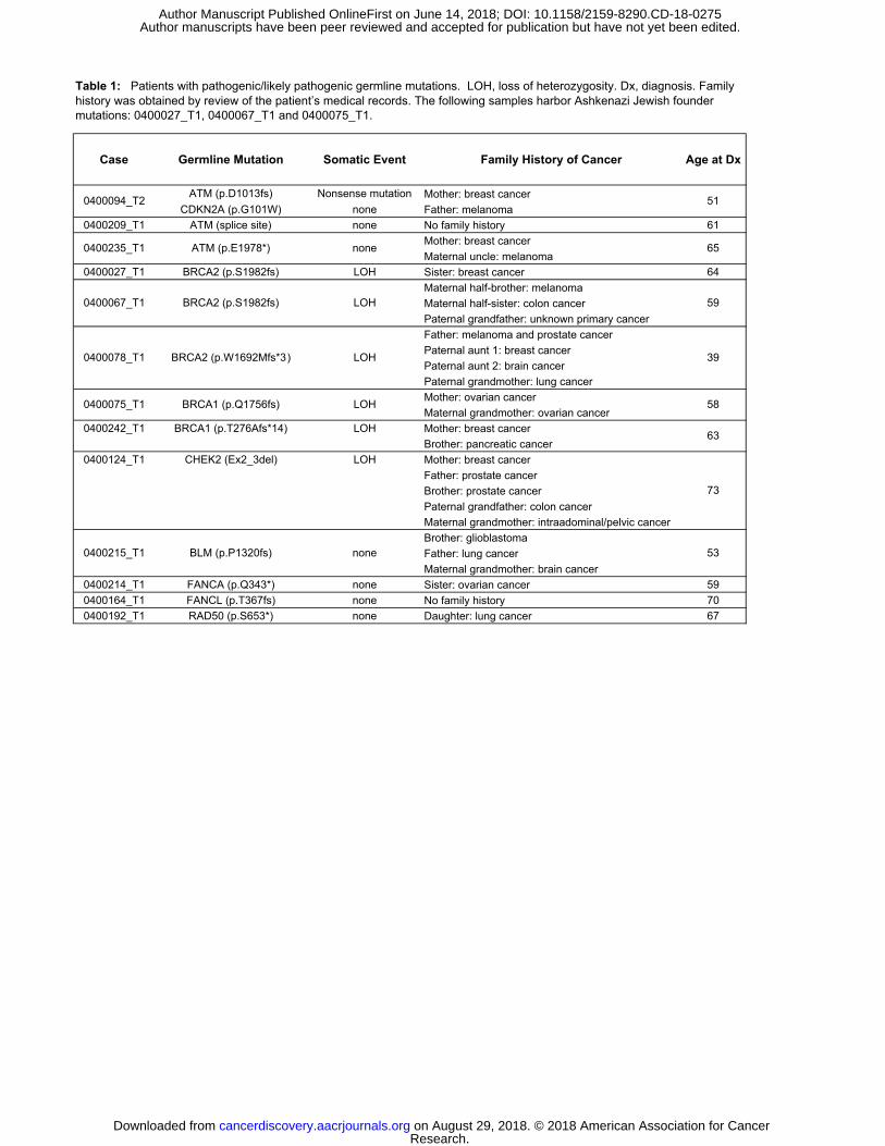
Case Germline Mutation Somatic Event Family History of Cancer Age at Dx
ATM (p.D1013fs) Nonsense mutation Mother: breast cancerCDKN2A (p.G101W) none Father: melanoma
0400209_T1 ATM (splice site) none No family history 61Mother: breast cancerMaternal uncle: melanoma
0400027_T1 BRCA2 (p.S1982fs) LOH Sister: breast cancer 64Maternal half-brother: melanomaMaternal half-sister: colon cancerPaternal grandfather: unknown primary cancerFather: melanoma and prostate cancerPaternal aunt 1: breast cancerPaternal aunt 2: brain cancerPaternal grandmother: lung cancerMother: ovarian cancerMaternal grandmother: ovarian cancer
0400242_T1 BRCA1 (p.T276Afs*14) LOH Mother: breast cancerBrother: pancreatic cancer
0400124_T1 CHEK2 (Ex2_3del) LOH Mother: breast cancerFather: prostate cancerBrother: prostate cancerPaternal grandfather: colon cancerMaternal grandmother: intraadominal/pelvic cancerBrother: glioblastomaFather: lung cancerMaternal grandmother: brain cancer
0400214_T1 FANCA (p.Q343*) none Sister: ovarian cancer 590400164_T1 FANCL (p.T367fs) none No family history 700400192_T1 RAD50 (p.S653*) none Daughter: lung cancer 67
BRCA2 (p.W1692Mfs*3) LOH 39
0400094_T2 51
0400235_T1 ATM (p.E1978*) none 65
Table 1: Patients with pathogenic/likely pathogenic germline mutations. LOH, loss of heterozygosity. Dx, diagnosis. Family history was obtained by review of the patient’s medical records. The following samples harbor Ashkenazi Jewish founder mutations: 0400027_T1, 0400067_T1 and 0400075_T1.
0400215_T1 BLM (p.P1320fs) none 53
0400075_T1 BRCA1 (p.Q1756fs) LOH 58
63
73
0400067_T1 BRCA2 (p.S1982fs) LOH 59
0400078_T1
Research. on August 29, 2018. © 2018 American Association for Cancercancerdiscovery.aacrjournals.org Downloaded from
Author manuscripts have been peer reviewed and accepted for publication but have not yet been edited. Author Manuscript Published OnlineFirst on June 14, 2018; DOI: 10.1158/2159-8290.CD-18-0275
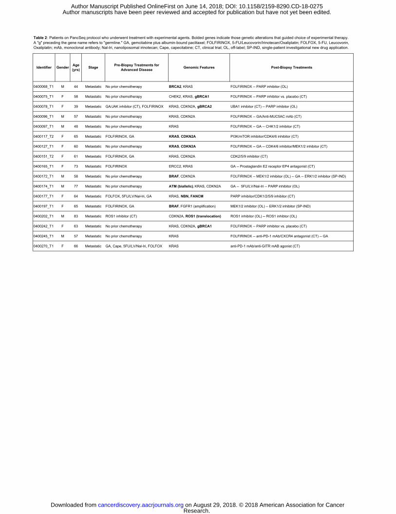
0400068_T1 M 44 Metastatic No prior chemotherapy BRCA2, KRAS FOLFIRINOX -- PARP inhibitor (OL)
0400075_T1 F 58 Metastatic No prior chemotherapy CHEK2, KRAS, gBRCA1 FOLFIRINOX -- PARP inhibitor vs. placebo (CT)
0400078_T1 F 39 Metastatic GA/JAK inhibitor (CT), FOLFIRINOX KRAS, CDKN2A, gBRCA2 UBA1 inhibitor (CT) -- PARP inhibitor (OL)
0400096_T1 M 57 Metastatic No prior chemotherapy KRAS, CDKN2A FOLFIRINOX -- GA/Anti-MUC5AC mAb (CT)
0400097_T1 M 48 Metastatic No prior chemotherapy KRAS FOLFIRINOX -- GA -- CHK1/2 inhibitor (CT)
0400117_T2 F 65 Metastatic FOLFIRINOX, GA KRAS, CDKN2A PI3K/mTOR inhibitor/CDK4/6 inhibitor (CT)
0400127_T1 F 60 Metastatic No prior chemotherapy KRAS, CDKN2A FOLFIRINOX -- GA -- CDK4/6 inhibitor/MEK1/2 inhibitor (CT)
0400151_T2 F 61 Metastatic FOLFIRINOX, GA KRAS, CDKN2A CDK2/5/9 inhibitor (CT)
0400165_T1 F 73 Metastatic FOLFIRINOX ERCC2, KRAS GA -- Prostaglandin E2 receptor EP4 antagonist (CT)
0400172_T1 M 58 Metastatic No prior chemotherapy BRAF, CDKN2A FOLFIRINOX -- MEK1/2 inhibitor (OL) -- GA -- ERK1/2 inhibitor (SP-IND)
0400174_T1 M 77 Metastatic No prior chemotherapy ATM (biallelic), KRAS, CDKN2A GA -- 5FU/LV/Nal-Iri -- PARP inhibitor (OL)
0400177_T1 F 64 Metastatic FOLFOX, 5FU/LV/Nal-Iri, GA KRAS, NBN, FANCM PARP inhibitor/CDK1/2/5/9 inhibitor (CT)
0400197_T1 F 65 Metastatic FOLFIRINOX, GA BRAF, FGFR1 (amplification) MEK1/2 inhibitor (OL) -- ERK1/2 inhibitor (SP-IND)
0400202_T1 M 83 Metastatic ROS1 inhibitor (CT) CDKN2A, ROS1 (translocation) ROS1 inhibitor (OL) -- ROS1 inhibtor (OL)
0400242_T1 F 63 Metastatic No prior chemotherapy KRAS, CDKN2A, gBRCA1 FOLFIRINOX -- PARP inhibitor vs. placebo (CT)
0400245_T1 M 57 Metastatic No prior chemotherapy KRAS FOLFIRINOX -- anti-PD-1 mAb/CXCR4 antagonist (CT) -- GA
0400270_T1 F 66 Metastatic GA, Cape, 5FU/LV/Nal-Iri, FOLFOX KRAS anti-PD-1 mAb/anti-GITR mAB agonist (CT)
Table 2: Patients on PancSeq protocol who underwent treatment with experimental agents. Bolded genes indicate those genetic alterations that guided choice of experimental therapy. A "g" preceding the gene name refers to "germline." GA, gemcitabine plus albumin-bound paclitaxel; FOLFIRINOX, 5-FU/Leucovorin/Irinotecan/Oxaliplatin; FOLFOX, 5-FU, Leucovorin, Oxaliplatin; mAb, monoclonal antibody; Nal-Iri, nanoliposomal irinotecan; Cape, capecitabine; CT, clinical trial; OL, off-label; SP-IND, single-patient investigational new drug application.
Stage Pre-Biopsy Treatments for Advanced Disease Genomic Features Post-Biopsy TreatmentsIdentifier Gender Age
(yrs)
Research. on August 29, 2018. © 2018 American Association for Cancercancerdiscovery.aacrjournals.org Downloaded from
Author manuscripts have been peer reviewed and accepted for publication but have not yet been edited. Author Manuscript Published OnlineFirst on June 14, 2018; DOI: 10.1158/2159-8290.CD-18-0275
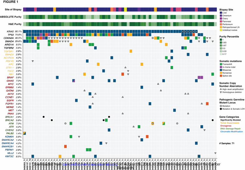
Site of Biopsy
ABSOLUTE Purity
H&E Purity0
40
02
53
_T
1
04
00
13
7_
T1
04
00
15
1_
T2
04
00
17
9_
T1
04
00
24
6_
T1
04
00
20
7_
T1
04
00
09
8_
T1
04
00
24
2_
T1
04
00
27
8_
T1
04
00
09
6_
T1
04
00
07
8_
T1
04
00
12
7_
T1
04
00
25
1_
T1
04
00
06
9_
T1
04
00
11
7_
T2
04
00
23
5_
T1
04
00
23
7_
T1
04
00
13
6_
T1
04
00
08
3_
T1
04
00
14
2_
T1
04
00
19
2_
T1
04
00
19
5_
T1
04
00
09
7_
T1
04
00
19
3_
T1
04
00
16
5_
T1
04
00
24
3_
T1
04
00
00
7_
T1
04
00
01
0_
T1
04
00
05
5_
T1
04
00
06
7_
T1
04
00
06
8_
T1
04
00
07
5_
T1
04
00
08
7_
T1
04
00
08
9_
T1
04
00
10
0_
T1
04
00
12
3_
T1
04
00
12
8_
T1
04
00
12
9_
T1
04
00
14
8_
T1
04
00
16
4_
T1
04
00
17
1_
T1
04
00
18
1_
T1
04
00
20
8_
T1
04
00
21
5_
T1
04
00
24
5_
T1
04
00
26
7_
T1
04
00
27
0_
T1
04
00
09
4_
T1
04
00
22
0_
T1
04
00
23
3_
T1
04
00
06
2_
T1
04
00
17
4_
T1
04
00
23
1_
T1
04
00
00
9_
T1
04
00
15
5_
T1
04
00
17
7_
T1
04
00
20
9_
T1
04
00
21
4_
T1
04
00
27
3_
T1
04
00
20
3_
T1
04
00
00
8_
T1
04
00
26
8_
T1
04
00
16
7_
T1
04
00
09
1_
T1
04
00
17
2_
T1
04
00
19
7_
T1
04
00
12
4_
T1
04
00
04
7_
T1
04
00
20
2_
T1
04
00
02
7_
T1
04
00
19
8_
T1
Purity Percentile1
0.83
0.67
0.5
0.33
0.17
04
00
25
3_
T1
04
00
13
7_
T1
04
00
15
1_
T2
04
00
17
9_
T1
04
00
24
6_
T1
04
00
20
7_
T1
04
00
09
8_
T1
04
00
24
2_
T1
04
00
27
8_
T1
04
00
09
6_
T1
04
00
07
8_
T1
04
00
12
7_
T1
04
00
25
1_
T1
04
00
06
9_
T1
04
00
11
7_
T2
04
00
23
5_
T1
04
00
23
7_
T1
04
00
13
6_
T1
04
00
08
3_
T1
04
00
14
2_
T1
04
00
19
2_
T1
04
00
19
5_
T1
04
00
09
7_
T1
04
00
19
3_
T1
04
00
16
5_
T1
04
00
24
3_
T1
04
00
00
7_
T1
04
00
01
0_
T1
04
00
05
5_
T1
04
00
06
7_
T1
04
00
06
8_
T1
04
00
07
5_
T1
04
00
08
7_
T1
04
00
08
9_
T1
04
00
10
0_
T1
04
00
12
3_
T1
04
00
12
8_
T1
04
00
12
9_
T1
04
00
14
8_
T1
04
00
16
4_
T1
04
00
17
1_
T1
04
00
18
1_
T1
04
00
20
8_
T1
04
00
21
5_
T1
04
00
24
5_
T1
04
00
26
7_
T1
04
00
27
0_
T1
04
00
09
4_
T1
04
00
22
0_
T1
04
00
23
3_
T1
04
00
06
2_
T1
04
00
17
4_
T1
04
00
23
1_
T1
04
00
00
9_
T1
04
00
15
5_
T1
04
00
17
7_
T1
04
00
20
9_
T1
04
00
21
4_
T1
04
00
27
3_
T1
04
00
20
3_
T1
04
00
00
8_
T1
04
00
26
8_
T1
04
00
16
7_
T1
04
00
09
1_
T1
04
00
17
2_
T1
04
00
19
7_
T1
04
00
12
4_
T1
04
00
04
7_
T1
04
00
20
2_
T1
04
00
02
7_
T1
04
00
19
8_
T1
KMT2C
MLL2
PBRM1
SMARCB1
SMARCA4
SMARCA2
KDM6A
PALB2
CHEK2
ATR
ATM
BRCA2
BRCA1
PAK4
MET
MDM2
FGFR1
EGFR
CCNE1
AKT2
GATA6
ERBB2
MYC
GNAS
BRAF
RB1
TSC2
STK11
APC
RNF43
ACVR2A
SMAD3
TGFBR2
ARID1A
SMAD4
CDKN2A
TP53
KRAS
KMT2C
MLL2
PBRM1
SMARCB1
SMARCA4
SMARCA2
KDM6A
PALB2
CHEK2
ATR
ATM
BRCA2
BRCA1
PAK4
MET
MDM2
FGFR1
EGFR
CCNE1
AKT2
GATA6
ERBB2
MYC
GNAS
BRAF
KRAS
TGFBR1
TGFBR2
ARID1A
SMAD4
CDKN2A
TP53
90.1%
74.6%
46.5%
29.6%
8.5%
5.6%
2.8%
2.8%
4.2%
4.2%
2.8%
2.8%
2.8%
5.6%
5.6%
7.0%
4.2%
1.4%
2.8%
5.6%
2.8%
1.4%
5.6%
1.4%
1.4%
2.8%
2.8%
5.6%
7.0%
4.2%
5.6%
1.4%
5.6%
1.4%
4.2%
1.4%
2.8%
1.4%
8.5%
Liver
Omentum
Ovary
Pancreas
Peritoneum
Retroperitoneal LN
Umbilical nodule
Somatic mutationsFrameshift
In−frame indel
Missense
Nonsense
Splice site
Somatic Copy
Number Aberration
High−level amplification
Homozygous deletion
Pathogenic Germline
Mutant Locus
Mutation
Mutation & Somatic LOH
Gene CategoriesSignificantly Mutated
Tumor Suppressors
Oncogenes
DNA−Damage Repair
Chromatin Modification
Biopsy Site
# Samples: 71# Samples: 71
FIGURE 1
Research. on August 29, 2018. © 2018 American Association for Cancercancerdiscovery.aacrjournals.org Downloaded from
Author manuscripts have been peer reviewed and accepted for publication but have not yet been edited. Author Manuscript Published OnlineFirst on June 14, 2018; DOI: 10.1158/2159-8290.CD-18-0275
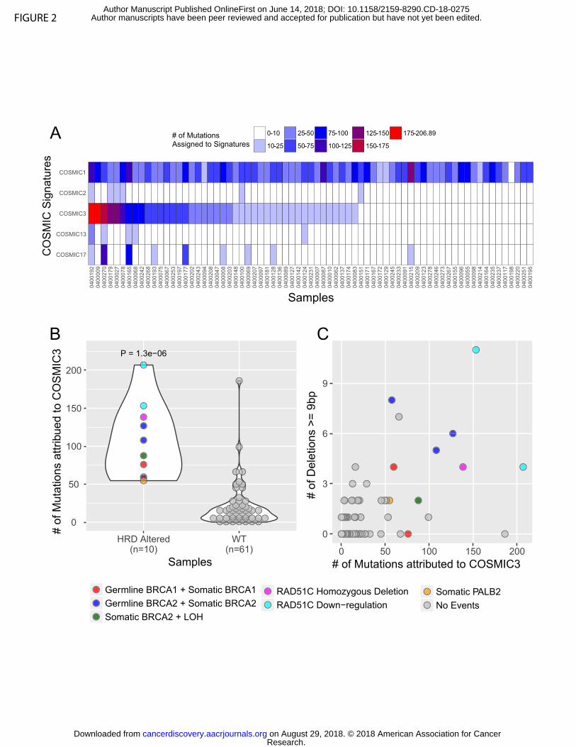
FIGURE 2
Research. on August 29, 2018. © 2018 American Association for Cancercancerdiscovery.aacrjournals.org Downloaded from
Author manuscripts have been peer reviewed and accepted for publication but have not yet been edited. Author Manuscript Published OnlineFirst on June 14, 2018; DOI: 10.1158/2159-8290.CD-18-0275
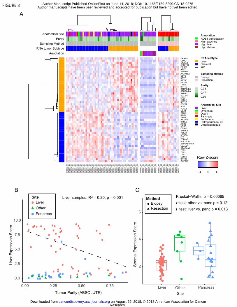
FIGURE 3
Research. on August 29, 2018. © 2018 American Association for Cancercancerdiscovery.aacrjournals.org Downloaded from
Author manuscripts have been peer reviewed and accepted for publication but have not yet been edited. Author Manuscript Published OnlineFirst on June 14, 2018; DOI: 10.1158/2159-8290.CD-18-0275
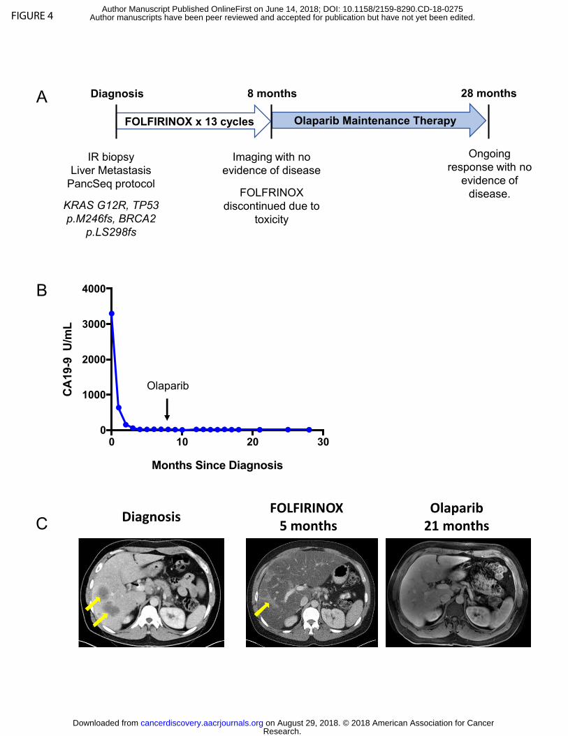
FIGURE 4
Research. on August 29, 2018. © 2018 American Association for Cancercancerdiscovery.aacrjournals.org Downloaded from
Author manuscripts have been peer reviewed and accepted for publication but have not yet been edited. Author Manuscript Published OnlineFirst on June 14, 2018; DOI: 10.1158/2159-8290.CD-18-0275
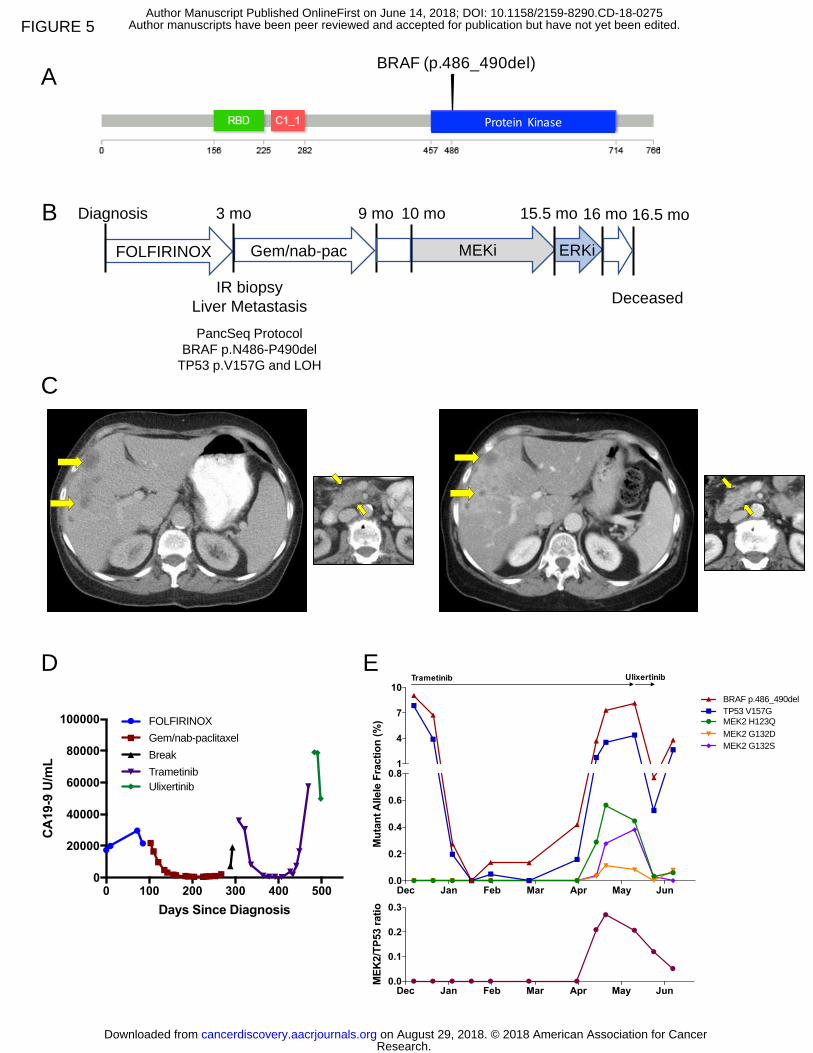
0 100 200 300 400 5000
20000
40000
60000
80000
100000
Days Since Diagnosis
CA
19-9
U/m
L
FOLFIRINOX
Gem/nab-paclitaxel
Break
Trametinib
Ulixertinib
IR biopsy
Liver Metastasis
PancSeq Protocol
BRAF p.N486-P490del
TP53 p.V157G and LOH
B
A
C
MEKi FOLFIRINOX
Deceased
Gem/nab-pac
D
Diagnosis 3 mo 15.5 mo 9 mo 16.5 mo 10 mo
E
ERKi
16 mo
Dec Jan Feb Mar Apr May Jun0.0
0.1
0.2
0.3
ME
K2/T
P53 r
ati
o
Dec Jan Feb Mar Apr May Jun0.0
0.2
0.4
0.6
0.81
4
7
10
Mu
tan
t A
llele
Fra
cti
on
(%
)
BRAF 1455_1470del
TP53 V157G
MEK2 H123Q
MEK2 G132D
MEK2 G132S
Trametinib Ulixertinib
Dec Jan Feb Mar Apr May Jun0.0
0.2
0.4
0.6
0.81
4
7
10
Mu
tan
t A
llele
Fra
cti
on
(%
)
BRAF p.486_490del
TP53 V157G
MEK2 H123Q
MEK2 G132D
MEK2 G132S
BRAF (p.486_490del)
ProteinKinase
FIGURE 5
Research. on August 29, 2018. © 2018 American Association for Cancercancerdiscovery.aacrjournals.org Downloaded from
Author manuscripts have been peer reviewed and accepted for publication but have not yet been edited. Author Manuscript Published OnlineFirst on June 14, 2018; DOI: 10.1158/2159-8290.CD-18-0275
Published OnlineFirst June 14, 2018.Cancer Discov Andrew J Aguirre, Jonathan A. Nowak, Nicholas D Camarda, et al. cancer to enable precision medicineReal-time genomic characterization of advanced pancreatic
Updated version
10.1158/2159-8290.CD-18-0275doi:
Access the most recent version of this article at:
Material
Supplementary
http://cancerdiscovery.aacrjournals.org/content/suppl/2018/06/14/2159-8290.CD-18-0275.DC1
Access the most recent supplemental material at:
Manuscript
Authoredited. Author manuscripts have been peer reviewed and accepted for publication but have not yet been
E-mail alerts related to this article or journal.Sign up to receive free email-alerts
Subscriptions
Reprints and
To order reprints of this article or to subscribe to the journal, contact the AACR Publications
Permissions
Rightslink site. Click on "Request Permissions" which will take you to the Copyright Clearance Center's (CCC)
.http://cancerdiscovery.aacrjournals.org/content/early/2018/06/14/2159-8290.CD-18-0275To request permission to re-use all or part of this article, use this link
Research. on August 29, 2018. © 2018 American Association for Cancercancerdiscovery.aacrjournals.org Downloaded from
Author manuscripts have been peer reviewed and accepted for publication but have not yet been edited. Author Manuscript Published OnlineFirst on June 14, 2018; DOI: 10.1158/2159-8290.CD-18-0275