Embed Size (px)
Citation preview
Washington University School of MedicineDigital Commons@Becker
Open Access Publications
2013
Proteomic analysis of Oesophagostomumdentatum (Nematoda) during larval transition, andthe effects of hydrolase inhibitors on developmentMartina OndrovicsUniversity of Veterinary Medicine Vienna
Katja SilbermayrUniversity of Veterinary Medicine Vienna
Makedonka MitrevaWashington University School of Medicine in St. Louis
Neil D. YoungUniversity of Melbourne
Ebrahim Razzazi-FazeliUniversity of Veterinary Medicine Vienna
See next page for additional authors
Follow this and additional works at: https://digitalcommons.wustl.edu/open_access_pubs
This Open Access Publication is brought to you for free and open access by Digital Commons@Becker. It has been accepted for inclusion in OpenAccess Publications by an authorized administrator of Digital Commons@Becker. For more information, please contact [email protected].
Recommended CitationOndrovics, Martina; Silbermayr, Katja; Mitreva, Makedonka; Young, Neil D.; Razzazi-Fazeli, Ebrahim; Gasser, Robin B.; and Joachim,Anja, ,"Proteomic analysis of Oesophagostomum dentatum (Nematoda) during larval transition, and the effects of hydrolase inhibitorson development." PLoS One.,. e63955. (2013).https://digitalcommons.wustl.edu/open_access_pubs/1494

AuthorsMartina Ondrovics, Katja Silbermayr, Makedonka Mitreva, Neil D. Young, Ebrahim Razzazi-Fazeli, Robin B.Gasser, and Anja Joachim
This open access publication is available at Digital Commons@Becker: https://digitalcommons.wustl.edu/open_access_pubs/1494
Proteomic Analysis of Oesophagostomum dentatum(Nematoda) during Larval Transition, and the Effects ofHydrolase Inhibitors on DevelopmentMartina Ondrovics1*, Katja Silbermayr1, Makedonka Mitreva2,3, Neil D. Young4, Ebrahim Razzazi-Fazeli5,
Robin B. Gasser4, Anja Joachim1
1 Department of Pathobiology, Institute of Parasitology, University of Veterinary Medicine Vienna, Vienna, Austria, 2 Genome Institute, Washington University School of
Medicine, St. Louis, Missouri, United States of America, 3 Division of Infectious Diseases, Department of Internal Medicine, Washington University School of Medicine, St.
Louis, Missouri, United States of America, 4 Faculty of Veterinary Science, The University of Melbourne, Parkville, Victoria, Australia, 5 VetCore Facility for Research,
University of Veterinary Medicine Vienna, Vienna, Austria
Abstract
In this study, in vitro drug testing was combined with proteomic and bioinformatic analyses to identify and characterizeproteins involved in larval development of Oesophagostomum dentatum, an economically important parasitic nematode.Four hydrolase inhibitors o-phenanthroline, sodium fluoride, iodoacetamide and 1,2-epoxy-3-(pnitrophenoxy)-propane(EPNP) significantly inhibited ($90%) larval development. Comparison of the proteomic profiles of the development-inhibited larvae with those of uninhibited control larvae using two-dimensional gel electrophoresis, and subsequent MALDI-TOF mass spectrometric analysis identified a down-regulation of 12 proteins inferred to be involved in various larvaldevelopmental processes, including post-embryonic development and growth. Furthermore, three proteins (i.e.intermediate filament protein B, tropomyosin and peptidyl-prolyl cis-trans isomerase) inferred to be involved in themoulting process were down-regulated in moulting- and development-inhibited O. dentatum larvae. This first proteomicmap of O. dentatum larvae provides insights in the protein profile of larval development in this parasitic nematode, andsignificantly improves our understanding of the fundamental biology of its development. The results and the approachused might assist in developing new interventions against parasitic nematodes by blocking or disrupting their keybiological pathways.
Citation: Ondrovics M, Silbermayr K, Mitreva M, Young ND, Razzazi-Fazeli E, et al. (2013) Proteomic Analysis of Oesophagostomum dentatum (Nematoda) duringLarval Transition, and the Effects of Hydrolase Inhibitors on Development. PLoS ONE 8(5): e63955. doi:10.1371/journal.pone.0063955
Editor: Andreas Hofmann, Griffith University, Australia
Received January 11, 2013; Accepted April 7, 2013; Published May 22, 2013
Copyright: � 2013 Ondrovics et al. This is an open-access article distributed under the terms of the Creative Commons Attribution License, which permitsunrestricted use, distribution, and reproduction in any medium, provided the original author and source are credited.
Funding: MO is recipient of a DOC-fFORTE-fellowship of the Austrian Academy of Sciences. Funding support to RBG from the Australian Research Council (ARC),the National Health and Medical Research Council (NHMRC) and the Alexander von Humboldt Foundation is gratefully acknowledged. Work at WashingtonUniversity is supported by National Institutes of Health grant AI081803 to MM. The funders had no role in study design, data collection and analysis, decision topublish, or preparation of the manuscript.
Competing Interests: The authors have declared that no competing interests exist.
* E-mail: [email protected]
Introduction
Parasitic roundworms (nematodes) of animals and humans are
of major socioeconomic importance worldwide [1–5]. Of these
nematodes, the soil-transmitted helminths (STHs) Ancylostoma
duodenale, Necator americanus, Trichuris trichiura and Ascaris spp. are
estimated to infect almost one sixth of the global human
population [6,7]. Also parasites of livestock, including species of
Haemonchus, Ostertagia, Teladorsagia, Trichostrongylus and Oesophagosto-
mum, collectively cause substantial economic losses estimated at
billions of dollars per annum, due to poor productivity, failure to
thrive, deaths and the cost of anthelmintic treatment [8–11]. In
addition to their socioeconomic impact, widespread resistance in
nematodes of livestock against the main classes of anthelmintics
[12–14] has stimulated research toward designing alternative
intervention and control strategies against these parasites. Central
to this effort should be the discovery of new drug targets through
an improved understanding of the fundamental biology of parasite
development.
Despite the advances in ‘-omics’ and computer-based technol-
ogies [15], and extensive studies of the free-living nematode
Caenorhabditis elegans [16–18], there is a paucity of information on
developmental processes in parasitic nematodes of animals,
particularly those of the order Strongylida, which are of major
socioeconomic importance. Numerous studies (reviewed in [19])
show that the porcine nodule worm, Oesophagostomum dentatum, is a
unique model for studying fundamental developmental and
reproductive processes in strongylid nematodes because of its
short life cycle and, particularly, an ability to maintain worms
in vitro for weeks through multiple moults.
The life cycle of O. dentatum is simple and direct [20].
Unembryonated eggs are released in host faeces and develop into
free-living, first- and second-stage larvae (L1s and L2s, respective-
ly). Feeding on nutrients and microbes in the faecal matter, they
develop into the infective, third-stage larvae (L3s) which are
protected within a cuticular sheath. These larvae migrate from the
faeces into the surrounding environment (pasture or soil), where
the porcine host ingests them. Once ingested, the L3s exsheath in
the small intestines of the pig en route to the large intestine. Upon
PLOS ONE | www.plosone.org 1 May 2013 | Volume 8 | Issue 5 | e63955

reaching the large intestine, they burrow into the mucosal layer of
the intestinal wall and subsequently produce lesions. Within the
submucosa, the L3s moult to fourth-stage larvae (L4s) [21] and
evoke an immune response that results in the encapsulation of the
larvae in raised nodular lesions, made up mainly of aggregates of
neutrophils and eosinophils [22]. Following the transition to the
L4s, the larvae emerge from the mucosa within 6–17 days. The
parasite undergoes another cuticular moult, subsequently matur-
ing to an adult. The pre-patent period of O. dentatum is ,17–20
days [23], although longer periods have been observed [20].
Recent transcriptomic studies [15,24] have provided first
insights into the molecular biology of different developmental
stages of O. dentatum, leading to the characterization of a range of
structural and functional molecules. In some studies of nematodes
[25–31], various hydrolases (including cysteine, metallo-, serine
and aspartic proteases, and pyrophosphatases) have been identi-
fied as key molecules likely to play essential and specific roles in
parasite development, cuticle collagen processing and/or moulting
processes and thus represent potential drug targets for nemato-
cides. Having available a practical in vitro culture system for O.
dentatum [32,33] provides a unique opportunity to assess the effects
of specific and selective inhibitors on protein expression in this
parasite. In the present study, we selected inhibitors of the most
relevant hydrolase groups involved in the development and
moulting of parasitic nematodes [25–27,30,31,34,35], based on
their ability to inhibit these processes without affecting viability
and motility. We investigated the effects of these hydrolase
inhibitors on the (phenotypic) proteomic profile of O. dentatum
during its transition from the L3 to L4 stage using an integrated
two-dimensional gel electrophoretic, mass spectrometric and
bioinformatic approach, taking advantage of all of the currently
available transcriptomic datasets for this parasitic nematode.
Materials and Methods
Ethics StatementExperiments were conducted in accordance with the Austrian
Animal Welfare Regulations and approved (permit GZ 68.205/
103-II/10b/2008) by the Animal Ethics Committee of the
University of Veterinary Medicine Vienna and the Ministry of
Science.
Parasite MaterialA monospecific strain (OD-Hann) of O. dentatum was maintained
routinely in experimentally infected pigs at the Institute of
Parasitology, University of Veterinary Medicine Vienna. The
faeces were collected to harvest L3s from coprocultures [23] and
stored in distilled water at 11uC for a maximum of six months.
Larval Development Inhibition AssayThe effects of seven different hydrolase inhibitors (Table 1) on
larval development were assessed; the inhibitors included o-
phenanthroline monohydrate (1,10-phenanthroline; Carl Roth,
Karlsruhe, Germany), a metalloprotease inhibitor; sodium fluoride
(Merck, Darmstadt, Germany), a pyrophosphatase inhibitor;
iodoacetamide (Sigma-Aldrich, St. Louis, USA), a cysteine
protease inhibitor; 1,2-epoxy-3-(pnitrophenoxy)-propane (EPNP;
Acros Organics, Geel, Belgium) and pepstatin A (Sigma-Aldrich),
two aspartic protease inhibitors; and 4-(2-Aminoethyl) benzene-
sulfonyl fluoride (AEBSF; Roche Applied Science, Basel, Switzer-
land) and aprotinin (Sigma-Aldrich), two serine protease inhibi-
tors. Ensheathed L3s were purified using a small-scale agar gel
migration technique [23,36] and then exsheathed in 12% (v/v)
hypochlorite at 22uC [23]. The exsheated L3s were maintained in
culture in 24-well plates (100 L3s/well) containing 1 ml of
cultivation medium containing LB broth and 10% pig serum
[37]. Plates were incubated at 38.5uC and 10% CO2 for 14 days,
with a change of medium on days 4 and 11. To determine the
effect of the different inhibitors on the development and moulting
of the L3 to L4 stage of O. dentatum, the seven inhibitors were
added (individually) to replicate cultures at ascending concentra-
tions (o-phenanthroline 3.125 mM–2 mM; sodium fluoride
0.5 mM–10 mM; iodoacetamide 5 mM–500 mM; EPNP
100 mM–4 mM; pepstatin A 0.05 mM–175 mM; AEBSF
250 mM–1 mM; aprotinin 0.77 mM–1.16 mM; see Table 1). The
percentage of developed L4s was determined on days 4, 7, 10 and
14. Phenotypes (viability, motility and mortality) were recorded at
100x magnification using an inverted light microscope (Diaphot
300, Nikon Corporation). Each inhibitor and each control were
run in triplicate, and the respective solvent controls were included
in each assay.
To test for the reversibility of inhibition of development, the
larvae were first cultured for seven days in medium containing
inhibitors at concentrations with a known inhibitory effect of
$90%. Then, the medium was replaced with inhibitor-free
medium following several washes, and the larvae maintained until
day 14.
Protein ExtractionL3s of O. dentatum (n = 500,000), cultured in vitro for four days
with or without the effective hydrolase inhibitors, were harvested,
washed three times in phosphate-buffered saline (PBS; pH 7.4),
snap frozen in liquid nitrogen and ground to fine powder with
mortar and pestle pre-frozen in liquid nitrogen. Proteins were
resuspended in ice-cold 10% (v/v) TCA in acetone at 220uC and
precipitated for 90 min. After precipitation, proteins were
centrifuged at 4uC at 17,500 g for 15 min. The supernatant was
discarded, and the pellet washed twice with chilled (220uC) 100%
acetone and centrifuged to remove any traces of TCA. Finally,
acetone was removed by evaporation at 22uC. Proteins were
resuspended overnight in 250–500 ml solubilisation buffer [7 M
urea, 2 M thiourea, 4% (w/v) 3-[(3-cholamidopropyl)dimethylam-
monio]-2-hydroxy-1-propanesulfonate (CHAPS; Carl Roth) and
30 mM Tris-Base (Carl Roth)] at 22uC. Insoluble material was
removed by centrifugation at 241,800 g at 20uC for 30 min. The
supernatant was collected and the total protein content of each
sample determined [38] using bovine serum albumin (BSA) as a
standard.
Two-dimensional ElectrophoresisFor separation in the first dimension, an aliquot of 120 mg of
parasite protein was diluted in a final volume of 300 ml of
rehydration solution [8 M urea, 2% (w/v) CHAPS, 12.7 mM
dithiothreitol (DTT), 2% immobilized pH gradient (IPG) buffer 3–
10 non-linear (GE Healthcare Life Sciences, Freiburg, Germany)]
and used to rehydrate 13 cm IPG strips with a non-linear gradient
pH 3–10 (Immobiline, GE Healthcare Life Sciences) for 18 h at
22–24uC. Isoelectric focusing (IEF) was carried out (300 V
ascending to 3,500 V for 90 min, followed by 3,500 V for 18 h)
using a Multiphor II electrophoresis chamber (GE Healthcare Life
Sciences). After IEF, the IPG strips were incubated at 22uC for
20 min in an equilibration buffer (6 M urea, 2% (w/v) sodium
dodecyl-sulphate (SDS), 30% (v/v) glycerol, 150 mM Tris-HCl,
pH 8.8, 64 mM DTT). A second equilibration was performed in
the same buffer, except that DTT was replaced by 135 mM
iodoacetamide. The IPG strips were then washed with deionized
water. In the second dimension, SDS-PAGE was performed in
vertical slab gels (1.5 mm; T = 12%, C = 2.6%, 1.5 M Tris-HCl,
Protein Profile of Nematode Development Inhibition
PLOS ONE | www.plosone.org 2 May 2013 | Volume 8 | Issue 5 | e63955
pH 8.8, 10% SDS) under reducing conditions at 15 mA for
15 min, followed by 25 mA in a Protean II electrophoresis
chamber (Bio-Rad Laboratories, Hercules, USA). Gels were
stained with silver [39]. Two technical replicates and one
biological replicate were run for each treatment. Gels were
scanned using the program ImageMasterTM 2D platinum v.7.0
(GE Healthcare Life Sciences). Proteins that were significantly
(p#0.05) differentially expressed were selected for further analyses
by mass spectrometry combined with bioinformatics.
Sample Preparation for Mass Spectrometric AnalysisProtein spots were excised manually from silver-stained two-
dimensional electrophoretic (2-DE) gels. ‘Negative’ control spots
were also excised from blank regions of each gel and processed in
parallel. Spots were washed, reduced with DTT and alkylated with
iodoacetamide. In-gel digestion with trypsin (Trypsin Gold, Mass
Spectrometry Grade, Promega, Madison, USA) was carried out
[40]. In order to enhance peptide ionisation and the sensitivity of
mass spectrometry, dried peptides were de-salted using Zip-Tips
mC18 (Millipore, Billerica, USA) according to the manufacturer’s
instructions.
Spotting and Mass SpectrometryPeptides were annotated using a Matrix Assisted Laser
Desorption Ionisation Tandem Time-of-Flight (MALDI-TOF/
TOF) mass spectrometer (Ultraflex II; Bruker Daltonics, Bremen,
Germany) in MS and MS/MS modes. De-salted peptides (0.5 ml)
were spotted on to a disposable AnchorChip MALDI target plate
pre-spotted with a-cyano-4-hydroxycinnamic acid (PAC target;
Bruker Daltonics). MS spectral data were acquired from the
samples and a MS/MS list was generated for further analysis
based on the most intense ions present (trypsin and major keratin
ions were excluded). Spectra processing and peak annotation were
carried out using FlexAnalysis and Biotools (Bruker Daltonics).
Data AnalysisAll peptide mass fingerprint (PMF) data were refined using MS/
MS data. Protein sequence matches with a significant MS/MS
score (p,0.05) were used for further analysis. Ion mass data and
amino acid sequences characterized from processed spectra were
compared with data in public eukaryotic databases, including an
inferred protein database of O. dentatum (www.nematode.net), the
UniProt database (http://www.uniprot.org/) and the National
Center for Biotechnology Information (NCBI) non-redundant (nr)
database (http://www.ncbi.nlm.nih.gov) using the following
search parameters: global modifications carbamido-methylation
on cysteine; variable modifications oxidation on methionine;
peptide mass tolerance = 100 ppm; MS/MS tolerance = 1 Da; one
missed cleavage allowed. The programs Proteinscape (v.2.1;
Bruker Daltonics) and MASCOT (www.matrixscience.com) were
used for data management and analyses. Proteins identified by MS
were annotated based on their closest homologue (BLASTp, E-
value cut-off #161025) in the UniProt database and Gene
Ontology (GO). Subsequent GO analyses were performed using
the AmiGO BLAST tool [41]. Protein sequences were also
mapped to known biological pathways (KOBAS algorithm) [42]
using BLASTp (E-value cut-off #1025) based on sequence
homology to molecules in the Kyoto Encyclopedia of Genes and
Genomes (KEGG) database [43].
Statistical AnalysisStatistical calculations were performed using SPSS (v.20.0) for
windows. To test for significant differences in the percentage of L4
development, the development-inhibited groups and the controls
were compared using the Kruskal-Wallis test and Mann-Whitney
U-test.
Results
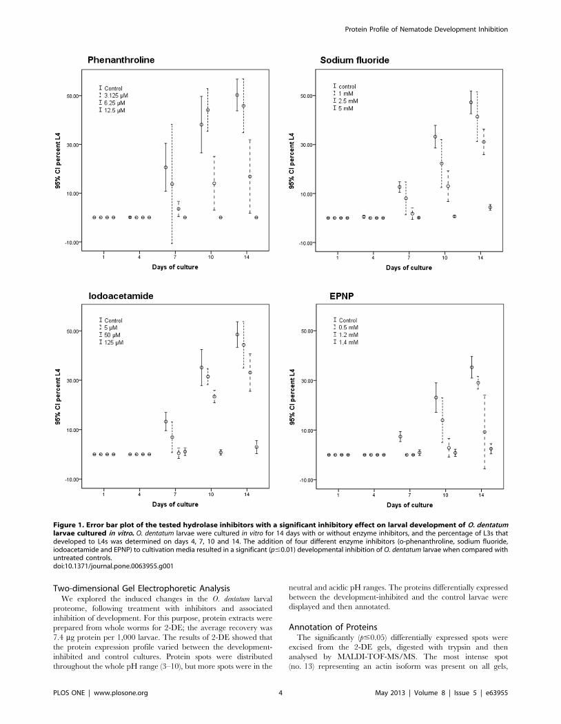
Testing of InhibitorsWe assessed the inhibitory effects of each compound on the
development from the exsheathed L3 to the L4 stage of O.
dentatum (Figure 1) by culturing larvae for 14 days in medium
containing seven different hydrolase inhibitors (Table 1). A
significant inhibitory effect (p#0.01) was detected for four of the
seven hydrolase inhibitors tested. By adding iodoacetamide
(125 mM), the development of L3s to L4s was inhibited by
93.965.1% compared with untreated controls. The addition of
1.4 mM EPNP resulted in 93.065.6% inhibition. Complete
inhibition of nematode development was achieved by adding
12.5 mM o-phenanthroline to the in vitro cultures. Sodium
fluoride (5 mM) resulted in a 90.863.2% inhibition. No
significant inhibitory effect was observed for the aspartic
protease inhibitor pepstatin A or either serine protease
inhibitors (AEBSF and aprotinin) tested (data not shown). The
addition of higher concentrations of each of the four inhibitory
compounds did not result in an increased inhibitory effect, but
did lead to a higher mortality of larvae. The reversibility of the
inhibitory effect of each inhibitor was assessed. The removal of
iodoacetamide, EPNP, o-phenanthroline and sodium fluoride
resulted in 90.5%, 85.6%, 89.5%, and 106.3%, respectively, of
L3s developing through to L4s with respect to the controls.
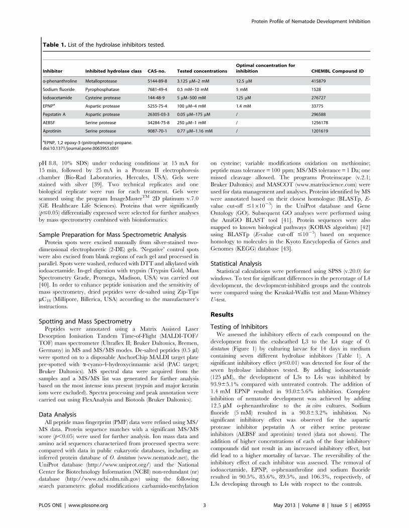
Table 1. List of the hydrolase inhibitors tested.
Inhibitor Inhibited hydrolase class CAS-no. Tested concentrationsOptimal concentration forinhibition CHEMBL Compound ID
o-phenanthroline Metalloprotease 5144-89-8 3.125 mM–2 mM 12.5 mM 415879
Sodium fluoride Pyrophosphatase 7681-49-4 0.5 mM–10 mM 5 mM 1528
Iodoacetamide Cysteine protease 144-48-9 5 mM–500 mM 125 mM 276727
EPNPa Aspartic protease 5255-75-4 100 mM–4 mM 1.4 mM 33775
Pepstatin A Aspartic protease 26305-03-3 0.05 mM–175 mM / 296588
AEBSF Serine protease 34284-75-8 250 mM–1 mM / 1256178
Aprotinin Serine protease 9087-70-1 0.77 mM–1.16 mM / 1201619
aEPNP, 1,2 epoxy-3-(pnitrophenoxy)-propane.doi:10.1371/journal.pone.0063955.t001
Protein Profile of Nematode Development Inhibition
PLOS ONE | www.plosone.org 3 May 2013 | Volume 8 | Issue 5 | e63955
Two-dimensional Gel Electrophoretic AnalysisWe explored the induced changes in the O. dentatum larval
proteome, following treatment with inhibitors and associated
inhibition of development. For this purpose, protein extracts were
prepared from whole worms for 2-DE; the average recovery was
7.4 mg protein per 1,000 larvae. The results of 2-DE showed that
the protein expression profile varied between the development-
inhibited and control cultures. Protein spots were distributed
throughout the whole pH range (3–10), but more spots were in the
neutral and acidic pH ranges. The proteins differentially expressed
between the development-inhibited and the control larvae were
displayed and then annotated.
Annotation of ProteinsThe significantly (p#0.05) differentially expressed spots were
excised from the 2-DE gels, digested with trypsin and then
analysed by MALDI-TOF-MS/MS. The most intense spot
(no. 13) representing an actin isoform was present on all gels,
Figure 1. Error bar plot of the tested hydrolase inhibitors with a significant inhibitory effect on larval development of O. dentatumlarvae cultured in vitro. O. dentatum larvae were cultured in vitro for 14 days with or without enzyme inhibitors, and the percentage of L3s thatdeveloped to L4s was determined on days 4, 7, 10 and 14. The addition of four different enzyme inhibitors (o-phenanthroline, sodium fluoride,iodoacetamide and EPNP) to cultivation media resulted in a significant (p#0.01) developmental inhibition of O. dentatum larvae when compared withuntreated controls.doi:10.1371/journal.pone.0063955.g001
Protein Profile of Nematode Development Inhibition
PLOS ONE | www.plosone.org 4 May 2013 | Volume 8 | Issue 5 | e63955
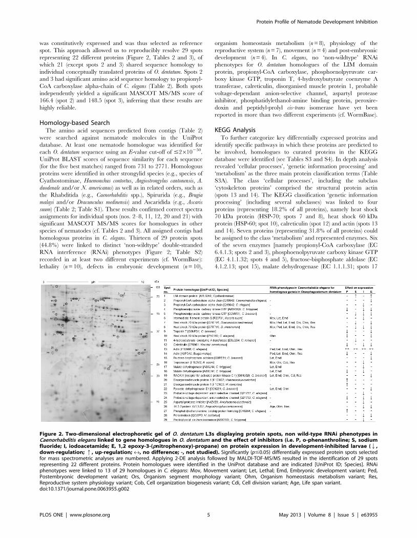
was constitutively expressed and was thus selected as reference
spot. This approach allowed us to reproducibly resolve 29 spots
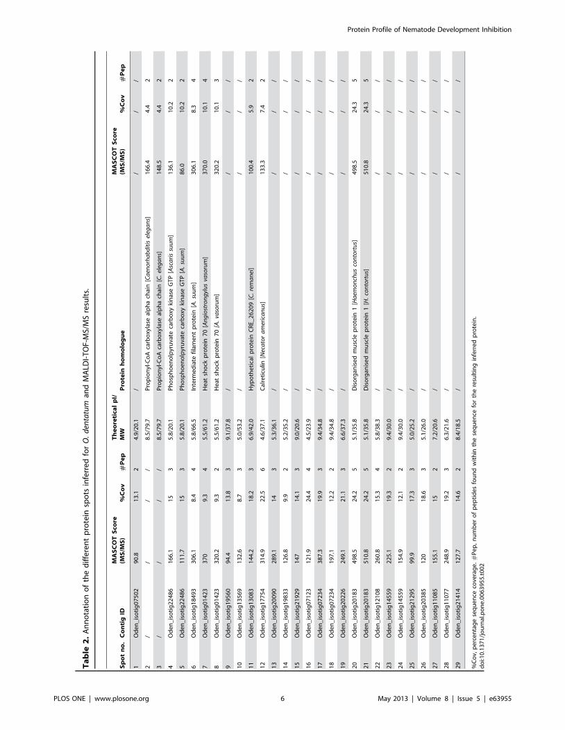
representing 22 different proteins (Figure 2, Tables 2 and 3), of
which 21 (except spots 2 and 3) shared sequence homology to
individual conceptually translated proteins of O. dentatum. Spots 2
and 3 had significant amino acid sequence homology to propionyl-
CoA carboxylase alpha-chain of C. elegans (Table 2). Both spots
independently yielded a significant MASCOT MS/MS score of
166.4 (spot 2) and 148.5 (spot 3), inferring that these results are
highly reliable.
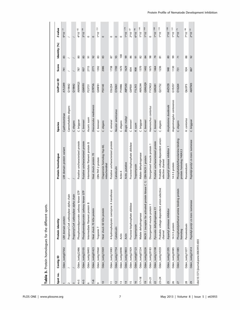
Homology-based SearchThe amino acid sequences predicted from contigs (Table 2)
were searched against nematode molecules in the UniProt
database. At least one nematode homologue was identified for
each O. dentatum sequence using an E-value cut-off of #2610250.
UniProt BLAST scores of sequence similarity for each sequence
(for the five best matches) ranged from 731 to 2771. Homologous
proteins were identified in other strongylid species (e.g., species of
Cyathostominae, Haemonchus contortus, Angiostrongylus cantonensis, A.
duodenale and/or N. americanus) as well as in related orders, such as
the Rhabditida (e.g., Caenorhabditis spp.), Spirurida (e.g., Brugia
malayi and/or Dracunculus medinensis) and Ascaridida (e.g., Ascaris
suum) (Table 2; Table S1). These results confirmed correct spectra
assignments for individual spots (nos. 2–8, 11, 12, 20 and 21) with
significant MASCOT MS/MS scores for homologues in other
species of nematodes (cf. Tables 2 and 3). All assigned contigs had
homologous proteins in C. elegans. Thirteen of 29 protein spots
(44.8%) were linked to distinct ‘non-wildtype’ double-stranded
RNA interference (RNAi) phenotypes (Figure 2; Table S2)
recorded in at least two different experiments (cf. WormBase):
lethality (n = 10), defects in embryonic development (n = 10),
organism homeostasis metabolism (n = 8), physiology of the
reproductive system (n = 7), movement (n = 4) and post-embryonic
development (n = 4). In C. elegans, no ‘non-wildtype’ RNAi
phenotypes for O. dentatum homologues of the LIM domain
protein, propionyl-CoA carboxylase, phosphoenolpyruvate car-
boxy kinase GTP, troponin T, 4-hydroxybutyrate coenzyme A
transferase, calreticulin, disorganised muscle protein 1, probable
voltage-dependant anion-selective channel, aspartyl protease
inhibitor, phosphatidylethanol-amine binding protein, peroxire-
doxin and peptidyl-prolyl cis-trans isomerase have yet been
reported in more than two different experiments (cf. WormBase).
KEGG AnalysisTo further categorize key differentially expressed proteins and
identify specific pathways in which these proteins are predicted to
be involved, homologues to curated proteins in the KEGG
database were identified (see Tables S3 and S4). In depth analysis
revealed ‘cellular processes’, ‘genetic information processing’ and
‘metabolism’ as the three main protein classification terms (Table
S3A). The class ‘cellular processes’, including the subclass
‘cytoskeleton proteins’ comprised the structural protein actin
(spots 13 and 14). The KEGG classification ‘genetic information
processing’ (including several subclasses) was linked to four
proteins (representing 18.2% of all proteins), namely heat shock
70 kDa protein (HSP-70; spots 7 and 8), heat shock 60 kDa
protein (HSP-60; spot 10), calreticulin (spot 12) and actin (spots 13
and 14). Seven proteins (representing 31.8% of all proteins) could
be assigned to the class ‘metabolism’ and represented enzymes. Six
of the seven enzymes [namely propionyl-CoA carboxylase (EC
6.4.1.3; spots 2 and 3), phosphoenolpyruvate carboxy kinase GTP
(EC 4.1.1.32; spots 4 and 5), fructose-bisphosphate aldolase (EC
4.1.2.13; spot 15), malate dehydrogenase (EC 1.1.1.31; spots 17
Figure 2. Two-dimensional electrophoretic gel of O. dentatum L3s displaying protein spots, non wild-type RNAi phenotypes inCaenorhabditis elegans linked to gene homologues in O. dentatum and the effect of inhibitors (i.e. P, o-phenanthroline; S, sodiumfluoride; I, iodoacetamide; E, 1,2 epoxy-3-(pnitrophenoxy)-propane) on protein expression in development-inhibited larvae (Q,down-regulation; q, up-regulation; «, no difference; -, not studied). Significantly (p#0.05) differentially expressed protein spots selectedfor mass spectrometric analyses are numbered. Applying 2-DE analysis followed by MALDI-TOF-MS/MS resulted in the identification of 29 spotsrepresenting 22 different proteins. Protein homologues were identified in the UniProt database and are indicated [UniProt ID; Species]. RNAiphenotypes were linked to 13 of 29 homologues in C. elegans: Mov, Movement variant; Let, Lethal; Emd, Embryonic development variant; Ped,Postembryonic development variant; Ors, Organism segment morphology variant; Ohm, Organism homeostasis metabolism variant; Res,Reproductive system physiology variant; Cob, Cell organization biogenesis variant; Cdi, Cell division variant; Age, Life span variant.doi:10.1371/journal.pone.0063955.g002
Protein Profile of Nematode Development Inhibition
PLOS ONE | www.plosone.org 5 May 2013 | Volume 8 | Issue 5 | e63955
Ta
ble
2.
An
no
tati
on
of
the
dif
fere
nt
pro
tein
spo
tsin
ferr
ed
for
O.
den
tatu
man
dM
ALD
I-T
OF-
MS/
MS
resu
lts.
Sp
ot
no
.C
on
tig
IDM
AS
CO
TS
core
(MS
/MS
)%
Co
v#
Pe
pT
he
ore
tica
lp
I/M
WP
rote
inh
om
olo
gu
eM
AS
CO
TS
core
(MS
/MS
)%
Co
v#
Pe
p
1O
de
n_
iso
tig
07
50
29
0.8
13
.12
4.9
/20
.1/
//
/
2/
//
/8
.5/7
9.7
Pro
pio
nyl
-Co
Aca
rbo
xyla
seal
ph
ach
ain
[Ca
eno
rha
bd
itis
eleg
an
s]1
66
.44
.42
3/
//
/8
.5/7
9.7
Pro
pio
nyl
-Co
Aca
rbo
xyla
seal
ph
ach
ain
[C.
eleg
an
s]1
48
.54
.42
4O
de
n_
iso
tig
22
48
61
66
.11
53
5.8
/20
.1P
ho
sph
oe
no
lpyr
uva
teca
rbo
xyki
nas
eG
TP
[Asc
ari
ssu
um
]1
36
.11
0.2
2
5O
de
n_
iso
tig
22
48
61
11
.71
53
5.8
/20
.1P
ho
sph
oe
no
lpyr
uva
teca
rbo
xyki
nas
eG
TP
[A.
suu
m]
86
.01
0.2
2
6O
de
n_
iso
tig
18
49
33
06
.18
.44
5.8
/66
.5In
term
ed
iate
fila
me
nt
pro
tein
[A.
suu
m]
30
6.1
8.3
4
7O
de
n_
iso
tig
01
42
33
70
9.3
45
.5/6
1.2
He
atsh
ock
pro
tein
70
[An
gio
stro
ng
ylu
sva
soru
m]
37
0.0
10
.14
8O
de
n_
iso
tig
01
42
33
20
.29
.32
5.5
/61
.2H
eat
sho
ckp
rote
in7
0[A
.va
soru
m]
32
0.2
10
.13
9O
de
n_
iso
tig
19
56
09
4.4
13
.83
9.1
/37
.8/
//
/
10
Od
en
_is
oti
g1
35
69
13
2.6
8.7
35
.0/5
3.2
//
//
11
Od
en
_is
oti
g1
30
83
14
4.2
18
.23
6.9
/42
.0H
ypo
the
tica
lp
rote
inC
RE_
26
20
9[C
.re
ma
nei
]1
00
.45
.92
12
Od
en
_is
oti
g1
77
54
31
4.9
22
.56
4.6
/37
.1C
alre
ticu
lin[N
eca
tor
am
eric
an
us]
13
3.3
7.4
2
13
Od
en
_is
oti
g2
00
90
28
9.1
14
35
.3/3
6.1
//
//
14
Od
en
_is
oti
g1
98
33
12
6.8
9.9
25
.2/3
5.2
//
//
15
Od
en
_is
oti
g2
19
29
14
71
4.1
39
.0/2
0.6
//
//
16
Od
en
_is
oti
g0
71
23
12
1.9
24
.44
4.5
/23
.9/
//
/
17
Od
en
_is
oti
g0
72
34
38
7.3
19
.93
9.4
/34
.8/
//
/
18
Od
en
_is
oti
g0
72
34
19
7.1
12
.22
9.4
/34
.8/
//
/
19
Od
en
_is
oti
g2
02
26
24
9.1
21
.13
6.6
/37
.3/
//
/
20
Od
en
_is
oti
g2
01
83
49
8.5
24
.25
5.1
/35
.8D
iso
rgan
ise
dm
usc
lep
rote
in1
[Ha
emo
nch
us
con
tort
us]
49
8.5
24
.35
21
Od
en
_is
oti
g2
01
83
51
0.8
24
.25
5.1
/35
.8D
iso
rgan
ise
dm
usc
lep
rote
in1
[H.
con
tort
us]
51
0.8
24
.35
22
Od
en
_is
oti
g1
21
08
26
0.8
15
.34
5.8
/38
.3/
//
/
23
Od
en
_is
oti
g1
45
59
22
5.1
19
.32
9.4
/30
.0/
//
/
24
Od
en
_is
oti
g1
45
59
15
4.9
12
.12
9.4
/30
.0/
//
/
25
Od
en
_is
oti
g2
12
95
99
.91
7.3
35
.0/2
5.2
//
//
26
Od
en
_is
oti
g2
03
85
12
01
8.6
35
.1/2
6.0
//
//
27
Od
en
_is
oti
g1
10
85
15
5.1
15
27
.2/2
0.6
//
//
28
Od
en
_is
oti
g1
10
77
24
8.9
19
.23
6.3
/21
.6/
//
/
29
Od
en
_is
oti
g2
14
14
12
7.7
14
.62
8.4
/18
.5/
//
/
%C
ov,
pe
rce
nta
ge
seq
ue
nce
cove
rag
e.
#P
ep
,n
um
be
ro
fp
ep
tid
es
fou
nd
wit
hin
the
seq
ue
nce
for
the
resu
ltin
gin
ferr
ed
pro
tein
.d
oi:1
0.1
37
1/j
ou
rnal
.po
ne
.00
63
95
5.t
00
2
Protein Profile of Nematode Development Inhibition
PLOS ONE | www.plosone.org 6 May 2013 | Volume 8 | Issue 5 | e63955
Ta
ble
3.
Pro
tein
ho
mo
log
ue
sfo
rth
ed
iffe
ren
tsp
ots
.
Sp
ot
no
.C
on
tig
IDP
rote
inid
en
tity
Pro
tein
ho
mo
log
ue
Sp
eci
es
Un
iPro
tID
Sco
reId
en
tity
[%]
E-v
alu
e
1O
de
n_
iso
tig
07
50
2LI
Md
om
ain
pro
tein
LIM
do
mai
np
rote
inva
rian
tC
yath
ost
om
inae
A7
LGW
97
41
83
9*1
02
77
2/
Pro
pio
nyl
-Co
Aca
rbo
xyla
seal
ph
ach
ain
/C
aen
orh
ab
dit
isel
ega
ns
Q1
98
42
//
/
3/
Pro
pio
nyl
-Co
Aca
rbo
xyla
seal
ph
ach
ain
/C
.el
ega
ns
Q1
98
42
//
/
4+5
Od
en
_is
oti
g2
24
86
Ph
osp
ho
en
olp
yru
vate
carb
oxy
kin
ase
GT
PP
uta
tive
un
char
acte
rize
dp
rote
inC
.b
rig
gsa
eA
8W
MQ
57
87
80
4*1
02
82
4+5
Od
en
_is
oti
g2
31
05
Ph
osp
ho
en
olp
yru
vate
carb
oxy
kin
ase
GT
PP
uta
tive
un
char
acte
rize
dp
rote
inC
.b
ren
ner
iG
0M
YR
19
37
83
3*1
02
99
6O
de
n_
iso
tig
18
49
3In
term
ed
iate
fila
me
nt
pro
tein
BIn
term
ed
iate
fila
me
nt
pro
tein
BA
sca
ris
suu
mP
23
73
12
71
59
10
7+8
Od
en
_is
oti
g0
14
23
He
atsh
ock
70
kDa
pro
tein
He
atsh
ock
pro
tein
70
Dra
cun
culu
sm
edin
ensi
sD
7R
TV
62
77
19
20
9O
de
n_
iso
tig
19
56
0T
rop
on
inT
CB
N-T
NT
-2p
rote
inC
.b
ren
ner
iG
0N
FB1
13
92
83
1*1
02
15
1
10
Od
en
_is
oti
g1
35
69
He
atsh
ock
60
kDa
pro
tein
Ch
ape
ron
inh
om
olo
gH
sp-6
0,
mit
och
on
dri
alC
.el
ega
ns
P5
01
40
20
86
83
0
11
Od
en
_is
oti
g1
30
83
4-h
ydro
xyb
uty
rate
coe
nzy
me
Atr
ansf
era
seP
uta
tive
un
char
acte
rize
dp
rote
inC
.re
ma
nei
E3LQ
S41
73
88
70
12
Od
en
_is
oti
g1
77
54
Cal
reti
culin
Cal
reti
culin
Nec
ato
ra
mer
ica
nu
sO
76
96
11
70
09
30
13
Od
en
_is
oti
g2
00
90
Act
inA
ctin
-4C
.el
ega
ns
P1
09
86
16
70
10
00
14
Od
en
_is
oti
g1
98
33
Act
inA
ctin
,p
uta
tive
Bru
gia
ma
layi
A8
P5
A0
16
29
99
1*1
02
17
9
15
Od
en
_is
oti
g2
19
29
Fru
cto
se-b
isp
ho
sph
ate
ald
ola
seFr
uct
ose
-bis
ph
osp
hat
eal
do
lase
C.
bre
nn
eri
G0
PE8
18
34
86
2*1
02
87
16
Od
en
_is
oti
g0
71
23
Tro
po
myo
sin
Tro
po
myo
sin
A.
suu
mF1
L3V
29
08
91
6*1
02
96
17
+18
Od
en
_is
oti
g0
72
34
Mal
ate
de
hyd
rog
en
ase
Mal
ate
de
hyd
rog
en
ase
C.
bri
gg
sae
A8
XLN
41
37
08
21
*10
21
49
19
Od
en
_is
oti
g2
02
26
RA
CK
-1(r
ece
pto
rfo
rac
tiva
ted
pro
tein
kin
ase
C1
)C
BN
-RA
CK
-1p
rote
inC
.b
ren
ner
iG
0N
UQ
91
49
28
41
*10
21
63
20
+21
Od
en
_is
oti
g2
01
83
Dis
org
anis
ed
mu
scle
pro
tein
1D
iso
rgan
ise
dm
usc
lep
rote
in1
Ha
emo
nch
us
con
tort
us
F1C
NG
31
67
59
80
22
Od
en
_is
oti
g1
21
08
Pyr
uva
ted
eh
ydro
ge
nas
eE1
Pu
tati
veu
nch
arac
teri
zed
pro
tein
C.
bre
nn
eri
G0
N2
81
14
64
79
1*1
02
16
0
23
+24
Od
en
_is
oti
g1
45
59
Pro
bab
levo
ltag
e-d
ep
en
de
nt
anio
n-s
ele
ctiv
ech
ann
el
Pro
bab
levo
ltag
e-d
ep
en
de
nt
anio
n-
sele
ctiv
ech
ann
el
C.
eleg
an
sQ
21
75
21
23
68
11
*10
21
33
25
Od
en
_is
oti
g2
12
95
Asp
arty
lp
rote
ase
inh
ibit
or
Asp
arty
lp
rote
ase
inh
ibit
or
1A
ncy
lost
om
ad
uo
den
ale
A4
ZV
Z8
94
88
01
*10
21
00
26
Od
en
_is
oti
g2
03
85
14
-3-3
pro
tein
14
-3-3
pro
tein
An
gio
stro
ng
ylu
sca
nto
nen
sis
G1
EUS7
11
30
99
1*1
02
12
1
27
Od
en
_is
oti
g1
10
85
Ph
osp
hat
idyl
eth
ano
l-am
ine
bin
din
gp
rote
inh
om
olo
gP
ho
sph
atid
yle
than
ola
min
e-b
ind
ing
pro
tein
ho
mo
log
F40
A3
.3C
.el
ega
ns
O1
62
64
73
17
12
*10
27
5
28
Od
en
_is
oti
g1
10
77
Pe
roxi
red
oxi
nP
ero
xire
do
xin
H.
con
tort
us
Q6
J3P
39
24
88
7*1
02
98
29
Od
en
_is
oti
g2
14
14
Pe
pti
dyl
-pro
lyl
cis-
tra
ns
iso
me
rase
Pe
pti
dyl
-pro
lyl
cis-
tra
ns
iso
me
rase
C.
bri
gg
sae
A8
XT
M3
86
79
22
*10
29
1
do
i:10
.13
71
/jo
urn
al.p
on
e.0
06
39
55
.t0
03
Protein Profile of Nematode Development Inhibition
PLOS ONE | www.plosone.org 7 May 2013 | Volume 8 | Issue 5 | e63955
and 18), pyruvate dehydrogenase E1 (EC 1.2.4.1; spot 22) and
peptidyl-prolyl cis-trans isomerase (EC 5.2.1.8; spot 29)] are
involved in various pathways linked to amino acid, carbohydrate
and/or energy metabolism.
The KEGG pathway classification included the biological
pathway terms ‘cellular processes’ (n = 4), ‘environmental infor-
mation processing’ (n = 2), ‘genetic information processing’ (n = 3),
‘metabolism’ (n = 5) and ‘organismal systems’ (n = 5) (Table S3B).
No annotation was found in the KEGG database for nine proteins
(representing 36.4% of all proteins), including LIM domain
protein (spot 1), intermediate filament protein B (spot 6), troponin
T (spot 9), 4-hydroxybutyrate coenzyme A transferase (spot 11),
tropomyosin (spot 16), receptor for activated protein kinase C 1
(RACK-1, spot 19), disorganised muscle protein 1 (DIM-1; spots
20 and 21), aspartyl protease inhibitor (spot 25) and phosphati-
dylethanol-amine binding protein (spot 27).
Gene Ontology (GO) AnalysisIn order to better understand the biological processes and
molecular functions in which the identified proteins are inferred to
be involved, GO analyses were performed using the AmiGO
BLAST tool. Of the 22 proteins, 19 (86.4%) could be assigned to
GO classifications associated with biological processes and 18
proteins (81.8%) were assigned to certain molecular functions.
Only GO annotations found in related nematode species
(particularly C. elegans) were taken into account using a cut-off of
p#2610225 (see Tables S5 and S6). GO classification analysis
revealed associations of the proteins identified to a range of
biological processes (Table S5A), including ‘reproduction’ (n = 8),
‘metabolic process’ (n = 10), ‘cellular process’ (n = 9), ‘multicellular
organismal growth’ (n = 11), ‘developmental process’ (n = 11),
‘growth’ (n = 11), ‘locomotion’ (n = 8), ‘response to stimulus’
(n = 8), ‘localization’ (n = 4) and ‘biological regulation’ (n = 8). Most
proteins assigned to the GO term ‘metabolic process’ related to
‘cellular metabolic process’ (representing 70% of the enzymes
identified). No GO classifications for biological processes were
found for three of the 22 proteins, namely DIM-1 (spots 20 and
21), aspartyl protease inhibitor (spot 25) and phosphatidylethanol-
amine binding protein (spot 27). ‘binding’ (n = 11) and ‘catalytic
activity’ (n = 10) were the two main functional GO categories
(Table S5B); additional GO categories included ‘structural
molecule activity’ (n = 2), ‘transporter activity’ (n = 1) and ‘antiox-
idant activity’ (n = 1). In depth analysis revealed that the majority
(63.6%) of proteins assigned to ‘binding’ are involved in ‘protein
binding’ processes. No GO annotation for molecular functions was
found for troponin T (spot 9), DIM-1 (spots 20 and 21), aspartyl
protease inhibitor (spot 25) or phosphatidylethanol-amine binding
protein (spot 27).
Discussion
Drug resistance represents a major concern in the control of
parasitic nematodes [12–14,44]. Therefore, much research is
directed towards the development of new agents in the treatment
of nematode infections. Proteins involved in fundamental devel-
opmental processes in nematodes represent promising targets for
the design of new and selective interventions. Hence, this study
aimed at identifying and characterizing proteins involved in the
larval development of O. dentatum, a model organism representative
of parasitic nematodes of major socioeconomic impact. We
applied an integrative approach combining in vitro drug testing
with proteomic and bioinformatic analyses to provide first insights
into larval development in O. dentatum.
To generate a development-inhibited phenotype for O. dentatum,
hydrolase inhibitors were selected based on their ability to impede
the moulting and development of larvae without affecting their
viability and motility. Enzyme inhibitors were tested to cover the
most relevant hydrolase classes known to be involved in the highly
sophisticated moulting process in various nematode species [25–
27,30,31,34,35]. Significant inhibition of moulting and develop-
ment has been described previously in a range of nematodes and
could be confirmed in O. dentatum for the three inhibitors, o-
phenanthroline [27,34,45], sodium fluoride [25,26] and iodoace-
tamide [28]. The inhibitory effect of EPNP had not been tested
before on the development of nematode larvae. In malaria
parasites, however, this protease inhibitor disintegrates gametocyte
membranes [46], and it has been used frequently as an inhibitor of
peptidases of the A1 family [47]. This is particularly interesting,
since the other aspartic protease inhibitor tested, pepstatin A, was
neither effective in our model nor in related nematodes [27,34],
probably due to its limited half-life.
Since the development of nematode larvae and the sophisticated
moulting process require a series of structural, biochemical,
metabolic and physiological changes, we assumed that the proteins
essential for fundamental developmental processes in develop-
ment-inhibited larvae are less abundantly expressed compared
with those of uninhibited controls in which normal development
occurred. Thus, protein spots that were significantly differentially
expressed between development-inhibited and control larvae were
subjected to mass spectrometric and bioinformatic analyses. Using
this approach, we identified 29 spots representing 22 different
proteins. Several spots located at different positions in the same gel
were inferred to be distinct protein isoforms or the same protein
with varying post-translational modifications. Furthermore, 27 out
of 29 identified spots were encoded in O. dentatum contigs derived
from ESTs generated by 454 sequencing (FLX GS20 sequencer)
[48] and analyzed using an advanced bioinformatic pipeline [49]
for the interference of key biological processes [50], such as
development and moulting. The O. dentatum contigs were further
subjected to homology-based functional annotation. All O. dentatum
amino acid sequences characterised had homologous proteins in
other nematodes, including species from the orders Strongylida,
Rhabditida, Spirurida, and Ascaridida.
The proteins identified and annotated included structural and
cytoskeletal proteins (intermediate filament protein B, troponin T,
actin, tropomyosin, DIM-1), enzymes involved in metabolism
(propionyl-CoA carboxylase, phosphoenolpyruvate carboxy kinase
GTP, 4-hydroxybutyrate coenzyme A transferase, fructose-bi-
sphosphate aldolase, malate dehydrogenase, pyruvate dehydroge-
nase E1, ‘probable voltage-dependent anion-selective channel’ and
peptidyl-prolyl cis-trans isomerase), stress-associated peptides (HSP-
60, HSP-70, calreticulin and peroxiredoxin), regulatory and
interacting peptides (LIM domain protein, RACK-1, 14-3-3
protein and phosphatidylethanol-amine binding protein) and one
protease-inhibiting protein (aspartyl protease inhibitor). The
expression of the vast majority (n = 19) of the 22 proteins identified
was down-regulated in the development-inhibited group com-
pared with controls, whereas three proteins (DIM-1, LIM domain
protein and phosphatidylethanol-amine binding protein) were
shown to be up-regulated. We functionally annotated 19 (86.4%)
of 22 protein sequences using GO and established pathway
associations for 14 (63.6%) of 22 sequences in KEGG. The
annotation of the 22 proteins revealed specific roles in larval
developmental processes for intermediate filament protein B,
HSP-70, troponin T, HSP-60, calreticulin, actin, fructose-bispho-
sphate aldolase, tropomyosin, malate dehydrogenase, RACK-1,
pyruvate dehydrogenase E1 and 14-3-3 protein. The expression of
Protein Profile of Nematode Development Inhibition
PLOS ONE | www.plosone.org 8 May 2013 | Volume 8 | Issue 5 | e63955
all 12 proteins was down-regulated in the development-inhibited
larvae compared with controls. This finding indicates important
roles for these proteins in nematode development.
KEGG analysis identified seven proteins (propionyl-CoA
carboxylase, phosphoenolpyruvate carboxy kinase GTP, fruc-
tose-bisphosphate aldolase, malate dehydrogenase, pyruvate
dehydrogenase E1, peroxiredoxin and peptidyl-prolyl cis-trans
isomerase) as enzymes involved in amino acid, carbohydrate
and/or energy metabolic pathways. Phosphoenolpyruvate carboxy
kinase GTP, fructose-bisphosphate aldolase, malate dehydroge-
nase and pyruvate dehydrogenase E1 are known to be involved in
the glucose associated energy metabolic pathways in nematodes
[51–54], whereas propionyl-CoA carboxylase is involved in fatty
acid synthesis [55]. The ‘probable voltage-dependent anion-
selective channel’ protein likely plays pivotal roles in the parasite’s
metabolisms, since it is involved in calcium signalling pathways
and ion transport [56]. Peroxiredoxins are a ubiquitous family of
antioxidant proteins and contribute to the oxidative-stress
response of multicellular organisms. In nematodes, antioxidant
enzymes and stress-associated proteins are central to the protec-
tion against oxygen radicals [57]. The expression of all enzymes
and all stress-associated proteins identified in our study was down-
regulated in development-inhibited larvae compared with larvae
exhibiting physiological moulting and growth patterns. We
hypothesize that the moulting process is accompanied by increased
energy consumption as well as augmented metabolic and
physiological activities. These changes were particularly evident
when the protein profiles of larvae with physiological develop-
mental patterns were compared with those whose moulting had
been inhibited with one of the abovementioned hydrolase
inhibitors, respectively.
Intermediate filament protein B, actin, troponin T, tropomyosin
and DIM-1 are structural proteins ubiquitously expressed in all
eukaryotic cells. They play essential roles in myofibril assembly
and muscle contraction [58–60], and provide structure and
stability [61–63]. In C. elegans (wild type strain of variation Bristol
N2), the silencing of the gene encoding for tropomyosin, lev-11,
results in embryonic lethality and worms that are paralyzed and
showed abnormal muscle filament assembly [64,65]. The over-
expression of DIM-1 in the development-inhibited larvae might be
a response to the reduced expression of the other structural
proteins identified. Thus, DIM-1 could be part of a regulatory
system and might be over-expressed to ensure stability and
structure in O. dentatum larvae. The multifaceted and crucial
functions of the abovementioned proteins indicate their impor-
tance in fundamental developmental processes. Interestingly, the
two structural proteins, namely intermediate filament protein B
and tropomyosin are known to play pivotal roles in the moulting
process of C. elegans by remodelling the attachments between the
muscle, the hypodermis and the exoskeleton [16,18]. Additionally,
the protein peptidyl-prolyl cis-trans isomerase is proposed to be
involved in the moulting process in nematodes [66]. Besides their
function as molecular chaperones and their involvement in stress
responses and cell signalling [67–69], peptidyl-prolyl cis-trans
isomerases of C. elegans are responsible for proper collagen
biosynthesis [66] during the moulting process. These proteins
represent a large multi-gene family which has also been identified
in B. malayi as well as in other parasitic and free-living nematodes,
including Onchocerca volvulus, H. contortus, Caenorhabditis briggsae and
C. elegans [67,70–72]. Interestingly, we observed a down-regulation
of expression of the proteins intermediate filament protein B,
tropomyosin and peptidyl-prolyl cis-trans isomerase, all homo-
logues of which are involved in the moulting process in C. elegans.
The moulting- and development-inhibited larvae expressed these
three proteins to a lesser extent compared with the control larvae,
which exhibited normal development and need the involvement of
the moulting-associated proteins to accomplish the initiation and
completion of their moulting. These three proteins, that are known
to be involved in the moulting process, represent particularly
promising candidate drug targets against parasitic nematodes,
since they are essential for one of the most determining parts in the
parasite’s development, appear to be conserved across nematode
species and are not present in the mammalian host (pig).
Two proteins, LIM domain protein and phosphatidylethanol-
amine binding protein (PEBP), are associated with ion and lipid
binding, respectively. LIM domain proteins are essential in a
variety of fundamental biological processes [73] by mediating
protein-protein interactions [74]. PEBPs are highly conserved and
function in lipid binding, serine protease inhibition [75] and the
regulation of several signalling pathways, such as the MAP kinase
[76] and the NF-kappaB [77] pathways. Members of the PEBP
family include the Ov-16 antigen of O. volvulus [78] and the
excretory-secretory antigen 26 of Toxocara canis [79]. In B. malayi, a
homologue of this protein is highly abundant in excretory/
secretory products [72,80]. Since both of these proteins were over-
expressed in the development-inhibited O. dentatum, we hypothe-
size that the addition of the hydrolase inhibitors might lead to an
up-regulation of specific interactions and signalling pathways, for
reasons that remain to be elucidated. These modified processes
might be associated with an increased involvement of LIM domain
protein and phosphatidylethanol-amine binding protein, in which
they might fulfil various protein interaction and/or pathway
regulatory functions.
ConclusionThe findings of this study support the hypothesis that, in
development-inhibited larvae of O. dentatum, proteins essential for
developmental processes are less abundantly expressed compared
with the uninhibited controls. Twelve proteins putatively involved
in various larval developmental processes as well as three proteins
involved in the moulting process were identified to be down-
regulated in the moulting- and development-inhibited O. dentatum
larvae. A better understanding of such key developmental
processes paves the way to new interventions against parasitic
nematodes by blocking or disrupting key biological pathways in
them. Thus, this study provides a foundation on which future
research on fundamental developmental processes in parasitic
nematodes could be built. Furthermore, the present findings
enable us to focus on specific proteins involved in the moulting
cascade in O. dentatum. In future studies, the protein expression of
exsheathed, ensheathed and moulting O. dentatum larvae could be
compared using DIGE technologies [81] and supported by real-
time PCR analysis of transcription. We aim to identify and
characterize proteins involved in the moulting process and assess
their merit as drug targets. The discovery of new drug targets is of
particular importance, considering the increasing prevalence and
distribution of drug resistance in parasitic nematodes of major
socioeconomic importance.
Supporting Information
Table S1 Detailed list of protein homologues includingthe five best matches for each protein identification. The
table lists the the ID number of the Oesophagostomum dentatum contig
(Contig ID) and the individual protein identities (Protein identity),
the five highest scoring putative proteins identified in homology-
based search using the UniProt database (Protein homologue), the
species in which these proteins occur (Species), the UniProt
Protein Profile of Nematode Development Inhibition
PLOS ONE | www.plosone.org 9 May 2013 | Volume 8 | Issue 5 | e63955
accession number of these proteins (UniProt ID), the score for
sequence similarity (Score), the per cent of sequence identity
(Identitiy [%]) and the expectation value (E-value).
(PDF)
Table S2 RNAi phenotypes in Caenorhabditis elegansfor homologous gene/s in Oesophagostomum dentatum.(PDF)
Table S3 A, B. KEGG pathway analysis of the proteinsidentified.(PDF)
Table S4 Detailed list of the protein assignments toKEGG BRITE protein families and to KEGG biologicalpathways.(PDF)
Table S5 A, B. Gene Ontology (GO) analysis of theproteins identified.(PDF)
Table S6 Gene Ontology (GO) assigments of proteinsidentified. The conceptually translated amino acid sequences of
the identified Oesophagostomum dentatum contigs were applied to GO
analyses using the AmiGO tool. Only annotations found in related
nematode species were taken into account using a cut-off of
p#2610225.
(PDF)
Acknowledgments
We thank Mag. Katharina Nobauer and Dr Karin Hummel (VetCore,
University of Veterinary Medicine Vienna, Vienna, Austria) for their
technical support in mass spectrometric analysis.
Author Contributions
Conceived and designed the experiments: MO KS AJ. Performed the
experiments: MO. Analyzed the data: MO NDY. Contributed reagents/
materials/analysis tools: MM ERF RBG AJ. Wrote the paper: MO KS
NDY RBG AJ.
References
1. De Silva NR, Brooker S, Hotez PJ, Montresor A, Engels D, et al. (2003) Soil-
transmitted helminth infections: Updating the global picture. Trends Parasitol
19: 547–551.
2. Artis D (2006) New weapons in the war on worms: Identification of putativemechanisms of immune-mediated expulsion of gastrointestinal nematodes.
Int J Parasitol 36: 723–733.
3. Bethony J, Brooker S, Albonico M, Geiger SM, Loukas A, et al. (2006) Soil-transmitted helminth infections: Ascariasis, trichuriasis, and hookworm. Lancet
367: 1521–1532.
4. Brooker S, Clements AC, Bundy DA (2006) Global epidemiology, ecology andcontrol of soil-transmitted helminth infections. Adv Parasitol 62: 221–261.
5. Nikolaou S, Gasser RB (2006) Prospects for exploring molecular developmental
processes in Haemonchus contortus. Int J Parasitol 36: 859–868.
6. Hotez PJ, Fenwick A, Savioli L, Molyneux DH (2009) Rescuing the bottom
billion through control of neglected tropical diseases. Lancet 373: 1570–1575.
7. Harhay MO, Horton J, Olliaro PL (2010) Epidemiology and control of humangastrointestinal parasites in children. Expert Rev Anti Infect Ther 8: 219–234.
8. Newton SE, Munn EA (1999) The development of vaccines against
gastrointestinal nematode parasites, particularly Haemonchus contortus. ParasitolToday 15: 116–122.
9. McLeod RS (1995) Costs of major parasites to the Australian livestock industries.
Int J Parasitol 25: 1363–1367.
10. Smith G (1997) The economics of parasite control: Obstacles to creating reliablemodels. Vet Parasitol 72: 437–449.
11. Coles GC (2001) The future of veterinary parasitology. Vet Parasitol 98: 31–39.
12. Sutherland IA, Leathwick DM (2011) Anthelmintic resistance in nematode
parasites of cattle: A global issue? Trends Parasitol 27: 176–181.
13. Kaplan RM (2004) Drug resistance in nematodes of veterinary importance: Astatus report. Trends Parasitol 20: 477–481.
14. Wolstenholme AJ, Fairweather I, Prichard R, von Samson-Himmelstjerna G,
Sangster NC (2004) Drug resistance in veterinary helminths. Trends Parasitol
20: 469–476.
15. Cantacessi C, Campbell BE, Jex AR, Young ND, Hall RS, et al. (2012)Bioinformatics meets parasitology. Parasite Immunol 34: 265–275.
16. Frand AR, Russel S, Ruvkun G (2005) Functional genomic analysis of C. elegans
molting. PLoS Biol 3: e312.
17. Jeong PY, Na K, Jeong MJ, Chitwood D, Shim YH, et al. (2009) Proteomicanalysis of Caenorhabditis elegans. Methods Mol Biol 519: 145–169.
18. Kamath RS, Fraser AG, Dong Y, Poulin G, Durbin R, et al. (2003) Systematic
functional analysis of the Caenorhabditis elegans genome using RNAi. Nature 421:231–237.
19. Gasser RB, Cottee PA, Nisbet AJ, Ruttkowski B, Ranganathan S, et al. (2007)
Oesophagostomum dentatum - potential as a model for genomic studies of strongylid
nematodes, with biotechnological prospects. Biotechnol Adv 25: 281–293.
20. Kotlan A (1948) Studies on the life-history and pathological significance ofOesophagostomum spp. of the domestic pig. Acta Vet Hung 1: 14–30.
21. McCracken RM, Ross JG (1970) The histopathology of Oesophagostomum dentatum
infections in pigs. J Comp Pathol 80: 619–623.
22. Stockdale PH (1970) Necrotic enteritis of pigs caused by infection withOesophagostomum spp. Br Vet J 126: 526–530.
23. Talvik H, Christensen CM, Joachim A, Roepstorff A, Bjørn H, et al. (1997)
Prepatent periods of different Oesophagostomum spp. isolates in experimentallyinfected pigs. Parasitol Res 83: 563–568.
24. Cantacessi C, Campbell BE, Gasser RB (2012) Key strongylid nematodes of
animals - impact of next-generation transcriptomics on systems biology and
biotechnology. Biotechnol Adv 30: 469–488.
25. Islam MK, Miyoshi T, Kasuga-Aoki H, Isobe T, Arakawa T, et al. (2003)
Inorganic pyrophosphatase in the roundworm Ascaris and its role in the
development and molting process of the larval stage parasites. Eur J Biochem
FEBS 270: 2814–2826.
26. Islam MK, Miyoshi T, Yamada M, Tsuji N (2005) Pyrophosphatase of the
roundworm Ascaris suum plays an essential role in the worm’s molting and
development. Infect Immun 73: 1995–2004.
27. Rhoads ML, Fetterer RH, Urban JF, Jr (1998) Effect of protease class-specific
inhibitors on in vitro development of the third- to fourth-stage larvae of Ascaris
suum. J Parasitol 84: 686–690.
28. Rhoads ML, Fetterer RH, Urban JF,Jr (2001) Cuticular collagen synthesis by
Ascaris suum during development from the third to fourth larval stage:
Identification of a potential chemotherapeutic agent with a novel mechanism
of action. J Parasitol 87: 1144–1149.
29. Lustigman S, McKerrow JH, Shah K, Lui J, Huima T, et al. (1996) Cloning of a
cysteine protease required for the molting of Onchocerca volvulus third stage larvae.
J Biol Chem 271: 30181–30189.
30. Guiliano DB, Hong X, McKerrow JH, Blaxter ML, Oksov Y, et al. (2004) A
gene family of cathepsin L-like proteases of filarial nematodes are associated with
larval molting and cuticle and eggshell remodeling. Mol Biochem Parasitol 136:
227–242.
31. Stepek G, McCormack G, Birnie AJ, Page AP (2011) The astacin
metalloprotease moulting enzyme NAS-36 is required for normal cuticle ecdysis
in free-living and parasitic nematodes. Parasitology 138: 237–248.
32. Joachim A, Ruttkowski B, Daugschies A (2001) Comparative studies on the
development of Oesophagostomum dentatum in vitro and in vivo. Parasitol Res 87: 37–
42.
33. Daugschies A, Watzel C (1999) The development of histotropic larvae of
Oesophagostomum dentatum under various conditions of cultivation. Parasitol Res
85: 158–161.
34. Rhoads ML, Fetterer RH, Urban JF,Jr (1997) Secretion of an aminopeptidase
during transition of third- to fourth-stage larvae of Ascaris suum. J Parasitol 83:
780–784.
35. Ford L, Guiliano DB, Oksov Y, Debnath AK, Liu J, et al. (2005)
Characterization of a novel filarial serine protease inhibitor, Ov-SPI-1, from
Onchocerca volvulus, with potential multifunctional roles during development of the
parasite. J Biol Chem 280: 40845–40856.
36. Daugschies A (1995) Oesophagostomum dentatum: Population dynamics and
synthesis of prostanoids by histotropic stages cultured in vitro. Exp Parasitol 81:
574–583.
37. Joachim A, Ruttkowski B (2008) Cytosolic glutathione S-transferases of
Oesophagostomum dentatum. Parasitology 135: 1215–1223.
38. Bradford MM (1976) A rapid and sensitive method for the quantitation of
microgram quantities of protein utilizing the principle of protein-dye binding.
Anal Biochem 72: 248–254.
39. Blum H, Beier H, Gross HJ (1987) Improved silver staining of plant proteins,
RNA and DNA in polyacrylamide gels. Electrophoresis 8: 93–99.
40. Shevchenko A, Wilm M, Vorm O, Mann M (1996) Mass spectrometric
sequencing of proteins silver-stained polyacrylamide gels. Anal Chem 68: 850–
858.
41. Carbon S, Ireland A, Mungall CJ, Shu S, Marshall B, et al. (2009) AmiGO:
Online access to ontology and annotation data. Bioinformatics 25: 288–289.
42. Xie C, Mao X, Huang J, Ding Y, Wu J, et al. (2011) KOBAS 2.0: A web server
for annotation and identification of enriched pathways and diseases. Nucleic
Acids Res 39: W316–22.
Protein Profile of Nematode Development Inhibition
PLOS ONE | www.plosone.org 10 May 2013 | Volume 8 | Issue 5 | e63955

43. Kanehisa M, Goto S (2000) KEGG: Kyoto encyclopedia of genes and genomes.
Nucleic Acids Res 28: 27–30.44. Roos MH (1997) The role of drugs in the control of parasitic nematode
infections: Must we do without? Parasitology 114: 137–144.
45. Hotez P, Haggerty J, Hawdon J, Milstone L, Gamble HR, et al. (1990)Metalloproteases of infective Ancylostoma hookworm larvae and their possible
functions in tissue invasion and ecdysis. Infect Immun 58: 3883–3892.46. Sologub L, Kuehn A, Kern S, Przyborski J, Schillig R, et al. (2011) Malaria
proteases mediate inside-out egress of gametocytes from red blood cells following
parasite transmission to the mosquito. Cell Microbiol 13: 897–912.47. Tang J (1971) Specific and irreversible inactivation of pepsin by substrate-like
epoxides. J Biol Chem 246: 4510–4517.48. Mitreva M, Mardis ER (2009) Large-scale sequencing and analytical processing
of ESTs. Methods Mol Biol 533: 153–187.49. Cantacessi C, Mitreva M, Jex AR, Young ND, Campbell BE, et al. (2010)
Massively parallel sequencing and analysis of the Necator americanus transcrip-
tome. PLoS Negl Trop Dis 4: e684.50. Mitreva M, Zarlenga DS, McCarter JP, Jasmer DP (2007) Parasitic nematodes -
from genomes to control. Vet Parasitol 148: 31–42.51. Klein RD, Winterrowd CA, Hatzenbuhler NT, Shea MH, Favreau MA, et al.
(1992) Cloning of a cDNA encoding phosphoenolpyruvate carboxykinase from
Haemonchus contortus. Mol Biochem Parasitol 50: 285–294.52. Inoue T, Yatsuki H, Kusakabe T, Joh K, Takasaki Y, et al. (1997) Caenorhabditis
elegans has two isozymic forms, CE-1 and CE-2, of fructose-1,6-bisphosphatealdolase which are encoded by different genes. Arch Biochem Biophys 339: 226–
234.53. Huang YJ, Walker D, Chen W, Klingbeil M, Komuniecki R (1998) Expression
of pyruvate dehydrogenase isoforms during the aerobic/anaerobic transition in
the development of the parasitic nematode Ascaris suum: Altered stoichiometry ofphosphorylation/inactivation. Arch Biochem Biophys 352: 263–270.
54. McElwee JJ, Schuster E, Blanc E, Thornton J, Gems D (2006) Diapause-associated metabolic traits reiterated in long-lived daf-2 mutants in the nematode
Caenorhabditis elegans. Mech Ageing Dev 127: 458–472.
55. Wakil SJ, Stoops JK, Joshi VC (1983) Fatty acid synthesis and its regulation.Annu Rev Biochem 52: 537–579.
56. De Pinto V, Messina A, Lane DJ, Lawen A (2010) Voltage-dependent anion-selective channel (VDAC) in the plasma membrane. FEBS Lett 584: 1793–1799.
57. Olahova M, Taylor SR, Khazaipoul S, Wang J, Morgan BA, et al. (2008) Aredox-sensitive peroxiredoxin that is important for longevity has tissue- and
stress-specific roles in stress resistance. Proc Nat Acad Sci USA 105: 19839–
19844.58. Krause M, Wild M, Rosenzweig B, Hirsh D (1989) Wild-type and mutant actin
genes in Caenorhabditis elegans. J Mol Biol 208: 381–392.59. Ohtsuki I, Maruyama K, Ebashi S (1986) Regulatory and cytoskeletal proteins of
vertebrate skeletal muscle. Adv Protein Chem 38: 1–67.
60. Myers CD, Goh PY, Allen TS, Bucher EA, Bogaert T (1996) Developmentalgenetic analysis of troponin T mutations in striated and nonstriated muscle cells
of Caenorhabditis elegans. J Cell Biol 132: 1061–1077.61. Francis R, Waterston RH (1991) Muscle cell attachment in Caenorhabditis elegans.
J Cell Biol 114: 465–479.62. Hapiak V, Hresko MC, Schriefer LA, Saiyasisongkhram K, Bercher M, et al.
(2003) Mua-6, a gene required for tissue integrity in Caenorhabditis elegans, encodes
a cytoplasmic intermediate filament. Dev Biol 263: 330–342.63. Rogalski TM, Gilbert MM, Devenport D, Norman KR, Moerman DG (2003)
DIM-1, a novel immunoglobulin superfamily protein in Caenorhabditis elegans, isnecessary for maintaining bodywall muscle integrity. Genetics 163: 905–915.
64. Williams BD, Waterston RH (1994) Genes critical for muscle development and
function in Caenorhabditis elegans identified through lethal mutations. J Cell Biol
124: 475–490.
65. Anyanful A, Sakube Y, Takuwa K, Kagawa H (2001) The third and fourth
tropomyosin isoforms of Caenorhabditis elegans are expressed in the pharynx and
intestines and are essential for development and morphology. J Mol Biol 313:
525–537.
66. Page AP, Johnstone IL (2007) The cuticle. WormBook: The Online Review of C.
elegans Biology: 1–15.
67. Page AP, MacNiven K, Hengartner MO (1996) Cloning and biochemical
characterization of the cyclophilin homologues from the free-living nematode
Caenorhabditis elegans. Biochem J 317 (Pt 1): 179–185.
68. Bell A, Monaghan P, Page AP (2006) Peptidyl-prolyl cis-trans isomerases
(immunophilins) and their roles in parasite biochemistry, host-parasite
interaction and antiparasitic drug action. Int J Parasitol 36: 261–276.
69. Galat A, Metcalfe SM (1995) Peptidylproline cis/trans isomerases. Prog Biophys
Mol Biol 63: 67–118.
70. Page AP, Winter AD (1998) A divergent multi-domain cyclophilin is highly
conserved between parasitic and free-living nematode species and is important in
larval muscle development. Mol Biochem Parasitol 95: 215–227.
71. Yatsuda AP, Krijgsveld J, Cornelissen AW, Heck AJ, de Vries E (2003)
Comprehensive analysis of the secreted proteins of the parasite Haemonchus
contortus reveals extensive sequence variation and differential immune recogni-
tion. J Biol Chem 278: 16941–16951.
72. Hewitson JP, Harcus YM, Curwen RS, Dowle AA, Atmadja AK, et al. (2008)
The secretome of the filarial parasite, Brugia malayi: Proteomic profile of adult
excretory-secretory products. Mol Biochem Parasitol 160: 8–21.
73. Bach I (2000) The LIM domain: Regulation by association. Mech Dev 91: 5–17.
74. Dawid IB, Breen JJ, Toyama R (1998) LIM domains: Multiple roles as adapters
and functional modifiers in protein interactions. Trends Genet 14: 156–162.
75. Hengst U, Albrecht H, Hess D, Monard D (2001) The phosphatidylethanol-
amine-binding protein is the prototype of a novel family of serine protease
inhibitors. J Biol Chem 276: 535–540.
76. Corbit KC, Trakul N, Eves EM, Diaz B, Marshall M, et al. (2003) Activation of
raf-1 signaling by protein kinase C through a mechanism involving raf kinase
inhibitory protein. J Biol Chem 278: 13061–13068.
77. Yeung KC, Rose DW, Dhillon AS, Yaros D, Gustafsson M, et al. (2001) Raf
kinase inhibitor protein interacts with NF-kappaB-inducing kinase and TAK1
and inhibits NF-kappaB activation. Mol Cell Biol 21: 7207–7217.
78. Lobos E, Altmann M, Mengod G, Weiss N, Rudin W, et al. (1990) Identification
of an Onchocerca volvulus cDNA encoding a low-molecular-weight antigen
uniquely recognized by onchocerciasis patient sera. Mol Biochem Parasitol 39:
135–145.
79. Gems D, Ferguson CJ, Robertson BD, Nieves R, Page AP, et al. (1995) An
abundant, trans-spliced mRNA from Toxocara canis infective larvae encodes a 26-
kDa protein with homology to phosphatidylethanolamine-binding proteins. J Biol
Chem 270: 18517–18522.
80. Bennuru S, Semnani R, Meng Z, Ribeiro JM, Veenstra TD, et al. (2009) Brugia
malayi excreted/secreted proteins at the host/parasite interface: Stage- and
gender-specific proteomic profiling. PLoS Negl Trop Dis 3: e410.
81. Alban A, David SO, Bjorkesten L, Andersson C, Sloge E, et al. (2003) A novel
experimental design for comparative two-dimensional gel analysis: Two-
dimensional difference gel electrophoresis incorporating a pooled internal
standard. Proteomics 3: 36–44.
Protein Profile of Nematode Development Inhibition
PLOS ONE | www.plosone.org 11 May 2013 | Volume 8 | Issue 5 | e63955