Embed Size (px)
Citation preview

30
Institute of Experimental Morphology, Pathology and Anthropology with Museum Bulgarian Anatomical SocietyActa morphologica et anthropologica, 24 (3-4)Sofia • 2017
Morphometric Featuresof Oesophagostomum dentatum, O. quadrispinulatum and Ascarops strongylina in Materials from Wild Boars from Bulgaria
Vassilena Dakova, Mariana Panayotova-Pencheva*
Institute of Experimental Morphologу, Pathology and Anthropology with Museum,Bulgarian Academy of Sciences, Sofia, Bulgaria
*Corresponding author: e-mail: [email protected]
A necropsy of wild boars from three regions of Southwest Bulgaria showed an infection with two oesophagostomid species (Oesophagostomum dentatum and O. quadrispinu-latum) and one gastric spirurid (Ascarops strongylina). The collected parasites were used for a morphometric description of the species. The results were compared with the existing ones in the literature. It was established that the morphological features of the species from wild boars were the same as those from domestic pigs given by other authors. Diversity in the metric data was observed. However, metric features specific for the species were within the limits given by other authors.
Key words: Oesophagostomum dentatum, Oesophagostomum quadrispinulatum, Ascarops strongylina, wild boars, morphometric description
Introduction
Pigs are even-toed ungulate animals from the family Suidae. They include the domestic pig and a number of other wild species, distributed all over the world. The European wild boar (Sus scrofa scrofa L.) is one of the most important game species in Europe, with populations that are increasing continuously [5]. The species can be found every-where in Bulgaria and it is important from an ecological point of view. The wild boar is also important for the game-breeding and the hunting reserve. As omnivorous animals wild boars are susceptible to invasion by numerous species of parasites [18]. The most infected organs are lungs (with metastrongylids), stomach (with strongylids, spirurids), small intestines (with ascarids, acanthocephalids), caecum and colon (with trichurids, oesophagostomids), and liver (with trematodes).
31
Many authors in Bulgaria have studied the helminthofauna of the domestic pig [1, 2, 3, 6, 7, 9, 10, 20]. The studies for the wild boar are less [8, 11, 12, 14, 15]. During our recent studies in this aspect we have established two species of oesophagostomids (Oesophagostomum dentatum, O. quadrispinulatum) and one gastric spirurid (Ascarops strongylina) parasitizing wild boars. The available literature data on the morphology of these species are based only on materials from domestic pigs (Sus scrofa domestica L.) [3, 13, 16, 17, 19]. There is, however, no such data in the literature based on materials derived from wild boars. Therefore, the aim of the present study was to fill up this gap with providing morphometrical data on these three parasites in materials from wild boars spread on the territory of Bulgaria.
Materials and Methods
The internal organs of 6 wild boars that were hunted during the shooting season in 2016 from three regions of Southwest Bulgaria were helminthologically necropsied. The discovered helminths were collected in a saline solution and after cleaning they were stored in 70% ethyl alcohol. Part of the helminths were enlightened in alcohol-glycerol sequence or with lactophenol, so that the taxonomic important features could be easier observed. After that the helminths were included in gelatin-glycerin and the edges of cover glasses were coated with Canadian balm for longer storage. The measurements were made with the help of classical parasitological methods or after shooting of sepa-rate structures of parasites with a Web camera Logitech 4000, which was attached to the “Amplival“ microscope, and their measuring with the picture analyzing computer program Image-Pro Plus-Version 6. Pictures were taken using a light microscope Leica DM5000 B, supplied with a camera and software (Leica Application Suite LAS v. 3.1). The obtained data were statistically analyzed according to Georgieva et al. [4]. Mea-surements in the tables are in µm.
Results and Discussion
We found two species of Oesophagostomum genus in the large intestines of two wild boars – O. dentatum and O. quadrispinulatum. In the stomach of one wild boar we found the gastric spirurid Ascarops strongylina. Further down data about their morpho-logical and metrical features are supplied.
Oesophagostomum spp. – general featuresOesophagostomids were white colored with a spindle shape. The two species had on their cranial end a vesicular cuticular dilatation. The dilatation begins from the buccal capsule and distally is restricted with a transversal cuticular line (Fig. 1a). The buccal capsule is formed around the mouth opening. It is relatively small, wider than longer and has two leaf crowns that consist of sharp lamellae (Fig. 1a). The outer crown is big-ger and its sharp lamellae are leaf-shaped. The inner crown is poorly developed and has numerous small lamellae. The oesophagus begins after the buccal capsule, in its second half it is dilated and assumes a bulb-like form.
The male specimens have well-developed bursa copulatrix, genital cone, equally long spicules and spade-shaped gubernaculum which is pointed distally. The female oesophagostomids possess well-developed ovejector. The vulva opens in the distal end of the body, before anus.

32
O. dentatum (Rudolphi, 1803), Molin, 186The buccal capsule has a cylindrical form (Fig. 1a). The first half of the oesophagus has the same width and the second half is widened like a bulb (Fig. 1b). Male. The bursa copulatrix is massive with three well-developed parts (Fig. 1c). The dorsal ray and exterodorsal rays come from one wide trunk. First from the trunk are separated the exterodorsal rays. They do not reach the bursal edge. The trunk continues in the dorsal
Fig. 1. Morphologic features of Oesophagostomum dentatum in materials from wild boars from Bul-garia. a) anterior end: 1- vesicular cuticular dilatation, 2 - buccal capsule; b) oesophagus; c) bursa copulatrix: 1 - exterodorsal rays, 2 - dorsal ray, 3 - anterolateral ray, 4 - medio- and posterolateral rays, 5 - ventral rays; d) posterior end of male: 1 - gubernaculum, 2 - distal spicule end; e) Posterior end of female: 1- ovijector, 2 - vulva; f) opening of the vulva

33
ray, which later is divided into two branches. At a half of their length those branches are also divided into one shorter lateral branch and one longer medial branch. Only the medial ones reach the bursal edge. The lateral rays also come out from one trunk. The anterolateral ray is the first separated from the trunk and it is the shortest from the lateral rays. The other two lateral rays are differentiated a little after that and stay adhering to each other until their end. The mediolateral ray is a little shorter than the posterolateral one. The ventral rays have the same length and also begin from one trunk. These rays differentiated early but stay stuck together for all of their length. The spicules are equal in length. Their proximal end is round and thickened and distal end is sharped (Fig. 1d). The gubernaculum looks like a spade directed distally (Fig. 1d). Female. The tail has a form of prolonged cone (Fig. 1e). The ovejector is chitinized and composed of two round parts and a channel system, which passes into the vulva (Fig. 1e, f). The metric data for this species in our materials together with the measurements from other authors that were available in the literature are shown in Table 1.
When we have compared our data from those by Ozerskaya [16] and Poelvoorde [17] who have used materials from domestic pigs, it can be seen that our male speci-mens as a whole are bigger. The mean values for the body length, spicules and guber-naculum exceed the maximal values pointed by those authors. The same is assigned to the distance between the anus and tail tip in females, measured by Ozerskaya [16]. However, Poelvoorde [17] has measured a longer distance between the anus and tail tip
Structure Ozerskaya1930
Georgiev & Kamburov, 1977
Poelvoorde (1978) Our data
Body length ♂ (in mm) 8.829 - 7.7 – 8.74 9.46± 0.28
(8.5-10)Body width in the end of oesophagus ♂ - - - 218 ± 14
(184-255)
Oesophagus length ♂ - - - 397 ± 10.4(364-428)
Max. oesophagus width ♂ - - - 110 ± 7,6(81-128)
Spicule length 896-937 1030-1205 1004-1043 1135 ± 30(1070-1250)
Gubernaculum length 101-122 92-138 - 127.7 ± 6.6(111-148)
Body length ♀ (in mm) 7.5-13.4 - 8.72 - 10.83 11.5 ± 0.6
(9-12.4)Body width in the end of oesophagus ♀ - - - 233.5 ± 14
(182-260)
Oesophagus length ♀ - - - 426.6 ± 10.2 (406-451)
Max. oesophagus width ♀ - - - 115.7 ± 5.8(101-126)
Vulva - anus 315-366 265-393 - 347 ± 15.8(280-370)
Anus - tail tip 255-265 241-324 313 293 ± 7.4(270-310)
Table 1. Metric data on O. dentatum by different authors
34
than us. Our metric data for this species are completely overlapped with those pointed by Georgiev and Kamburov [3] in materials from domestic pigs from four regions of Bulgaria, including the Sofia region.
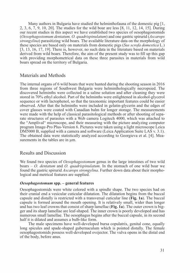
O. quadrispinulatum Marcone, 1901The buccal capsule (Fig. 2a) has a form of a truncated cone with the basis directed at the oesophagus. The oesophagus (Fig. 2a) has two dilatations: one small round widen-ing at the beginning and a bulbous widening in its second half. The oesophagus canal is surrounded by a row of round light-refracting cells.
Male. The bursa copulatrix, spicules and gubernaculums have the same morphol-ogy as O. dentatum (Fig. 2 b, c). The genital cone is well-developed and there are some papillae on its surface (Fig. 2c). Female. The tail is thin and styliform. The ovejector is similar to this of O. dentatum (Fig. 2d). Our metric data and the available literature data on this species are shown in Table 2.
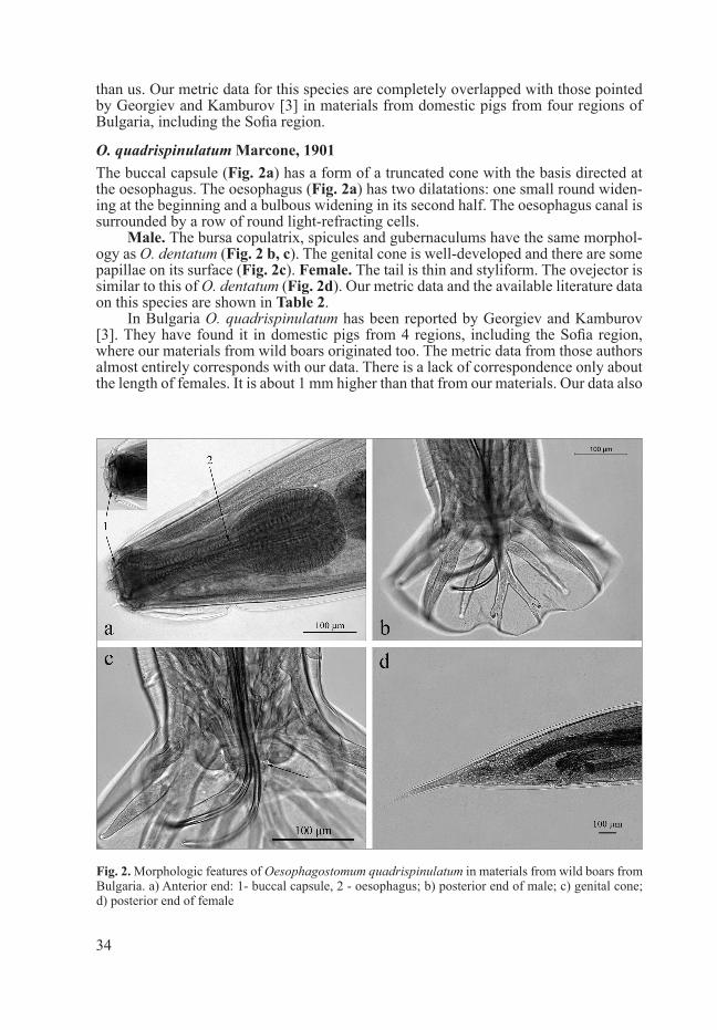
In Bulgaria O. quadrispinulatum has been reported by Georgiev and Kamburov [3]. They have found it in domestic pigs from 4 regions, including the Sofia region, where our materials from wild boars originated too. The metric data from those authors almost entirely corresponds with our data. There is a lack of correspondence only about the length of females. It is about 1 mm higher than that from our materials. Our data also
Fig. 2. Morphologic features of Oesophagostomum quadrispinulatum in materials from wild boars from Bulgaria. a) Anterior end: 1- buccal capsule, 2 - oesophagus; b) posterior end of male; c) genital cone; d) posterior end of female
35
correspond to those pointed by Mapelstone [13] in materials from domestic swine from Bengal. There is only one exception concerning the distance between the anus and tail tip in females, which is longer in our specimens.
The two oesophagostomid species are distinguished by some metric and morpho-logical features. O. dentatum has a longer body than O. quadrispinulatum. The spicules of O. dentatum are longer too. The proportion between the thin and leaf-shaped parts of the gubernaculum in O. dentatum is 1:1. In O. quadrispinulatum this ratio is 1:2. O. quadrispinulatum specimens have longer distances between the vulva and anus and between the anus and tail tip than these of O. dentatum. The oesophagus of O. dentatum usually has not light-refracting structures or these structures can be found only in its be-ginning. The oesophagus of O. quadrispinulatum has a small, but clearly visible second dilatation right after the buccal capsule.
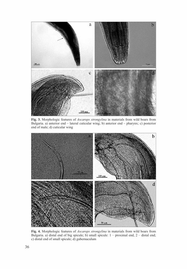
Ascarops strongylina Rudolphi, 1819This is a brown nematode with fusiform form. It has one narrow lateral cuticular wing (Fig. 3a). From the mouth opening begins the pharynx, which is consisted of spiral rings (Fig. 3b). The oesophagus has two parts – the anterior part is muscular and short and posterior is longer and glandular.
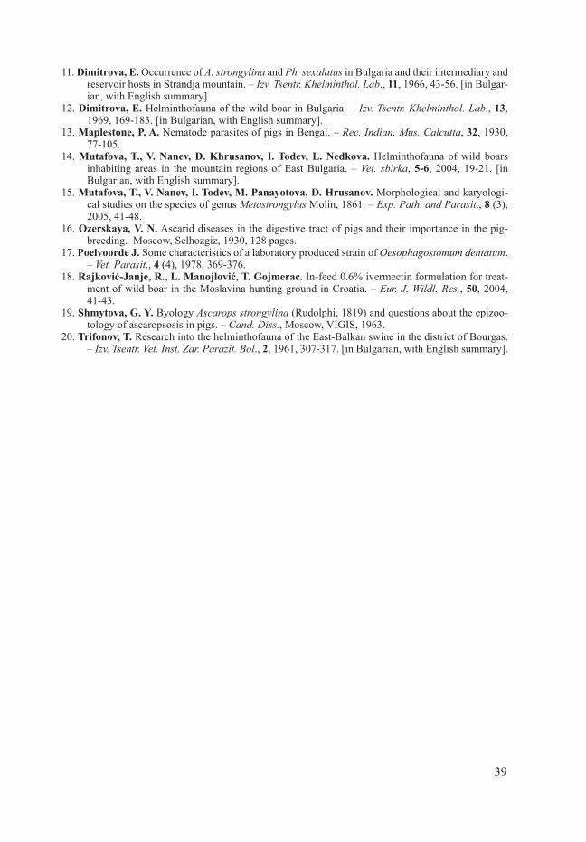
Male. There is no bursa copulatrix. The posterior end is curved ventrally. Two asymmetric, transversally furrowed wings are visible on it, as one is wider than another (Fig. 3c, d). Around the cloaca are several pairs of genital papillae. The spicules are different in length and form. The one spicule is long and thin. Its distal end is lancet-shaped (Fig. 4a).
The second spicule is shorter and wider, its proximal end is thickened and distal end looks like an arrow-head (Fig. 4b, c). The gubernaculum is situated around the cloaca and looks like an elongated lamella (Fig. 4d). Female. The vagina is tubular and
Table 2. Metric data on O. quadrispinulatum by different authors
Structure Mapelstone, 1930
Georgiev & Kamburov, 1977 Our data
Body length ♂ (in mm) 6.56-8.85 7.8-8.8 8.13 ± 0.53 (6.8-9)
Body width in the end of oesophagus ♂ – – 217 ± 15.7 (180-243)
Oesophagus length ♂ – 380-420 402 ± 13 (371-420)Max. oesophagus width ♂ – – 115 ± 7.7 (105-125)Spicule length 780-880 765-955 843 ± 63 (760-990)Gubernaculum length 104-120 85-128 105.6 ± 5.6 (93-115)Body length ♀ (in mm) 8.33-10.36 9.6 - 12.1 9.93 ± 0.62 (8-11.1)
Body width in the end of oesophagus ♀ – – 266.8 ± 24.8 (210-330)
Oesophagus length ♀ – 426 - 462 439.8 ± 8 (416-456)Max. oesophagus width ♀ – – 133 ± 5.6 (118-156)Vulva – anus 380-520 400 - 552 445.4 ± 19.2 (400-500)
Anus – tail tip 360-370 458 - 588 530.1 ± 22 (460-600)
36
Fig. 3. Morphologic features of Ascarops strongylina in materials from wild boars from Bulgaria. a) anterior end – lateral cuticular wing; b) anterior end – pharynx; c) posterior end of male; d) cuticular wing
Fig. 4. Morphologic features of Ascarops strongylina in materials from wild boars from Bulgaria. a) distal end of big spicule; b) small spicule: 1 – proximal end, 2 – distal end; c) distal end of small spicule; d) gubernaculum

37
Fig. 5. Morphologic features of Ascarops strongylina in materials from wild boars from Bulgaria. a) vagina and vulva; b) posterior end of female
Table 3. Metric data on Ascarops strongylina by different authors
Structure Shmytova, 1963 Our dataBody length ♂ (in mm) 10.1-14.9 12Body width in the end of oesophagus ♂ 270 - 360 330Anterior end - beginning of lateral wing ♂ 210 - 290 211Length of the pharynx ♂ 80-100 86Width of the pharynx ♂ 30 32Number of pharynx’s spiral rings in ♂ 14-18 17Length of muscular oesophagus ♂ 300-400 350Length of glandular oesophagus ♂ 2340-3700 3200Oesophagus width ♂ - 119 Small spicule length 500-600 575Big spicule length 2300-2900 2100Gubernaculum length 66-80 55Gubernaculum width 50-69 38Width of the bigger caudal wing ♂ 200-230 290Cloaca – tail tip - 253 Body length ♀ (in mm) 12.4-23.7 16.52 ± 2.76 (14-20)Body width in the end of oesophagus ♀ 230 - 430 357.8 ± 37.9 (300-382)Anterior end - beginning of lateral wing ♀ 230 - 340 340 ± 67 (288-420)Length of the pharynx ♀ 90-105 83 ± 8.2 (74-92)Width of the pharynx ♀ 30-40 31 ± 1.8 (29-33)Number of pharynx’s spiral rings in ♀ 14-18 15-16
Length of muscular oesophagus ♀ 290-540 359 ± 12.1 (345-370)
Length of glandular oesophagus ♀ 2730-3800 3360 ± 400 (2900-3800)Oesophagus width ♀ - 124 ± 13.6 (108-137)Anterior length - vulva 6800-1110 8120 ± 1340 (6800-9400)Anus - tail tip 230-300 232 ± 5.9 (219-240)

38
situated in the middle of the body. At first it goes parallel to the body, afterwards turns perpendicular and opens in the vulva (Fig. 5a). The eggs in the vagina are embryonated. The body’s caudal end is straight and the tail is short with a rounded tip (Fig. 5b).
We compared our metric data for A. strongylina with those from description by Shmytova [19]. Comparative data are shown in Table 3.
Our results about most of the measurements correspond with hers. There are few differences in the females: the lateral cuticular wing of specimens from our materials starts backwards, the pharynx is shorter and its width is in the lower limits pointed by Shmytova [19]. More important differences are observed in the male sexual structures. Our male specimen has shorter big spicule and gubernaculum, whereas its bigger cu-ticular wing on the caudal end is wider.
Conclusion
The morphological features of the established by us species O. dentatum, O. quadrispi-nulatum and A. strongylina in wild boars from Bulgaria completely correspond to those described by other authors in materials from domestic pigs. Diversity in the metric features is observed, which could be due to differences in methods of preparation and observation of the helminths as well as to the peculiarities of the separate parasite popu-lations from different parts of the world. The measurements about taxonomic important features, however, are in the limits, which have been pointed by other authors.
Acknowledgements: The authors express their gratitude to engineer Dragomir Penchev of State Hunting Enterprise “Vitoshko - Studena” for his collaboration in the obtaining of internal organs from wild boars.
R e f e r e n c e s
1. Genov, T. Helminthofauna of domestic and wild hoofed mammals of the region of Silistra town. – Nauch. Trud. Inst. Zooteh. Vet. Med., 22, 1971, 465-477 [in Bulgarian, with English summary].
2. Georgiev, B. Helminths and helminthoses on large pig-raising farms. First communication. Hel-minthofauna and helminthocenosis on large pig-raising farms. – Vet. Med. Nauk., 7(5), 1970, 35-40. [in Bulgarian, with English summary].
3. Georgiev, M., P. Kamburov. Occurrence of Oesophagostomum quadrispinulatum Marcone, 1901 (syn. O. longicaudum Goodey, 1925) in domestic pigs in Bulgaria. – Khelmintologiya, 4, 1977, 11-16. Sofia. [in Bulgarian, with English summary].
4. Georgieva, Y., N. Minkovski, R. Damyanova, I. Аpostolova. Handbook for laboratory exercises in physic and biophysic. Sofia, 2006, 10-16 [in Bulgarian].
5. Gortázar, C., J. Herrero, R. Villafuerte, J. Marco. Historical examination of the status of large mammals in Aragon, Spain. – Mammalia, 64, 2000, 411-422.
6. Denev, Y. Studies on metastrongylosis in pigs. I. Distribution and dynamics of the invasion in areas in north-eastern Bulgaria. – Izv. Tsentr. Vet. Inst. Zar. Parazit. Bol., 5, 1962, 145-150. [in Bulgarian, with English summary].
7. Denev, Y. Experiments to treat metastrongyloidiasis in pigs. Second communication. – Vet. Med. Nauk., 1 (10), 1964, 15-19. [in Bulgarian, with English summary].
8. Dimitrova, E. On the problem of the distribution of Metastrongyli in the lung of the boar (Sus scrofa L.). – Izv. Tsentr. Khelminthol. Lab., 7, 1962, 33-41 [in Bulgarian, with English summary].
9. Dimitrova, E. Trematodes among swine in Bulgaria. – Izv. Tsentr. Khelminthol. Lab., 8, 1963, 55-67. [in Bulgarian, with English summary].
10. Dimitrova, E. Helminthofauna of the mangalitsa swine in Bulgaria. – Izv. Tsentr. Khelminthol. Lab., 9, 1964, 57-68. [in Bulgarian, with English summary].
39
11. Dimitrova, E. Occurrence of A. strongylina and Ph. sexalatus in Bulgaria and their intermediary and reservoir hosts in Strandja mountain. – Izv. Tsentr. Khelminthol. Lab., 11, 1966, 43-56. [in Bulgar-ian, with English summary].
12. Dimitrova, E. Helminthofauna of the wild boar in Bulgaria. – Izv. Tsentr. Khelminthol. Lab., 13, 1969, 169-183. [in Bulgarian, with English summary].
13. Maplestone, P. A. Nematode parasites of pigs in Bengal. – Rec. Indian. Mus. Calcutta, 32, 1930, 77-105.
14. Mutafova, T., V. Nanev, D. Khrusanov, I. Todev, L. Nedkova. Helminthofauna of wild boars inhabiting areas in the mountain regions of East Bulgaria. – Vet. sbirka, 5-6, 2004, 19-21. [in Bulgarian, with English summary].
15. Mutafova, T., V. Nanev, I. Todev, M. Panayotova, D. Hrusanov. Morphological and karyologi-cal studies on the species of genus Metastrongylus Molin, 1861. – Exp. Path. and Parasit., 8 (3), 2005, 41-48.
16. Ozerskaya, V. N. Ascarid diseases in the digestive tract of pigs and their importance in the pig-breeding. Moscow, Selhozgiz, 1930, 128 pages.
17. Poelvoorde J. Some characteristics of a laboratory produced strain of Oesophagostomum dentatum. – Vet. Parasit., 4 (4), 1978, 369-376.
18. Rajković-Janje, R., L. Manojlović, T. Gojmerac. In-feed 0.6% ivermectin formulation for treat-ment of wild boar in the Moslavina hunting ground in Croatia. – Eur. J. Wildl. Res., 50, 2004, 41-43.
19. Shmytova, G. Y. Byology Ascarops strongylina (Rudolphi, 1819) and questions about the epizoo-tology of ascaropsosis in pigs. – Cand. Diss., Моscow, VIGIS, 1963.
20. Trifonov, T. Research into the helminthofauna of the East-Balkan swine in the district of Bourgas. – Izv. Tsentr. Vet. Inst. Zar. Parazit. Bol., 2, 1961, 307-317. [in Bulgarian, with English summary].