Embed Size (px)
Citation preview
FULL PAPERwww.afm-journal.de
© 2018 WILEY-VCH Verlag GmbH & Co. KGaA, Weinheim1805777 (1 of 7)
Protein-Based Hydrogels that Actuate Self-Folding Systems
Conor M. Gomes, Chang Liu, Jeffrey A. Paten, Samuel M. Felton, and Leila F. Deravi*
An approach to build a chemomechatronic system inspired by self-folding robots is described. This system, which comprises a protein-based hydrogel bound to a low-profile laminate, responds to different aqueous environments by undergoing geometric transformations. This response is dependent on the thickness and stiffness of the templating hydrogel, which directly regu-lates the diffusion of water into and out of the platform to initiate its revers-ible shape changes. When modified to include more complex geometries, these controllable shape changes can also be used to selectively trigger multiple folding events, illustrating a new platform for chemically initiated mechatronic devices. Together, these data show how compositionally discrete components are physically, chemically, and mechanically coupled together to generate a new actuator for biohybrid self-folding systems.
DOI: 10.1002/adfm.201805777
C. M. Gomes, Dr. J. A. Paten, Prof. L. F. DeraviDepartment of Chemistry and Chemical BiologyNortheastern UniversityBoston, MA 02115, USAE-mail: [email protected]. Liu, Prof. S. M. FeltonDepartment of Mechanical and Industrial EngineeringNortheastern UniversityBoston, MA 02115, USA
The ORCID identification number(s) for the author(s) of this article can be found under https://doi.org/10.1002/adfm.201805777.
to overcome the dependency, especially as devices continue to miniaturize and increase in complexity.
Soft, biomimetic hydrogels are an attractive alternative for actuation that have facile fabrication techniques with increased biocompatibility.[14] Hydrogels can absorb ≈90% of their mass in water, resulting in significant changes to its 3D architecture[15] that can be tuned to respond to a wide range of external stimuli including changes in pH,[16] tem-perature,[17] and analyte concentration.[18] Hydrogel mechanics can also be modu-lated via cross-linking,[19] interpenetrating networks,[20] and/or nanoparticle integra-tion,[21] to reduce actuation times (<1 s)[22]
while increasing the complexity of achievable shapes upon hydration/dehydration.[23] In this paper, we evaluated a protein-based hydrogel as the active component in a self-folding com-posite. We designed a new biohybrid system that incorporated a bilayer material comprising aluminum chemically coupled to the hydrogel. Gelatin was used as a model protein for our system because of its robustness, low cost, and ability to spon-taneously form a hydrogel in ambient conditions.[24] We inves-tigated how changes in hydrogel mechanics, thickness, and cross-linking density influenced the kinetics of folding (i.e., actuation) under different aqueous environments.
Actuation was achieved through dehydrating the biohybrid system in polyethylene glycol (PEG) and reversed via hydration in sodium chloride (NaCl). PEG, a commonly used biodegrad-able and biocompatible polymer,[25] also exhibits deliquescence, a feature that can offer precise control over the steady-state dehy-dration of the hydrogel to effectively eliminate effects associ-ated with humidity-induced variability.[26] While the use of PEG has been successfully implemented for applications ranging from concentrating collagen molecules in solution,[27] driving optical clearing in skin tissue,[28] and regulating collagen fibril diameter and spacing in the cornea,[29] it has yet to be used to stimulate hydrogel actuation. Thus, we explored whether these properties could be leveraged to selectively remove the water from within a hydrogel without compromising the network. We monitored dimensional changes exhibited by the hydrogels during actuation using bright-field microscopy and analyzed the kinetic behavior of these changes using a modified lumped-capacitance model.[30] From this model, we quantified the time constants associated with folding and unfolding, as well as identified how the presence of water influenced folding velocity. Our data indicate the utility of biohybrid actuators that control self-folding within aqueous environments supported with new methods to relate the hydration state of the bound hydrogel to the degree of actuation.
Actuators
1. Introduction
Self-folding systems offer a robust platform for assembling complex, multicomponent devices across multiple spatial scales[1] ranging from macroscopic robots[2] to microscale sur-gical tools.[3] The components of a self-folding technology include a rigid substrate, a flexible region that serves as a hinge, and a material responsive to one or multiple forms of stimula-tion (current,[4] voltage,[5] temperature,[6] light,[7] pressure,[8] or pH[9]). Self-folding technologies also show great potential for generating reconfigurable biomaterial interfaces which can be useful in delivering cargo[3a,10] or navigating complex geom-etries[11] in the body; however, these applications introduce new design considerations such as the need for biocompatible materials and compact, long term power sources. A popular material used to overcome the barrier of biocompatibility is poly -dimethylsiloxane (PDMS), which achieves shape-based changes via pneumatic-based actuators[12] designed to mimic human functions such as muscle contraction.[13] Although PDMS is biocompatible, it relies on pneumatic actuation that translates poorly for long-term in vivo applications due to its dependence on an external power source. Thus, new materials are required
Adv. Funct. Mater. 2018, 1805777
www.afm-journal.dewww.advancedsciencenews.com
1805777 (2 of 7) © 2018 WILEY-VCH Verlag GmbH & Co. KGaA, Weinheim
2. Results and Discussion
2.1. Characterization of the Effect of Initial Gelatin Thickness on Actuation
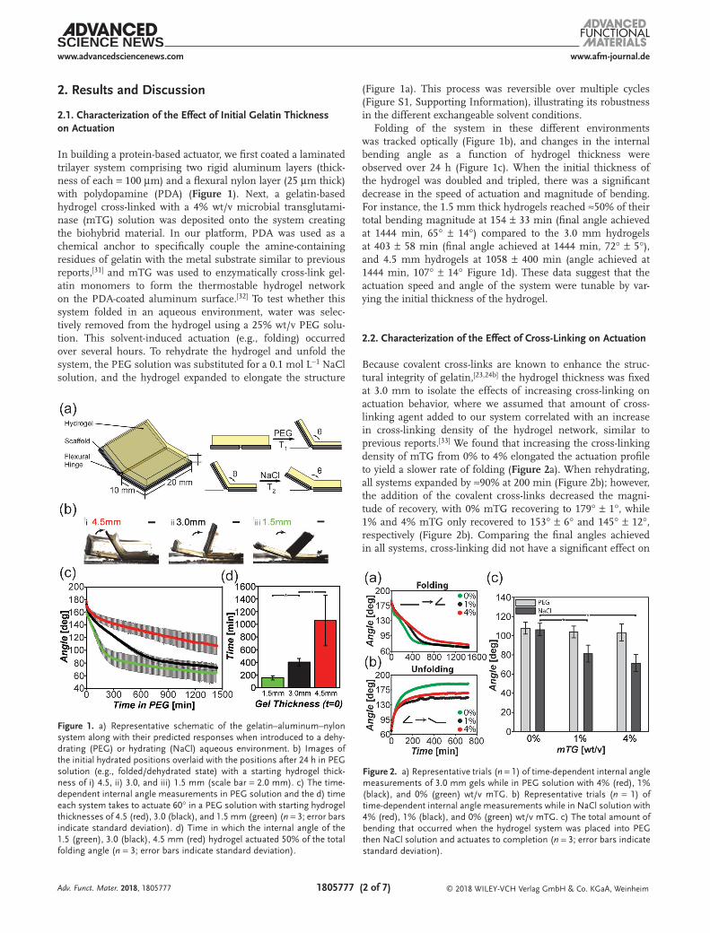
In building a protein-based actuator, we first coated a laminated trilayer system comprising two rigid aluminum layers (thick-ness of each = 100 µm) and a flexural nylon layer (25 µm thick) with polydopamine (PDA) (Figure 1). Next, a gelatin-based hydrogel cross-linked with a 4% wt/v microbial transglutami-nase (mTG) solution was deposited onto the system creating the biohybrid material. In our platform, PDA was used as a chemical anchor to specifically couple the amine-containing residues of gelatin with the metal substrate similar to previous reports,[31] and mTG was used to enzymatically cross-link gel-atin monomers to form the thermostable hydrogel network on the PDA-coated aluminum surface.[32] To test whether this system folded in an aqueous environment, water was selec-tively removed from the hydrogel using a 25% wt/v PEG solu-tion. This solvent-induced actuation (e.g., folding) occurred over several hours. To rehydrate the hydrogel and unfold the system, the PEG solution was substituted for a 0.1 mol L−1 NaCl solution, and the hydrogel expanded to elongate the structure
(Figure 1a). This process was reversible over multiple cycles (Figure S1, Supporting Information), illustrating its robustness in the different exchangeable solvent conditions.
Folding of the system in these different environments was tracked optically (Figure 1b), and changes in the internal bending angle as a function of hydrogel thickness were observed over 24 h (Figure 1c). When the initial thickness of the hydrogel was doubled and tripled, there was a significant decrease in the speed of actuation and magnitude of bending. For instance, the 1.5 mm thick hydrogels reached ≈50% of their total bending magnitude at 154 ± 33 min (final angle achieved at 1444 min, 65° ± 14°) compared to the 3.0 mm hydrogels at 403 ± 58 min (final angle achieved at 1444 min, 72° ± 5°), and 4.5 mm hydrogels at 1058 ± 400 min (angle achieved at 1444 min, 107° ± 14° Figure 1d). These data suggest that the actuation speed and angle of the system were tunable by var-ying the initial thickness of the hydrogel.
2.2. Characterization of the Effect of Cross-Linking on Actuation
Because covalent cross-links are known to enhance the struc-tural integrity of gelatin,[23,24b] the hydrogel thickness was fixed at 3.0 mm to isolate the effects of increasing cross-linking on actuation behavior, where we assumed that amount of cross-linking agent added to our system correlated with an increase in cross-linking density of the hydrogel network, similar to previous reports.[33] We found that increasing the cross-linking density of mTG from 0% to 4% elongated the actuation profile to yield a slower rate of folding (Figure 2a). When rehydrating, all systems expanded by ≈90% at 200 min (Figure 2b); however, the addition of the covalent cross-links decreased the magni-tude of recovery, with 0% mTG recovering to 179° ± 1°, while 1% and 4% mTG only recovered to 153° ± 6° and 145° ± 12°, respectively (Figure 2b). Comparing the final angles achieved in all systems, cross-linking did not have a significant effect on
Adv. Funct. Mater. 2018, 1805777
Figure 1. a) Representative schematic of the gelatin–aluminum–nylon system along with their predicted responses when introduced to a dehy-drating (PEG) or hydrating (NaCl) aqueous environment. b) Images of the initial hydrated positions overlaid with the positions after 24 h in PEG solution (e.g., folded/dehydrated state) with a starting hydrogel thick-ness of i) 4.5, ii) 3.0, and iii) 1.5 mm (scale bar = 2.0 mm). c) The time-dependent internal angle measurements in PEG solution and the d) time each system takes to actuate 60° in a PEG solution with starting hydrogel thicknesses of 4.5 (red), 3.0 (black), and 1.5 mm (green) (n = 3; error bars indicate standard deviation). d) Time in which the internal angle of the 1.5 (green), 3.0 (black), 4.5 mm (red) hydrogel actuated 50% of the total folding angle (n = 3; error bars indicate standard deviation).
Figure 2. a) Representative trials (n = 1) of time-dependent internal angle measurements of 3.0 mm gels while in PEG solution with 4% (red), 1% (black), and 0% (green) wt/v mTG. b) Representative trials (n = 1) of time-dependent internal angle measurements while in NaCl solution with 4% (red), 1% (black), and 0% (green) wt/v mTG. c) The total amount of bending that occurred when the hydrogel system was placed into PEG then NaCl solution and actuates to completion (n = 3; error bars indicate standard deviation).

www.afm-journal.dewww.advancedsciencenews.com
1805777 (3 of 7) © 2018 WILEY-VCH Verlag GmbH & Co. KGaA, Weinheim
the total magnitude of folding, but it did significantly reduce the ability to recover when rehydrated (Figure 2c). Despite these changes, varying the amount of covalent cross-linking did not correlate with the percent recovered, as the 1% and 4% hydro-gels were not significantly different.
2.3. Model Fitting and Analysis
In the folding profiles reported in Figures 1c and 2a, we observed that the folding behavior of this biohybrid system was not a simple exponential decay. Instead, two inflection points were pre-sent, which indicated multiple kinetic steps within our system. To better characterize this behavior, the transition point between these steps was identified as the maximum convexity between the two concave regions of the curve (Figure S2, Supporting Infor-mation). From this analysis, we interpolated transition points by using a modified lumped-capacitance model,[30] represented as
if
if
1 1
2 1
11
22
t a e t t
t a e t t
t
t
θ θ
θ θ
( )
( )
= + <
= + >
τ
τ
∞
−
∞
− (1)
where θ(t) is the internal angle (deg) at time t (min), t1 is the transition point (min), 1θ∞ and 2θ∞ are the plateau angles of the first and second fits (deg), and a1 and a2 are constants. The time constants, τ1 and τ2 (min), were extrapolated from each condition, where τ2 was associated with the approach to the theoretical minimum. When increasing the initial thick-ness of the hydrogel, the transition point was significantly delayed, resulting in a 175% increase in time between the 1.5 and 4.5 mm thick hydrogels (Figure 3a). Because the
lumped-capacitance model is thickness dependent,[30a] the time constants could not be compared across all thicknesses directly. However, we did note that τ1 was always smaller than τ2 across all conditions (Figure 3b), suggesting a “fast” and “slow” phase of contraction during dehydration in PEG.
When thickness was fixed at 3.0 mm and cross-linking was increased, we also observed an increase in the time to reach the transition points (Figure 3c). Time constants for the 0%, 1%, and 4% cross-linked samples were similar (τ1 = 139 ± 51, 166 ± 82, and 186 ± 12 min for 0%, 1%, and 4%, respectively; τ2 = 193 ± 16 and 195 ± 11 min for 0% and 1%, respectively), with only τ2 of 4% (321 ± 32 min) being sig-nificantly different from the 0% and 1% cross-linked samples (Figure 3d). The final bending angles of all conditions were similar, averaging 70° ± 13° (Figure 2a), suggesting that the increase in covalent cross-linking only affected the time it took to achieve the minimum angle during actuation.
Rehydrating the systems occurred much faster than the dehydrating processes and did not exhibit a transition point (Figure 2b), thus was fit to a single function
10t a et
θ θ( ) = + −
τ−
(2)
where θ0 is the angle (deg) at t = 0, with τ values significantly lower than those derived from the contraction. Rehydration τ values were similar (e.g., 93 ± 11 and 91 ± 8 min, for 0% and 4% mTG, respectively, Figure 3d), indicating that while the magnitude of recovery differed, the time to recover was the same regardless of cross-linking density.
2.4. Characterizing Gel Thickness Changes During Actuation
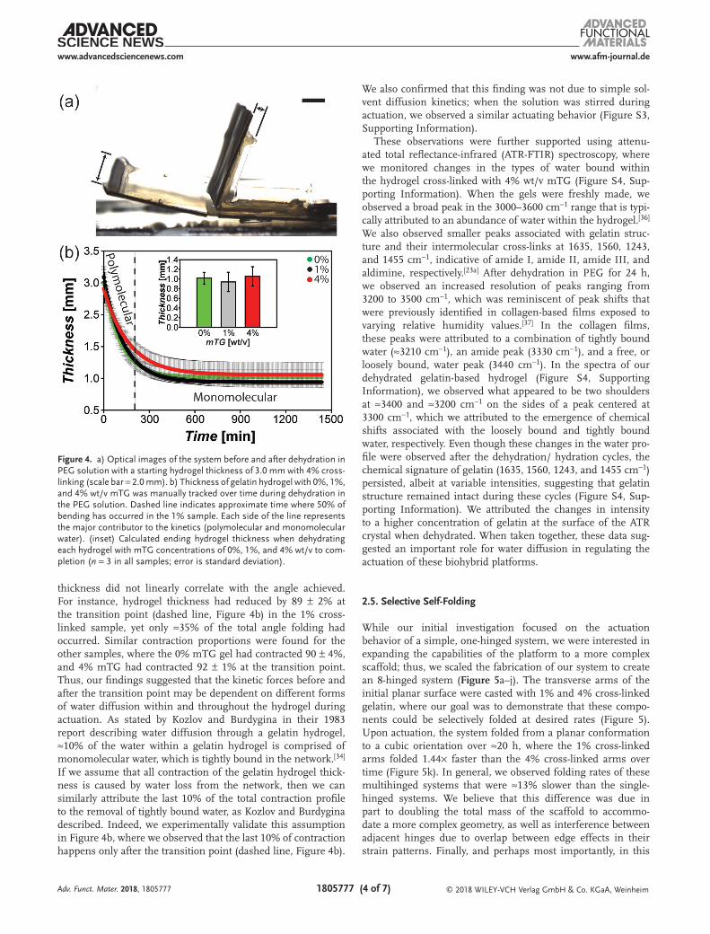
To better understand why cross-linking affected the kinetics but not the total magnitude of folding, we investigated the dimensional change that the hydrogels exhibited during dehy-dration in PEG. We hypothesized that the kinetic behavior of contraction was regulated by water diffusion through the hydrogel, where τ1 was associated with water that could easily be liberated (referred to as polymolecular water), and τ2 was associated with tightly integrated water (referred to as struc-tural and monomolecular water) within the hydrogel, similar to previous reports.[34] To test this hypothesis in our system, we monitored the change in hydrogel thickness as a func-tion of cross-linking density, which is a known parameter that regulates water diffusion.[35] Starting with an initial thickness of 3.0 mm, we manually tracked the profile of the contracting hydrogels in PEG over time (Figure 4a) and observed no signifi-cant differences in the thicknesses that each system achieved using the different cross-linking densities (Figure 4b, inset). Each hydrogel ultimately plateaued to a final thickness aver-aging 1.0 ± 0.3 mm. In our analysis, we also observed no tran-sition point during the contraction; instead, each parameter followed a single decay function with τ values of 111 ± 13, 131 ± 9, and 165 ± 13 min (Figure 4b).
When comparing the relationship between the angles achieved during contraction (plotted in Figure 2) with the hydrogel thicknesses reported in Figure 4, the variations in
Adv. Funct. Mater. 2018, 1805777
Figure 3. a) Calculated transition points when dehydrating each hydrogel (cross-linked with 4% w/v mTG) with initial starting thickness of 1.5, 3.0, and 4.5 mm. b) Calculated time constants when dehydrating each hydrogel with initial starting thickness of 1.5, 3.0, and 4.5 mm. c) Calcu-lated transition points when dehydrating each hydrogel with mTG con-centrations of 0%, 1%, and 4% wt/v. d) Calculated time constants when dehydrating each hydrogel with mTG concentrations of 0%, 1%, and 4% wt/v (n = 3 for all samples; error bars indicate standard deviation).
www.afm-journal.dewww.advancedsciencenews.com
1805777 (4 of 7) © 2018 WILEY-VCH Verlag GmbH & Co. KGaA, Weinheim
thickness did not linearly correlate with the angle achieved. For instance, hydrogel thickness had reduced by 89 ± 2% at the transition point (dashed line, Figure 4b) in the 1% cross-linked sample, yet only ≈35% of the total angle folding had occurred. Similar contraction proportions were found for the other samples, where the 0% mTG gel had contracted 90 ± 4%, and 4% mTG had contracted 92 ± 1% at the transition point. Thus, our findings suggested that the kinetic forces before and after the transition point may be dependent on different forms of water diffusion within and throughout the hydrogel during actuation. As stated by Kozlov and Burdygina in their 1983 report describing water diffusion through a gelatin hydrogel, ≈10% of the water within a gelatin hydrogel is comprised of monomolecular water, which is tightly bound in the network.[34] If we assume that all contraction of the gelatin hydrogel thick-ness is caused by water loss from the network, then we can similarly attribute the last 10% of the total contraction profile to the removal of tightly bound water, as Kozlov and Burdygina described. Indeed, we experimentally validate this assumption in Figure 4b, where we observed that the last 10% of contraction happens only after the transition point (dashed line, Figure 4b).
We also confirmed that this finding was not due to simple sol-vent diffusion kinetics; when the solution was stirred during actuation, we observed a similar actuating behavior (Figure S3, Supporting Information).
These observations were further supported using attenu-ated total reflectance-infrared (ATR-FTIR) spectroscopy, where we monitored changes in the types of water bound within the hydrogel cross-linked with 4% wt/v mTG (Figure S4, Sup-porting Information). When the gels were freshly made, we observed a broad peak in the 3000–3600 cm−1 range that is typi-cally attributed to an abundance of water within the hydrogel.[36] We also observed smaller peaks associated with gelatin struc-ture and their intermolecular cross-links at 1635, 1560, 1243, and 1455 cm−1, indicative of amide I, amide II, amide III, and aldimine, respectively.[23a] After dehydration in PEG for 24 h, we observed an increased resolution of peaks ranging from 3200 to 3500 cm−1, which was reminiscent of peak shifts that were previously identified in collagen-based films exposed to varying relative humidity values.[37] In the collagen films, these peaks were attributed to a combination of tightly bound water (≈3210 cm−1), an amide peak (3330 cm−1), and a free, or loosely bound, water peak (3440 cm−1). In the spectra of our dehydrated gelatin-based hydrogel (Figure S4, Supporting Information), we observed what appeared to be two shoulders at ≈3400 and ≈3200 cm−1 on the sides of a peak centered at 3300 cm−1, which we attributed to the emergence of chemical shifts associated with the loosely bound and tightly bound water, respectively. Even though these changes in the water pro-file were observed after the dehydration/ hydration cycles, the chemical signature of gelatin (1635, 1560, 1243, and 1455 cm−1) persisted, albeit at variable intensities, suggesting that gelatin structure remained intact during these cycles (Figure S4, Sup-porting Information). We attributed the changes in intensity to a higher concentration of gelatin at the surface of the ATR crystal when dehydrated. When taken together, these data sug-gested an important role for water diffusion in regulating the actuation of these biohybrid platforms.
2.5. Selective Self-Folding
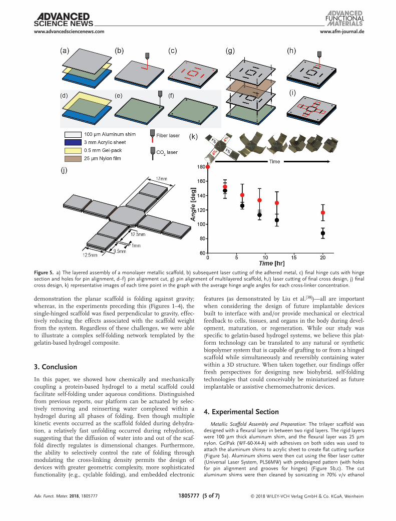
While our initial investigation focused on the actuation behavior of a simple, one-hinged system, we were interested in expanding the capabilities of the platform to a more complex scaffold; thus, we scaled the fabrication of our system to create an 8-hinged system (Figure 5a–j). The transverse arms of the initial planar surface were casted with 1% and 4% cross-linked gelatin, where our goal was to demonstrate that these compo-nents could be selectively folded at desired rates (Figure 5). Upon actuation, the system folded from a planar conformation to a cubic orientation over ≈20 h, where the 1% cross-linked arms folded 1.44× faster than the 4% cross-linked arms over time (Figure 5k). In general, we observed folding rates of these multihinged systems that were ≈13% slower than the single-hinged systems. We believe that this difference was due in part to doubling the total mass of the scaffold to accommo-date a more complex geometry, as well as interference between adjacent hinges due to overlap between edge effects in their strain patterns. Finally, and perhaps most importantly, in this
Adv. Funct. Mater. 2018, 1805777
Figure 4. a) Optical images of the system before and after dehydration in PEG solution with a starting hydrogel thickness of 3.0 mm with 4% cross-linking (scale bar = 2.0 mm). b) Thickness of gelatin hydrogel with 0%, 1%, and 4% wt/v mTG was manually tracked over time during dehydration in the PEG solution. Dashed line indicates approximate time where 50% of bending has occurred in the 1% sample. Each side of the line represents the major contributor to the kinetics (polymolecular and monomolecular water). (inset) Calculated ending hydrogel thickness when dehydrating each hydrogel with mTG concentrations of 0%, 1%, and 4% wt/v to com-pletion (n = 3 in all samples; error is standard deviation).
www.afm-journal.dewww.advancedsciencenews.com
1805777 (5 of 7) © 2018 WILEY-VCH Verlag GmbH & Co. KGaA, Weinheim
demonstration the planar scaffold is folding against gravity; whereas, in the experiments preceding this (Figures 1–4), the single-hinged scaffold was fixed perpendicular to gravity, effec-tively reducing the effects associated with the scaffold weight from the system. Regardless of these challenges, we were able to illustrate a complex self-folding network templated by the gelatin-based hydrogel composite.
3. Conclusion
In this paper, we showed how chemically and mechanically coupling a protein-based hydrogel to a metal scaffold could facilitate self-folding under aqueous conditions. Distinguished from previous reports, our platform can be actuated by selec-tively removing and reinserting water complexed within a hydrogel during all phases of folding. Even though multiple kinetic events occurred as the scaffold folded during dehydra-tion, a relatively fast unfolding occurred during rehydration, suggesting that the diffusion of water into and out of the scaf-fold directly regulates is dimensional changes. Furthermore, the ability to selectively control the rate of folding through modulating the cross-linking density permits the design of devices with greater geometric complexity, more sophisticated functionality (e.g., cyclable folding), and embedded electronic
features (as demonstrated by Liu et al.[38])—all are important when considering the design of future implantable devices built to interface with and/or provide mechanical or electrical feedback to cells, tissues, and organs in the body during devel-opment, maturation, or regeneration. While our study was specific to gelatin-based hydrogel systems, we believe this plat-form technology can be translated to any natural or synthetic biopolymer system that is capable of grafting to or from a hinged scaffold while simultaneously and reversibly containing water within a 3D structure. When taken together, our findings offer fresh perspectives for designing new biohybrid, self-folding technologies that could conceivably be miniaturized as future implantable or assistive chemomechatronic devices.
4. Experimental Section
Metallic Scaffold Assembly and Preparation: The trilayer scaffold was designed with a flexural layer in between two rigid layers. The rigid layers were 100 µm thick aluminum shim, and the flexural layer was 25 µm nylon. GelPak (WF-60-X4-A) with adhesives on both sides was used to attach the aluminum shims to acrylic sheet to create flat cutting surface (Figure 5a). Aluminum shims were then cut using the fiber laser cutter (Universal Laser System, PLS6MW) with predesigned pattern (with holes for pin alignment and grooves for hinges) (Figure 5b,c). The cut aluminum shims were then cleaned by sonicating in 70% v/v ethanol
Adv. Funct. Mater. 2018, 1805777
Figure 5. a) The layered assembly of a monolayer metallic scaffold, b) subsequent laser cutting of the adhered metal, c) final hinge cuts with hinge section and holes for pin alignment, d–f) pin alignment cut, g) pin alignment of multilayered scaffold, h,i) laser cutting of final cross design, j) final cross design, k) representative images of each time point in the graph with the average hinge angle angles for each cross-linker concentration.
www.afm-journal.dewww.advancedsciencenews.com
1805777 (6 of 7) © 2018 WILEY-VCH Verlag GmbH & Co. KGaA, Weinheim
(Sigma) for 1 h and dried. A second set of acrylic with GelPak on top (Figure 5d) was cut using the CO2 laser cutter (Figure 5e), with holes for pin alignment during assembly process (Figure 5f). Nylon film was sandwiched between two cleaned aluminum shims with adhesive, and the three layers were pin-aligned to adhere on the GelPak (Figure 5g). A release cut was used to cut out the outline (Figure 5h,i).
After final assembly, the scaffold was sonicated in 70% v/v ethanol and dried. After drying, the scaffold was placed in a 0.25% w/v Tris base (Sigma) for 10 min while stirring. Dopamine HCl (Sigma) was added while stirring to achieve a 0.2% wt/v solution. The scaffold was left in solution for 24 h, removed and sonicated in deionized water for 1 h, then dried under vacuum overnight. This process enabled PDA complexes to form with metallic atoms, as well as spontaneous bonds to form with amine-containing residues of gelatin,[31] ensuring covalent coupling of the protein hydrogel and the scaffold.
Hydrogel Preparation: Gelatin powder (Type A, 300 bloom, Electron Microscopy Sciences) was dissolved in deionized water at 60 °C to achieve 20% wt/v. T1 Transglutaminase Formula (Modernist Pantry) was dissolved in deionized water at 30 °C to achieve a desired % wt/v solution. The two solutions were mixed 1:1 before being applied to the PDA-coated metallic scaffold. The gel was cured for 1 h before any solvent was introduced to a PEG or NaCl solution, which is in agreement with previously published reports illustrating that 1 h is a sufficient curing time for the mTG cross-linked gelatin hydrogels.[39]
Imaging and Data Production: The scaffold was imaged using a Nikon SMZ18 stereoscope with an Imaging Source DFK 33UX174 camera. Images were captured using Nikon NIS Elements D Imaging Software. The scaffold demo was imaged using a GoPro Hero5 Session. Images were compiled and analyzed using ImageJ and MATLAB, respectively. Manual measurements of the gelatin hydrogel thickness were recorded using the imaging software ImageJ.
Modeling and Statistical Analysis: A lumped capacitance method[30] was used to model the folding behavior of the system. When dehydrating the gel in PEG, one or more exponential functions were fit to the data and the τ values and the plateau values were compared. The lumped capacitance equation is shown in Equation (1). When hydrating in 0.1 m NaCl, the exponential function was fitted, shown in Equation (2).
Student’s one-tail t-test was used to calculate a p value. If p < 0.05, the data were considered to be significantly different (notated by asterisk).
Supporting InformationSupporting Information is available from the Wiley Online Library or from the author.
AcknowledgementsThe authors gratefully acknowledge the financial support from the Northeastern University Tier 1 Program and the National Science Foundation (Grant No. DMR-1700720, CMG LFD).
Conflict of InterestThe authors declare no conflict of interest.
Keywordsbiohybrid, chemomechatronic, protein actuator, protein hydrogel, self-folding
Received: August 19, 2018Revised: October 12, 2018
Published online:
[1] a) M. E. W. Nisser, S. M. Felton, M. T. Tolley, M. Rubenstein, R. J. Wood, presented at 2016 IEEE/RSJ IROS, Daejeon, South Korea, October, 2016; b) S. H. Kang, M. D. Dickey, MRS Bull. 2016, 41, 93.
[2] a) C. Liu, S. M. Felton, presented at 2017 IEEE/RSJ Int. Conf. on Intel-ligent Robots and Systems (IROS), Vancouver, Canada, September, 2017, https://doi.org/10.1109/IROS.2017.8206017; b) S. Felton, M. Tolley, E. Demaine, D. Rus, R. Wood, Science 2014, 345, 644.
[3] a) A. Ghosh, C. Yoon, F. Ongaro, S. Scheggi, F. M. Selaru, S. Misra, D. H. Gracias, Front. Mech. Eng. 2017, 3; b) C. Pacchierotti, F. Ongaro, F. van der Brink, C. Yoon, D. Prattichizzo, D. H. Gracias, S. Misra, IEEE Trans. Autom. Sci. Eng. 2018, 15, 290.
[4] A. Tselev, J. D. Budai, E. Strelcov, J. Z. Tischler, A. Kolmakov, S. V. Kalinin, Nano Lett. 2011, 11, 3065.
[5] J. Kaushik, T. J. Noah, D. Neel, G. Ben, J. W. Robert, Smart Mater. Struct. 2018, 27, 065028.
[6] A. Bhattacharyya, D. C. Lagoudas, Y. Wang, V. K. Kinra, Smart Mater. Struct. 1995, 4, 252.
[7] M. P. M. Dicker, A. B. Baker, R. J. Iredale, S. Naficy, I. P. Bond, C. F. J. Faul, J. M. Rossiter, G. M. Spinks, P. M. Weaver, Sci. Rep. 2017, 7, 9197.
[8] I. V. Uvarov, A. V. Postnikov, V. B. Svetovoy, IOP Conf. Ser.: Mater. Sci. Eng. 2016, 108, 012032.
[9] N. Bassik, B. T. Abebe, K. E. Laflin, D. H. Gracias, Polymer 2010, 51, 6093.
[10] a) W. Xu, K. S. Kwok, D. H. Gracias, Acc. Chem. Res. 2018, 51, 436; b) J. C. Breger, C. Yoon, R. Xiao, H. R. Kwag, M. O. Wang, J. P. Fisher, T. D. Nguyen, D. H. Gracias, ACS Appl. Mater. Interfaces 2015, 7, 3398.
[11] a) S. Russo, T. Ranzani, J. Gafford, C. J. Walsh, R. J. Wood, presented at 2016 IEEE Int. Conf. on Robotics and Automation (ICRA), Stockholm, Sweden, May, 2016, https://doi.org/10.1109/ICRA.2016.7487203; b) J. H. Na, A. Evans Arthur, J. Bae, C. Chiappelli Maria, D. Santangelo Christian, J. Lang Robert, C. Hull Thomas, C. Hayward Ryan, Adv. Mater. 2015, 27, 79.
[12] a) Y. Hwang, R. N. Candler, Lab Chip 2017, 17, 3948; b) O. Jeong, S. Konishi, Sens. Actuators, A 2007, 135, 849.
[13] A. Miriyev, K. Stack, H. Lipson, Nat. Commun. 2017, 8, 596.[14] a) J. Stieghorst, T. Doll, IEEE Trans. Biomed. Eng. 2016, 63, 2294;
b) O. Ordeig, S. Y. Chin, S. Kim, P. V. Chitnis, S. K. Sia, Sci. Rep. 2016, 6, 22803; c) E. Popa, V. Santo, M. Rodrigues, M. Gomes, Polymers 2016, 8, 28.
[15] E. M. Ahmed, J. Adv. Res. 2015, 6, 105.[16] a) J. P. Best, M. P. Neubauer, S. Javed, H. H. Dam, A. Fery,
F. Caruso, Langmuir 2013, 29, 9814; b) H. Li, G. Go, S. Y. Ko, J.-O. Park, S. Park, Smart Mater. Struct. 2016, 25, 027001; c) T. Traitel, J. Kost, presented at Biomaterials, Boston, MA, 2004.
[17] a) H. Warren, M. i. h. Panhuis, G. M. Spinks, D. L. Officer, J. Polym. Sci., Part B: Polym. Phys. 2018, 56, 46; b) X. Zhang, C. L. Pint, M. H. Lee, B. E. Schubert, A. Jamshidi, K. Takei, H. Ko, A. Gillies, R. Bardhan, J. J. Urban, M. Wu, R. Fearing, A. Javey, Nano Lett. 2011, 11, 3239.
[18] a) Z. Mohammadi, S.-X. Xie, E. Peltier, M. Veisi, C. Berkland, Eur. Polym. J. 2011, 47, 1485; b) H. F. El-Sharif, Q. T. Phan, S. M. Reddy, Anal. Chim. Acta 2014, 809, 155.
[19] a) V. Yesilyurt, M. Ayoob Andrew, A. Appel Eric, T. Borenstein Jeffrey, R. Langer, G. Anderson Daniel, Adv. Mater. 2017, 29, 1605947; b) M. Pakulska Malgosia, K. Vulic, Y. Tam Roger, S. Shoichet Molly, Adv. Mater. 2015, 27, 5002.
[20] E. S. Dragan, Chem. Eng. J. 2014, 243, 572.[21] a) M. Biondi, A. Borzacchiello, L. Mayol, L. Ambrosio, Gels 2015, 1,
162; b) A. A. Hani, M. A. Hussain, A. Musab, N. Fozia Al, A.-H. Faten, O. Rahmi, K. Ali, Biomed. Mater. 2016, 11, 014104.
[22] a) G. Stoychev, L. Guiducci, S. Turcaud, W. C. Dunlop John, L. Ionov, Adv. Funct. Mater. 2016, 26, 7733; b) E. Lee, D. Kim, H. Kim, J. Yoon, Sci. Rep. 2015, 5, 15124.
Adv. Funct. Mater. 2018, 1805777

www.afm-journal.dewww.advancedsciencenews.com
1805777 (7 of 7) © 2018 WILEY-VCH Verlag GmbH & Co. KGaA, WeinheimAdv. Funct. Mater. 2018, 1805777
[23] a) J. Guan, H. He, D. J. Hansford, L. J. Lee, J. Phys. Chem. B 2005, 109, 23134; b) X. Peng, T. Liu, C. Jiao, Y. Wu, N. Chen, H. Wang, J. Mater. Chem. B 2017, 5, 7997.
[24] a) A. Maddaus, P. Curley, M. A. Griswold, B. D. Costa, S. Hou, K. J. Jeong, E. Song, L. F. Deravi, Biointerphases 2016, 11, 041002; b) M. L. McCain, A. Agarwal, H. W. Nesmith, A. P. Nesmith, K. K. Parker, Biomaterials 2014, 35, 5462.
[25] a) P. Jaipan, A. Nguyen, R. J. Narayan, MRS Commun. 2017, 7, 416; b) B. J. Klotz, D. Gawlitta, A. J. W. P. Rosenberg, J. Malda, F. P. W. Melchels, Trends Biotechnol. 2016, 34, 394.
[26] a) J. A. Baird, R. Olayo-Valles, C. Rinaldi, L. S. Taylor, J. Pharm. Sci. 2010, 99, 154; b) J. Zhu, Biomaterials 2010, 31, 4639; c) J. Ulbricht, R. Jordan, R. Luxenhofer, Biomaterials 2014, 35, 4848; d) S. Gou, T. Yin, Q. Xia, Q. Guo, RSC Adv. 2015, 5, 32064.
[27] J. A. Paten, G. E. Tilburey, E. A. Molloy, R. Zareian, C. V. Trainor, J. W. Ruberti, Biomaterials 2013, 34, 2577.
[28] D. K. Tuchina, V. D. Genin, A. N. Bashkatov, E. A. Genina, V. V. Tuchin, Opt. Spectrosc. 2016, 120, 28.
[29] S. Hayes, T. White, C. Boote, C. S. Kamma-Lorger, J. Bell, T. Sorenson, N. Terrill, O. Shebanova, K. M. Meek, J. R. Soc., Interface 2017, 14, 20170062.
[30] a) F. P. Incropera, D. P. DeWitt, Fundamentals of Heat and Mass Transfer, Wiley, New York 1990; b) C. R. Ruivo, J. J. Costa, A. R. Figueiredo, Int. J. Therm. Sci. 2008, 47, 282.
[31] a) Y. Liu, H. Meng, S. Konst, R. Sarmiento, R. Rajachar, B. P. Lee, ACS Appl. Mater. Interfaces 2014, 6, 16982; b) J. Yang, V. Saggiomo, A. H. Velders, M. A. Cohen Stuart, M. Kamperman, PLoS One 2016, 11, e0166490; c) Z. Xu, Sci. Rep. 2013, 3, 2914.
[32] G. Yang, Z. Xiao, H. Long, K. Ma, J. Zhang, X. Ren, J. Zhang, Sci. Rep. 2018, 8, 1616.
[33] C. W. Yung, L. Q. Wu, J. A. Tullman, G. F. Payne, W. E. Bentley, T. A. Barbari, J. Biomed. Mater. Res., Part A 2007, 83A, 1039.
[34] P. V. Kozlov, G. I. Burdygina, Polymer 1983, 24, 651.[35] Y. Wu, S. Joseph, N. R. Aluru, J. Phys. Chem. B 2009, 113, 3512.[36] K. S. Finnie, D. J. Cassidy, J. R. Bartlett, J. L. Woolfrey, Langmuir
2001, 17, 816.[37] S. Kudo, H. Ogawa, E. Yamakita, S. Watanabe, T. Suzuki,
S. Nakashima, Appl. Spectrosc. 2017, 71, 1621.[38] C. Liu, C. M. Gomes, J. K. McDonald, L. F. Deravi, S. M. Felton,
Proc. SMASIS2018 2018, 2, V002T02A005.[39] G. Yang, Z. Xiao, X. Ren, H. Long, H. Qian, K. Ma, Y. Guo, PeerJ
2016, 4, e2497.