Embed Size (px)
Citation preview
Journal of Bioscience, 19(1), 115–135, 2008
BACILLUS CEREUS IN FOOD: PARTIAL PURIFICATION AND CHARACTERIZATION OF HEMOLYSIN 1Shobana C S*, 2Lakkumi Venmal A K and 1Amsaveni P
1Faculty of Biotechnology, International University College of Technology Twintech, Bandar Sri Damansara, 52200 Kuala Lumpur, Malaysia 2 Department of Microbiology, Dr. G. R. Damodaran College of Science, Coimbatore, 641014 India Abstract: Bacillus cereus is an aerobic spore former commonly found in soil, vegetables and in many raw, and processed foods. Consumption of foods that contain large numbers of B. cereus may result in food poisoning. Although, certain physiological and cultural characteristics are necessary for identifying B. cereus, its enterotoxigenicity indicates whether a suspect strain may be a public health hazard. Out of 75 food samples tested, we obtained 57 hemolytic strains of B. cereus. On further screening based on the antibiotic susceptibility pattern, three strains viz., B. cereus PCBC-06, B. cereus PCBC-52 and B. cereus PCBC-57 were selected and investigated for their growth pattern. Of all the three strains, B. cereus PCBC-06 exhibited good growth pattern. Test strains grew after the treatment at 40, 50, 60, 70, 80 and 90ºC and were killed only after treating at 121ºC for 20 min at 15 lb/sq. in. The optimum pH and glucose concentration for the growth of all strains were between 7.0 and 9.0, and 0.5%, respectively. B. cereus PCBC-06, B. cereus PCBC-52 and B. cereus PCBC-57 optimally grew at salt concentration of 0.75%, 0.5% and 0.25%, respectively. The heat resistance measurements exhibited that the test strains failed to grow at 2°C and the spore D90 value ranged from 5.40 to 5.50 min. B. cereus PCBC-06 was subjected to protein studies and its protein accumulation was directly proportional to its growth. In gel diffusion assay, discontinuous pattern of hemolysis by the crude toxin was observed and the zones measured 0.6 cm, 0.8 cm and 0.9 cm at 20 min, 65 min and 100 min, respectively. Hemolysin was produced by dialyzing membrane technique and partially purified hemolysin by ammonium sulphate precipitation had protein content of 44 μg/ml of sample. SDS-PAGE of the crude hemolysin showed four conspicuous bands of molecular weight 70 000, 68 000, and 57 000 and 43 000. Keywords: Food, B. cereus, Growth, Hemolysin, Gel Diffusion, Partial Purification INTRODUCTION Foods that are nutritional value to humans are often ideal culture media for microbial growth. The foods that we eat are rarely sterile, carry microbial association whose composition depends upon which organism gain access and how they grow, survive and interact in the food overtime. Three gram-positive rods viz., Clostridium botulinum, C. perfringens, and Bacillus cereus are known to cause food intoxications. B. cereus is one of the 60 representatives of the widely varied genus Bacillus and found frequently as a saprophyte in soil, water, vegetation and air (Stanier et al. 1976). Two distinct types of illnesses are caused by B. cereus depending on the type of toxin produced: an emetic illness characterized by nausea and vomiting with an incubation time of 1 to 6 h, and a *Corresponding author: [email protected]
115

Shobana C S et al.
diarrheal type, with incubation time of 4 to 16 h. The emetic type is often associated with boiled or fried rice, while the diarrheal type is associated with a wider range of foods (Pelczar & Reid 1972; Davis & Walkinson 1973; Goepfert et al. 1973). Present routine detection methods for B. cereus rely on standard plate counting which require, approximately 4 days to be performed, including confirmatory testing. This is time-consuming when inspecting products with short shelf-live. The presence of B. cereus strains that cause food poisoning can also be indicated by detection of their toxins (Torkar & Mozina 2000). The purposes of the current study are to screen various food types for the presence of B. cereus, to isolate and to correlate various microenvironments for its growth, sporulation, detection, production and partial purification of hemolysin. MATERIALS AND METHODS Samples Food samples were collected in and around the area of Peelamedu, Coimbatore, India from December 2003 to February 2004. Each sample unit consisted of at least 100 ml/g. Liquid food was subjected for continuous or periodic mixing and the samples were withdrawn into a sterile container, transported to lab and mixed thoroughly once again before pipetting the amounts required for investigation. Sampling of solids and semisolids was performed using sterile scalpels and spoons depending on the nature of material. Foods were examined by taking deep samples as well as surface sample. Deep sample was taken with care to minimize contamination from superficial levels. These large samples were treated separately, each being mixed thoroughly in the laboratory before processing. The samples were transported and stored under condition, which inhibited changes in microbial numbers. Frozen foods were kept frozen. Chilled/refrigerated foods were kept at 4°C and not frozen. Dried foods and canned foods were not cooled but stored and transported at a temperature less than 40°C. Insulated containers were used to hold and transport chilled or frozen samples. Samples were transported to the laboratory as soon as possible, preferably within 2 h. A total number of 75 samples were collected and designated from S1 to S75 (Table 1). Table 1: Details of the food samples collected for isolation of Bacillus spp.
Sample No. Type of sample Name of sample No. of collected sample 1 Milk 7 2 Apple juice 4 3 Coffee 6 4 Cooler water 8 5
I Liquid
Bottled drink 2
(continue on next page)
116

Bacillus cereus in Food
Table 1 (continued)
S. No. Type of sample Name of sample No. of collected sample 6 Rose milk 5 7 Lassie 3 8 Drink in Carton 1 9 Paneer butter masala 2 10 Sambar 11 11 Fish curry 6 12 Beef curry 3 13
II Semi solid
Egg curry 7 14 Ghee rice 2 15 Vada 5 16
III Solid Plain rice 3
Total 75
Aerobic plate count (APC) and Enumeration of presumptive Bacillus species. Serial dilutions from 10-2 to 10-6 were prepared from homogenized food samples (Andrews & Hammack 1998). Plate count agar was used for APC (Rhodehamel & Harmon 1998) while selective and differential media namely Polymyxin pyruvate egg yolk mannitol bromothymol blue agar (PPEMBA) was used (Lancette & Harmon 1980) for the presumptive enumeration of B. cereus. Appropriate dilutions were spread on to the plates of plate count agar and PPEMBA. The plates were incubated in an inverted position for 48 h ± 4 h at 37°C. Colonies were counted immediately after the incubation period and observed for the presumptive B. cereus colonies (Rhodehamel & Harmon 1998). Isolation and confirmation of B. cereus isolates Five colonies with the typical morphology of B. cereus were isolated from PPEMBA plates from each sample and subcultured on Mannitol egg yolk agar (MYP). Colonies having the characteristic appearance of B. cereus were selected and subcultured on J agar (JA) plates (Choma et al. 2000). Identification and confirmation of the isolates to B. cereus group was done by Gram-staining, inoculation in B. cereus motility medium (BCMM), hanging drop method for motility identification, rhizoid growth, test for protein toxin crystals, growth in anaerobic medium, endospore staining and starch hydrolysis. Biochemical characterization of Bacillus species was performed by glucose fermentation, Voges-Proskauer (VP) test, citrate utilization test, catalase test and nitrate reduction test. Test for extracellular protein activity of the Bacillus spp. was performed by streaking the test strains on egg yolk agar, blood agar, starch agar and milk agar, followed by incubation at 37°C for 1 to 4 days and observation for lecithinase production, hemolysis, starch and casein hydrolysis, respectively (Claus & Berkeley 1986; Cappuccino & Sherman 1996; Atlas 1997). Based on
117

Shobana C S et al.
these analyses a total number of 57 hemolytic isolates were identified and strain numbers were designated as B. cereus PCBC-01 to 57. Antibiogram pattern of the isolates of Bacillus species B. cereus strains were further screened on the basis of antibiotic susceptibility as per the method of Bauer et al. (1966). Commonly used antibiotics for the treatment of B. cereus infection viz: ampicillin (A), cephalothin (Ch), cephotoximine (Ce), chloramphenicol (C), ciproflaxacin (Cf), clindamycin (Cd), erythromycin (E), gentamycin (G), lincomycin (L), neomycin (N), oxacillin (Ox), streptomycin (S) and tetracycline (T) each at the concentration of 30 µg/ml were used for the test. Growth pattern of the selected isolates of B. cereus Depending on antibiotic susceptibility, three stains of B. cereus viz., PCBC-06, PCBC-52 and PCBC-57 were selected for further analysis. About 10 ml of the 24 h old culture was inoculated into 90 ml of sterile nutrient broth and incubated in a shaker at 37°C. Optical density (OD) of the cultures was measured periodically at 660 nm in ELICO, SL-159 UV-VIS spectrophotometer. Effect of various growth conditions on Bacillus species. Temperature B. cereus suspension in nutrient broth was placed in the water bath for 10 min at different temperatures viz., 40°C, 50°C, 60°C, 70°C, 80°C and 90°C and these cultures were inoculated on nutrient agar and incubated at 37oC for 48 h. The plate with no growth was considered as thermal death point. pH Sterile nutrient broth was prepared with varying pH of 5.0, 6.0, 7.0, 8.0, and 9.0, inoculated with 0.5 ml of the test culture and incubated at 37°C for 24 h. OD was measured at 660 nm in ELICO, SL-159 UV spectrophotometer. Sodium chloride concentration Sterile nutrient broth with different concentration sodium chloride (NaCl) viz., 0.25%, 0.50%, 0.75%, and 1.00% was inoculated with 0.1 ml of the 24 h old culture, incubated at 37°C for 24 h and OD was measured at 660 nm in ELICO, SL-159 UV-VIS spectrophotometer. Glucose concentration Sterile nutrient broth with different glucose concentration (0%, 0.25%, 0.5%, 0.75%, and 1%) was inoculated with 0.1 ml of the 24 h old culture of test strains of B. cereus, incubated at 37°C for 24 h and OD was measured at 660 nm for each tube in ELICO, SL-159 UV VIS spectrophotometer.
118

Bacillus cereus in Food
Growth of B. cereus at various temperatures J broth (JB), inoculated with the 24 h old culture of B. cereus was incubated at 2°C, 5°C, 30°C, 37°C and 42°C and observed for growth on 2, 3, 5, 7, 14 and 21 days. Positive cultures on JA were further analysed for spore production and spore morphology.
Detection of heat resistant spores of B. cereus Sterile soil extract agar plates were inoculated with the test strains of B. cereus and incubated at room temperature of 28°C for five days. The spores were collected from the agar surface with a sterile cotton swab, suspended in 1.5 ml sterile distilled water and placed in ice for 45 min. The spore suspension was submerged in hot water at 90°C for 3, 5, 7, 10 and 15 min respectively, streaked onto JA plates and incubated at 30°C for 24 h. Growth and protein accumulation of B. cereus About 10 ml of the test strains of B. cereus PCBC-06 was inoculated into 90 ml of sterile nutrient broth and incubated at 37°C. Periodical estimation of the protein in the broth culture was carried out by Lowry et al. (1951). Extraction and estimation of whole cell protein from B. cereus Nutrient broth containing the 24 h old culture of B. cereus PCBC-06 was centrifuged and pellet was washed with phosphate buffer saline (pH 7.2). The pellet was incubated in boiling water bath with the sample buffer for 10 min and subjected to Lowry et al. (1951) method of protein estimation. Hemolysin production by B. cereus Aqueous suspension of B. cereus PCBC-06 was inoculated into brain heart infusion glucose (BHIG) broth and the turbidity was made equivalent to No. 1 on McFarland nephelometer scale (McFarland 1907). The broth was shaken at 3 ± 2oC at 125 rev min-1 for 12 h in environmental shaker. The contents were centrifuged at 10 000 rpm for 10 min. The supernatant was examined for the presence of toxin. Gel diffusion assay for hemolysin activity Brain heart infusion broth supplemented with 0.1% glucose (BHIG) was inoculated with 1% (v/v) of an overnight stationary seed culture of B. cereus PCBC-06, grown in BHIG at 30°C and incubated on a rotary shaker at 200 rev min-1 for 5 h. Cells were removed by centrifugation at 10 000 rpm for 10 min and the supernatant was stored at –20°C. Using a gel cutter, 3.0 mm wells were made in the center of sterile blood agar plates (Beecher & Wong 1994), loaded with 10 μl of the supernatant and incubated at 37°C. Hemolysis was monitored at 15 min intervals. Partial purification of extracellular hemolysin from B. cereus Preparation of crude hemolysin was produced by dialysis membrane technique (Murphy & Haque 1967). Precipitation of hemolysin was accomplished with saturated solution of ammonium sulphate (pH 5.0) at 4°C overnight. The
119
Shobana C S et al.
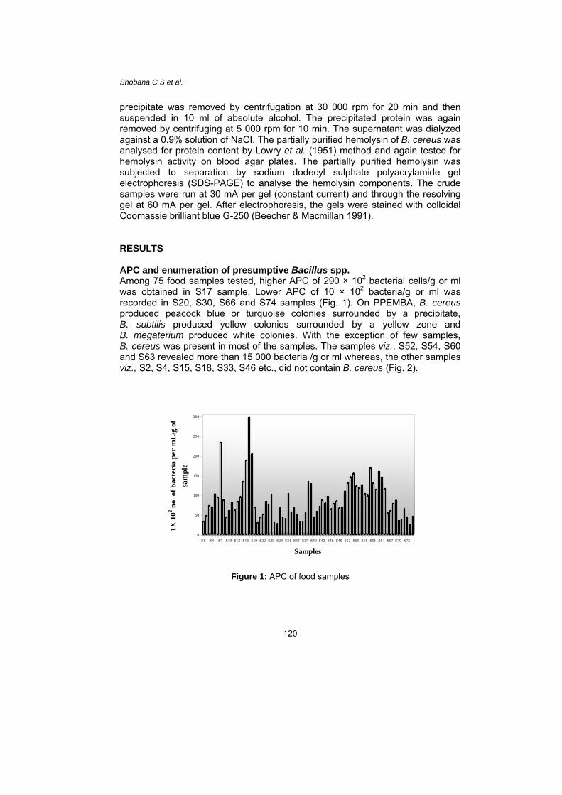
precipitate was removed by centrifugation at 30 000 rpm for 20 min and then suspended in 10 ml of absolute alcohol. The precipitated protein was again removed by centrifuging at 5 000 rpm for 10 min. The supernatant was dialyzed against a 0.9% solution of NaCI. The partially purified hemolysin of B. cereus was analysed for protein content by Lowry et al. (1951) method and again tested for hemolysin activity on blood agar plates. The partially purified hemolysin was subjected to separation by sodium dodecyl sulphate polyacrylamide gel electrophoresis (SDS-PAGE) to analyse the hemolysin components. The crude samples were run at 30 mA per gel (constant current) and through the resolving gel at 60 mA per gel. After electrophoresis, the gels were stained with colloidal Coomassie brilliant blue G-250 (Beecher & Macmillan 1991). RESULTS APC and enumeration of presumptive Bacillus spp. Among 75 food samples tested, higher APC of 290 × 102 bacterial cells/g or ml was obtained in S17 sample. Lower APC of 10 × 102 bacteria/g or ml was recorded in S20, S30, S66 and S74 samples (Fig. 1). On PPEMBA, B. cereus produced peacock blue or turquoise colonies surrounded by a precipitate, B. subtilis produced yellow colonies surrounded by a yellow zone and B. megaterium produced white colonies. With the exception of few samples, B. cereus was present in most of the samples. The samples viz., S52, S54, S60 and S63 revealed more than 15 000 bacteria /g or ml whereas, the other samples viz., S2, S4, S15, S18, S33, S46 etc., did not contain B. cereus (Fig. 2).
0
50
100
150
200
250
300
1X 1
02 no.
of b
acte
ria
per
mL
/g o
f sa
mpl
e
S1 S4 S7 S10 S13 S16 S19 S22 S25 S28 S31 S34 S37 S40 S43 S46 S49 S52 S55 S58 S61 S64 S67 S70 S73
Samples
Figure 1: APC of food samples
120

Bacillus cereus in Food
0
5000
10000
15000
20000
25000
S1 S4 S7S10
S13S16
S19S22
S25S28 S3
1S34
S37S40
S43S46
S49S52
S55S58
S61 S64S67
S70S73
Sample
No.
of B
acill
us sp
p. p
er m
L/g
of s
ampl
e
Figure 2: Presumptive enumeration of B. cereus
Isolation and confirmation of B. cereus isolates The presumptive colonies of B. cereus were identified and differentiated from other species of Bacillus by morphological and biochemical characterization (Table 2). A total number of 57 hemolytic isolates of B. cereus were obtained which were designated from B. cereus PCBC-01 to 57. Table 2: Morphological and biochemical characterization for the identification of Bacillus spp.
Sample No.
Test B. cereus B. subtilis B. megaterium
1
2 3 4 5 6 7 8 9
10
Gram staining Motility Rhizoid growth Protein toxin crystals Spore staining Glucose fermentation VP test Citrate utilization test Catalase test Nitrate test
Gram positive
+ _ _ + A + + + +
Gram positive
_ _ _ _ A + + + +
Gram positive
_ _ _ _ A _ + + +
+ – Positive reaction - – Negative reaction A – Acid reaction
121

Shobana C S et al.
Antibiogram pattern of the isolates of Bacillus cereus Antibiogram pattern of the selected isolates of B. cereus demonstrated 89.47%, 91.22%, 7.00%, 77.20%, 15.79%, 89.47%, 17.54%, 85.90%, 15.79%, 94.70%, 8.77%, 10.53% and 82.46% of the strains were resistant to Ce, L, Cf, A, C, Ch, G, T, S, Cd, N, E and Ox, respectively. Based on the antibiotic susceptibility pattern the isolates viz., B. cereus PCBC-06, B. cereus PCBC-52 and B. cereus PCBC-57 were selected for further investigation as they were resistant to most of the antibiotic used when compared to other strains (Table 3). Table 3: Antibiogram pattern of Bacillus species
Strains Ce L Cf A C Ch G T S Cd N E Ox
B1 R R S R R R S R R R R R R
B2 R R S R S R R R R R S S R
B3 R R S R S R S R R R S S R
B4 R R S R S R S R R R S S R
B5 R R S R R R S R R R S R R
B6 R R R R S R R R R R R R R
B7 R R R R R R S R R R R R R
B8 R R S R R R R R R R S S R
B9 R R S R S R R R R R S S R
B10 R R S R S R S R R R S S R
B11 R R S R S S S S R R S S R
B12 R R S R S R S R R R S S R
B13 R R S R S R S R R S S S R
B14 R R S R S R S R R R S S R
B15 R S S R S R S R R R S S R
B16 R R S R S R S R R R S S R
B17 R R S R S R S R R R S S R
B18 R R S R S R S R R R S S R
B19 R S S S R S S S S S S S S (continue on next page)
122

Bacillus cereus in Food
Table 3 (continued)
Strains Ce L Cf A C Ch G T S Cd N E Ox
B20 R R S S S R R S R R S S S
B21 R R S S R R S R R R S S S
B22 R R S R S R R R R R S S R
B23 R R S R S R S R R R S S R
B24 R R S R S R S R R R S S R
B25 R R S R S R S R R R S S R
B26 R R S R S S S R R R S S R
B27 R R S R S R S S R R S S R
B28 R R S R S R S R R R S S R
B29 R R S R S R S R R R S S R
B30 R S S R R R S S R R S S R
B31 R R S R R R R R R R S S R
B32 R R S R S R R R R R S S R
B33 R R S R S R S S R R S S R
B34 R R S R S R S R R R S S R
B35 R R S R S R S R R R S S R
B36 R R S R S R S R R R S S R
B37 R R S R S R S R R R S S R
B38 R R S R S S S S R R S S R
B39 R R S R S R S R R R S S R
B40 R R S S S R S R R R S S S
B41 R R S R S R S R R R S S R
B42 R R S R S R S R R R S S R
B43 R R S R S R S R R R S S R
(continue on next page)
123

Shobana C S et al.
Table 3 (continued)
Strains Ce L Cf A C Ch G T S Cd N E Ox
B44 R R S R S R S R R R S S R
B45 R R S R S R S R R R S S R
B46 R R S R S R S R R R S S R
B47 S R S R S R S R R R S S R
B48 S R S S S S S R S R S S S
B49 S S S S S R S R S S S S S
B50 R R S S S R S R S R S S S
B51 R R S S S R S R S R S S S
B52 R R R R R R S R R R R R R
B53 S R S S S R R R S R S S S
B54 S R S S S S S S S R S S S
B55 S S S S S R S R S R S S S
B56 S S S S S R S R S R S S S
B57 R R R S R R R R R R R R R R–Resistant; S–Sensitive
Ce – Cephalothin; L – Lincomycin; Cf – Ciprofloxacin; A`– Ampicillin, C – Chloramphenicol; Ch – Cephalothin; G – Gentamycin; T – Tetracycline, S – Streptomycin; Cd – Clindamycin; N – Neomycin; E – Erythromycin; Ox – Oxacillin
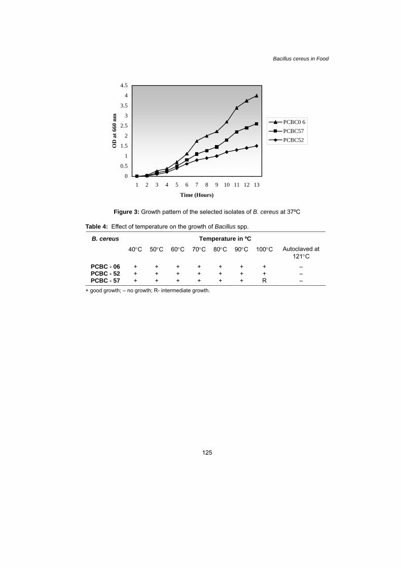
Growth experiments B. cereus PCBC-06 exhibited good growth pattern compared to the other two isolates viz., B. cereus PCBC-52 and B. cereus PCBC-57 (Fig. 3). All the three test strains had good growth after the treatment at 40ºC, 50ºC, 60ºC, 70ºC, 80ºC and 90ºC. The culture of B. cereus PCBC-57 showed slight retardation in growth after the treatment at 100ºC whereas the other two strains grew normally. The test cultures exposed to 121ºC for 20 min at 15 lb/sq.in. did not show any growth (Table 4). The optimum pH for growth was between 7.0 and 9.0 for all the test strains. The optimum salt concentration for the strains B. cereus PCBC-06, B. cereus PCBC-56 and B. cereus PCBC-57 were 0.75%, 0.5% and 0.25%, respectively. All the three strains had optimum requirement of 0.5% of glucose (Table 5).
124
Bacillus cereus in Food
0
0.5
1
1.5
2
2.5
3
3.5
4
4.5
1 2 3 4 5 6 7 8 9 10 11 12 13
Time (Hours)
OD
at 6
60 n
mPCBC0 6PCBC57PCBC52
Figure 3: Growth pattern of the selected isolates of B. cereus at 37ºC
Table 4: Effect of temperature on the growth of Bacillus spp.
Temperature in ºC B. cereus
40°C 50°C 60°C 70°C 80°C 90°C 100°C Autoclaved at 121°C
PCBC - 06 + + + + + + + – PCBC - 52 + + + + + + + – PCBC - 57 + + + + + + R –
+ good growth; – no growth; R- intermediate growth.
125

Shobana C S et al.
Table 5: Effect of various growth conditions on growth of B. cereus strains
OD at 660 nm Test strains Growth Conditions
B. cereus PCBC-06
B. cereus PCBC-52
B. cereus PCBC-57
1.0 0.10 0.10 0.10
2.0 0.20 0.22 0.20
3.0 0.25 0.28 0.25
4.0 0.29 0.35 0.28
5.0 0.30 0.40 0.30
6.0 0.35 0.45 0.34
7.0 0.45 0.50 0.37
8.0 0.44 0.48 0.36
9.0 0.39 0.44 0.32
pH
10.0 0.37 0.39 0.30
0.00 0.15 0.35 0.30
0.25 0.20 0.25 0.28
0.50 0.23 0.20 0.29
0.75 0.45 0.15 0.18
Concentration of NaCl (%)
1.00 0.25 0.20 0.20
0.00 0.20 0.21 0.20
0.25 0.30 0.40 0.23
0.50 0.50 0.80 0.45
0.75 0.45 0.60 0.40
Concentration of glucose (%)
1.00 0.40 0.50 0.38
126
Bacillus cereus in Food
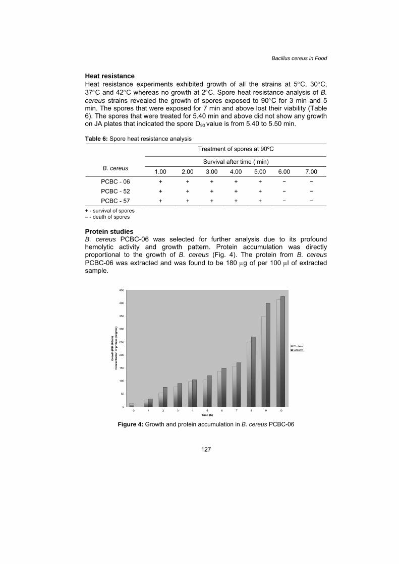
Heat resistance Heat resistance experiments exhibited growth of all the strains at 5°C, 30°C, 37°C and 42°C whereas no growth at 2°C. Spore heat resistance analysis of B. cereus strains revealed the growth of spores exposed to 90°C for 3 min and 5 min. The spores that were exposed for 7 min and above lost their viability (Table 6). The spores that were treated for 5.40 min and above did not show any growth on JA plates that indicated the spore D90 value is from 5.40 to 5.50 min.
Table 6: Spore heat resistance analysis
Treatment of spores at 90ºC
Survival after time ( min)
B. cereus 1.00 2.00 3.00 4.00 5.00 6.00 7.00 PCBC - 06 + + + + + – –
PCBC - 52 + + + + + – –
PCBC - 57 + + + + + – –
+ - survival of spores – - death of spores Protein studies B. cereus PCBC-06 was selected for further analysis due to its profound hemolytic activity and growth pattern. Protein accumulation was directly proportional to the growth of B. cereus (Fig. 4). The protein from B. cereus PCBC-06 was extracted and was found to be 180 μg of per 100 μl of extracted sample.
0
50
100
150
200
250
300
350
400
450
0 1 2 3 4 5 6 7 8 9 10
Time (h)
Gro
wth
(OD
660
nm)
Con
cent
ratio
n of
pro
tein
(mcg
/mL)
ProteinGrowth
Figure 4: Growth and protein accumulation in B. cereus PCBC-06
127

Shobana C S et al.
Gel diffusion assay for hemolysin activity The supernatant containing the hemolysin produced hemolysis on blood agar in 30 min, indicating the production of hemolysin by B. cereus PCBC-06. Discontinuous hemolytic pattern of hemolysin was observed (Fig. 5). The diameter of zone of hemolysis was 0.6 cm, 0.8 cm and 0.9 cm on 20 min, 65 min and 100 min respectively (Table 7).
Figure 5: Hemolytic patterns produced by culture supernatants from cultures of B. cereus PCBC-06 in gel diffusion assay
Table7: Gel diffusion assay for activity of hemolysin from B. cereus
Time ( min) Diameter of zone of hemolysis 0 0.00 20 0.60 50 0.70 65 0.80 80 0.85
100 0.90
Partial purification and characterization of extracellular hemolysin from B. cereus Partially purified hemolysin by ammonium sulphate precipitation produced clear zone of hemolysis around the point of application in 20 min on blood agar. The protein estimation revealed 44 μg/ml of sample. SDS-PAGE of the crude hemolysin showed four conspicuous bands with molecular weight of 70 000, 68 000, 57 000 and 43 000 (Fig. 6).
128

Bacillus cereus in Food
70,000 68,000 57,000 43,000
Figure 6: SDS-PAGE showing protein bands of hemolysin of B. cereus DISCUSSION Bacillus spp., including B. cereus are environmental microorganisms, present in a variety of foods. Among spore forming bacteria B. cereus has been responsible for food poisoning (Granum 1997) and frequently isolated from raw and processed food products such as rice, milk and dairy products, spices, vegetables (Roberts et al. 1982), meat products and farinaceous foods (Kramer & Gilbert 1989). A total number of 75 samples of food were collected and assessed for its microbial population with special reference to Bacillus spp. The aerobic plate count ranged from 1 000 to 29 000 bacteria per g/ml of the food sample. If those samples/foodstuff are held without proper storage or inadequate keeping temperature it may lead to severe consequences. The presumptive Bacillus count ranged from 2 500 to 16 000 bacteria per g/ml of sample. B. cereus was detected by Choma et al. (2000) in 20% of unstored vegetable purees pasteurized in their final package at less than 10 cfu/g. Hobbs and Gilbert (1974) have suggested that large numbers (≥ 105 cfu/g) of B. cereus are required to cause food poisoning. On the other hand, Dack et al. (1954) observed that the presence of large numbers of bacteria in food does not always result in illness.
On PPEMBA, peacock blue, yellow and white colonies were produced by B. cereus, B. subtilis and B. megaterium respectively. The population of B. cereus were determined after enumeration of the colonies having the characteristic appearance of B. cereus i.e., precipitation of hydrolyzed lecithin and failure to utilize mannitol (Lancette & Harmon 1980). After presumptive identification the isolates were characterized based on their morphological and biochemical properties. A provisional serotyping scheme was used by Gilbert and Parry (1977) to type cultures of B. cereus from 84 outbreaks of food poisoning in seven countries. Phylogenetic diversity of the genus Bacillus has been demonstrated by many researches on the basis of the 16S rRNA gene sequences (Rossler et al. 1991; Farrow et al. 1992; Ash et al. 1993; Nielsen et al.
129

Shobana C S et al.
1994; Suzuki & Yamasato 1994). By detailed morphological and biochemical characterization, 57 isolates of hemolytic B. cereus were obtained. Extracellular enzyme activity was performed and it was interesting to observe better hemolysis, lecithinase production, starch and casein hydrolysis by B. cereus PCBC-06 when compared to the other two strains. Kramer and Gilbert (1989) stated that emetic strains of B. cereus are usually unable to degrade starch. Identification of B. cereus was confirmed by catalase reaction and anaerobic growth (Claus & Berkeley 1986). To investigate bacteria resistant to antimicrobial drugs which penetrate into the human population with foods, antibiotic susceptibility test was conducted according to Bauer et al. (1966). This test was performed for two reasons. First, the population may represent a possible source of drug resistance for human pathogenic agents (Shryock 1999; Teuber et al. 1999) and second, to select the strains for further analysis. Based on the results of the susceptibility pattern strains PCBC-06, PCBC-52 and PCBC-57 were selected for further analyses.
As a preliminary investigation, effect of various microenvironments viz., temperature, pH, NaCl and glucose on growth of B. cereus strains were observed. The observation of heat resistance revealed that, B. cereus PCBC-06 and PCBC-52 were highly resistant as they could be killed only after autoclaved at 121°C. Temperature affects the kinetics of all reactions taking within a sporulating cell. Bacillus is usually isolated from unheated or heated samples. Some species produce heat resistant spores and determined after heating for 30 min at 100°C (Franklin et al. 1956). The effect of temperature on the maximal specific growth rate was studied by Choma et al. (2000) in B. cereus between 5°C and 40°C cultivated in courgette broth and growth was observed below 10°C. The optimum pH for growth was between 7.0 and 9.0 for all the test strains. Cultures of B. cereus strain T, grown in an unbuffered glucose-yeast extract-mineral salt medium and in the same medium buffered at pH 6.4, 7.0, or 7.4, were examined by Nakata (1963). Differences between microorganisms in respect of their pH tolerance can be screened qualitatively by using pH gradient plates (Sacks 1956). Goepfert et al. (1973) stated that the range of pH permitting the growth of B. cereus in laboratory media to be 4.9 to 9.3. The optimum salt concentration for the strains B. cereus PCBC-06, B. cereus PCBC-56 and B. cereus PCBC-57 were 0.75%, 0.5% and 0.25% respectively. All the three strains had optimum requirement of 0.5% of glucose. The effects of NaCl, pH, and water activity on the ability of vegetative cells of B. cereus to initiate aerobic growth in brain heart infusion broth at 30°C were studied by Raevuori and Genigeorgis (1975) and they observed decreased growth rate of B. cereus occurs when exposed to media with NaCl concentration increasing from 0 to 10%. Brain-heart infusion medium, modified by 1.0% w/v glucose supplement, 1.0% w/v soluble starch supplement, pH adjustment to 8.8 or to 5.0, was used by Garcia-Arribas and Kramer (1990) to investigate the influence of glucose, starch and pH on growth, enterotoxin and hemolysin production by B. cereus.
The isolates were grown at various temperatures and the growth was also observed at 5°C proving the ability of the strain to thrive even during refrigeration. A Bacillus food poisoning episode usually occurs because spores still survive during cooking or pasteurization and then germinate and multiply
130
Bacillus cereus in Food
when the food is inadequately refrigerated. Anon (1991) reported that B. cereus should not be a hazard whenever refrigeration is properly maintained throughout the shelf life of the product. Choma et al. (2000) stated that bacteria were not detected on selective media in products stored at 4°C, as 4°C is recommended storage temperature for seeds, cereals, flour etc. Jaaskelainen et al. (2003) found cereulide, an emetic toxin, in products stored at refrigerator temperature (4–8°C) and was not inactivated by heating. Spore heat resistance was measured and the D value ranged somewhere between 5.50 min to 6.0 min. Hence, proper cooking is essential to destroy the spores present. Sporulation generally occurs rapidly and with highest spore yields at or near the optimum growth temperature, the percentage of sporulation being reduced by unfavorable growth temperatures (Murrell 1961). The heat resistance of spores is also influenced by their growth temperature. B. cereus spores had the heat resistance when produced at 30°C (Murrell & Warth 1965). However, other work (Lechovich & Ordal 1960; Murrell 1961; Roberts & Hitchins 1969; Warth 1978; Khoury et al. 1987) has shown increased heat resistance with higher sporulation temperature. More analyses have to be performed so that the nature of the isolates can further be explored. From this point, the investigation was preferentially narrowed down to B. cereus PCBC-06 due to its hemolysis, better growth pattern and antibiotic resistance.
Hemolysin BL (HBL) is a diarrheal enterotoxin produced by Bacillus cereus. HBL, a membrane-lytic system composed of three antigenically distinct proteins thought to contribute to diarrheal food poisoning and necrotizing
infections. Separately, the HBL components are nontoxic, but when combined they exhibit a variety of toxic activities including hemolysis, cytotoxicity, vascular
permeability, dermonecrosis, enterotoxicity, and ocular toxicity. When HBL diffuses from a bacterial colony or a well in blood agar, it produces an unusual discontinuous hemolysis pattern (Beecher & Wong 1997). In the Gel Diffusion assay for hemolysin activity, there was a comprehensible revelation of hemolysis by HBL within 20 min of incubation and a similar pattern of hemolysis was noticed. Hemolysin was obtained by dialysis membrane technique suggested by Murphy and Haque (1967) and was partially purified by ammonium sulphate precipitation. Cereolysin, a hemolytic toxin was purified by Cowell et al. (1976) to apparent homogeneity by using ammonium sulphate fractionation, hydrophobic chromatography with AH-Sepharose, isoelectric focusing and gel filtration. In the present study SDS-PAGE of the partially purified hemolysin displayed four distinct components with molecular weight of 20 5000, 57 400, 68 000, and 43 000. Thompson et al. (1984) and, Bitsaev and Ezepchuk (1987) reported a multicomponent diarrheal enterotoxin from B. cereus. In the prototype strain F837/76, HBL is composed of three antigenically distinct proteins designated B, L1, and L2 that have molecular weight of 37.5, 38.2, and 43.5 kDa, respectively. All three proteins are required for biological activity (Beecher & Wong 1994).
To conclude, hemolytic enterotoxin seems to be broadly distributed among strains of B. cereus group and relates neither to a specific strain nor to a specific environment. The detection of toxin production is useful in determining the significance of isolates. The gel diffusion assay of hemolysin can be a good detection method for B. cereus in food and it is evaluated for many strains of B.
131

Shobana C S et al.
cereus in food and other samples by Beecher and Wong (1994). The advantage of this technique is that samples can be stored frozen and assayed when desired. The consequences of microbiological findings for food safety considerations are yet to be evaluated. REFERENCES Andrews W H and Hammack T S. (1998). Salmonella: Bacteriological analytical manual
(online version). 8th Edition. Gaithersburg, MD: AOAC International. Anon. (1991). Guide de bonnes pratiques hygiéniques des plats préparés réfrigérés
élaboré par le Syndicat national des fabricants de plats préparés. Bulletin Officiel de la Concurrence, de la Consommation et de la Répression des Fraudes 6 September, 261–269.
Ash C, Priest F G and Collins M D. (1993). Molecular identification of rRNA group 3 bacilli
(Ash, Farrow, Wallkbans and Collins) using a PCR probe test: Proposal for the creation of new genus Paenibacillus. Antonie van Leeuwenhhoek 64: 253–260.
Atlas R M. (1997). Handbook of microbiological media. 2nd ed. In L C Parks (ed.). US:
CRC Press Inc, 224. Bauer A W, Kirby W M, Sherris J C and Turck M. (1966). Antibiotic susceptibility testing by
a standardized single disk method. Am. J. Clin. Pathol. 45(4): 493–496. Beecher D J and Macmillan J D. (1991). Characterization of the components of hemolysin
BL from Bacillus cereus. Infect. Immun. 59: 1778–1784. Beecher D J and Wong A C L. (1994). Improved purification and characterization of
hemolysin BL, a hemolytic dermonecrotic vascular permeability factor from B. cereus. Infect. Immun. 62(3): 980–986.
_____. (1997). Tripartite hemolysin BL from Bacillus cereus. Hemolytic analysis of
component interactions and a model for its characteristic paradoxical zone phenomenon. J. Biol. Chem. 272: 233–239.
Bitsaev A R and Ezepchuk Y V. (1987). The molecular nature of the pathogenic effect
induced by B. cereus. Mol. Genet. Mikrobiol. Virusol. 7: 18–23. Cappuccino J G and Sherman N. (1996). Microbiology: A lab manual. 4th ed. Redwood
City, CA: The Benjamin / Cummings Publication Company. 59–471. Choma C, Clavel H, Dominguez H, Razafindramboa N, Soumille H, Nguyen-the C and
Schmitt P. (2000). Effect of temperature on growth characteristics of B. cereus TZ415. Int J Food Microbiol. 55(1–3): 73–77.
Claus D and Berkeley R C W. (1986). Genus Bacillus Cohn 1872, 174AL. In P H A Sneath,
N S Mair, M E Sharpe and J G Holt (eds.). Bergy's manual of systematic bacteriology. Baltimore: Williams and Wilkins, 1105–1139.
132

Bacillus cereus in Food
Cowell J L, Grushoff-Kosyk P S and Bernheimer A W. (1976). Purification of cereolysin and the electrophoretic separation of the active (reduced) and inactive (oxidized) forms of the purified toxin. Infect Immun. 14(1): 144–54.
Dack G M, Sugiyama H, Owens F J and Kirsner J B. (1954). Failure to produce illness in
human volunteers fed with Bacillus cereus and Clostridium perfringens. J. Infect. Dis. 94(1): 34–38.
Davis F L and Walkinson G. (1973). B. cereus in milk dairy products. In The
Microbiological safety of food. London: Academic Press, 57–67. Farrow J A E, Walkbanks S and Collins M D. (1992). Phylogenitic analysis of the genera
Planococcus, Marinococcus, and Sporosacina and their relationships to members of the genus Bacillus. FEMS Microbiol. Lett. 93: 167–172.
Franklin J G, Williams D J and Clegg L F L. (1956). A survey of the number and type of
aerobic mesophilic spores in the milk before and after commercial sterilization. J. Appl. Bacteriol. 19: 46–53.
Garcia-Arribas M L and Kramer J M. (1990). The effect of glucose, starch, and pH on
growth, enterotoxin and hemolysin production by strains of Bacillus cereus associated with food poisoning and non-gastrointestinal infection. Int. J. Food Microbiol. 11(1): 21–33.
Gilbert R J and Parry J M. (1977). Serotypes of Bacillus cereus from outbreaks of food
poisoning and from routine foods. J. Hyg. 78(1): 69–74. Goepfert J M, Spira W M and Glatz B A. (1973). Pathogenicity of B. cereus. In The
microbiological safety of food. London: Academic Press, 69–75. Granum P E. (1997) Bacillus cereus. In P Doyle, L R Beuchat and T J Montville (eds.).
Food microbiology, fundamentals and frontiers. Washington D C: ASM Press, 327–336.
Hobbs B C and Gilbert R J. (1974). Microbiological counts in relation to food poisoning.
Proccedings of the IVth International Congress of Food Science and Technology III, 159.
Jaaskelainen E L, Haggblom M M, Andersson M A, Vanne L and Salkinoja- Salonen M S.
(2003). Potential of Bacillus cereus for producing an emetic toxin, cereulide, in bakery products: Quantitative analysis by chemical and biological methods. J. Food Prot. 66(6): 1047–54.
Khoury P H, Lombardi S J and Slepecky R A. (1987). Perturbation of the heat resistance
of bacterial spores by sporulation temperature and ethanol. Current Microbiology 5: 15–19.
Kramer J M and Gilbert R J. (1989). Bacillus cereus and other Bacillus species. In M P
Doyle (ed.). Foodborne bacterial pathogens. New York: Marcel Dekker, 21–70. Lancette G A and Harmon S N. (1980). Enumeration and confirmation of Bacillus cereus in
foods: Collaborative study. J. Assoc. Off. Anal. Chem. 63: 580–586.
133

Shobana C S et al.
Lechovich R V and Ordal Z J. (1960). The influence of sporulation temperature on the thermal resistance and chemical composition of bacterial endospores. Bacteriological Proceedings 60: 44–45.
Lowry O H, Rosebrough N J, Farr A L and Randall R J. (1951). Protein measurement with
the folin phenol reagent. J. Biol Chem. 193: 265–75. McFarland J. (1907). The nephelometer: An instrument for estimating the number of
bacteria in suspensions used for calculating the opsonic index and for vaccines. J. Am. Med. Assoc. 49: 1176.
Murphy R A and Haque R. (1967). Purification and properties of Staphylococcal δ-
Hemolysin I: Production of δ-Hemolysin. J. Bacteriol. 94(5): 1327–1333. Murrell W G. (1961). Spore formation and germination as a microbial reaction to the
environment. Symp. Soc. Gen. Microbiol. 11: 100–150. Murrell W G and Warth A D. (1965). Composition and heat resistance of bacterial spores.
In L L Campbell and H H Halvorson (eds.). Spore III. American Society for Microbiology, 1–24.
Nakata H M. (1963). Effect of pH on intermediates produced during growth and sporulation
of Bacillus cereus. J. Bacteriol. 86(3): 577–581. Nielsen P, Rainey F A, Outtrup F A, Priest F G and Fritze D. (1994). Comparitive 16S
rDNA sequence analysis of some alkaliphilic bacilli and the establishment of a sixth rRNA group within the genus Bacillus. FEMS Microbiol. Lett. 117: 61–66.
Pelczar M L and Reid R D. (1972). Microbiology. New York: McGraw-Hill Book Company,
662–663. Raevuori M and Genigeorgis C. (1975). Effect of pH and sodium chloride on growth of B.
cereus in laboratory media and certain foods. J. of Appl. Microbiol. 29: 68–73. Rhodehamel E J and Harmon S M. (1998). Bacillus cereus. Chapter 14. In Bacteriological
analytical manual (online version). Gaithersburg, MD: AOAC International. Roberts T A and Hitchins A D. (1969). Resistance of spores. In G W Gould and A Hurst
(eds.). The bacterial spore. London: Academic Press, 611–670. Roberts D, Watson G N and Gilbert R J. (1982). Contamination of food plants and plant
products with bacteria of public health significance. In M E Rhodes-Roberts and F A Skinner (eds.). Bacteria and plants. United Kingdom, London: Academic Press, 169–195.
Rossler D, Ludwig W, Schleifer K H, Lin C, McGill T J, Wisotzkey J D, Jurtshuk Jr P and
Fox G E. (1991). Phylogenetic diversity in the genus Bacillus as seen in 16S rRNA sequencing studies. Syst. Appl. Microbiol. 14(3): 266–269.
Sacks L E. (1956). A pH gradient agar plate. Nature 178: 269–270.
134

Bacillus cereus in Food
Shryock T R. (1999). Relationship between usage of antibiotics in food producing animals and the appearance of antibiotic resistant bacteria. Int. J. Antimicrob. Agents. 12: 275–278.
Stanier R Y, Ingraham J L and Adelberg E A. (1976). The microbial world. 4th ed.
Englewood Cliffs, NJ: Prentice Hall Inc. Suzuki T and Yamasato K. (1994). Phylogeny of spore-forming lactic acid bacteria based
on 16S rRNA gene sequences. FEMS Microbiol. Lett. 115: 13–18. Teuber M, Meile L and Schwarz F. (1999). Acquired antibiotic resistance in lactic acid
bacteria from food. Antonie van Leuwenhoek 76: 115–137. Thompson N E, Ketterhagen M J, Bergdoll M S and Shantz E J. (1984). Isolation and
some properties of an enterotoxin produced by Bacillus cereus. Infect. Immun. 43: 887–894.
Torkar K G and Mozina S S. (2000). Differentiation of Bacillus cereus isolates. Food
Technol. Biotechnol. 38(2): 135–142. Warth A D. (1978). Relationship between the heat resistance of spores and the
optimum and maximum growth temperatures of Bacillus species. J. Bacteriol. 134: 699–705.
135