Embed Size (px)
Citation preview

1
Proceedings of the
held in Rome the 8th
-9th
of April 2016
Editors:
Giovanni Brandimarte,MD
Francesco Di Mario,MD,PhD
Carmelo Scarpignato,MD, FRCP
Antonio Tursi,MD
Preface
List of contributors
CONTENTS
Organized by:
The Triumph Italy S.r.l
2
CONTENTS
Preface 6
List of Contributors 7
PART I: PATHOGENESIS
Genetics and inflammation - a paradigm for complex diseases
M. Walker 12
Neuromuscular function abnormalities
G. Bassotti 14
Role of microbiota in diverticular disease
A. Gasbarrini 18
PART II: DIAGNOSIS Colonoscopy
R. Conigliaro 22
Ultrasonography
G. Maconi 24
Computerized tomography
N. Flor, Soldi S, Sbaraini S, Pesapane F, Balestra F, Pickhardt PJ 27
Diverticular disease and IBS: overlapping or misunderstanding?
R.C. Spiller 32
Accuracy of biomarkers of past and present inflammation
G. Cammarota 34
How to differentiate segmental colitis associated with diverticulosis and inflammatory bowel
diseases?
C. Cassieri, L. Di Cesare, W. Elisei, P.G. Lecca, E. Goni, A. Penna, M. Picchio 37
Diverticular disease and colorectal cancer: incidental diagnosis or real association?
J. Regula 40
PART III: COURSE OF THE DISEASE Epidemiology
A. Papa 43
Miths and evidences
M. Bafutto, E. Chaves Oliveira 46

3
Prognostic role of the endoscopic classification DICA
A. Tursi, G. Brandimarte, F. Di Mario, W. Elisei, C. Scarpignato, M. Picchio 50
PART IV: MEDICAL TREATMENT OF DIVERTICULAR DISEASE Antibiotics
A. Lanas 54
Aminosalicylates
G. Barbara, M.R. Barbaro, C. Cremon, L. Bellacosa, V. Stanghellini 58
Probiotics
C. Scarpignato, A. Bertelè 62
PART V: SURGICAL MANAGEMENT OF DIVERTICULAR DISEASE Open or laparoscopic treatment: differences and outcomes
E. Chaves Oliveira, M. Bafutto, JR Almeida 67
Long-term outcome of elective surgery
R. Persiani, A. Biondi, F. Santullo, V. Fico, D. D‟Ugo 70
Peritonitis in Diverticular Disease. From Hartmann to Peritoneal Lavage
R. Escalante 73
PART VI: DIVERTICULAR DISEASE IN PRIMARY CARE SETTING How to make the diagnosis in primary care
P. Hungin 75
Management and referral of patients with symptomatic diverticular disease in primary care
R. De Bastiani, G. Sanna P. Fracasso, M. D‟Urso, E. Benedetto, A. Tursi 77
PART VII: DIVERTICULAR DISEASE AROUND THE WORLD
Statistic dance on diverticular disease
W. Elisei 80
Italian guidelines
B. Annibale, R. Cuomo 82
Scandinavian guidelines
J. Schultz 84
Australian guidelines
M. Walker 87
4
ORAL PRESENTATIONS
Symptomatic uncomplicated diverticular disease: evaluation of mesalamine and/or probiotics
treatment in fecal calprotectin
M. Bafutto, M. Bafutto Gomes Costa, E. H. Ferreira Bafutto, A. A. Ferreira Bafutto,
J.R. de Almeida, E. Chaves de Oliveira 89
Role of CT colonography in early follow-up of acute complicated diverticulitis
M. Bassi, L. Scagliarini, M. Tilli, G. Anania, F. Agresta, R. Rizzati, G. Benea 90
Effect of high perioperative oxygen fraction on surgical site infection following surgery for acute
sigmoid diverticulitis. a prospective, randomized, double-blind, controlled, monocentric trial
S. Colozzi, M. Schietroma, B. Pessia, G. Cianca, M. Clementi, F. Carlei, G. Amicucci 92
Assessment of fecal microbiota and fecal metabolome in symptomatic uncomplicated diverticular
disease of the colon
P. Mastromarino, D. Capobianco, W. Elisei, A. Miccheli, G. Capuani, M. Picchio, G. Giorgetti,
F. Fabiocchi, M. Di Fonzo, G. Brandimarte, A. Tursi 93
The effect of a liquid multi-strain probiotic in symptomatic diverticular disease - a randomised
double-blind controlled trial
C.L. Kvasnovsky, S. Papagrigoriadis, N. Donaldson, I. Bjarnason 94
POSTER
Association between colonic diverticula and colorectal polyps and cancer
F. Ciccone, G. Valerii, D. Gabrieli, S. Calcina, A. Capannolo, A.Viscido, D. Melideo,
S. Necozione, G. Frieri, G. Latella 96
Complicated diverticular disease: our experience and current management
S. Colozzi, M. Schietroma, G. Cianca, M. Clementi, F. Carlei, G. Amicucci 98
Prevalence of colonic diverticulosis in patients affected by ulcerative colitis: a prospective study
C. Cassieri, R. Pica, E. V. Avallone, M. Zippi, P. Crispino, D. De Nitto, P. Paoluzi,
G. Brandimarte, P. G. Lecca, L. Di Cesare, W. Elisei, M. Picchio, A. Tursi 99
The efficacy of lactibiane IKI®
(bifidobacterium lactis LA304, lactobacillus salivarius LA302,
lactobacillus acidophilus LA201) in reducing abdominal symptoms and inflammatory biomarkers
in acute uncomplicated diverticulitis
V. Ojetti, C. Petruzziello, D. Sinatti, F. Franceschi 101
Age, BMI and severity of acute diverticulitis: myths or facts?
D. Tartaglia, V. Coli , F. Arces, S. Raffaele, A. Bertolucci, M. Modesti, L. Cobuccio, I. Cengeli, M.
Pucciarelli, C. Galatioto, M. Chiarugi 102
Evaluation of nutritional status in patients with symptomatic uncomplicated diverticular disease
versus patients with irritable bowel syndrome
V. Vecchiarelli, A. Cingolani, P. Usai 103
Surgical treatment of complicated acute diverticulitis (Hinchey III). Results of 9 years of experience
E. Molinari, A. Talamini, P. Tedesco, F. Tollini, P. Pirani, F. Panzeri, R. Rossato, E. Laterza 105
5
Budesonide for the treatment of segmental colitis associated with diverticulosis (SCAD):
preliminary experience
A. Penna, A. Tursi, T. Luisi, C. Licci, R. Fornaro 106
Mesalazine for treatment of symptomatic uncomplicated diverticular disease of the colon and for
primary prevention of diverticulitis: a systematic review of randomized clinical trials
M. Picchio, A. Tursi, , W. Elisei, G. Brandimarte, F. Di Mario, P. Malfertheiner,
C. Scarpignato 107
Nutritional risk screening (nrs2002) system in hospitalized patients with acute diverticular disease
in a medicine department: implementation of a large scale
G. Giorgetti, F. Fabiocchi, G. Brandimarte, A. Tursi 108
6
PREFACE
Welcome to the Proceeding of the 2nd
International Symposium Diverticular disease of the colon.
For those who do not know about this Symposium, it is the second edition of this international
meeting on this topic. Diverticulosis of the colon is a very common anatomical condition. In the
Western World, it affects more than 70% of over 65rs population, and its prevalence is now
increasing even in developing countries. We know that most of patients remain asymptomatic for
all their life, but about 25% of them will experience symptoms (the so-called ―Diverticular
Disease‖), and about 5% will develop acute inflammation of the diverticula (the so-called ―Acute
Diverticulitis‖). This means extremely high direct and indirect costs for the National Health
Systems. Despite this condition, diverticular disease of the colon, with all its clinical aspects, is still
today an ignored and disregarded pathology. However, in the last years there has been some
important change both in patho-physiological and therapeutic areas: these news allow to understand
more about the causes of the disease and to start new therapeutic courses, even if not always shared
by all the experts. This 2nd
International Symposium Diverticular disease of the colon represents the
perfect place for debate and link between the research and clinic, a chance for the growth of new
generations and opportunity of constructive discussion. The event envisaged the participation of
400 national and international experts in order to analyze the state of the art of this important
disease, and to share common diagnostic and therapeutic pathways.
We are very grateful to the speakers who, in addition of delivering excellent lectures, kindly
accepted to increase their already intense workload by writing a short overview of their speeches.
Also, we would like to thank the team of The Triumph Italy S.r.l. for their excellent work as
technical secretariat and dedication to this publication. Any defect in this CD is the editors‘
responsibility, not theirs.
Once again, welcome to the Proceeding of the 2nd
International Symposium Diverticular disease of
the colon. For those who attended the course they will be an opportunity for virtually going back to
the meeting room. For those who did not attend, these Proceedings hopefully would be a major
reason to apply for future editions.
Rome, April 2016
Giovanni Brandimarte,MD
Francesco Di Mario,MD,PhD
Carmelo Scarpinato,MD, FRCP
Antonio Tursi,MD

7
LIST OF CONTRIBUTORS
Jose Alberto Almeida
Dep. Internal Medicine, School of Medicine, Universidade Federal de Pernambuco,
Brazil
Bruno Annibale
Department of Digestive and Liver Disease, “Sant'Andrea” Hospital, University “Sapienza”,
Rome,
Italy
Mauro Bafutto
Consultório R, 46 - 25 Clin de Gastroentero (St Coimbra) Goiânia, Goiás (GO)
Brazil
Federica Balestra
Department of Radiology, “S. Paolo” Hospital, Milan
Italy
Giovanni Barbara
Department of Clinical Medicine, Section of Gastroenterology, “S. Orsola” Hospital, University of
Bologna, Bologna,
Italy
Maria Raffaella Barbaro
Department of Clinical Medicine, Section of Gastroenterology, “S. Orsola” Hospital, University of
Bologna, Bologna,
Italy
Gabrio Bassotti
Department of Clinical and Experimental Medicine, Gastroenterology and Hepatology Section,
“Santa Maria della Misericordia” Hospital, University of Perugia, San Sisto (PG)
Italy
Lara Bellacosa
Department of Clinical Medicine, Section of Gastroenterology, “S. Orsola” Hospital, University of
Bologna, Bologna,
Italy
Anna Bertelé
Laboratory for GI Functional Investigations, Maggiore University Hospital, Parma
Italy
Alberto Biondi
Department of Surgical Sciences, First General Surgery Unit, Catholic University - “A. Gemelli”
University Hospital, Rome
Italy
Giovanni Brandimarte
Division of Internal Medicine and Gastroenterology, “Cristo Re” Hospital, Rome
Italy

8
Giovanni Cammarota
Digestive Endoscopy Unit, Catholic University - "A. Gemelli" University Hospital, Rome
Italy
Claudio Cassieri
Division of Internal Medicine and Gastroenterology, “Cristo Re” Hospital, Rome
Italy
Rita Conigliaro
Division of Gastroenterology and Digestive Endoscopy, “Nuovo Ospedale S. Agostino Estense”
Hospital, Baggiovara (MO)
Italy
Cesare Cremon
Department of Clinical Medicine, Section of Gastroenterology, “S. Orsola” Hospital, University of
Bologna, Bologna,
Italy
Rosario Cuomo
Department of Clinical Medicine and Surgery, Physiopathology and Therapy of the
Gastrointestinal Motility Unit, “Federico II” University, Naples
Italy
Domenico D’Ugo
Department of Surgical Sciences, First General Surgery Unit, Catholic University - “A. Gemelli”
University Hospital, Rome
Italy
Rudi De Bastiani Family Physician and Gastroenterologist, Italian Association for the study of Gastroenterology in
Primary Care (GICA-CP), Feltre (BL)
Italy
Luigi Di Cesare
Division of Internal Medicine and Gastroenterology, “Cristo Re” Hospital, Rome
Italy
Francesco Di Mario Gastroenterology Unit, “Ospedale Maggiore” University Hospital, University of Parma, Parma
Italy
Walter Elisei
Division of Gastroenterology, ASL Roma H, Albano Laziale (Roma)
Italy
Ricardo Escalante
Universidad Central de Venezuela, Loira Medical Center, Caracas
Venezuela
9
Valeria Fico
Department of Surgical Sciences, First General Surgery Unit, Catholic University - “A. Gemelli”
University Hospital, Rome
Italy
Nicola Flor
Department of Radiology, “S. Paolo” Hospital, Milan
Italy
Antonio Gasbarrini
Division of Internal Medicine and Gastroenterology, “A. Gemelli” Hospital, Catholic University,
Rome - Italy
Elisabetta Goni
Department of Gastroenterology, Hepatology and Infectious Diseases, Otto-von-Guericke
University Hospital, Magdeburg
Germany
Pali Hungin
Wolfson Research Institute, Center for Health Studies, Queen's Campus, Durham University,
Durham
United Kingdom
Angel Lanas
Service of Digestive Diseases, “Lozano Blesa” University Hospital, IIS Aragón, CIBEREHD,
University of Zaragoza
Spain
Piera Giuseppina Lecca
Division of Internal Medicine and Gastroenterology, “Cristo Re” Hospital, Rome
Italy
Loris Riccardo Lopetuso
Division of Internal Medicine and Gastroenterology, “A. Gemelli” Hospital, Catholic University,
Rome - Italy
Enio Chavez de Oliveira
Av, 470 - Hosp Ort de Goiania (St Aeroporto) , Goiânia, Goiás (GO)
Brazil
Alfredo Papa
Division of Internal Medicine and Gastroenterology, CI “Columbus”, Catholic University - “A.
Gemelli” University Hospital, Rome
Italy
Antonio Penna
Division of Gastroenterology, “S. Paolo” Hospital, Bari
Italy
10
Roberto Persiani
Department of Surgical Sciences, First General Surgery Unit, Catholic University - “A. Gemelli”
University Hospital, Rome
Italy
Filippo Pesapane
Department of Radiology, “S. Paolo” Hospital, Milan
Italy
Marcello Picchio
Division of General Surgery, “P. Colombo” Hospital, ASL Roma H, Velletri (Roma)
Italy
Perry J. Pickhardt
Department of Radiology, University of Wisconsin School of Medicine & Public Health, E3/311
Clinical Science Center, 600 Highland Ave, Madison, WI 53792-3252
USA
Jaroslaw Regula
Department of Gastroenterology, Hepatology and Clinical Oncology, Medical Postgraduate
Education Centre; Department of Gastrointestinal Oncology, Maria Sklodowska-Curie Clinical
Oncology Institute, Warsaw
Poland
Francesco Santullo
Department of Surgical Sciences, First General Surgery Unit, Catholic University - “A. Gemelli”
University Hospital, Rome
Italy
Sara Sbaraini
Department of Radiology, “S. Paolo” Hospital, Milan
Italy
Carmelo Scarpignato
Clinical Pharmacology & Digestive Pathophysiology Unit, Department of Clinical & Experimental
Medicine, University of Parma
Italy
Simone Soldi
Department of Radiology, “S. Paolo” Hospital, Milan
Italy
Robin C. Spiller
Nottingham Digestive Diseases, Biomedical Research Unit, University Hospital, Queen„s Medical
Centre, Nottingham
United Kingdom
Vincenzo Stanghellini
Department of Clinical Medicine, Section of Gastroenterology, “S. Orsola” Hospital, University of
Bologna, Bologna,
Italy

11
Antonio Tursi
Gastroenterology Service, ASL BAT, Andria (BT)
Italy
Marjorie Walker
University of Newcastle, Faculty of Health and Medicine, School of Medicine & Public Health
Australian Gastrointestinal Research Alliance, Newcastle
Australia

12
GENETICS AND INFLAMMATION – A PARADIGM FOR COMPLEX
DISEASES
Marjorie M Walker
School of Medicine and Public Health, University of Newcastle, NSW 2308, Australia
Inflammation is the key component of symptom generation in complex chronic gut syndromes (1),
the major example being inflammatory bowel disease (IBD). Inflammation can be triggered by
infection, as seen in another chronic gut syndrome - post infectious irritable bowel syndrome (PI-
IBS) (2). Patterns of inflammation give rise to the disease phenotype, seen on biopsy.
The sequence of events in these syndromes is likely dependent upon a myriad of host and
environmental factors including genetic, environment (diet, early life and stress) in conjunction with
an inherent inflammatory response of the gut mucosa to pathogens and
composition of the microbiome.
These conditions may commonly co-exist. In terms of disease phenotype, there are some
similarities, with both IBD and IBS patients exhibiting similar inflammatory responses (3).
Importantly, the major immune-related genes identified as polymorphisms in PI-IBS patients are
also important in IBD. For instance genetic variations in the TNFSF15 gene have been identified as
a risk factor for IBD (4). SNPs in this gene are also associated with a significant risk of IBS,
particularly IBS constipation (5) and also diverticulitis (6).
Both IBS and IBD are characterized by dysbiosis. The IBD field has for some time investigated the
hypothesis that IBD is initiated by a single enteric pathogen but given the similarities in terms of
immune gene risk loci, it is more likely that the loss of immune homeostasis observed in IBD
patients is a more complex and severe manifestation of the immunopathology present in PI-IBS
patients (7). The inflammation observed in functional gastrointestinal disorders is consistent with
low-grade immune activation and may be indicative of loss of homeostasis rather than organic
immune activation (8).
Another key player may be the change in the gut microbiome as a result of infection or
environment. The complexity of the interaction goes beyond the constituents of the microbiome as
it is becoming apparent that the host genome also influences the composition of the microbial
milieu. Understanding of how the microbiome orchestrates gut architecture is evolving and it is now
Mucosal
inflammation
Chronic gut syndromes
Genetics
Environment Microbiome
13
recognized that microbial-derived metabolites are capable of initiating epigenetic changes that
influence gut homeostasis (9).
Overlap of chronic gut syndromes also occurs in diverticular disease (DD), as inflammation
(diverticulitis) in some patients leads to IBS- like symptoms, referred to as post-diverticulitis-IBS
(10). Similarly, in DD environmental factors pose a risk to develop disease and studies of
heritability of DD in twins shows conclusive evidence that genetic susceptibility occurs. A
challenge is to tease out relative contributions of the host genetic susceptibility, the environment,
such as dietary induced changes in microbiota and the host inflammatory response which may
underlie the subsequent development of diverticulitis in DD (11).
References
1) Habtezion A, Nguyen LP, Hadeiba H et al. Leukocyte trafficking to the small intestine and colon.
Gastroenterology 2016 ;150:340-54
2) Spiller R, Garsed K. Postinfectious irritable bowel syndrome. Gastroenterology 2009;136:1979-
88
3) Barbara G, Cremon C, Stanghellini V. Inflammatory bowel disease and irritable bowel
syndrome: similarities and differences. Curr Opin Gastroenterol 2014;30:352-8
4) Yamazaki K, McGovern D, Ragoussis J et al. Single nucleotide polymorphisms in TNFSF15
confer susceptibility to Crohn's disease. Hum Mol Genet 2005;14:3499-506
5) Zucchelli M, Camilleri M, Andreasson AN et al. Association of TNFSF15 polymorphism with
irritable bowel syndrome. Gut 2011;60:1671-7
6) Connelly TM, Berg AS, Hegarty JP et al. The TNFSF15 gene single nucleotide polymorphism
rs7848647 is associated with surgical diverticulitis. Ann Surg. 2014;259:1132-7
7) Lidar M, Langevitz P, Shoenfeld Y. The role of infection in inflammatory bowel disease:
initiation, exacerbation and protection. Isr Med Assoc J 2009;11:558-63
8) Keely S, Walker MM, Marks E, Talley NJ. Immune dysregulation in the functional
gastrointestinal disorders. Eur J Clin Invest 2015;45:1350-9
9) Wang MH, Achkar JP. Gene-environment interactions in inflammatory bowel disease
pathogenesis. Curr Opin Gastroenterol 2015 ;31:277-82
10) Cohen E, Fuller G, Bolus R, et al Increased risk for irritable bowel syndrome after acute
diverticulitis. Clin Gastroenterol Hepatol 2013;11:1614-9
11) Tursi A. TNFSF15 Polymorphism and Diverticulitis of the Colon: Another step toward a more
tailored approach in a complex disease. Ann Surg 2015 Oct 22

14
NEUROMUSCULAR FUNCTION ABNORMALITIES
Gabrio Bassotti
Gastroenterology & Hepatology Section, Department of Medicine, University of Perugia Medical
School, Perugia, Italy
Introduction
Colonic diverticulosis is an age-related disorder of the large bowel featuring outpouching of the
colonic wall, is relatively frequent in the general population [1], and represents the fifth most
important gastrointestinal disease in terms of health-care costs in Western countries [2]. Although
factors such as genetic predisposition, intrinsic anatomic features of the large bowel, colonic wall
modifications with aging, and dietary fiber are likely to contribute to the formation of diverticula, it
is commonly thought that abnormal colonic motility might play an important pathophysiologic role
[3].
Rectosigmoid motor activity in colonic diverticulosis
Most data are relatively old, and carried out with suboptimal techniques in the distal colon (rectum,
rectosigmoid junction) with blind or rigid rectoscopy positioning of catheters/electrodes,
positioning that actually often did not surpass the rectosigmoid junction. Thus, at least some of such
studies may have missed the diverticular area, and account for the discrepancies between different
studies (see below). Early radiologic and motility studies data showed that patients with
diverticulosis had exaggerated motility, both basally and after eating, and suggested that high
pressures in the affected segments might be responsible for the formation of diverticula [4,5].
Subsequent studies, by means of electromyographic [6] or manometric [7] techniques, confirmed
the presence of similar abnormalities, abnormalities also documented in right-sided colonic
diverticulosis [8]. Other authors, however, were not able to demonstrate significant differences in
rectosigmoid motility between controls and patients [9,10]. These discrepancies probably justify the
different results obtained by surgical procedures focused on the correction of these dysmotilities,
showing no [11] or positive effects [12] in improving abnormal colonic motility.
Colonic motor and sensory activity in colonic diverticulosis
The introduction of research techniques that allow records of colonic motor activity for 24 hours or
more in the entire colon [13], and the possibility of evaluating visceral perception in the
rectosigmoid area [14] have yielded interesting information on the basic pathophysiologic aspects
of the motor abnormalities in patients with colonic diverticulosis. In a study comparing 24-hr
recordings of colonic motility between healthy controls and patients with diverticulosis, the latter
showed a significant increase of motility in the diverticular segments [15]. The motor response to
physiological stimuli (meals) [16] was also altered in the patients‘ group, featuring a sort of spastic
activity, especially in the sigmoid colon. In addition, compared to controls, patients displayed a
significant increase of high-amplitude propagated contractions, the manometric equivalent of mass
movements [17]; about 20% of this activity was retropropagated. This suggests that in the sigmoid a
local non-dominant pacemaker may take over and initiate oral spreading of contractions along the
less active proximal colonic segments [15]. Another 24-hr colonic motility study in patients with
symptomatic uncomplicated diverticular disease (SUDD) demonstrated a significant increase of
regular contractile patterns in the diverticular segments; more than 80% of this was represented by
a 2-3 cycles per minute pattern. In addition, more than two thirds of patients, but none of the
controls, reported abdominal pain while occurring a regular contractile pattern; this association was
statistically significant according to symptom association probability criteria [18]. Concerning
visceral perception, one study is available on colonic sensory activity in patients with colonic
diverticula. This study compared data obtained in patients with diverticulosis, SUDD, and controls
[19]. Perception of rectal distention was increased in SUDD compared to the other two groups,

15
whereas rectal compliance was similar between the three groups. Sigmoid perception was increased
in SUDD (before and after meals) compared to controls, but not to diverticulosis, and the
compliance was similar in the three groups. The colonic response to eating did not show significant
differences between groups. Thus, SUDD patients, but not those with diverticulosis, have increased
perception of distention not only in the affected (sigmoid) segment, but also in the unaffected
rectum, and that this increase of perception is not due to abnormal wall compliance. These results
suggest that diverticular patients have colonic motor/perceptive abnormalities, likely responsible
for, or related to, some of the symptoms complained by these patients.
Pathophysiology of colonic neuromuscular dysfunction in patients with colonic diverticular
disease
The motor and perceptive abnormalities of the large bowel observed in these patients might be
reconducted to the presence of subtle anatomic and physiological alterations of the properties of the
viscus, acting synergically to cause its malfunction. These abnormalities include the muscular
thickening often found in the diverticular areas [20], and probably due to elastosis causing abnormal
longitudinal muscle relaxation [21], abnormal myogenic activity in vitro [22], and a marked
decrease of contractile responses to tachykinins [23]. Moreover, diverticular patients display an
altered pattern of factors involved in smooth muscle contractility [24]. In addition, there is recent
evidence that patients with diverticular disease may have discrete pathological abnormalities,
involving one or more components of the enteric nervous system [25-27]. Another interesting point
is the relationship between low-grade mucosal inflammation and enteric neurosignaling; mucosal
neurotransmitters may play a role in the dysmotility of these patients [28], as shown by an increased
number of serotonin-containing cells [29], the decreased serotonin transporter expression and
function in patients with recent acute diverticulitis [30], the increased mucosal neuropeptides in
SUDD, expression of a previous resolved inflammation [31], and the increased number of colonic
mast cells [32].
Conclusions
The pathogenesis of colonic diverticulosis features several basic mechanisms; however, several
evidences suggest an important role played by neuromuscular dysfunction. Of course, more studies
are needed in this area, to establish in a firm manner the true role of these abnormalities in
diverticulosis.
References
1. Cuomo R, Barbara G, Pace F, et al. Italian consensus conference for colonic diverticulosis
and diverticular disease. United European Gastroenterol J 2014, 2:413-442
2. Sandler RS, Everhart JE, Donowitz M, et al. The burden of selected digestive diseases in the
United States. Gastroenterology 2002, 122:1500-1511
3. Bassotti G, Chistolini F, Morelli A. Pathophysiological aspects of diverticular disease of
colon and role of large bowel motility. World J Gastroenterol 2003, 9:2140-2142
4. Parks TG, Connell AM. Motility studies in diverticular disease of the colon. Gut 1969,
10:534-542
5. Ritchie JA. Movement of segmental constrictions in the human colon. Gut 1971, 12:350-355
6. Suchowiecky M, Clarke DD, Bhasker M, Perry RJ, Snape WJ Jr. Effect of secoverine on
colonic myoelectric activity in diverticular disease of the colon. Dig Dis Sci 1987, 32:833-
840
7. Cortesini C, Pantalone D. Usefulness of colonic motility study in identifying patients at risk
for complicated diverticular disease. Dis Colon Rectum 1991, 34:339-342
8. Sugihara K, Muto T, Morioka Y. Motility study in right sided diverticular disease of the
colon. Gut 1983, 24:1130-1134
16
9. Katschinski M, Lederer P, Ellermann A, Ganzleben R, Lux G, Arnold R. Myoelectric and
manometric patterns of human rectosigmoid colon in irritable bowel syndrome and
diverticulosis. Scand J Gastroenterol 1990, 25:761-768
10. Viebig RG, Pontes JF, Michelsohn NH. Electromanometry of the rectosigmoid in colonic
diverticulosis. Arq Gastroenterol 1994,31:135-144
11. Parks TG. Rectal and colonic studies after resection of the sigmoid for diverticular disease.
Gut 1970, 11:121-125
12. Cortesini C, Bruno L, Pantalone D. Motility effects of anterior resection of the rectum
performed for diverticular disease. Ital J Surg Sci 1989,19:369-373
13. Bassotti G, Crowell MD, Whitehead WE. Contractile activity of the human colon: lessons
from 24 hour studies. Gut 1993, 34:129-133
14. Camilleri M, Bharucha AE, di Lorenzo C, et al. American Neurogastroenterology and
Motility Society consensus statement on intraluminal measurement of gastrointestinal and
colonic motility in clinical practice. Neurogastroenterol Motil 2008, 20:1269-1282
15. Bassotti G, Battaglia E, Spinozzi F, Pelli MA, Tonini M. Twenty-four hour recordings of
colonic motility in patients with diverticular disease: evidence for abnormal motility and
propulsive activity. Dis Colon Rectum 2001, 44:1814-1820
16. Bassotti G, Betti C, Imbimbo BP, Pelli MA, Morelli A. Colonic motor response to eating: a
manometric investigation in proximal and distal portions of the viscus in man. Am J
Gastroenterol 1989, 84:118-122
17. Bassotti G, Gaburri M. Manometric investigation of high-amplitude propagated contractile
activity of the human colon. Am J Physiol 1988, 255:G660-G664
18. Bassotti G, Battaglia E, De Roberto G, Morelli A, Tonini M, Villanacci V. Alterations in
colonic motility and relationship to pain in colonic diverticulosis. Clin Gastroenterol
Hepatol 2005, 3:248-253
19. Clemens CH, Samsom M, Roelofs J, van Berge Henegouwen GP, Smout AJ. Colorectal
visceral perception in diverticular disease. Gut 2004, 53:717-722
20. Whiteway J, Morson BC. Pathology of the ageing--diverticular disease. Clin Gastroenterol
1985, 14:829-846
21. Golder M, Burleigh DE, Ghali L, et al. Longitudinal muscle shows abnormal relaxation
responses to nitric oxide and contains altered levels of NOS1 and elastin in uncomplicated
diverticular disease. Colorectal Dis 2007, 9:218-228
22. Huizinga JD, Waterfall WE, Stern HS. Abnormal response to cholinergic stimulation in the
circular muscle layer of the human colon in diverticular disease. Scand J Gastroenterol 1999,
34:683-688
23. Fornai M, Colucci R, Antonioli L, et al. Role of cyclooxygenase isoforms in the altered
excitatory motor pathways of human colon with diverticular disease. Br J Pharmacol 2014,
171:3728-3740
24. Mattii L, Ippolito C, Segnani C, et al. Altered expression pattern of molecular factors
involved in colonic smooth muscle functions: an immunohistochemical study in patients
with diverticular disease. PLoS One 2013, 8:e57023
25. Bassotti G, Battaglia E, Bellone G, et al. Interstitial cells of Cajal, enteric nerves, and glial
cells in colonic diverticular disease. J Clin Pathol 2005, 58:973-977
26. Wedel T, Büsing V, Heinrichs G, et al. Diverticular disease is associated with an enteric
neuropathy as revealed by morphometric analysis. Neurogastroenterol Motil 2010, 22:407-
414
27. Bassotti G, Villanacci V, Sidoni A, et al. Myenteric plexitis: A frequent feature in patients
undergoing surgery for colonic diverticular disease. United European Gastroenterol J 2015,
3:523-528
17
28. Jeyarajah S, Papagrigoriadis S. Review article: the pathogenesis of diverticular disease--
current perspectives on motility and neurotransmitters. Aliment Pharmacol Ther 2011,
33:789-800
29. Banerjee S, Akbar N, Moorhead J, et al. Increased presence of serotonin-producing cells in
colons with diverticular disease may indicate involvement in the pathophysiology of the
condition. Int J Colorectal Dis 2007, 22:643-649
30. Costedio MM, Coates MD, Danielson AB, et al. Serotonin signaling in diverticular disease.
J Gastrointest Surg 2008, 12:1439-1445
31. Simpson J, Sundler F, Humes DJ, Jenkins D, Scholefield JH, Spiller RC. Post inflammatory
damage to the enteric nervous system in diverticular disease and its relationship to
symptoms. Neurogastroenterol Motil 2009, 21:847-e58
32. Bassotti G, Villanacci V, Nascimbeni R, et al. The role of colonic mast cells and myenteric
plexitis in patients with diverticular disease. Int J Colorectal Dis 2013, 28:267-272

18
ROLE OF MICROBIOTA IN COLONIC DIVERTICULAR DISEASE
Antonio Gasbarrini, Loris R. Lopetuso
Division of Internal Medicine and Gastroenterology, “A. Gemelli” Hospital, Catholic University,
Rome - Italy
The gastrointestinal (GI) tract represents a dynamic network where several players cross-talk
forming a functional unit organized as a semipermeable multi-layer ecosystem. This unit is
constituted by two main parts: a superficial physical barrier, which prevents bacterial adhesion and
regulates paracellular diffusion to the underlying host tissues, and a deeper functional barrier, which
is able to discriminate commensal bacteria from pathogens and is responsible for immunological
tolerance to commensal and immune response to pathogen microorganisms [1]. Various gut
mucosal cells and their defense molecules, the immune system, food particles, and the resident
microbiota are able to allow the absorption of nutrients and macromolecules required for human
metabolic processes and, on the other hand, protect the individual from potentially invasive
microorganisms [2-4]. This complex habitat harbors around 1kg of commensal microbes that
include more than 3 million of genes [5, 6]. They belong to the three domains of life, Bacteria,
Archaea and Eukarya [7-9], as well as to viral particles [10, 11]. Recent advances in metagenomic
pyrosequencing on human mucosal biopsies, luminal contents and feces, have found that four major
microbial phyla, (Firmicutes, Bacteroides, Proteobacteria and Actinobacteria), represent 98% of
the intestinal microbiota and fall into three main groups of strict extremophile anaerobes:
Bacteroides, Clostridium cluster XIVa (also known as the Clostridium Coccoides group), and
Clostridium cluster IV (also known as the Clostridium leptum group) [4, 7, 8, 12-19].
A complex and mutualistic symbiosis regulates the relationship between the host and the gut
microbiota [13, 20-22]. The mucosal immune system participates in the maintenance of gut
microbial communities by directly monitoring the luminal environment through the constant
sampling through M-cells that overlie lymphoid follicles and by dendritic cells that resides within
the lamina propria. The interaction of these cellular components sustains the delicate equilibrium to
maintain intestinal homeostasis, establishing a state of immunological tolerance towards antigens
from food and commensal bacteria. This interplay is constantly challenged with several factors
such as rapid turnover of the intestinal epithelium and overlaying mucus, exposure to peristaltic
activity, food molecules, gastric, pancreatic and biliary secretions, defense molecules, drugs, pH
and redox potential variations, and exposure to transient bacteria from the oral cavity and
esophagus, and can lead to the collapse of the microbial community structure [19, 22]. On the other
hand, resident microbes perform several useful functions, including maintaining barrier function,
synthesis and metabolism of nutrients, drug and toxin metabolism, and behavioral conditioning [1].
Gut microbiota is also involved in the digestion of energy substrates, production of vitamins and
hormones [23], protection from pathogenic bacteria by consuming nutrients and producing
molecules that inhibit their growth [24-26], production of nutrients for mucosal cells [27-29],
augmenting total and pathogen-specific mucosal IgA levels upon infection [30, 31], and in
modulating immune system development and immunological tolerance [32].
Unfavorable perturbation of microbiota composition, known as dysbiosis, has been associated to
chronic, and perhaps also systemic, immune disorders of the gut, such as in the pathogenesis of
inflammatory bowel diseases (IBD), and other gastrointestinal disorders, including gastritis, peptic
ulcer, irritable bowel syndrome and even gastric and colon cancer [16, 33-35]. Changes in intestinal
microbiota composition may play a role in the development of Diverticular disease (DD) and its
complications. This may be due to an uninhibited activation of intestinal immune responses.
Interestingly, a chronic low-grade inflammation can be found in patients with asymptomatic
diverticulosis. Changed stability control factors or genetic variations could have led to these
changes in intestinal microbiota composition. The onset of inflammation in diverticulitis shows
similarities to the induction of inflammation in IBD. Deficiencies of host immune defenses and

19
dysfunction of the barrier effect result in increased mucosal adherence of bacteria and promote
translocation. A pathogenic immune response is activated and inflammation induced by the
formation and topical release of proinflammatory cytokines. Inflammatory and/ or functional
changes lead to abdominal symptoms, such as lower abdominal pain/discomfort, bloating,
tenesmus, and diarrhea. Evidence that supports the assumption that microbiota and low-grade
inflammation play important roles in DD derives from studies demonstrating the efficacy of
rifaximin, 5-aminosalicylic acid, and probiotics in achieving symptom relief and disease remission
[36]. In particular, Rifaximin, a poorly absorbable antibiotic, decreases the metabolic activity of the
intestinal bacterial flora and the degradation of dietary fiber. Cyclic administration of rifaximin with
dietary fiber supplementation is more effective in reducing both symptom and complication
frequency than simple dietary fiber supplementation in patients with DD [37]. However, a solitary
role for microbiota is not likely. The pathogenesis is more likely multifactorial and the result of
complex interactions. There may well be some missing links, yet to be discovered, other than a
changed microbiome and subsequent activation of immune responses that are necessary for the
development of DD and/or its complications. The pathophysiologic significance of these changes in
gut microbiome is still uncertain. Importantly, it needs to be determined whether changes in the gut
microbiome indeed are a cause or just a consequence of DD. If the exact role of gut microbiota in
DD is determined, this could be of great clinical value in the diagnosis and prevention of disease,
treatment options, targeting of treatment, and in measuring the effect of therapy.
References
1. Scaldaferri F et al. The gut barrier: new acquisitions and therapeutic approaches. J Clin
Gastroenterol 2012;46 Suppl: S12-7
2. Walker WA et al. Intestinal uptake of macromolecules. III. Studies on the mechanism by
which immunization interferes with antigen uptake. J Immunol 1975; 115(3): 854-61
3. Walker WA et al. Intestinal uptake of macromolecules. IV.--The effect of pancreatic duct
ligation on the breakdown of antigen and antigen-antibody complexes on the intestinal surface.
Gastroenterology 1975;69(6): 1223-9
4. Lopetuso LR et al. Commensal Clostridia: leading players in the maintenance of gut
homeostasis. Gut Pathog 2013;5(1): 23
5. Leser TD, Molbak L. Better living through microbial action: the benefits of the mammalian
gastrointestinal microbiota on the host. Environ Microbiol 2009 ;11(9): 2194-206
6. Neish AS. Microbes in gastrointestinal health and disease. Gastroenterology 2009;136(1):
65-80
7. Eckburg PB et al. Diversity of the human intestinal microbial flora. Science
2005;308(5728): 1635-8
8. Gill SR et al. Metagenomic analysis of the human distal gut microbiome. Science 2006;
312(5778): 1355-9
9. Scanlan PD, Marchesi JR. Micro-eukaryotic diversity of the human distal gut microbiota:
qualitative assessment using culture-dependent and -independent analysis of faeces. ISME J
2008 ;2(12): 1183-93
10. Zhang T et al. RNA viral community in human feces: prevalence of plant pathogenic
viruses. PLoS Biol 2006;4(1): p. e3
11. Breitbart M et al. Viral diversity and dynamics in an infant gut. Res Microbiol 2008;159(5):
367-73
12. Hold GL et al. Assessment of microbial diversity in human colonic samples by 16S rDNA
sequence analysis. FEMS Microbiol Ecol 2002;39(1): 33-9
13. Backhed F et al. Host-bacterial mutualism in the human intestine. Science 2005;307(5717):
1915-20
14. Ley RE et al. Obesity alters gut microbial ecology. Proc Natl Acad Sci USA 2005;102(31):
11070-5

20
15. Ley RE, Peterson DA, Gordon JI. Ecological and evolutionary forces shaping microbial
diversity in the human intestine. Cell 2006;124(4): 837-48
16. Frank DN et al. Molecular-phylogenetic characterization of microbial community
imbalances in human inflammatory bowel diseases. Proc Natl Acad Sci USA 2007;104(34): 13780-
5
17. Rajilic-Stojanovic M, Smidt H, de Vos WM. Diversity of the human gastrointestinal tract
microbiota revisited. Environ Microbiol 2007;9(9): 2125-36
18. Tap J et al. Towards the human intestinal microbiota phylogenetic core. Environ Microbiol
2009 ;11(10): 2574-84
19. Manson JM, Rauch M, Gilmore MS. The commensal microbiology of the gastrointestinal
tract. Adv Exp Med Biol 2008;635: 15-28
20. McCracken VJ, Lorenz RG. The gastrointestinal ecosystem: a precarious alliance among
epithelium, immunity and microbiota. Cell Microbiol 2001;3(1): 1-11
21. Lievin-Le Moal V, Servin AL. The front line of enteric host defense against unwelcome
intrusion of harmful microorganisms: mucins, antimicrobial peptides, and microbiota. Clin
Microbiol Rev 2006;19(2): 315-37
22. Lopetuso LR et al. The gastrointestinal microbiome - functional interference between
stomach and intestine. Best Pract Res Clin Gastroenterol 2014;28(6): 995-1002
23. Sekirov I, Russell SL, Antunes LC, Finlay BB. Gut mircobiota in health and disease. Physiol
Rev 2010;90: 859-904
24. Silva AM, Barbosa FH, Duarte R et al., Effect of Bifidobacterium longum ingestion on
experimental salmonellosis in mice. J Appl Microbiol 2004;97: 29-37
25. Truusalu K, Mikelsaar RH, Naaber P et al. Eradication of Salmonella Typhimurium
infection in a murine model of typhoid fever with the cimbination of probiotic Lactobacillus
fermentum ME-3 and ofloxacin. BMC Microbio 2008;8: 132
26. Searle LE et al. A mixture containing galactooligosaccharide, produced by the enzymic
activity of Bifidobacterium bifidum, reduces Salmonella enterica serovar Typhimurium infection in
mice. J Med Microbiol 2009;58(Pt 1): 37-48
27. Martens EC, Roth R, Heuser JE, Gordon JI. Coordinate regulation of glycan degradation and
polysaccharide capsule biosynthesis by a prominent gut symbiont. J Biol Chem 2009;284: 18445-
18457
28. Burger van Paassen N, Vincent A, Puiman PJ, van der Sluis M et al. The regulation of
intestinal mucin MUC2 expression by short chain fatty acid: implications for epithelial pretection.
Biochem J 2009;420: 211-219
29. Dharmani P, Srivastava V, Kissoon-Singh V, Chadee K. Role of intestinal mucins in innate
host defense mechanisms against pathogens. J Innamte Immun 2009;1: 123-135
30. Galdeano CM, Perdigon G. The probiotic bacterium Lactobacillus casei induces activation
of the gut mucosal immune system through innate immunity. Clin Vaccine Immunol 2006;13(2):
219-26
31. Leblanc J, Fliss I, Matar C. Induction of a humoral immune response following an
Escherichia coli O157:H7 infection with an immunomodulatory peptidic fraction derived from
Lactobacillus helveticus-fermented milk. Clin Diagn Lab Immunol 2004;11(6): 1171-81
32. Allen CA, Torres AG. Host-microbe communication within the GI tract. Adv Exp Med Biol
2008;635: 93-101
33. Swidsinski A et al. Mucosal flora in inflammatory bowel disease. Gastroenterology
2002;122(1): 44-54
34. Hill DA, Artis D. Intestinal bacteria and the regulation of immune cell homeostasis. Annu
Rev Immunol 2010 ;28: 623-67
35. Sartor RB. Microbial influences in inflammatory bowel diseases. Gastroenterology
2008;134(2): 577-94

21
36. Daniels L, Philipszoon LE, Boermeester MA. A hypothesis: important role for gut
microbiota in the etiopathogenesis of diverticular disease. Dis Colon Rectum 2014 ;57(4): 539-43
37. Bianchi M et al. Meta-analysis: long-term therapy with rifaximin in the management of
uncomplicated diverticular disease. Aliment Pharmacol Ther 2011;33(8): 902-10
22
COLONOSCOPY Rita Luisa Conigliaro
Gastroenterology and Digestive Endoscopy Unit, “Nuovo Ospedale S. Agostino Estense” Hospital,
Azienda Unità Sanitaria Locale, Modena - Italy
Colonic diverticula is one of the most frequent conditions found during the endoscopic examination
of the lower digestive tract. Although the absolute prevalence is difficult to quantify, it is possible to
determine that in the Western world, after 80 years old more than 70% of patients has diverticula.
Colonscopy is still the most widely used diagnostic tool for the patients with colonic diverticula,
for this reason the role of endoscopy in the diagnosis and management of diverticular disease is
now evolving.
1) Colonscopy is not indicated in order to confirm acute diverticulitis diagnosed with Abdominal
CT (gold standard).
2) Colonoscopy is debated in the following conditions:
- to confirm diverticular disease suspect with clinical examination or other imaging test;
- after the resolution of an episode of acute diverticulitis, in patient without colonscopy in the last 3
years
3) Colonoscopy is instead mandatory in case of persistence of symptoms after 10 days of treatment
during diverticulitis, in order to exclude other diseases.
4) Urgent Colonscopy is indicated in case of suspected diverticular bleeding
Regarding the timing of endoscopic procedure, AGA raccomandes colonscopy after at least 6 weeks
of resolution of acute diverticulitis episode, neverthless recent data showed that earlier approach
(after 7-10 day to clinical resolution) do not increase the percentage of endoscopic-related adverse
event.
Endoscopic examination has also the role of ruling out other intestinal diseases that can go in the
differential diagnosis of acute diverticulitis, such as colon cancer, ischemic colitis, infective colitis,
inflammatory bowel disease in the acute phase, especially when radiologic test (CT Abdomen) are
not pathognomonic.
Different pictures can be found during colonscopy in patient with diverticula: non flogistic
diverticula, segmental colitis associated to diverticulosis (SCAD), diverticulitis with or without
complications, bleeding diverticula. In addition, colonoscopy may reveal indirect signs of previous
acute diverticulitis, as the rigidity of the colonic wall and the sub-stenosis or stenosis of the
intestinal lumen.
Colonoscopy, in patients with diverticular disease, can take an advanced skill, both in recognizing
situations of particular risk (acute diverticulitis with or without perforation), or in special situations
such as massive diverticulosis with virtual colonic lumen, the presence of narrow angles and rigid
fixed lumen, or to passing stenosis. The use of endoscopes with different caliber and stiffness can
be useful in some cases, giving further help to endoscopist. The treatment of diverticular bleeding is
also a challenge for the endoscopist.
Recently we introduced and validated the DICA score (Diverticular inflamation and Complications
Assessment), to establish, with objective and reproducible score, the severity of the disease
associated with the diverticula. The DICA score consists of purely endoscopic parameters as the
number of diverticula (in the right and left colon), the presence of inflammatory sign
(edema/hyperemia, erosions, SCAD) and the presence of complications of stigmata, such as
stiffness or the luminal stenosis and the presence of complications such as bleeding and the
presence of pus.
The main aim of DICA score is to predict the future development of complications and the global
outcome of the disease, deciding whether a medical therapy is needed.

23
In summary, the role of endoscopy in diverticular disease is of prime importance in the staging of
the severity, the complications and to choice appropriate medical therapy.
References
Tursi A. The Role of Colonoscopy in Managing Diverticular Disease of the Colon. J Gastrointestin
Liver Dis 2015;24: 85-93
Daniels L, Ünlü Ç, de Wijkerslooth TR, Dekker E, Boermeester MA. Routine colonoscopy after
left-sided acute uncomplicated diverticulitis: a systematic review. Gastrointest Endosc 2014;79:379-
89
Stollman N, Smalley W, Hirano I; AGA Institute Clinical Guidelines Committee. American
Gastroenterological Association Institute guideline on the management of acute diverticulitis.
Gastroenterology 2015;149:144-9
Perry AF, Sandler RS. Diverticular disease: Reconsidering Conventional Wisdom. Clin
Gastroenterol Hepatol 2013;11:1532-1537
Tursi A, Brandimarte G, Di Mario F et al. Development and validation of an endoscopic
classification of diverticular disease of the colon: the DICA classification. Dig Dis 2015;33: 68-76

24
ULTRASONOGRAPHY
Giovanni Maconi
Gastroenterology Unit – Department of Biomedical and Clinical Sciences, “Luigi Sacco”
University Hospital – Milan, Italy
Introduction
Diverticulosis, colonic diverticular disease and acute diverticulitis are common clinical
conditions, with increasing burden in ambulatory visits and diagnostic procedures, hospital
admission and mortality, in particular in industrialised countries, in both elderly and young
patients [1,2]. The role of diagnostic imaging has became very important for the diagnosis of
these conditions, to differentiate them from other symptomatic diseases and from other
inflammatory conditions and to tailor the best treatment, providing information on potential
outcome and optimising the follow up of patients with diverticular disease and diverticulitis.
Among diagnostic examinations, ultrasound has several advantages. It is non invasive, of
ready and quick use, repeatable and accurate. All these features could make of ultrasound – in
specific circumstances – the natural extension of the patients‘ physical examination, with
positive repercussion on heath of patients and social costs.
Thereafter, the main role of ultrasonography in diverticular disease of the colon and acute
diverticulitis, in particular in detecting these conditions and their complication, and in
optimising the treatment and follow up will be discussed.
Ultrasonography in colonic diverticulosis
The term colonic diverticulosis simply reflects the presence of diverticula, regardless to
symptoms. It is a common condition in the West, with a prevalence <5% under age of 40, and
>65% over 80 [3], and has a strong predilection for the sigmoid and descending colon where
it may be associated the thickening of the muscularis propria, mainly of the circular smooth
muscle, a condition that may be well observed by ultrasound.
The diverticula may appear at ultrasound as external hyperechoic pockets with shadows (due
to internal coprolites) of the colonic wall. The colonic wall maintains its normal stratification
even if frequently associated with thickening of the muscolaris propria (usally >2 mm)
(Figure 1). All these features are lacking in the right-sided diverticulosis, more common in
Asia, and not easy to observe by ultrasound, unless they are complicated.
Ultrasonographic detection of asymptomatic diverticula, as occurs during screening
colonoscopy, may occur in clinical practice, but it does not have a relevant impact on the
outcome of the patients and does not necessarily require any treatment or change in dietary
habit.

25
Figure 1
Sonographic feature of diverticulosis (a) and diverculitis (b) of the sigmoid colon. Note the
thickening of muscolaris propria and the diverticula that appear as hyperechoic pockets with
shadowing (due to internal coprolites) in diverticulosis (a). In acute diverticulitis, note the
hypoechoic periintestinal area, associated with thickening of the bowel, irregularity of the external
margin and hypertrophy of the mesenteric fat (b).
a
b
Ultrasonography in Symptomatic Uncomplicated Diverticular Disease
The role of ultrasound in chronic uncomplicated symptomatic diverticular disease is debated
and largely un-investigated. Abdominal ultrasound is currently used as first exam in patients
with chronic abdominal complaints. In this context ultrasound with bowel investigation has
been proved to be very useful in detecting inflammatory disorders, like Crohn‘s disease and
ulcerative colitis and acute abdominal conditions, like epiploic appendagitis. However, the
usefulness of ultrasound to distinguish these conditions from diverticular disease and other
functional disorders such as irritable bowel syndrome, has not been still fully investigated.
Anyway, in patients that for several reasons do not require or necessitate a prompt invasive
investigation of the colon, ultrasound could be a useful preliminary investigation.
Ultrasonography in Acute diverticulitis
Ultrasound is widely considered a front-line imaging test for acute diverticulitis being safe,
widely available, and easily accessible within the emergency department. It is a fast, low-cost
and non-invasive examination. In particular, ultrasound may be a reasonable consideration in
thin patients and in young females, where radiation exposure is best avoided. Another
advantage of ultrasound is the ability to correlate imaging findings with the region of greatest
tenderness in real time, providing in such instances useful information for the differential
diagnosis(e.g. ovarian cysts, stones in the urinary tract, epiploic appendagitis, ecc..).
At ultrasound acute diverticulitis appear as hypoechoic periintestinal area, associated with
marked thickening of the bowel, irregularity of the external margin at the level of diverticula
and hypertrophy of the mesenteric fat, where maximum is the tenderness complained by the
patient, in particular during the compression with the probe (Figure 1b). However, it has to be
acknowledged that these features may be difficult to detect if the inflamed diverticulum is
deeply seated in the abdomen and in the pelvis, especially in obese patients and may be more
difficult to be appreciated by non expert investigators.
When performed by expert examiners, ultrasound can be reasonably effective [4], in particular
when used as a preliminary test in a sequential diagnostic strategy that includes CT as a
confirmatory test in negative or un-conclusive examinations. Two

26
meta-analyses have reported that ultrasound and CT have comparable accuracy in the
evaluation of acute diverticulitis [5,6] and other studies have shown that contrast-enhanced
ultrasound (CEUS) could further increase the detection of acute diverticulitis and well as the
diagnosis and differentiation of its complications like fistulas or covered perforations,
inflammatory masses and abscesses [7].
References
1. Fong SS, Tan EY, , Foo A, Sim R, Cheong DMO. The changing trend of diverticular
disease in a developing Nation. Colorectal disease 2011:312-6. doi: 10.1111/j.1463-
1318.2009.02121.x.
2. Sheth AA, Longo W, Floch MH. Diverticular disease and diverticulitis. Am J Gastroenterol
2008;103:1550–1556
3. Ferzoco LB, Raptopoulos V, Silen W. Acute diverticulitis. N Engl J Med 1998;338:1521–
1526
4. Toorenvliet BR, Bakker RF, Breslau PJ et al. Colonic diverticulitis: a pro- spective analysis
of diagnostic accuracy and clinical decision-making. Colorectal disease 2010; 12: 179 – 186
5. Lameris W, van Randen A, Bipat S et al. Graded compression ultrasonography and
computed tomography in acute colonic diverticulitis: meta-analysis of test accuracy. European
Radiology 2008; 18: 2498– 2511
6. Liljegren G, Chabok A, Wickbom M et al. Acute colonic diverticulitis: a systematic review
of diagnostic accuracy. Colorectal disease 2007; 9: 480 – 488
7. Girlich C, Schacherer D, Lamby P, Scherer MN, Schreyer AG, Jung EM. Innovations in
contrast enhanced high resolution ultrasound improve sonographic imaging of the intestine.
Clin Hemorheol Microcirc. 2010;45:207-15.
COMPUTED TOMOGRAPHY

27
Nicola Flor1, Simone Soldi
1, Sara Sbaraini
2, Filippo Pesapane
2, Federica Balestra
2, Perry J
Pickhardt3
1Unità Operativa di Radiologia Diagnostica e Interventistica, Azienda Ospedaliera San Paolo,
Milan - Italy
Dipartimento di Scienze della Salute, Università degli Studi di Milano, Milan - Italy 2Scuola di Specializzazione in Radiodiagnostica, Facoltà di Medicina e Chirurgia, Università degli
Studi di Milano, Milan - Italy 3
Department of Radiology, University of Wisconsin School of Medicine & Public Health, E3/311
Clinical Science Center, Madison, WI 53792-3252 - USA
Acute diverticulitis
The evaluation of patients with acute diverticulitis includes medical history, physical examination,
and laboratory testing, but cross-sectional imaging often plays a pivotal role in verifying the
diagnosis. In practice, clinical diagnosis without imaging confirmation is unreliable [1-2].
Different radiologic tests can be applied for the diagnosis of acute diverticulitis, including
ultrasound, computed tomography (CT), and magnetic resonance (MR) imaging. Both CT
colonography (CTC) and the double contrast barium enema (DCBE) are contraindicated in the
setting of suspected acute diverticulitis.
In some cases, CT may be deemed necessary to confirm suspected US-guided diagnosis, and to
assess for complications. Two meta-analyses have reported that ultrasound and CT have
comparable accuracy in the evaluation of acute diverticulitis [3-4], although these data may be
somewhat biased.
All the limitations associated with ultrasound can be overcome by conventional abdominal CT,
which is generally considered by most as the preferred front-line radiologic test for evaluating
patients with suspected acute diverticulitis. Strengths of CT examination include its reproducibility,
operator independence, wide availability, and high accuracy for diagnosing acute disease [3-4]. CT
allows for comprehensive evaluation, including the grading of severity and detection of
complications that affect therapeutic management. Diagnosis can be directly made on the basis of
localized bowel wall thickening that is centered on an inflamed diverticulum, with surrounding
peridiverticular inflammation of the pericolonic fat. As diverticulitis is primarily an extraluminal
disease, cross-sectional imaging holds a distinct advantage over luminal studies. Covered or free
perforations can be rapidly and reliably diagnosed by the direct detection of air inclusions outside
the intestinal lumen, often associated with mesenteric fasciae thickening and free fluid.
CT evaluation is valuable for its appraisal of disease severity, which impacts therapeutic
management. In particular, different severity scores and guidelines [5-9] strive to divide patients
into two main categories, namely uncomplicated and complicated acute diverticulitis. In
uncomplicated cases, the CT findings are generally limited to phlegmonous reaction of pericolonic
fat tissue, whereas complicated features include peridiverticular abscess, significant
pneumoperitoneum, and diffuse peritonitis. Moreover, CT grading of acute diverticulitis has
prognostic significance in terms of disease recurrence after an initial episode of acute disease [10].
In addition to being highly accurate for acute diverticulitis itself, CT is also the most accurate test
for diagnosing alternative conditions [11-12], including acute appendicitis [13].
CTC and DCBE are contraindicated in patients with acute diverticulitis, adding no additional useful
information to conventional CT evaluation for acute management. Since both examinations include
active colonic distention with either room air or carbon dioxide, there is at least a theoretical
concern for extension of the typical microperforation associated with acute diverticulitis to more
frank perforation and peritonitis. DCBE in particular is an obsolete test and should be abandoned,
regardless of the clinical scenario. This test has a lower accuracy than CTC and optical
colonoscopy for colorectal evaluation [14], is associated with higher ionizing radiation exposure
[15], and is less acceptable for patients [16]. On occasion, findings of unsuspected mild acute or
28
subacute diverticulitis may be encountered at CTC in patients with only minimal or no apparent
symptoms.
MR imaging currently does not play an important role in the work-up of patients with suspected
acute diverticulitis, but can be considered in selected cases, such as pregnant women. Although
there are some advantages compared with other radiologic tests (e.g., lack of ionizing radiation
exposure and high intrinsic contrast resolution), MR availability in the emergency department is
currently limited in most hospital settings. To date, there is relatively little evidence regarding the
accuracy of MR for acute diverticulitis, limited to small select patient cohorts [17-18]. However,
due to the rapid technological progress in terms of MR imaging speed and resolution, and the
increasing availability of MR, its role in the setting of the non-traumatic acute abdomen appears to
be rapidly expanding. The imaging findings of MR are analogous to CT, but there may be a
learning curve in diagnostic interpretation [19].
Chronic diverticular disease
In contrast to acute diverticulitis, the role of imaging in chronic diverticular disease is in evolution
and still subject to debate. Among the radiologic exams, CTC has the potential to play a pivotal role
due to the unique 2D/3D combination that allows for comprehensive endoluminal and extraluminal
evaluation. In particular, CTC looks promising in evaluating patients who have recently recovered
from an episode of acute diverticulitis, representing a natural extension of the imaging performed in
during acute phase. One major strength of CTC over DBCE, US, and MR is related to the ability to
confirm the diagnosis of diverticular disease or suggesting superimposed CRC. CTC can also
explain persistent symptoms due to unknown complications such as peridiverticular abscesses or
fistulas, and determine the severity of disease, which may impact therapeutic management
decisions. Moreover, a high quality CTC examination can generally be obtained even in cases of
severe luminal stenosis [20-21], allowing adequate accuracy in diagnosing proximal colonic polyps
and CRC [22-23]. This has particular value in the setting of right-sided advanced neoplasia, which
could be ignored for a prolonged period of time due to an incomplete optical colonoscopy.
With CTC, diverticula can be easily recognized as outpouchings of the colonic wall, which can be
air-filled, contrast-filled, or impacted with stool. Due to colonic distention, CTC is also able to
demonstrate the presence of associated wall thickening and luminal stenosis. Wall thickening can
reach 10-15 mm and typically involves a long colonic segment. Short-segment wall thickening
should raise concern for CRC in the differential diagnosis, although most cases represent
pseudotumoral diverticular masses or less commonly, mucosal prolapse.
To reduce both the risk of perforation risk and of the likelihood of a residual acute inflammatory
component, CTC should be carried out at least two or three months after the acute episode of
diverticulitis.
In our opinion, it may be advisable to modify the standard CTC protocol slightly in the setting of
known complicated diverticular disease. For example, it can be useful to perform the CTC
examination with IV contrast. In particular, a contrast-enhanced regimen should be considered in
the presence of severe wall thickening and luminal stenosis, when the differential diagnosis between
diverticular disease and CRC is more relevant. Another scenario generally requiring IV contrast is
when there is potential concern for diverticular complication such as abscesses or fistula [24]. To
optimize distention of the entire colon, which is critical for high quality examination, automated
carbon dioxide insufflation is preferred [25]. In addition, a spasmolytic agent may help optimize
distention as well.
It is unreliable to describe the degree of severity of diverticular disease in a subjective manner.
Recently, a diverticular disease severity score (DDSS) based on CTC findings [24] has been
proposed. The score is based on the varying degrees of two CTC findings, wall thickening and
lumen stenosis, and consists of four grades (DDSS 1-4). In the case of DDSS grade 4, where
marked wall thickening is associated with severe luminal stenosis, surgical options should be
considered. In practice, the simultaneous presence of severe stenosis and the inability to exclude

29
CRC are both potential indications for surgery [26]. Moreover, this validated CTC-based DDSS
score seems to have prognostic value in the follow-up of acute diverticulitis [27].
In patients with diverticular disease, it can be challenging to recognize a superimposed colorectal
cancer (CRC), but these two entities are both relatively common in elderly patients, and can
therefore coexist. This differential diagnosis is particularly tricky in cases of marked wall
thickening and severe luminal stenosis from diverticular disease. Some authors [28-29] have
described a number of CTC findings as being useful in differentiating these two disease entities. Of
these various findings, the absence of diverticula in the affected segment and the presence of a
shoulder phenomenon are the two most important findings for CRC. Other CTC signs in favor of
cancer include shorter length with straightening of the involved segment, absence of mesenteric
fascia thickening, presence of distorted folds, and the presence of prominent local lymph nodes.
The above mentioned criteria are useful in ruling out CRC, but sometimes the CTC findings will
overlap. In these selected cases, referral to optical colonoscopy or flexible sigmoidoscopy may be
necessary to allow for direct mucosal evaluation and biopsy. In other cases, the surgical option may
be indicated regardless of underlying cause. There are a variety of treatment options for patients
with chronic diverticular disease, leading to some controversy in the surgical guidelines [26]. In
particular, the surgical option takes into account multiple factors, including patient age, number of
recurrent episodes of acute diverticulitis, and presence of complications. Before elective surgery,
surgeons could benefit from detailed anatomic information regarding the entire colon, and CTC in
our opinion represents the test of choice in providing this. In this regard, CTC is clearly superior to
both optical colonoscopy and the barium enema. In particular, CTC provides detailed information
on colon anatomy, total number and distribution of diverticula, and the degree of wall thickening
and luminal stenosis. Surgical treatment is often considered when CTC detects unsuspected
complications, such as abscess or fistula. CTC can also guide clinicians and surgeons when the
appropriate therapeutic management is uncertain. For example, CTC diagnosis of unsuspected
severe luminal stenosis could be a key factor in deciding on a surgical option. The surgical
approach is generally laparoscopic, and surgeons could benefit from information about the vascular
map derived from CTC [30-31]. Of course, to obtain this level of detail requires a contrast-
enhanced CTC protocol, adding an arterial contrast phase to the standard portal venous phase. In
general, the initial position (e.g., prone) is obtained prior to IV contrast, allowing for assessment of
enhancement.
Bibliography
1) Laurell H, Hansson LE, Gunnarsson U. Acute diverticulitis – clinical presentation and
differential diagnostics. Colorectal disease 2007; 9: 496 – 501
2) Toorenvliet BR, Bakker RF, Breslau PJ et al. Colonic diverticulitis: a prospective analysis of
diagnostic accuracy and clinical decision-making. Colorectal disease: the official journal of the
Association of Coloproctology of Great Britain and Ireland 2010; 12: 179 – 186
3) Lameris W, van Randen A, Bipat S et al. Graded compression ultrasonography and computed
tomography in acute colonic diverticulitis: meta-analysis of test accuracy. European Radiology
2008; 18: 2498– 2511
4) Liljegren G, Chabok A, Wickbom M et al. Acute colonic diverticulitis: a systematic review of
diagnostic accuracy. Colorectal disease 2007; 9: 480 – 488
5.)Hinchey EJ, Schaal PG, Richards GK. Treatment of perforated diverticular disease of the colon.
Adv Surg 1978;12:85–109
6) Klarenbeek BR, de Korte N, van der Peet DL, Cuesta MA. Review of current classifications for
diverticular disease and a traslation into clinical practice. Int J Colorectal Dis 2012;27:207-214
7) Tursi A, Brandimarte G, Di Mario F, Andreoli A, Annunziata ML, et al. Development and
validation of an endoscopic classification of diverticular disease of the colon: the DICA
classification. Dig Dis 2015;33: 68-76

30
8) Sartelli A, Moore FA, Ansaloni L, Di Saverio S, Coccolini F et al. A proposal for a CT driven
classification of left colon acute diverticulitis. World J Emerg Surg 2015;10:3
9) Schreyer AG, Layer G. S2k Guidlines for Diverticular Disease and Diverticulitis: Diagnosis,
Classification, and Therapy for the Radiologist. Fortschr Röntgenstr 2015;187: 676–684
10) Ambrosetti P. Value of CT for Acute Left-Colonic Diverticulitis: The Surgeon‘s View. Dig Dis
2012;30:51–55
11) Stoker J, van Randen A, Lameris W, Boermeester MA. Imaging Patients with Acute Abdominal
Pain. Radiology 2009;253:31-46
12) van Randen A, Lameris W, Nio CY et al. Inter-observer agreement for abdominal CT in
unselected patients with acute abdominal pain. Eur Radiol 2009;19:1394-1407
13) Pooler BD, Lawrence AM, Pickhardt PJ. Alternative diagnoses to suspected acute appendicitis
at CT. Radiology 2012;265:733-742
14) Halligan S, Wooldrage K, Dadswell E, Kralj-Hans I, von Wagner C et al. Computed
tomographic colonography versus barium enema for diagnosis of colorectal cancer or large polyps
in symptomatic patients (SIGGAR): a multicentre randomised trial. Lancet 2013;6;381:1185-93
15) Neri E, Faggioni L, Cerri F, Turini F, Angeli S, et al. CT colonography versus double-contrast
barium enema for screening of colorectal cancer: comparison of radiation burden. Abdom Imaging
2010;35:596-601
16) Stevenson G. Colon imaging in radiology departments in 2008: goodbye to the routine double
contrast barium enema. Can Assoc Radiol J 2008;59:174-82
17) Oh KY, Gilfeather M, Kennedy A, et al. Limited abdominal MRI in the evaluation of acute
right upper quadrant pain. Abdom Imaging 2003;28:643–651
18) Heverhagen JT, Sitter H, Zielke A et al. Prospective evaluation of the value of magnetic
resonance imaging in suspected acute sigmoid diverticulitis. Diseases of the colon and rectum 2008;
51: 1810 – 1815
19) Bannas P, Pickhardt PJ. MR Evaluation of the Nontraumatic Acute Abdomen with CT
Correlation. Radiol Clin North Am 2015;53:1327-1339
20) Hjern F, Jonas E, Holmstrom B, Josephson T, Mellgren A, Johansson C. CT colonography
versus colonoscopy in the follow-up of patients after diverticulitis. A prospective, comparative
study. Clin Radiol 2007;62:645–650
21) Flor N, Rigamonti P, Di Leo G et al. Technical quality of CT colonography in relation with
diverticular disease. Eur J Radiol 2012; 81:e250-4
22)Sanford M, Pickhardt PJ: Diagnostic performance of primary 3-dimensional computed
tomography colonography in the setting of colonic diverticular disease. Clin Gastroenterol Hepatol
2006; 4:1039–1047
23)Flor N, Sardanelli F, Pickhardt PJ Diagnostic accuracy of CT colonography for the detection of
polyps in the diverticular disease. Scand J Gastroenterol 2014;49:383-4
24)Flor N, Rigamonti P, Pisani Ceretti A et al.. Diverticular disease severity score based on CT
colonography. Eur Radiol 2013; 23:2723-9
25) Shinners TJ, Pickhardt PJ, Taylor AJ, Jones DA, Olsen CH. Patient-controlled room air
insufflation versus automated carbon dioxide delivery for CT colonography. AJR 2006;186:1491-
1496
26) Köhler L, Sauerland S, Neugebauer R, et al. Diagnosis and treatment of diverticular disease
Results of a consensus development conference. Surg Endosc 1999;13:430-6
27) Flor N, Maconi G, Sardanelli S, et al. Prognostic Value of the Diverticular Disease Severity
Score Based on CT Colonography: Follow-up in Patients Recovering from Acute Diverticulitis.
Acad Radiol 2015:22:1503-1509
28) Gryspeerdt S, Lefere P. Chronic diverticulitis vs. colorectal cancer: findings on CT
colonography. Abdom Imaging 2012;37:1101-9
29) Lips LM, Cremers PT, Pickhardt PJ et al. Sigmoid cancer versus chronic diverticular disease:
differentiating features at CT colonography. Radiology 2015;275:127-35

31
30) Matsuki M, Okuda J, Kanazawa S, Kanamoto T, Inada Y, Tatsugami F et al. Virtual CT
colectomy by three-dimensional imaging using multidetector-row CT for laparoscopic colorectal
surgery. Abdom Imaging 2005;30:698-707
31) Flor N, Campari A, Ravelli A, Lombardi MA, Pisani Ceretti A et al. Vascular Map Combined
with CT Colonography for Evaluating Candidates for Laparoscopic Colorectal Surgery. Korean J
Radiol 2015;16:821-826
DIVERTICULAR DISEASE AND IBS: OVERLAPPING OR
MISUNDERSTANDING?

32
Robin Spiller
Nottingham Digestive Diseases Biomedical Research Centre, University of Nottingham, United
Kingdom
The Irritable bowel syndrome is characterised by abdominal pain and disturbed bowel habit, often
with bloating in a patient in whom other significant diagnoses has been excluded. Since IBS lacks
biomarker it cannot be easily distinguished from other disorders which cause similar symptoms.
Current definitions exclude structural or biochemical abnormalities (1) but this lags behind research
which shows subgroups of IBS have objective abnormalities, both central and peripheral. Peripheral
abnormalities include increased gut permeability (2), immune activation, increased mast cells (3),
abnormal serotonin availability (4), altered enteric nerves (5) and abnormal gut microbiota (6).
Central abnormalities include elevated anxiety, depression and somatisation (7) and impaired
descending inhibitory control mechanisms leading to abnormal pain processing. Central and
peripheral factors often interact for example psychological stress can impair gut barrier function.
These abnormalities may occur together or separately indicating that IBS patients are heterogenous
and in the future are likely to be subdivided according to dominant underlying mechanism.
Diverticular associated disorders can be divided into 2 patterns. Type 1 is characterised by isolated
episodes of pain, fever and disturbed bowel habit lasting 1-2 weeks with long symptom-free
periods. Type 2 is characterised by recurrent pain often associated with disturbed bowel habit
several times per week lasting a few hours to days, a pattern more typical of IBS. Type 1 may
evolve into Type 2 in some patients. Some of the mechanisms described in IBS are relevant to
patients with symptomatic diverticular disease. Thus immune activation (8), increased serotonin
availability and abnormal enteric nerves (9) have all been demonstrated though so far none have
been proven to be relevant therapeutic targets. Abnormal central processing of painful stimuli and
anxiety, depression and somatisation (10) are also important predictors of symptoms as are social
factors like bereavement and social isolation (11).
All these factors should be considered when deciding on the individualised treatment our patients
deserve.
References
1) Longstreth GF, Thompson WG, Chey WD, Houghton LA, Mearin F, Spiller RC. Functional
bowel disorders. Gastroenterology 2006;130(5):1480-91.
2) Dunlop SP, Hebden J, Campbell E, Naesdal J, Olbe L, Perkins AC et al. Abnormal intestinal
permeability in subgroups of diarrhea-predominant irritable bowel syndromes. Am J Gastroenterol
2006;101(6):1288-94.
3) Barbara G, Stanghellini V, De Giorgio R, Cremon C, Cottrell GS, Santini D et al. Activated
mast cells in proximity to colonic nerves correlate with abdominal pain in irritable bowel syndrome.
Gastroenterology 2004;126(3):693-702.
4) Foley S, Garsed K, Singh G, Duroudier NP, Swan C, Hall IP et al. Impaired uptake of
serotonin by platelets from patients with irritable bowel syndrome correlates with duodenal immune
activation. Gastroenterology 2011;140(5):1434-43.
5) Akbar A, Yiangou Y, Facer P, Walters JR, Anand P, Ghosh S. Increased capsaicin receptor
TRPV1-expressing sensory fibres in irritable bowel syndrome and their correlation with abdominal
pain. Gut 2008;57(7):923-9.
6) Jeffery IB, O'Toole PW, Ohman L, Claesson MJ, Deane J, Quigley EM et al. An irritable
bowel syndrome subtype defined by species-specific alterations in faecal microbiota. Gut
2012;61(7):997-1006.
7) Spiller RC, Humes DJ, Campbell E, Hastings M, Neal KR, Dukes GE et al. The Patient
Health Questionnaire 12 Somatic Symptom scale as a predictor of symptom severity and consulting

33
behaviour in patients with irritable bowel syndrome and symptomatic diverticular disease. Aliment
Pharmacol Ther 2010;32(6):811-20.
8) Humes DJ, Simpson J, Smith J, Sutton P, Zaitoun A, Bush D et al. Visceral hypersensitivity
in symptomatic diverticular disease and the role of neuropeptides and low grade inflammation.
Neurogastroenterol Motil 2012;24(4):318-e163.
9) Simpson J, Sundler F, Humes DJ, Jenkins D, Scholefield JH, Spiller RC. Post inflammatory
damage to the enteric nervous system in diverticular disease and its relationship to symptoms.
Neurogastroenterol Motil 2009;21(8):847-58.
10) Humes DJ, Simpson J, Neal KR, Scholefield JH, Spiller RC. Psychological and colonic
factors in painful diverticulosis. Br J Surg 2008;95(2):195-8.
11) Simpson J, Neal KR, Scholefield JH, Spiller RC. Patterns of pain in diverticular disease and
the influence of acute diverticulitis. Eur J Gastroenterol Hepatol 2003;15(9):1005-10.
ACCURACY OF BIOMARKERS OF PAST AND PRESENT
INFLAMMATION

34
Giovanni Cammarota
Department of Internal Medicine and Surgery, Catholic University of Rome, Italy
Biomarkers may be useful tools in managing diverticular disease (DD). In particular, they may be
useful in assessing disease‘s activity, in predicting and preventing clinical relapse of the disease, in
predicting and preventing surgery, and finally in evaluating the response to therapy. Unfortunately,
biomarkers have not been deeply Investigated in DD, and only in the last years some data have been
published.
C-Reactive Protein
As an acute-phase reactant, CRP is increased in acute diverticulitis and it seems to be the best
biomarker in that form of disease. The CRP values expressed in diverticulitis may be useful in
distinguishing between acute uncomplicated diverticulitis (AUD) and acute complicated
diverticulitis (ACD). In fact, the mean values of CRP are 2.50 mg/dl (range 1.0–3.50) and 20.50
mg/dl (range 15.0–33.50) in AUD and ACD, respectively (p=0.005) [1]. On the other hand, values
>50 mg/dl are strongly suggestive of acute diverticulitis, especially in association with direct
tenderness only in the left lower quadrant, and the absence of vomiting [2].
CRP is also the stronger marker colonic perforation in acute diverticulitis as well. Käser et al. [3]
found recently that a CRP below 50 mg/l suggests a perforation to be unlikely in acute sigmoid
diverticulitis, whereas a CRP higher than 200 mg/l is a strong indicator of perforation. Finally, CRP
seems to be the best marker to assess response to therapy.
Ridgway et al. [4] performed recently a clinical trial assessing the role of intravenous or oral
antibiotics in obtaining remission in AUD. They found that intravenous and oral antibiotics are both
effective in obtaining resolution of symptoms in those patients. Moreover, they found that serial
decrease CRP in both groups was noted, and that CRP strongly correlated with the resolution of left
lower quadrant tenderness by day 6 of treatment (r = 0.40).
Other Serological Markers in DD
Assessment of white blood cells (WBCs) has been the mainstay in the diagnosis of DD. Increased
WBC count is considered one of the key clinical factors to pose the diagnosis of acute diverticulitis
(associated with left lower quadrant pain and abdominal tenderness) [5]. However, it is not a useful
marker of disease activity in clinical practice as there are many factors besides disease activity
(systemic glucocorticosteroids, immunosuppressants, presence of abscess) that affect it.
As in IBD, WBC count values are related to the severity of the diverticulitis, showing higher values
in complicated diverticulitis [1,3] . However, WBC count shows low sensitivity and specificity than
CRP both in diagnosis [3,6] and monitoring acute diverticulitis after treatment [4,5,7].
A Promising Fecal Marker in DD: Fecal Calprotectin
FC is a cytoplasmic antimicrobial compound prominent in granulocytes, monocytes, and
macrophages. It accounts for approximately 60% of the total cytosolic protein. It is released from
cells during cell activation or death, and it is stable in feces for several days after excretion [8,9].
After the first report about a slightly increasing FC levels in diverticulosis than in healthy controls
[10], a study assess the role of FC in colonic DD, comparing it with IBS patients and healthy
controls. Moreover, FC levels in different degrees of DD were compared, and FC in symptomatic
DD before and after treatment was assessed as well [11]. FC was not increased in healthy controls
or IBS patients and no difference was found between asymptomatic diverticulosis, healthy controls,
and IBS patients. Higher FC values were found in AUD and in SUDD than in healthy controls or in
IBS patients.
FC values correlated with inflammatory infiltrate. FC decreased after treatment to normal values
both in AUD, and in symptomatic uncomplicated DD (SUDD) after treatment [11]. These results

35
have been confirmed by another study, which used the same semiquantitative method to assess FC
[12].
These results are very interesting for the clinical practice. First of all, FC measuring seems to be
related to the severity of the disease. So, detecting of higher values may be synonymous of severity
of diverticulitis. The second important point is that we can be able in monitoring the therapeutic
response to the treatment only assessing FC, which decreases according to the therapeutic response.
But the most important point is that FC seems to be able in discriminating between SUDD and IBS,
which shared a lot of symptoms. The is a key point, due to the higher risk of over- or
undertreatment in patients suffering from abdominal paint and incorrectly classified.
Other Biomarkers in DD
Other markers may be identified as biomarkers in DD. In particular, we have some
immunohistochemical markers or cytokines that represent the stage of the disease. Among other, it
is worth of noting the matrix metalloproteinase and their inhibitors, tachykinins and some
cytokines.
Conclusions
Although biomarkers have not been completely investigated in DD, we know today that serological
marker may be helpful in managing DD [13]. In particular, CRP seems to be the most sensitive
marker of disease activity and response to therapy. Unfortunately, CRP, as well as other
inflammatory markers, by definition are not increased in SUDD. In this field, FC seems to be a
promising tool, in particular in differentiating between IBS and SUDD [14]. Further, larger studies
are needed to understand the role of other biomarkers, from tachykinins to cytokines, in the clinical
setting of DD.
References
1) Tursi A, Brandimarte G, Giorgetti G, Elisei W, Maiorano M, Aiello F. The clinical picture of
uncomplicated versus complicated diverticulitis of the colon. Dig Dis Sci 2008; 53: 2474–2479
2) Laméris W, van Randen A, van Gulik TM, et al. A clinical decision rule to establish the
diagnosis of acute diverticulitis at the emergency department. Dis Colon Rectum 2010; 53: 896–904
3) Käser SA, Fankhauser G, Glauser PM, Toia D, Maurer CA. Diagnostic value of inflammation
markers in predicting perforation in acute sigmoid diverticulitis. World J Surg 2010; 34: 2717–2722
4) Ridgway PF, Latif A, Shabbir J, et al. Randomized controlled trial of oral vs. intravenous therapy
for the clinically diagnosed acute uncomplicated diverticulitis. Colorectal Dis 2009; 11: 941–946
5) Hall JF, Roberts PL, Ricciardi R, et al. Long-term follow-up after an initial episode of
diverticulitis: what are the predictors of recurrence? Dis Colon Rectum 2011; 54: 283–288
6) Tursi A, Elisei W, Brandimarte G, Giorgetti GM, Aiello F. Predictive value of serologic markers
of degree of histologic damage in acute uncomplicated colonic diverticulitis. J Clin Gastroenterol
2010; 44: 702–706
7) Etzioni DA, Chiu VY, Cannom RR, Burchette RJ, Haigh PI, Abbas MA. Outpatient treatment of
acute diverticulitis: rates and predictors of failure. Dis Colon Rectum 2010; 53: 861–865
8) Costa F, Mumolo MG, Bellini M, et al. Role of faecal calprotectin as a non-invasive marker of
intestinal inflammation. Dig Liver Dis 2003; 35: 642–647
9) Tibble J, Teahon K, Thjodleifssan B, et al. A simple method for assessing intestinal
inflammation in Crohn‘s disease. Gut 2000; 47: 506–513
10) Pezzilli R, Barrassi A, Morselli Labate AM, Tinozzi S, Fantini I, Gizzi G, Latzniker M, Villani
V, Melzi D‘Eril G, Corinaldesi R: Fecal calprotectin levels in patients with colonic polyposis. Dig
Dis Sci 2008; 53: 47–51
11) Tursi A, Brandimarte G, Elisei W, Giorgetti GM, Inchingolo CD, Aiello F: Faecal calprotectin
in colonic diverticular disease: a casecontrol study. Int J Colorectal Dis 2009; 24:49–55

36
12) Dumitru E, Alexandrescu L, Suceveanu AI, Dumitru IM, Tofolean IT: Fecal calprotectin in
diagnosis of complicated colonic diverticular disease. Gastroenterology 2010; 138(suppl 1):S365
13) Tursi A. Biomarkers in diverticular diseases of the colon. Dig Dis 2012; 30:12-18
14) Tursi A, Elisei W, Picchio M, Brandimarte G. Increased faecal calprotectin predicts recurrence
of colonic diverticulitis. Int J Colorectal Dis 2014;29:931–935
HOW TO DIFFERENTIATE SEGMENTAL COLITIS ASSOCIATED WITH
DIVERTICULOSIS AND INFLAMMATORY BOWEL DISEASES?

37
Claudio Cassieri1, Luigi Di Cesare
1, Walter Elisei
2, Piera Giuseppina Lecca
1, Elisabetta Goni
3,
Antonio Penna4, Marcello Picchio
5
1Division of Internal Medicine and Gastroenterology, “Cristo Re” Hospital, Rome – Italy;
2Division of Gastroenterology, ASL RMH, Albano Laziale (Roma) – Italy;
3Department of
Gastroenterology, Hepatology and Infectious Diseases, Otto-von-Guericke University Hospital,
Magdeburg - Germany; 4Division of Gastroenterology, “S. Paolo” Hospital, Bari - Italy;
5Division
of Surgery, “P. Colombo” Hospital, ASL RMH, Velletri (Rome) - Italy
What is Segmental Colitis Associated with Diverticulosis?
Diverticular disease of the colon affects the majority of the Western population older than 60 year
(1, 2). For many years, the inflammatory symptoms of diverticular disease of the colon have been
divided into two main categories:
1. Acute diverticulitis of the colon. This is a form of inflammation which is mainly of diverticular
origin. In this type of inflammation, the inflammatory process originates from the diverticula and
extends to the colonic mucosa around the diverticula, but usually does not involve the
interdiverticular mucosa (3, 4).
2. The so-called ―segmental colitis associated with diverticulosis‖, better known by its acronym
―SCAD‖. This is a chronic inflammatory process localized in the colonic area presenting
diverticulosis, and therefore mainly in the sigmoid colon (5). By definition, both the rectum and the
right colon are spared from any inflammation both endoscopically and histologically (6).
Recent data have hypothesized that SCAD may be an independent clinical entity within the IBD set
of diseases. Several characteristics seem to support this hypothesis:
1. Similar to that occurs in IBD patients, SCAD patient present much higher levels of tumor
necrosis factor-alpha (TNF-α) compared with control population (matched patients with irritable
bowel syndrome) (7,8);
2. Histopathology study found recently that histological inflammation was surprising similar
between UC and SCAD, and cytoarchitectural structure was altered in both diseases as well.
However, mucin depletion and increased number of plasma cells, lymphocytes and histioid cells
was found in UC than in SCAD (9).
3. In patients with SCAD refractory to conventional treatment, infliximab seems to be a good
therapeutic option (10);
How to differentiate beween diverticular disese and SCAD
Both diverticular disease and SCAD affect the same colonic district, having similarities well
identified (in example, old ages affected in both diseases and a benign course, since surgery is
generally required only for severe form of the diseases) (11,12). However, there are specific
differences that help in posing the correct diagnosis (see table 1):
1. The main characteristic is the endoscopic appearance of SCAD, which is quite different from that
of DD. In particular, the endoscopic appearance of SCAD always shows inflammatory involvement
of the interdiverticular mucosa, with sparing of the peridiverticular mucosa, which may be involved
only in cases of severe inflammation (4,13). In almost all patients we can see sparing of
peridiverticular mucosa, whilst involvement of this region is typical of diverticular inflammation.
On the contrary, inflammation in diverticulitis always affects peridiverticular mucosa, and
involvement of interdiverticular mucosa may occur only in case of severe disease. Moreover, the
endoscopic appearance of SCAD is often similar to that of IBD. Even if not applicable in all cases,
most SCAD patients present an endoscopic picture similar to UC and CD, endoscopically defined
as ‗‗ulcerative colitis like‘‘ and ‗‗Crohn‘s colitis like‘‘ (4,13);

38
2. Histopathology is quite differing between SCAD and diverticular disease. SCAD shows always
active inflammatory infiltrate, often resembling UC. On the contrary, diverticular disease often
shows unspecific inflammatory infiltrate, sometimes active, but never similar to that of IBD
(14,15).
References
1) Tursi A. Diverticulosis today: unfashionable and still under-researched. Therap Adv
Gastroenterol 2016;9: 213-28
2) Tursi A, Papa A, Danese S. Review article: the pathophysiology and medical management of
diverticulosis and diverticular disease of the colon. Aliment Pharmacol Ther 2015;42: 664-84
3) Tursi A. New physiopathological and therapeutic approaches to diverticular disease disease of
the colon. Expert Opin Pharmacoter 2007;8:299–307
4) Tursi A, Elisei W, Brandimarte G, Giorgetti GM, Lecca PG, Di Cesare L, Inchingolo CD, Aiello
F. The endoscopic spectrum of segmental colitis associated with diverticulosis. Colorectal Dis
2010;12: 464-70
5) Harpaz N, Sachar DB. Segmental colitis associated with diverticular disease and other IBD look-
alikes. J Clin Gastroenterol 2006;40:S132–S135
6) Lamps LW, Knapple WL. Diverticular disease-associated segmental colitis. Clin Gastroenterol
Hepatol 2007;5:27–31
7) Ierardi E, Meucci G, Hassan C et al. Tumour necrosis factor alpha in segmental colitis associated
with diverticula. Dig Dis Sci 2008;53:1865–1868
8) Tursi A, Inchingolo CD, Nenna R et al. Pattern of mucosal tumor necrosis factor-alpha
expression in segmental colitis associated with diverticula suggests a truly autonomous clinical
entity Inflamm Bowel Dis 2008;14:1315–1317
9) Tursi A, Inchingolo CD, Picchio M et al. Histopathology of segmental colitis associated with
diverticulosis resembles Inflammatory Bowel Diseases. J Clin Gastroenterol 2015;49: 350-1
10) Hassan C, Zullo A, Ierardi E, Burattini O, De Francesco V, Morini S. Tumour necrosis factor
alpha downregulation and therapeutic response to infliximab in a case of segmental colitis
associated with diverticula. Gut 2006;55:589–590
11) Imperiali G, Meucci G, Alvisi C et al. Segmental colitis associated with diverticula: a
prospective study. Gruppo di Studio per le Malattie Infiammatorie Intestinali (GSMII). Am J
Gastroenterol 2000;95:1014–1016
12) Tursi A, Elisei W, Giorgetti GM, Inchingolo CD, Nenna R, Picchio M, Brandimarte G.
Segmental colitis associated with diverticulosis: a 5-year follow-up. Int J Colorectal Dis
2012;27:179-85
13) Koutroubakis IE, Antoniou P, Tzardi M, Kouroumalis EA. The spectrum of segmental colitis
associated with diverticulosis. Int J Colorectal Dis 2005;20:28–32
14) Goldstein NS, Leon-Armin C, Mani A. Crohn‘s colitis-like changes in sigmoid diverticulitis
specimens is usually an idiosyncratic inflammatory response to the diverticulosis rather than
Crohn‘s disease. Am J Surg Pathol 2000;24:668–675
15) Tursi A, Brandimarte G, Elisei W et al. Assessment and grading of mucosal inflammation in
colonic diverticular disease. J Clin Gastroenterol 2008;42:699–703
Table 1. SCAD and diverticular disease: similarities and differences
________________________________________________________________________________

39
Similarities
Older ages affected
May affect the same colonic regions (sigmoid, descending colon)
Endoscopic lesions are limited to colonic regions harboring diverticula
Endoscopic and histological sparing in other colonic areas without diverticula
Differences
Endoscopic lesions in SCAD are typical (‗‗crescentic fold disease‘‘ or SCAD type A), or similar to
UC (SCAD types B and D) or to CD (SCAD type C); endoscopic lesions of DD are often
nonspecific
Inflammatory infiltrate in SCAD resembles IBD in most of cases (active and chronic inflammatory
infiltrate, glandular distortion, transmucosal inflammation, cryptic abscesses); inflammatory
infiltrate in DD is chronic in most patients, and only in acute diverticulitis can an active,
unstructured inflammatory infiltrate be found
TNF-α overexpressed in SCAD, values higher than those detected in DD
________________________________________________________________________________
SCAD segmental colitis associated with diverticulosis, DD diverticular disease, IBD inflammatory
bowel diseases, TNF-α tumor necrosis factoralpha, UC ulcerative colitis, CD Crohn‘s disease
DIVERTICULAR DISEASE AND COLORECTAL CANCER: INCIDENTAL
40
DIAGNOSIS OR REAL ASSOCIATION?
Jaroslaw Regula
Department of Gastroenterology and Hepatology, Medical Center for Postgraduate Education and
the Maria Sklodowska-Curie Memorial Cancer Center and Institute of Oncology, Warsaw, Poland
INTRODUCTION
Associations between diverticular disease of the colon and the colorectal cancer has been studied
for more than 60 years. Theoretically, many options are possible including:
these two diseases may be associated due to causal relationship
they may be associated due to common risk factors and similar causes
these are two separate entities, unrelated to each other
one may mask the other due to similar localization (in sigmoid colon) and cause
diagnostic difficulties.
Below available the most recent studies dealing with this problem are presented.
RESULTS FROM AVAILABLE STUDIES
The earliest studies published in the 80-ties, that were using barium enema as well as old-
type endoscopy for diagnosis of the diverticulas suggested that as many as one-third to even half of
patients with diverticulas finally had a colorectal cancer. Such studies initiatied the notion that
diverticulosis leads to colon cancer. Furthermore, such conclusion was stressed by the similar
epidemiology of both diseases; both with increasing frequency with age. Also similar trends were
observed concerning populations living the so called western style. The hypothesis of strong
relationship was supported by endoscopic, radiologic and autopsy studies proving coexistence of
both diseases. However, over time the body of evidence was not so convincing anymore, with more
and more studies showing no or a negative association. This was present especially in studies in
which the first year after diagnosis of diverticulosis was excluded from analysis. The data from the
older studies were gathered by Morini et al. in their extensive review paper (1). The same authors
also provided extensive review retrieving older studies showing relationship between diverticulosis
and colorectal adenomas, including autopsy studies. Furthermore, studies documenting microscopic
and macroscopic findings in the area of diverticulosis were extracted (1).
The most recent studies using large databases were published trying to finally solve the
dilemma. The first important study was done in Sweden by Granlund et al (2). This was a huge
nation-wide case control study involving 41,037 patients with colorectal cancer identified by
Swedish Cancer Registry. Each case was matched with 2 controls without cancer. Cases and
controls were then searched for the episode of hospitalizations with the diagnosis of diverticular
disease. Odds ratios for receiving the diagnosis of colon cancer was calculated after hospital
discharge for diverticular disease. Further, cancer mortality was calculated for those cases with and
without diverticular disease. Results are important and meaningful. The odds ratio for receiving the
diagnosis of colon cancer was extremely high 31,49 (95% CI 19,00- 52,21) within 6 months
following hospitalization for diverticular disease. The risk of diagnosis of colon cancer was,
however, not increased 12 month after hospitalization. Among patients with colon cancer the
mortality was not different between patients with and without diverticular disease. Authors

41
additionally gave strong recommendation that patients with diverticular disease should have a high
quality diagnostic work-up within first 12 months after initial episode of diverticular disease.
The second large study was performed in Taiwan (3). The methodology was different.
Authors, first retrieved a cohort of 41,359 patients with diverticular disease from National Health
Research Institute database. Of those patients, 28,909 had diverticulitis and 12, 450 had
diverticulosis. These patients were matched with 4 controls comprising 165,436 individuals without
diverticular disease. In the initial analysis the risk of colorectal cancer was significantly increased
(adjusted HR:4,54, 95%CI 4,19-4.91). However, in the proper analysis performed after excluding
the first 12 month of follow up, the adjusted hazard ratio was not increased at all (HR 0,98, 95% CI
0,85-1,13). He risk was also not increased for both sub-cohorts with diverticulosis and diverticulitis.
Authors drew similar conclusions as in Swedish study: diverticular disease does not increase the
risk of colorectal cancer. The risk is only increased within the first year after diagnosis of
diverticular disease which rather suggests the misclassification and misdiagnosis of diverticular
disease.
The third large study comes from United States and deals with separate problem of interval
cancer following colonoscopy (4). Authors first, using SEER and Medicare databases retrieved
299,260 patients aged 69 years or more with colorectal cancer diagnosed between 1994 and 2005.
After exclusions the study sample consisted of 57,839 patients with colorectal cancer who had a
colonoscopy documented. Of those, 53,647 underwent colonoscopy within 6 months of cancer
diagnosis (most probably it was diagnostic colonoscopy). The remaining 4,192 patient had their
colonoscopy within 6-36 month period, which represented the so called interval cancer
(presumably missed cancers). The main result was that 23,6% of patients from the first group had
the diverticulosis diagnosed earlier, while much more, 51,2% of patients of the second group
(interval cancer group) – had the diverticular disease diagnosed earlier. Authors concluded that
diverticulosis was strongly associated with interval colorectal cancer in all segments of the large
bowel. Because interval colorectal cancer was present in all segments, this suggests that the
explanation of this finding does not lay in impaired visualization of lesion during colonoscopy in
patients with diverticulosis. In conclusion findings of this large study is a bit puzzling and requires
further explanations.
Interestingly, some authors suggested protective role of diverticular disease in populations
undergoing screening for colorectal cancer using FOBT- due to the fact that positive result is more
frequently expected in those with diverticulas leading to colonoscopy. This special clinical situation
and interesting hypothesis needs further exploration and validation.
SUMMARY
Diverticular disease, as most recent large population based studies suggest, does not increase the
risk of colon cancer after the first year of diagnosis. Within the first year of diagnosis the
association is strong, most probably due to difficulties with differential diagnosis and
misclassifications and shared symptoms. All experts also agree that colon cancer has to be excluded
using modern techniques after the first episode of suspected diverticulitis.
BIBLIOGRAPHY
1) Morini S, Zullo A, Hassan C, Tomao S, Campo SMA. Diverticulosis and colorectal cancer.
Between lights and shadows. J Clin Gastroenterol 2008; 42: 763-770.
2) Granlund J, Svensson T, Granath F et al. Diverticular disease and the risk of colon cancer- a
population-based case-control study. Aliment Pharmacol Ther 2011; 34: 675-681.
3) Huang W-Y, Lin C-C, Jen Y-M et al. Association between colonic diverticular disease and
colorectal cancer: a nationwide population-based study. Clin Gastroenterol and Hepatol 2014; 12:
42
1288–1294
4) Cooper GS, Xu F, Schluchter MD, Koroukian SM, Barnholtz Sloan JS. Diverticulosis and the
risk of interval colorectal cancer. Dig Dis Sci 2014; 59: 2765- 2772.
5) Tsiamoulos JZ, Peake STC, Nickerson C, Rutter MD, Saunders BP. Does diverticular disease
protect against sigmoid colon cancer? Colorectal Dis 2013; 16: 70-71

43
EPIDEMIOLOGY OF DIVERTICULOSIS AND DIVERTICULAR DISEASE
Alfredo Papa
Fondazione Policlinico A. Gemelli, Università Cattolica del S. Cuore, Roma - Italy
Diverticular disease (DD) and its complications represent a burden for the health systems all over
the world. Recent data obtained from the 2010 National Ambulatory Medical Care Survey and
National Hospital Ambulatory Medical Care Survey (United States), report that diverticular disease
is the 8th most frequent outpatient gastrointestinal diagnosis with 2,7 million of clinic visits (1). In
addition, inpatients gastrointestinal, liver and pancreatic discharge diagnoses from the 2012
Nationwide Inpatients Sample (NIS) reported that diverticulitis without hemorrhage admissions are
more than 200,000 with an increase of 21% when compared to 2003 data, with an aggregate cost of
2,2 billion of USD (1). Diverticular hemorrhage (included in gastrointestinal hemorrhage diagnosis)
has an adjunctive burden of admissions and costs. When we consider the causes of death for
gastrointestinal, liver and pancreatic disease in the US in 2012, the rank of diverticular disease is 16
with a crude rate of 0.9 per 100,000 patients (1). Data obtained from the Scottish Morbidity Records
confirm that DD is an increasing burden on health service resources, particularly in younger age
groups (2). The study cohort included all patients with a hospital admission and a primary diagnosis
of diverticular disease (DD). There were 90,990 admissions for DD from 2000 to 2010 with an
average annual increase per year of 4.5% (2). Standardized mortality ratio (SMR) among patients
having an operative primary management for DD was 4.95 in case of emergency admission route
and 2.80 for patients with elective admission route in the group of age ≤ 55 years old (2). The
worldwide prevalence of diverticulosis differs according to the different geographical areas. In
particular, the prevalence was higher in Western Europe, North America, Japan and Australia and
lower in Africa and Middle East, reflecting different dietary habits and life-style. The prevalence of
asymptomatic diverticulosis increases with age. In a recent study from Taiwan, the prevalence of
asymptomatic diverticulosis ranges from 4.7% in subjects with age less than 39 years old to 24.4%
in subjects aged more than 70 years old (3). The prevalence rate of diverticulitis in patients with
diverticulosis is not as high as it was believed in the past. Indeed, in a cohort of 433 patients with
diverticulosis diagnosed with endoscopy (index colonoscopy between 1998 and 2000) and followed
for a mean time of 14.1 years 30 cases of diverticulitis (7%) occurred (4). Of these, 19 were mild
diverticulitis (medically treated) and 11 were severe diverticulitis (needing surgical intervention).
The overall prevalence was of 4.8 cases of diverticulitis per 1000 patient-year (4). Another study
performed in the Tokyo area included 1514 patients with endoscopy-confirmed asymptomatic
diverticulosis (5). The study end-point was a bleeding event with a median follow-up time of 46
months (5). Bleeding events occurred in 35 patients with an overall incidence of 0.46 per 1000
patient-year; age ≥70 and bilateral diverticulosis were significant risk factors for bleeding (5).
In table 1 are reported the most important risk factors involved in the occurrence of diverticulosis
and DD.

44
It should be emphasized that the contribution of genetic factors in the development of DD in twins
ranges from 40 to 50% (6). In table 2 are reported some life-style modifications that could reduce
the risk of diverticulitis in patients with diverticulosis.
Further studies are needed to better understand the natural history of diverticular disease and its
complications and to better define the risk factors involved in the different stages of diverticular
disease.

45
References
1) Peery AF, Crockett SD, Barritt AS, et al. Burden of Gastrointestinal, Liver, and Pancreatic
Diseases in the United States. Gastroenterology 2015;149:1731-41
2) Paterson HM, Arnott ID, Nicholls RJ, et al. Diverticular disease in Scotland: 2000-2010.
Colorectal Disease 2014; 17:329-34
3) Loffeld RJLF. Long-term follow-up and development of diverticulitis in patients diagnosed with
diverticulosis of the colon. Int J Colorectal Dis 2016; 31:15-17
4) Wang F-W, Chuang H-Y, Tu M-S, et al. Prevalence and risk factors of asymptomatic colorectal
diverticulosis in Taiwan. BMC Gastroenterology 2015; 15:40
5) Niikura R, Nagata N, Shimbo T, et al. Natural history of bleeding risk in colonic diverticulosis
patients: a long-term colonoscopy-based cohort study. Aliment Pharmacol Ther 2015; 41:888-94
6) Reichert MC, Lammert F. The genetic epidemiology of diverticulosis and diverticular disease:
emerging evidence. United European Gastroenterology Journal 2015; 3:409-18

46
MYTHS AND EVIDENCES IN DIVERTICULAR DISEASE
Mauro Bafutto1, Enio Chaves Oliveira
2
1 Dep. Internal Medicine, School of Medicine, Universidade Federal de Goias, Brazil
2 Dep. Surgery, School of Medicine, Universidade Federal de Goias, Brazil
Diverticular disease is a very common entity. At epidemiologic studies, it affects 50% of people at
age 50 years, 60% at age 60 years, and almost 70% of people in theirs 70s and 80s. From this high
frequency and prevalence and based on a neglected little understood disease, it raised a lot of myths
between patients and doctors that managed colonic diverticular disease. Let's talk about some of the
myths and some myth-busters that have been provided by the evidence from a series of studies that
have come out recently.
Epidemiology
Myth
Diverticulosis Frequently Progresses to Diverticulitis
It is a very frequent concern in medicine practice. The literature says that 10%-25% of patients with
diverticulosis will develop diverticulitis. A retrospective epidemiologic study using the Veterans
Affairs of Greater Los Angeles database included more than 2200 patients and looked at a period of
11 years. The incidence of diverticulitis wasn't 10%-25% - it was 4.3%. In fact, when they used the
diagnosis of established and provocatively ironclad diverticulitis (meaning surgery or CT-
established diverticulitis), the incidence rate was even lower, at 1% over 11 years. This number is a
lot lower than what we thought, at least according to this database, and whether that extrapolates to
and is corroborated by other populations remain to be seen (1)
.
Pathophysiology
Myth
- Fiber Intake Prevents the Development of Diverticulosis
- Constipation Raises the Risk for Diverticulosis
- Nuts and Seeds Increase the Risk of Diverticulitis
On the basis of the pathophysiological considerations, could be argue that a diet rich in fiber and
low in meat might prevent the formation of diverticula. In support, one study comparing non-
vegetarians and long-term vegetarians (who consumed twice as much fiber) showed an almost three
times higher rate of diverticulosis in non-vegetarians (2)
. In other study that support this concept,
immigrants from countries with a low prevalence of diverticular disease reach the same prevalence
level as individuals in their new home country after adopting Western eating habits (3)
.
However, Peery et al.(4)
failed to show a preventive effect of fiber intake on the development of
diverticulosis but found that a high-fiber diet was associated with increased prevalence of
asymptomatic diverticulosis. Furthermore, in patients with diverticulosis, we would think that
patients who have less frequent stools would be at higher risk, but in fact, the risk for diverticulosis
was reduced compared with the people who had stools daily or more frequently. They also looked
at hard stools and straining to have a bowel movement. The odds ratios were 25% lower for
diverticulosis in these individuals compared with those who reported having "normal" bowel
consistency. Based on this study low fiber intake and constipation are not associated with a higher
frequency of diverticulosis
We have data that dietary fiber is useful in the prevention of constipation, and this may be
beneficial from a quality-of-life standpoint, but as it relates to the prevention of diverticulosis, we
just don't have the evidence to support that. Also, two large prospective cohort studies have shown a
protective effect of high fiber when symptomatic diverticular disease was used as an outcome
measure. In the Health Professionals Follow-up Study (HPFS), men within the highest quintile of
fiber intake had a 42% risk reduction for the development of symptomatic diverticular disease (5)
.
47
Similarly, the Oxford-based EPIC study of >47,000 participants (of whom 33% were vegetarians),
yielded almost identical results with a 41% risk reduction in the group with the highest fiber
intake(6)
.
Traditionally, patients have been advised to avoid foods that leave rough, undigested particles in
the stool, such as nuts, corn or popcorn. In the HPFS study, consumption of nuts or corn was
associated with a reduced risk of diverticulitis or diverticular bleeding. Those foods don't seem to
make any difference in the development of diverticulosis or diverticulitis (7)
.
Management of diverticular disease
Myth
Diverticulitis Can Not Be Predicted.
There is evidence from a study of a cohort in Boston, in which vitamin D levels were
assessed as a possible predictor of diverticulitis in patients with diverticulosis. Patients with low
serum hydroxyvitamin D levels had a much higher likelihood of having diverticulitis or
complications of diverticulitis. Vitamin D has a role in maintaining colonic homeostasis and
mucosal integrity and in modulating inflammation in the gut. It makes sense that lower vitamin D
levels might be associated with a lower incidence of diverticulitis. Check the patient's vitamin D
levels and replace if necessary, may help for lots of reasons and has lots of good health implications
.We don't have any longitudinal prevention data, but certainly the retrospective data are at least
suggestive.
Fecal calprotectin (FC) may have a role in predicting recurrence of diverticulitis. A prospective
cohort study was performed on 54 patients suffering from acute uncomplicated diverticulitis
diagnosed by computerized tomography. After remission, patients underwent to clinical follow-up
every 2 months. After remission and during the follow-up, FC was analyzed. The mean follow-up
was 20 months (range 12-24 months). Forty-eight patients were available for the final evaluation,
and six patients were lost to follow-up. During follow-up, increased FC was detected in 17 (35.4 %)
patients and diverticulitis recurred in eight patients (16.7 %). Diverticulitis recurred in eight (16.7
%) patients: seven (87.5 %) patients showed increased FC during the follow-up, and only one (12.5
%) patient with recurrent diverticulitis did not show increased FC. Diverticulitis recurrence was
strictly related to the presence of abnormal FC test during follow-up (9)
.
A periodic analysis and control of FC levels may be an option for patients with diverticular
disease. We recently evaluated the effects of mesalazine and/or probiotics in FC levels in patients
with symptomatic uncomplicated diverticular disease (SUDD).(10)
163 patients with SUDD and >
FC 150microg / gr were selected. Patients received mesalazine 800mg b.i.d. for 60 days and made
new FC dosage. 115 patients (71%) presented FC <150microgr / g (normal range). 48 (29%)
patients presented CF> 150 microgr / g and were divided into 3 groups: 1) MP group: 16 patients
using mesalazine 800mg bid and a mix of probiotics (L. acidophilus, L. casei, L. lactis, B. lactis, B.
bifidum) bid for 8 days; 2) Group P: 16 patients using mix of probiotics (L. acidophilus, L. casei, L.
lactis, B. lactis, B. bifidum) b.i.d. for eight days; and 3) M3 group: 16 patients with mesalazine 800
mg tid for 8 days. There was a statistically significant decrease (51.8%) in FC levels after combined
treatment (MP group), p<0.002. Statistically significant reduction was also seen in FC levels
(42.2%) after treatment with higher doses of mesalamine (M3 group), p<0.01. No significant
reduction in FC levels were seen in probiotic group (Group P).
Myth
Recurrent Diverticulitis Is Inevitable. The patient with diverticulosis and a history of
diverticulitis, nothing can be done to prevent the recurrence of diverticulitis.
A recent published study(11)
looked at 210 patients who had recurrent diverticulitis. They
were randomly assigned to 1 of 4 groups:
• Placebo;
• Mesalamine 1.6 g/day;

48
• Lactobacillus DG 24 billion units daily; or
• Mesalamine 1.6 g/day and lactobacillus DG 24 billion units daily.
At 1 year, the recurrence rate was 46% in the placebo group (unusually high), approximately
14% in each of the mesalamine and lactobacillus DG groups, and 0% in the combination group. We
know from other trials that 5-aminosalicylic acid (5-ASA) drugs may have a preventive benefit, but
now it seems to be apparent that the combination with a probiotic could be better. We know that
recurrent diverticulitis is associated with exposure to nonsteroidal anti-inflammatory drugs
(NSAIDs), so avoidance of NSAIDs and the use of a 5-ASA drug combined with a probiotic may
make some sense in patients with relapsing disease.
Treatment
Myth
In Diverticulitis Recurrence Surgery is Indicated
Diverticulitis Must be Treated with Antibiotics
Until a decade ago, two episodes of recurrence—especially in combination with elevated
markers of inflammation indicative of type 3b diverticular disease—prompted the recommendation
of elective sigmoid resection(12)
to prevent severe complications. Studies from the past decade have
suggested that the risk of severe complicated disease is highest during the first attack of acute
diverticulitis and decreases with subsequent bouts (13,14)
Moreover, a considerable proportion of
patients (6–30%) report persistence of symptoms after elective sigmoid resection (15,16,17,18,19,20,21)
.
The general recommendation of elective sigmoid resection after two attacks of diverticulitis has,
therefore, been abandoned in favour of a more individual approach based on the patient's risk level
and medical conditions.
There are several recent studies that have prompted a re-evaluation of using antibiotics for
acute diverticulitis. There were two prospective trials to support this paradigm shift (22,23)
.
One recently published trial included approximately 670 patients from Sweden and Iceland
who were allocated on an inpatient basis to receive 7 days of antibiotics or intravenous fluids only.
The trial found no significant difference in the time to resolution of symptoms, complications,
duration of hospital stay, or the risk for recurrence.
A second trial included approximately 530 patients from The Netherlands who had a first
occurrence of imaging-confirmed acute uncomplicated diverticulitis.[10]
Patients, now inpatients,
were allocated to receive either 10 days of antibiotics or observation. Similarly, this trial found no
significant difference in time to resolution of symptoms, complications, duration of hospital stay, or
risk for recurrence.
There are four countries in Europe (Dutch, Danish, German, and Italian society consensus
reports) that have already changed their national guideline recommendations regarding the use of
antibiotics. Now say that antibiotics can be omitted in patients without risk factors who have
uncomplicated disease, but these patients should be monitored closely.
References
1) Shahedi K, Fuller G, Bolus R et al. Long-term risk of acute diverticulitis among patients with
incidental diverticulosis found during colonoscopy. Clin Gastroenterol Hepatol 2013;11:1609-1613
2) Gear JS et al. Symptomless diverticular disease and intake of dietary fiber. Lancet 1979;1:511–
514
3) Hjern F, Johansson C, Mellgren A, Baxter NN, Hjern A. Diverticular disease and migration—the
influence of acculturation to a Western lifestyle on diverticular disease. Aliment Pharmacol
Ther 2006;23:797–805
3) Peery AF, Sandler RS, Ahnen DJ et al. Constipation and low-fiber diet are not associated with
diverticulosis. Clin Gastroenterol Hepatol 2013;11:1622-1627
49
4) Aldoori WH et al. A prospective study of diet and the risk of symptomatic diverticular disease in
men. Am J Clin Nutr 1994;60:757–764
5) Crowe FL, Appleby PN, Allen NE, Key TJ. Diet and risk of diverticular disease in Oxford cohort
of European Prospective Investigation into Cancer and Nutrition (EPIC): prospective study of
British vegetarians and non-vegetarians. BMJ 2011;343:d413
6) Strate LL, Liu YL, Syngal S, Aldoori WH, Giovannucci EL. Nut, corn, and popcorn
consumption and the incidence of diverticular disease. JAMA 2008;300:907-914
7) Maguire LH, Song M, Strate LE, Giovannucci EL, Chan AT. Higher serum levels of vitamin D
are associated with a reduced risk of diverticulitis. Clin Gastroenterol Hepatol 2013;11:1631-1635
8) Tursi A, Elisei W, Picchio M, Brandimarte G. Increased faecal calprotectin predicts recurrence of
colonic diverticulitis. Int J Colorectal Dis 2014;29:931-5
9) Bafutto M, Bafutto AA, Gomes Costa MB, Bafutto EH, Oliveira EC, Resende Filho J.
Evaluation of mesalamine and/or probiotics treatment in fecal calprotectin levels in patients with
Symptomatic Uncomplicated Diverticular Disease. Program and abstracts of the 2016 Digestive
Disease Week; May 21-24, 2016; San Diego, USA .
10) Tursi A, Brandimarte G, Elisei W et al. Randomised clinical trial: mesalazine and/or probiotics
in maintaining remission of symptomatic uncomplicated diverticular disease - a double-blind,
randomised, placebo-controlled study. Aliment Pharmacol Ther 2013;38:741-751
11) Wong WD et al. Practice parameters for the treatment of sigmoid diverticulitis - supporting
documentation. The Standards Task Force. The American Society of Colon and Rectal
Surgeons. Dis. Colon Rectum 2000;43: 290–297
12) Chapman JR, Dozois EJ, Wolff BG, Gullerud RE, Larson DR. Diverticulitis: a progressive
disease? Do multiple recurrences predict less favorable outcomes? Ann Surg 2006;243:876–830
13) Ritz JP et al. Outcome of patients with acute sigmoid diverticulitis: multivariate analysis of risk
factors for free perforation. Surgery 2011;149 606–613
14) Benn PL, Wolff BG, Ilstrup DM. Level of anastomosis and recurrent colonic diverticulitis. Am
J Surg 1986;151 269–271
15) Breen RE, Corman ML, Robertson WG, Prager ED. Are we really operating on
diverticulitis? Dis Colon Rectum 1986;29: 174–176
16) Egger B, Peter MK, Candinas D. Persistent symptoms after elective sigmoid resection for
diverticulitis. Dis Colon Rectum 2008;51: 1044–1048
17) Leigh JE, Judd ES, Waugh JM. Diverticulitis of the colon. Recurrence after apparently adequate
segmental resection. Am J Surg 1962;103:51–54
18) Mueller MH et al. Long-term outcome of conservative treatment in patients with diverticulitis
of the sigmoid colon. Eur J Gastroenterol Hepatol 2005;17:649–654
19) Munson KD, Hensien MA, Jacob LN, Robinson AM, Liston WA. Diverticulitis. A
comprehensive follow-up. Dis Colon Rectum 1996;39: 318–322
20) Parks TG, Connell AM. The outcome in 455 patients admitted for treatment of diverticular
disease of the colon. Br J Surg 1970;57: 775–778
21) Chabok A, Påhlman L, Hjern F, Haapaniemi S, Smedh K; AVOD Study Group. Randomized
clinical trial of antibiotics in acute uncomplicated diverticulitis. Br J Surg 2012;99:532-539
22) Daniels L, Ünlü Ç, de Korte N et al; Collaborators of the DIABOLO Trial. A randomized
clinical trial of observational versus antibiotic treatment for a first episode of uncomplicated acute
diverticulitis. Program and abstracts of the 2014 United European Gastroenterology Week; October
18-22, 2014; Vienna, Austria. Abstract OP004.

50
PROGNOSTIC ROLE OF THE ENDOSCOPIC CLASSIFICATION “DICA”
Antonio Tursi1, Giovanni Brandimarte
2, Francesco Di Mario
3, Walter Elisei
4, Carmelo
Scarpignato5, Marcello Picchio
6
1Gastroenterology Service , ASL BAT, Andria (BT) – Italy;
2Division of Internal Medicine and
Gastroenterology, “Cristo Re” Hospital, Rome – Italy; 3Department of Clinical & Experimental
Medicine, Gastroenterology Unit, University of Parma, Parma – Italy; 4Division of
Gastroenterology , ASL RMH, Albano Laziale (Roma) – Italy; 5Laboratory of Clinical
Pharmacology, Division of Gastroenterology, University of Parma, Parma - Italy; 6Division of
Surgery, “P. Colombo” Hospital, ASL RMH, Velletri (Rome) - Italy
Introduction
Diverticulosis of the colon is the most frequent structural alteration of the colon diagnosed at
colonoscopy (1). However, an endoscopic classification of diverticulosis and DD was absent until
now. This is surprising if we consider the high number of colonoscopies performed worldwide, that
diverticulosis is the most frequently recognized alteration at colonoscopy (1), and that endoscopic
signs of diverticular inflammation are found in 0.48-1.7% of patients undergoing colonoscopy (2,3).
Furthermore, some characteristic of the colon harbouring diverticula has been already identified as
predictor of the outcome of the disease. In example, radiology found diverticulosis extension as one
of the strongest predictors of recurrence of diverticulitis (4). However, it is poorly known whether
specific endoscopic findings are able to influence the outcome of DD, and patients may differ from
each other. For example, having scattered sigmoid diverticula may be different from having diffuse
diverticulosis and rigidity of the colon at inflation, but whether this difference has a prognostic
significance, is poorly known.
The first endoscopic classification for diverticular disease: the DICA classification
The first endoscopic classification of DD of the colon, called ―DICA‖ (Diverticular Inflammation
and Complication Assessment), has been recently developed. It takes into account few endoscopic
findings of the colon with diverticula (5), consisting of 4 endoscopic items (figure 1):
a. Diverticulosis extension: left, right colon. Two points are assigned to diverticulosis located in the
left colon because in Western world diverticulosis (and therefore diverticulitis) occurs more
frequently in the left than in the right colon.
b. Number of diverticula (in each district): up to 15 (grade I), more than 15 (grade II).
c. Presence of inflammation: Edema/Hyperemia; Erosions; Segmental Colitis Associated with
Diverticulosis. When different degrees of inflammation were detected, the most severe grade of
inflammation had to be reported.
d. Complications:
- Rigidity of the colon: poor distension of the diverticular district to inflation, also including a
mild stenosis allowing a standard colonoscope to pass through the narrowed lumen;
- Stenosis: a stenosis not allowing a standard colonoscope to pass or narrowed lumen with
elevated risk of perforation due to presence of some anatomical characteristics (e.g. a
several diverticula at the splenic flexure);
- Pus: purulent material passing from the diverticular opening.
- Bleeding.
Therefore, DICA was classified as DICA 1 (up to 3 points); DICA 2 (4 to 7 points); DICA 3 (more
than 7 points).

51
Predictive value of the DICA endoscopic classification: results from an international study
This was a multicenter, international retrospective cohort study. In order to have results coming
from real life, patients with DD were identified from tertiary, secondary and primary clinical
centers. Twenty-one centers were from Italy, 2 from Brazil, 1 from Venezuela and 1 from Norway.
From December 31, 2014, patients with endoscopic diagnosis of DD were selected if they met the
following criteria: (1) cases were at the first endoscopic diagnosis of diverticulosis/diverticular
disease; 2) properly recorded on electronic database; (2) complete clinical and endoscopic data
available. If clinical follow-up data were incomplete, they could be completed by telephone
interview.
The following information was recorded from each patient: age at the time of diagnosis of DD,
DICA score at diagnosis, presence of abdominal pain, months of follow-up, C-reactive protein
>5mg/L and fecal calprotectin test positivity at diagnosis (if available), therapy (if any) during
follow-up to maintain remission, time to occurrence/recurrence of diverticulitis; need for colonic
surgery.
The study enrolled 1651 patient. According to DICA score, patients were distributed as follows:
939 (56.9%) patients in DICA 1, 501 (30.3%) patients in DICA 2 and 211 (12.8) patients in DICA
3. However, in the subgroup of 68 coming from Brazil and Venezuela 52 (76.5%) patients were
DICA 1, 13 (19.1%) were DICA 2 and 3 (4.4%) were DICA 3. A significant difference was present
in this subgroup compared with the entire study group (p=0.005).
Mean age did not differ between the three groups, while males were significantly lesser in DICA 3
(p=0.003). Either presence of abdominal pain, C-reactive protein positivity and fecal calprotectin
positivity were significantly related to the DICA score. In particular, abdominal pain ranged from
less than 40% of DICA 1 to more than 85% of DICA 3, CRP positivity ranged from about 18% of
DICA 1 to about 90% of DICA 3, fecal calprotectin positivity ranged from less than 49% of DICA
1 to more than 93% of DICA 3 (p<0.0001 for each parameter).
Primary endpoints
The median (interquartile range) follow-up was 24 (9-138) months. Acute diverticulitis (AD)
occurred/recurred in 263 (15.9%) patients. At each level of DICA classification a significant
increase of AD occurrence/recurrence was detected. In particular, AD occurred in 34 (3.8%)
patients in DICA 1, and recurred in 110 (21.9%) patients in DICA 2, and in 119 (56.4%) patients in
DICA 3. Acute complicated diverticulitis occurred in 23 (1.4%) patients, and was significantly
more frequent in DICA 3: it occurred in one DICA 1 patient group, in 7 DICA 2 patients group,
and in 15 DICA 3 patients group (p=0.038).
Surgery was necessary in 57 (3.5%) of those cases. At each level of DICA classification a
significant increase of surgery were detected. In particular, it was necessary in 3 (0.3%) patients in
DICA 1, in 21 (4.2%) patients in DICA 2, and in 33 (15.6%) patients in DICA 3. The reason for
surgery differ but not significantly between the 3 groups. In particular, urgent surgery occurred in
32 (1.9%) of cases, and it was necessary in 1 (33.3%) patient in DICA 1, in 11 (52.4%) patients in
DICA 2, and in 20 (60.6%) patients in DICA 3 (p=0.333).
DICA was the only factor significantly associated to the occurrence/recurrence of AD and surgery
either at univariate or multivariate analysis.
Secondary endopoint
Therapy with various regimens was taken by 883 (53.5%) patients during the follow-up. In
particular rifaximin based therapy was taken by 337 (38.1%) subjects, mesalazine based therapy by
298 (33.7%), rifaximin combined with mesalazine by 111 (12.6%) and other therapies by 137
(15.5%). Being on therapy was effective to prevent occurrence/recurrence of AD in DICA 2 ones
with a HR (95% CI) of 0.598 (0.391 to 0.914) (p=0.006, log rank test). No significant effect was
52
detected either in DICA 1 patients (p=0.109, log rank test) or in DICA 3 ones (p=0.437, log rank
test). Therapeutic regimens including mesalazine were the only effective therapies to reduce
diverticulitis occurrence/recurrence compared to no therapy.
How endoscopic classification DICA impacts on our clinical practice
Since diverticulosis is the most common finding at colonoscopy (1), and since endoscopic signs of
inflammation may be frequently detected in those patients (2,3), it is hypothesized that endoscopic
characteristics may be predictive of the outcome of the disease. DICA classification has been
developed and validated in order to fulfil the following end-points: to use a common language in
describing the colon harbouring diverticula and to identify endoscopic findings predictive of disease
outcome. While the first end-point has been already reached (5), the second end-point has been
reached in this study.
The first finding of this large, retrospective, multicentre, international study is that clinical
characteristics of the people harbouring diverticula are linked to DICA score. In particular, we
found that severity of abdominal pain, CRP and fecal calprotectin expression were significantly
expressed according to DICA score. Moreover, we found that DICA classification is the only
predictor for occurrence/recurrence of AD. Thus, patients with simple diverticulosis, and without
signs of active or past inflammation, namely DICA 1 patients, are at lower risk of developing
inflammatory complications, while patients having diverticulosis with signs of active or past
inflammation, namely DICA 3, are at higher risk. The same seems to occur when we consider
surgery for those patients: DICA 1 patients are at lower, while DICA 3 ones are at higher risk of
surgery.
The second finding of this study is that we identified for the first time a specific subgroup of
patients having diverticulosis, who require scheduled treatment in order to prevent
occurrence/recurrence of complications. Outcomes were not influenced by scheduled treatment in
DICA 1 and 3 patients. Hence, to advice or not scheduled therapy in those patients does not
influence the risk to develop occurrence/recurrence of AD or need of surgery: DICA 1 patients
persist being at lower, while DICA 3 patient persist being at higher risk. On the contrary, DICA 2 is
the only subgroup in which treatment influences the outcome of the disease. These patients showed
lower occurrence/recurrence of AD or need of surgery under scheduled treatment than patients
under simple clinical observation without therapy.
The third finding of this study is that mesalazine-based therapies were identified as the best
treatments to reach these outcomes. The beneficial effect of mesalazine occurs in those patients
with signs of mild inflammation (i.e., edema or hyperemia), that can be easily managed with an
anti-inflammatory drug (6-8). It is hypothesized that those patients are on DICA 2 score. On the
contrary, mesalazine is completely ineffective in patients with recurrent attack of acute
uncomplicated diverticulitis, patients that are more likely to be on DICA 3 score. We can speculate
on this different mesalazine behaviour in these subgroups. In DICA 2, inflammation may be
confined in colonic mucosa, where mesalazine is able to work (9). On the contrary, in DICA 3 we
have finding of past (rigidity/stenosis) or active (pus) inflammation involving the entire colonic
wall, where mesalazine, a drug working only in the mucosa, is probably no more effective (9).
Of course, the retrospective design of this study has some limitations on the interpretation of these
finding. Results of the prospective study currently are therefore particularly welcomed to confirm
these results.
References
1) Strate LL, Modi R, Cohen E et al. Diverticular disease as a chronic illness: evolving
epidemiologic and clinical insights. Am J Gastroenterol 2012;107: 1486-93
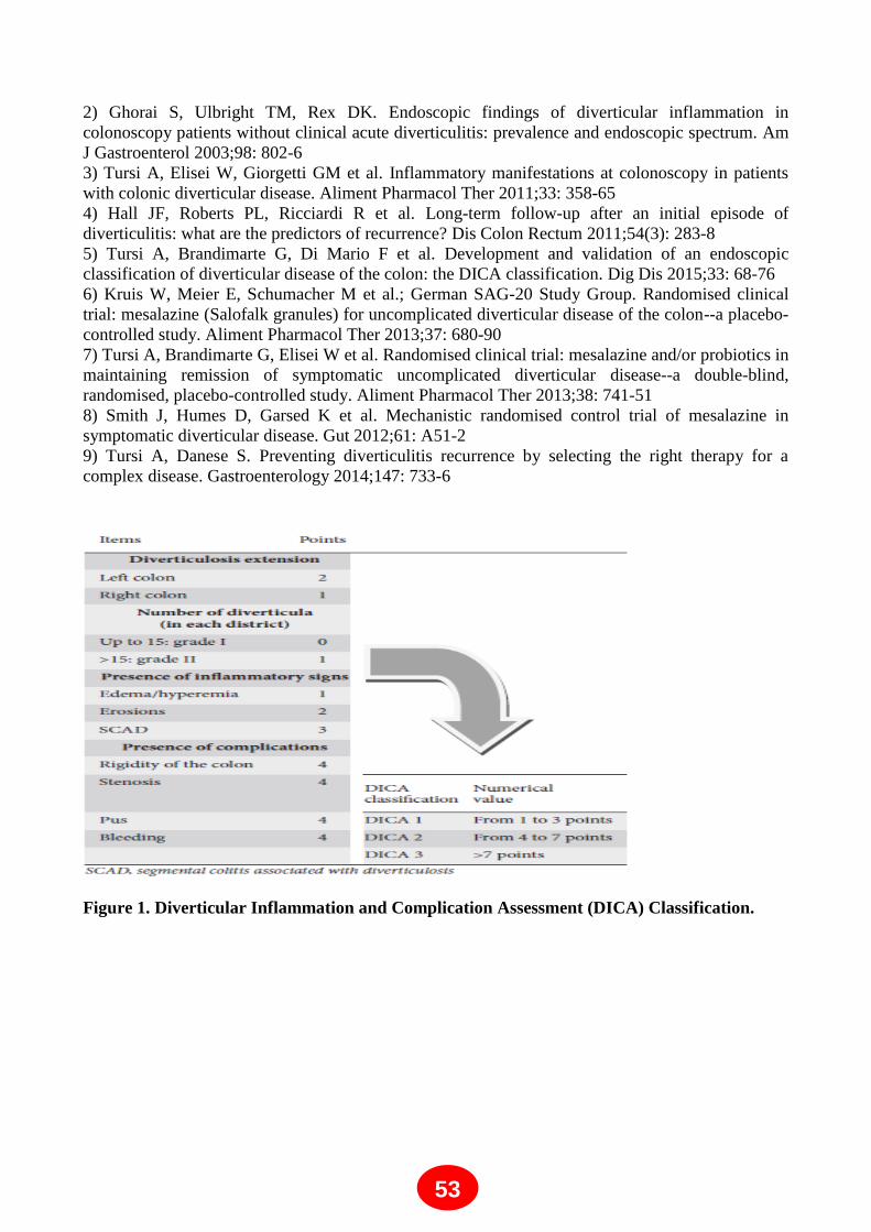
53
2) Ghorai S, Ulbright TM, Rex DK. Endoscopic findings of diverticular inflammation in
colonoscopy patients without clinical acute diverticulitis: prevalence and endoscopic spectrum. Am
J Gastroenterol 2003;98: 802-6
3) Tursi A, Elisei W, Giorgetti GM et al. Inflammatory manifestations at colonoscopy in patients
with colonic diverticular disease. Aliment Pharmacol Ther 2011;33: 358-65
4) Hall JF, Roberts PL, Ricciardi R et al. Long-term follow-up after an initial episode of
diverticulitis: what are the predictors of recurrence? Dis Colon Rectum 2011;54(3): 283-8
5) Tursi A, Brandimarte G, Di Mario F et al. Development and validation of an endoscopic
classification of diverticular disease of the colon: the DICA classification. Dig Dis 2015;33: 68-76
6) Kruis W, Meier E, Schumacher M et al.; German SAG-20 Study Group. Randomised clinical
trial: mesalazine (Salofalk granules) for uncomplicated diverticular disease of the colon--a placebo-
controlled study. Aliment Pharmacol Ther 2013;37: 680-90
7) Tursi A, Brandimarte G, Elisei W et al. Randomised clinical trial: mesalazine and/or probiotics in
maintaining remission of symptomatic uncomplicated diverticular disease--a double-blind,
randomised, placebo-controlled study. Aliment Pharmacol Ther 2013;38: 741-51
8) Smith J, Humes D, Garsed K et al. Mechanistic randomised control trial of mesalazine in
symptomatic diverticular disease. Gut 2012;61: A51-2
9) Tursi A, Danese S. Preventing diverticulitis recurrence by selecting the right therapy for a
complex disease. Gastroenterology 2014;147: 733-6
Figure 1. Diverticular Inflammation and Complication Assessment (DICA) Classification.

54
ANTIBIOTICS
Angel Lanas
Digestive Diseases Service. University Clinic Hospital Lozano Blesa. Zaragoza. Spain. Aragon
Health Research Institute (IIS Aragón). Zaragoza. Spain. 3University of Zaragoza. Spain.
CIBERehd. Zaragoza. Spain
Introduction:
Diverticular disease of the colon represents the most common disease affecting the colon in the
Western societies and its prevalence is increasing because of changes in lifestyle, overweight,
physical inactivity and low fiber diet. It is uncommon in subjects under 40 years of age increase up
to 65% in those aged 65 years or more whereas 80% of patients who present with diverticulitis are
50 or older. Diverticula can present in number from solitary to hundreds, they are typically 5-10
mm in diameter. Diverticulosis occurs primarily in the sigmoid and descending colon in more than
90% of patients, but may be prevalent in varying degrees in the rest of the colon [1]. Eighty percent
of patients with diverticular disease will remain asymptomatic for their lifespam, but 15-20% will
develop symptoms, and approximately 1/4 of them will eventually have an episode of symptomatic
painful diverticular disease without inflammation; up to 10-25% if patients with diverticula will
have an episode of acute diverticulitis, 1-2% will require hospitalization and 0.5% will require
surgery. Diverticula are also responsible for the majority of episodes of lower gastrointestinal
bleeding [2].
In this summary the presence of diverticula in the colon in the absence of overt inflammation will
be called diverticulosis or uncomplicated diverticular disease (UDD). It may be symptomatic or
asymptomatic. The term ―acute colonic diverticulitis‖ (ACD) is used to describe inflammation of
the diverticula, wich may or may not progress to complications (complicated ACD). There is also
chronic diverticulitis, because of recurrent diverticulitis or because of the development of a
segmental colitis associated with the diverticula [3]. Figure 1. Summary, the clinical spectrum of
diverticular disease is wide.
Management of uncomplicated diverticular disease (UDD)
In patients with asymptomatic UDD [2] , a high fiber diet may be recommended because its
possible prophylactic benefit in preventing symptomatic UDD and complications. There is no
evidence that other drugs are useful in these patients. The rationale for the use of antibiotics in
symptomatic UDD is not clearly established. Recent studies suggest that changes in gut microbiota
(intestinal bacterial overgrowth) could contribute to symptoms development due to excessive
production of bowel gas through carbohydrate fermentation. In order to avoid systematic effects,
poorly absorbed antimicrobials act against enteric pathogens but have minimal risk of systematic
toxicity or side effects. Rifaximin may decrease metabolic activity of bowel flora, increasing fecal
mass, and may also eradicate bacterial overgrowth. Three open and two double blind RCT [4-8]
have examined the effectiveness of cyclic administration of rifaximn and fiber in reducing
symptoms compared with fiber alone. A systematic review and two meta-analysis have analyzed
theses trials [9-10]. They concluded that combined treatment is effective in obtaining symptom
relief at 1 year in patients with UDD; 35% of patients treated with fiber alone were asymptomatic
compared with 64% in group of combined treatment. The number needed to treat was three for
rifaximin vs placebo to relieve symptom and nine to avoid complications. Summary, the best results

55
have been obtained using a combination of soluble fiber, such as glucomannan, and rifaximin 1
week every month.
Treatment of acute colonic diverticulitis (ACD)
In patients with mild symptoms and without signs of complicated ACD, the combination of pain in
lower left abdomen, the absence of vomiting and a C reative protein > 50mg/l, may be sufficient for
the diagnosis. If imaging is indicated, a conditional strategy with ultrasound as first line technique
and followed by computerized tomography (CT), if ultrasound is inconclusive may represent the
most effective approach. Hinchey et al. proposed a classification of ACD, which distinguishes five
stages; stage 0: clinically mild diverticulitis, stage Ia: pericolic inflammation; Ib: abscess < 5 cm in
the proximity of the primary inflammation, stage II: intra-abdominal, pelvic or retroperitoneal
abscess or abscess distant from the primary inflammation; stage III: generalized purulent peritonitis
and stage IV: fecal peritonitis.
HINCHEY stage 0 or Ia: The majority of uncomplicated ACD with this Hinchey stage can be
safely treated conservatively with success rate between 70-100% [11]. In cases of uncomplicated
ACD, criteria for inpatient management are significant inflammation (include presence of fever or
peritonitis), intolerance to oral fluids, age over 80-85 years, immune-suppression, or severe
comorbidities. In most cases, a short hospital stay will be sufficient. There is not evidence that
dietary restrictions influenced treatment outcomes, although most physicians usually recommend
clear liquid diet. A recent Cochrane review found that the best available data do no support the
routine use of antibiotics [12]. Antibiotics neither accelerate recovery nor prevent complications or
recurrence. Therefore, the use of antibiotics in this case is questionable. Probably, they would be
appropriate in selected patients (generalized infection and in immunocompromised patients).
Various antibiotics may be used, ranging from ampicillin to third generation cephalosporins, as long
as it is effective against gram positive, gram negative and anaerobic bacteria. The combination of
ciprofloxacin and metronidazole is probably the most prescribed oral treatment. Recent data
have shown that there are no advantages of intravenous over oral antibiotics and of intravenous 4
days treatment over 7 days treatment [13-15] .Usually, clinical improvement is observed within 3-4
days of treatment.
ACD HINCHEY Ib or II with Abscess: Approximately 15% of patients with ACD will develop
an abscess. Hospitalization is indicated. Small abscesses (< 4 cm), need conservative treatment with
broad spectrum antibiotics, which is successful in up to 70% [16]. When conservative treatment
fails or in larger abscesses, percutaneous drainage should be performed, which is successful in up to
80%. Surgery will be a rescue treatment when previously mentioned treatments fail.
ACD HINCHEY III or IV and purulent or faecal peritonitis: Peritonitis is the most serious
complication, with a mortality of 14%. Although there is no evidence, early surgery is considered
standard therapy for these patients. In critical ill patients with hemodynamic instability, Hartmann´s
procedure is recommended. However, in hemodynamically stable patients, primary anastomosis
with or without proximal faecal diversion has to be considered a preferable choice.
Prevention of recurrent ACD
The evidence to define the optimal treatment following an ACD episode in order to prevent a new
episode is scarce. There are three recent systematic reviews that assessed the role of cyclic
rifaximin in preventing recurrence of ACD, but did not show a clear benefit [10, 17,18]. From a
pathophysiologic point of view, a plausible explanation of ineffectiveness of rifaximin in preventing

56
recurrences could be that a cyclic treatment may not control the colonic bacterial population during
full month, because colonic bacterial population recovers within 7-14 days after the end of
rifaximin. However, a recent Spanish open RCT has shown that cyclic rifaximin can improve
symptoms and maintain periods of remission following ACD [19]. Recurrences occurred in 10, 4%
of patients given rifaximin plus fiber vs 19, 3% of fiber alone. Moreover, patients first diagnosed
since ≥ 1 year had a higher risk of exacerbation (OR 3, 34, 95% CI: 0.01-12.18). But, further
studies are needed since- at present time- no recommendations can be made. There is not evidence
to support the use of other antibiotics in this setting.
Summary
In symptomatic UDD, the aims of treatment are to prevent complications and reduce symptoms.
According to current evidence, fiber plus cyclic seems to be an effective therapy. In the ACD,
antibiotics seem to remain the mainstay of treatment and an outpatient management is considered
the optimal approach in the vast majority of patients with uncomplicated ACD. However, inpatient
management and intravenous antibiotics are necessary in complicated ACD. Currently, the role of
emergency surgery is changing. Most diverticulitis-associated abscesses can be treated with
intravenous antibiotics and/or percutaneous drainage and emergency surgery will be considered
standard treatment only in patients with peritonitis. Finally, elective surgery after recovery from
ACD should be made on a case-by-case basis
REFERENCES
1. Gargallo C, Sopeña F, Lanas A. Colonic Diverticular Disease. Treatment and prevention.
Gastroenterol Hepatol 2016 (in press).
2. Stollman N, Raskin JB. Diverticular disease of the colon. Lancet. 2004;363:631-9.
3. Humes DJ, Spiller RC. The pathogenesis and management of acute colonic diverticulitis.
Aliment Pharmacol Ther 2014;39:359-370.
4. Colecchia A, Vestito A, Pasqui F,et al. Efficacy of long term cyclic administration of the
poorly absorbed antibiotic Rifaximin in symptomatic, uncomplicated colonic diverticular
disease. World J Gastroenterol 2007;13:264-9
5. Papi C et al. Efficacy of rifaximin on symptoms of uncomplicated diverticular disease of the
colon. A pilot multicentre open trial. Diverticular Disease Study Group. Ital J Gastroenterol
1992;24:452-6
6. Papi C, Ciaco A, Koch M et al. Efficacy of rifaximin in the treatment of symptomatic
diverticular disease of the colon. A multicentre double-blind placebo-controlled trial.
Aliment Pharmacol Ther 1995;9:33-9
7. Latella G, Pimpo MT, Sottili S et al. Rifaximin improves symptoms of acquired
uncomplicated diverticular disease of the colon. Int J Colorectal Dis 2003;18:55-62
8. D'Incà R, Pomerri F, Vettorato MG et al. Interaction between rifaximin and dietary fibre in
patients with diverticular disease. Aliment Pharmacol Ther 2007;25:771-9
9. Latella G, Scarpignato C Rifaximin in the management of colonic diverticular disease.
Expert Rev Gastroenterol Hepatol 2009;3: 585-598
10. Bianchi M, Festa V, Moretti A et al. Meta-analysis: long-term therapy with rifaximin in the
management of uncomplicated diverticular disease. Aliment Pharmacol Ther 2011;33:902-
10
11. Rafferty J, Shellito P, Hyman NH, et al. Standards Committee of American Society of
Colon and Rectal Surgeons. Practice parameters for sigmoid diverticulitis. Dis Colon
Rectum 2006;49:939-44

57
12. Shabanzadeh DM, Wille-Jørgensen P. Antibiotics for uncomplicated diverticulitis. Cochrane
Database Syst Rev 2012;11:CD009092.
13. Moya P, Arroyo A, Pérez-Legaz J et al Applicability, safety and efficiency of outpatient
treatment in uncomplicated diverticulitis. Tech Coloproctol 2012;16:301-7
14. Etzioni DA, Chiu VY, Cannom RR et al. Outpatient treatment of acute diverticulitis: rates
and predictors of failure. Dis Colon Rectum 2010;53:861-5
15. Andeweg CS, Mulder IM, Felt-Bersma RJ et al. Guidelines of diagnostics and treatment of
acute left-sided colonic diverticulitis. Dig Surg 2013;30:278-92
16. Ambrosetti P, Chautems R, Soravia C et al. Long-term outcome of mesocolic and pelvic
diverticular abscesses of the left colon: a prospective study of 73 cases. Dis Colon Rectum
2005;48:787-91
17. Zullo A, Hassan C, Maconi G et al. Cyclic antibiotic therapy for diverticular disease: a
critical reappraisal. J Gastrointestin Liver Dis 2010;19:295-302
18. Maconi G, Barbara G, Bosetti C, et al. Treatment of diverticular disease of the colon and
prevention of acute diverticulitis: a systematic review. Dis Colon Rectum 2011;54:1326-38
19. Lanas A, Ponce J, Bignamini A, et al. One year intermittent rifaximin plus fibre
supplementation vs. fibre supplementation alone to prevent diverticulitis recurrence: a
proof-of-concept study. Dig Liver Dis 2013;45:104-9.
Figure 1: Algorithm for management of acute colonic diverticulitis. Taken from reference 1. Figure
4:
Acute colonic diverticulitis ( ACD)
Complicated ACDUncomplicated ACD
• Inmunocompromised patient?
• Signs of generalized infection?
• Affected general condition?
• Severe comorbidity?
• Advanced age?
yesno
• Outpatient treatment
• Oral ATB?
• Outpatient or inpatient treatment
• Broad spectrum ATB ( oral vs iv)
ACD Hinchey Ib or II ACD Hinchey III or IV
• Small abcesses ( < 4.-5 cm) : iv ATB
• Larger abcesses; iv ATB plus percutaneous
drainage
• Emergency surgery
Nota: ATB: Antibiotics, iv; intravenous

58
AMINOSALICYLATES
Giovanni Barbara, Maria Raffaella Barbaro, Cesare Cremon, Lara Bellacosa, Vincenzo
Stanghellini
Department of Medical and Surgical Sciences, St. Orsola-Malpighi Hospital, Bologna - Italy
Colonic diverticulosis is common in Western industrialized countries, increases with age and occurs
in up to 65% of people over the age of 60 years. Although in most cases colonic diverticula do not
generate troubles during lifetime, 10-25% of subjects, develop symptoms, such as abdominal pain
and changes in bowel habit, a condition termed symptomatic uncomplicated diverticular disease
(SUDD). Diverticulitis is the most common complication, affecting 10-25% of patients, of which
15% develop further complications including bleeding, abscesses, fistulae, stenosis or peritonitis.
The related morbidity and mortality, the use of imaging techniques, the requirement of expensive
medical and surgical treatments account for a substantial social and economic burden.1, 2
Little is known about the natural history of diverticular disease. However, one study suggests that
the risk of recurrent attacks of diverticulitis after a first episode is estimated between 7% and 45%.3
Although prophylactic surgery has been recommended after 2 attacks of diverticulitis, this view has
changed in recent years and has given more space to the conservative approach.4 This has created a
large population of post-diverticulitis patients at potential risk for subsequent attacks, raising the
important clinical question as to whether any medical therapy can alter their natural history.
Although no disease modifying therapies are as yet approved to manage SUUD symptoms or
prevent acute diverticulitis several dietary and pharmacological approaches have been proposed
although their efficacy has not always been tested in controlled trials. These approaches include,
among others, a high fiber diet, avoidance of seeds and popcorn, the use of anti-spasmodics, for the
management of pain, non-absorbable antibiotics and probiotics based on the dogma that bacterial
overgrowth in the diverticulum is the initiatior of a pathogenetic cascade of events eventually
culminating in diverticulitis and complications. The rational for the use of aminosalicylates, such as
mesalazine or balsazide, is based on the evidence that inflammation may underlie symptoms and
diverticulitis in patients with diverticular disease.5, 6
AMYNOSALICYLATES 5-Aminosalicylic acid (mesalazine) is an anti-inflammatory agent widely used in the treatment of
ulcerative colitis. After oral or rectal administration, mesalazine, is absorbed by colonic epithelial
cells and its efficacy is related to its mucosal concentration.7 The anti-inflammatory mechanism of
mesalazine is not completely understood, but believed to be dependent on: 1) reduction in the
synthesis of prostaglandins and pro-inflammatory cytokines, including interleukin-1 and tumor
necrosis factor-alpha; 2) inhibition of the chemiotaxis of neurophils; 3) inhibition of the activation
of nuclear factor Kappa-B transcription factor family; 4) the activation of nuclear receptors (i.e. the
gamma form of peroxisome proliferator-activated receptors), which downregulate inflammation and
reduce inflammatory cytokine release; 5) promotion of mucosal barrier effect; 6) change in colonic
luminal pH which would favor the growth of beneficial bacteria such as Bifidobacteria and
Lactobacilli; 6) antibacterial activity with inhibition of expression of bacterial genes involved in
invasiveness, epithelial adherence proliferation, antibiotic resistance.7-10
Some of these effects need
to be confirmed in further studies.
MESALAZINE IN DIVERTICULAR DISEASE
The clinical scenarios in which the efficacy and safety of mesalazine alone or in combination with
probiotics have been studied include, SUDD, the prevention of diverticulitis and the prevention of
recurrent diverticulitis. There are some limitations related to the currently available Literature.
These drawbacks are related to the heterogeneity of study design, the frequently use of an open
59
label design, the use of combinations of therapies in the same trial, the sometimes small sample size
and insufficient details in the reported data. These limitations reduce the overall quality of the
results and the level of recommendation in clinical practice.11
Given these limitations, the
conclusion of a systematic review of the Literature was that medical treatment showed some
evidence of improvement in symptoms in patients with SUDD, but its role in the prevention of
acute diverticulitis remains to be defined.11
Two recent large controlled studies on the efficacy of
mesalazine have been published and helped only in part the numerous questions that the clinician
faces when approaching the patient with diverticular disease. The first study addressed the effect of
mesalazine in the control of symptoms in patients with SUDD12
and the second evaluated the
therapeutic role of mesalazine in the prevention of recurrent diverticulitis and will be briefly
mentioned below.13
Symptomatic uncomplicated diverticular disease.
The effect of mesalazine in SUUD has been investigated in numerous open label studies as well as
in three double-blind, placebo-controlled studies. One of these controlled studies showed that lower
abdominal pain was significantly ameliorated in patients treated with mesalazine over patients
treated with placebo. However, a statistical significance was reached only in the per-protocol
analysis (P=0.05) but not in the intention to treat evaluation (P=0.374).14
Several factors contributed
to this inconsistency, including an unbalanced distribution of patients, particularly for the use of
medications in the placebo and active drug arms. Interestingly, in ad hoc analyses, the authors found
that the presence of diverticula in the descending colon represented a confounding factor, indeed
after normalization of this and other factors the statistical analysis almost reached significance.
Another confounder may be represented by the heterogeneity of the population under study and in
particular the possible inclusion of patients with irritable bowel syndrome. Unfortunately, all these
studies did not include patients according to the presence of low-grade intestinal inflammation that
have been indicated to underlie symptom development in at least subgroups of patients with SUDD. 5, 15, 16
Another multicenter double-blind, placebo-controlled study assessed the efficacy of pulse treatment
with mesalazine with or without probiotics (10 days per month for 12 months) on the recurrence of
abdominal pain in SUUD patients. The results showed that the combination of mesalazine with
Lactobacillus casei was superior to placebo in the maintenance of remission in SUDD. While these
studies are promising, there is a need for very large studies powered enough to identify predictors of
response and biomarkers useful to identify patients who merit one over another type of treatment. In
the recent mesalazine trial in patients with irritable bowel syndrome there was clearly a subgroup of
patients that showed a sustained marked response to mesalazine, further suggesting the need to
identify the phenotype and predictors of the responders17
.
Prevention of diverticulitis. A double-blind placebo controlled study assessed the prevention of acute diverticulitis in patients
receiving mesalazine (1.6 g/day for 10 days/month), a probiotic (L. casei subgroup DG, 24
billion/day for 10 days/month), mesalazine plus probiotic or placebo. The results showed that
mesalazine was better than placebo in preventing acute diverticulitis.6 In line with these results
Gaman et al., randomized patients to receive either mesalazine (n=68) or placebo (n=52). Patients
on mesalazine were significantly less likely to experience a flare over a mean follow-up of 40.5
months.18
These studies are highly demanding as diverticulitis is a relatively uncommon
complication and long follow-up studies are needed to detect recurrences. However, in contrast with
these promising data are the results on the efficacy and safety of multimatrix mesalazine vs placebo
in the prevention of recurrent diverticulitis. This outcome was assessed in two large identically
designed studies: PREVENT1 conducted in 590 patients and PREVENT2 in 592 patients. Adult
patients with ≥ 1 episodes of acute diverticulitis in the previous 24 months that resolved without
surgery were randomized to receive mesalazine 1.2 g, 2.4 g, or 4.8 g or placebo, once daily for two

60
years. The results showed that mesalazine did not reduce the rate of recurrence or time to
recurrence. The authors concluded that mesalazine is not recommended for this indication.
Conclusions
In conclusion, evidence suggests some efficacy of mesalazine in the control of symptoms in patients
with uncomplicated disease. The largest study so far published on the efficacy of mesalazine in the
prevention of diverticulitis showed that mesalazine was not superior than placebo. However, some
argue that this is not the last word as many issues remain open and unresolved.
REFERENCES
1. Delvaux M. Diverticular disease of the colon in Europe: epidemiology, impact on citizen
health and prevention. Aliment Pharmacol Ther 2003;18 Suppl 3:71-4.
2. Stollman N, Raskin JB. Diverticular disease of the colon. Lancet 2004;363:631-9.
3. Bartho L, Benko R, Patacchini R, et al. Effects of capsaicin on visceral smooth muscle: a
valuable tool for sensory neurotransmitter identification. Eur J Pharmacol 2004;500:143-57.
4. Cuomo R, Barbara G, Pace F, et al. Italian consensus conference for colonic diverticulosis
and diverticular disease. United European Gastroenterol J 2014;2:413-42.
5. Tursi A, Brandimarte G, Elisei W, et al. Assessment and grading of mucosal inflammation
in colonic diverticular disease. J Clin Gastroenterol 2008;42:699-703.
6. Tursi A, Brandimarte G, Elisei W, et al. Randomised clinical trial: mesalazine and/or
probiotics in maintaining remission of symptomatic uncomplicated diverticular disease--a
double-blind, randomised, placebo-controlled study. Aliment Pharmacol Ther 2013;38:741-
51.
7. Desreumaux P, Ghosh S. Review article: mode of action and delivery of 5-aminosalicylic
acid - new evidence. Aliment Pharmacol Ther 2006;24 Suppl 1:2-9.
8. Barbara G, Stanghellini V, Cremon C, et al. Aminosalicylates and other anti-inflammatory
compounds for irritable bowel syndrome. Dig Dis 2009;27 Suppl 1:115-21.
9. Andrews CN, Griffiths TA, Kaufman J, et al. Mesalazine (5-aminosalicylic acid) alters
faecal bacterial profiles, but not mucosal proteolytic activity in diarrhoea-predominant
irritable bowel syndrome. Aliment Pharmacol Ther 2011;34:374-83.
10. Rousseaux C, Lefebvre B, Dubuquoy L, et al. Intestinal antiinflammatory effect of 5-
aminosalicylic acid is dependent on peroxisome proliferator-activated receptor-gamma. J
Exp Med 2005;201:1205-15.
11. Barbara G, Wang B, Stanghellini V, et al. Mast cell-dependent excitation of visceral-
nociceptive sensory neurons in irritable bowel syndrome. Gastroenterology 2007;132:26-37.
12. Kruis W, Meier E, Schumacher M, et al. Randomised clinical trial: mesalazine (Salofalk
granules) for uncomplicated diverticular disease of the colon--a placebo-controlled study.
Aliment Pharmacol Ther 2013;37:680-90.
13. Raskin JB, Kamm MA, Jamal MM, et al. Mesalamine did not prevent recurrent diverticulitis
in phase 3 controlled trials. Gastroenterology 2014;147:793-802.
14. Barbara GS, Grunewald B, Paute S, et al. Study of nicotinic acetylcholine receptors on
cultured antennal lobe neurones from adult honeybee brains. Invert Neurosci 2008;8:19-29.
15. Simpson J, Sundler F, Humes DJ, et al. Post inflammatory damage to the enteric nervous
system in diverticular disease and its relationship to symptoms. Neurogastroenterol Motil
2009;21:847-e58.
16. Humes DJ, Simpson J, Smith J, et al. Visceral hypersensitivity in symptomatic diverticular
disease and the role of neuropeptides and low grade inflammation. Neurogastroenterol Motil
2012;24:318-e163.
17. Barbara G, Xing Z, Hogaboam CM, et al. Interleukin 10 gene transfer prevents experimental
colitis in rats. Gut 2000;46:344-9.
61
18. Gaman A TR, Georgescu EF, Tataru Abagiu M. . Prophylactic effects of mesalamine in
diverticular disease. Hypothesis and facts: Germany: Falk Symposium, 2011.

62
PROBIOTICS
Carmelo Scarpignato1, Anna Bertelé
1,2
1Clinical Pharmacology & Digestive Pathophysiology Unit, Department of Clinical &
Experimental Medicine, University of Parma, Italy 2Laboratory for GI Functional Investigations, Maggiore University Hospital, Parma, Italy
In an era when inflammatory bowel disease (IBD) represents a fertile ground for research, enjoying
the fruits of innovative therapeutic approaches, and irritable bowel sindrome (IBS) witnesses
increasing interest by both basic scientists and clinicians, colonic diverticela and their complications
remain relatively forgotten. Diverticulosis however is common and, as our populations age, its
prevalence is steadily increasing [1].
Despite the pathogenesis of chronic symptoms in the aftermath of acute diverticulitis as well as the
link between uncomplicated diverticulosis and symptoms (leading to the so-called symptomatic
uncomplicated diverticular disease, SUDD) are complex and still not fully understood [2], low-
grade inflammation appears to be pivotal in the pathophysiology of symptoms in both clinical
settings [3]. This intriguing hypothesis must be viewed in the context of bacteria-induced immune
activation and consequent inflammation. Indeed, while changes in the colonic microbiota are clearly
critical to the pathogenesis of diverticular complications, such as diverticulitis and peri-diverticular
abscesses, more subtle changes in microbiota composition may well be important to the more
chronic manifestations of diverticulosis [4].
Alteration in bacterial flora in diverticular disease occurs primarily as a result of fecal stasis, which
Is a result of slow colonic transit and stagnation of fecal material in the diverticula themselves. The
―blind-pouch‖ theory, which implicates fecal stasis and bacterial overgrowth in the pathogenesis of
clinical conditions such as appendicitis and pouchitis, has been similarly used to explain the
development of diverticular inflammation [5]. Altered bacterial flora in these regions trigger
intestinal inflammation by impairing mucosal barrier function and up-regulating inflammatory
cytokine release [4].
However, only few studies have focused on the microbiological characterization of the microbiota
in diverticular disease. Some studies have shown the presence of bacterial overgrowth in subgroups
of patients with diverticular disease [6,7]. However, traditional methods of investigation are
nowadays inadequate, since a large portion of the resident bacterial flora is not cultivable and few
data are available to date. The most commonly isolated organisms are anaerobes (Bacteroides,
Peptostreptococcus, Clostridium, and Fusobacterium), while the most often cultivated ones are
Gram-negatives, particularly Escherichia coli, and facultative Gram-positives, such as Streptococci.
The recent development of culture-independent approaches, such as metagenomics, is greatly
contributing to the understanding of gut microbiota composition. Gueimonde et al. [8] collected and
analysed biopsy specimens of patients with colorectal cancer (CRC), IBD or diverticulitis and found
that strains of the genus Bifidobacterium were present in all samples of patients with diverticulitis
or IBD, but only in a 76% of CRC patients. Bifidobacterium longum and Bifidobacterium animalis
were found in significantly higher proportions in patients with diverticulitis compared to the other
two groups, while Bifidobacterium adolescentis and Bifidobacterium catenulatum were absent in
patients with diverticulitis. A recent analysis of fecal specimens found an increase of Proteobacteria
and Actinobacteria in patients with SUDD compared with healthy controls, with most represented
species being Collinsella Aerofacien [9]. Despite these interesting observations, there is no definite
evidence allowing correlating diverticular disease and its complications to a specific bacterial strain.
While both systemic and poorly absorbed antibiotics are often prescribed to patients with colonic
diverticular disease [5,10], relatively few studies have attempted the manipulation of gut microbiota
as a therapeutic target. Compared with antimicrobial therapy, less invasive and more physiologic
approaches to modulate intestinal microecology are represented by probiotics, prebiotics and
symbiotics.

63
According to the original FAO/WHO definition [11], probiotics are ―live microorganisms, which –
when administered in adequate amounts – confer a health benefit on the host‖. The rationale for the
use of probiotics for the treatment of gut microbiota-related diseases is the restoration of intestinal
homeostasis by beneficial microbes. Most probiotics consist of Lactobacilli and Bifidobacteria, but
also yeasts such as Saccharomyces boulardii have been used with good clinical outcomes.
Probiotics have the ability to restore balance to enteric flora, primarily by decreasing numbers of
pathogenic gram-negative bacteria [12]. By competitively inhibiting pathogenic bacterial
overgrowth at the mucosal level, probiotics improve mucosal defense by enhancing tight junction
integrity [13] decreasing bacterial translocation, and down-regulating pro-inflammatory cytokines
such as tumor necrosis factor (TNF) [14].
A significant proportion of patients with DD complain of symptoms resembling or overlapping
those of IBS, making a clear differentiation between these two clinical conditions challenging [15-
17]. Despite the considerable debate regarding whether trials of different probiotics should be
pooled into a meta-analysis [18], several systematic reviews and meta-analyses [19-24] have
consistently shown the efficacy of (single or multi-strain) probiotic treatment on IBS symptom
cluster. Some early [25, 26] and one, very recent and comprehensive [27], systematic reviews have
also tried to summarize the available literature concerning the use of probiotics, alone or as add-on
medication, for the treatment of SUDD.
Although several investigations evaluating the clinical efficacy of probiotics have been performed,
no definitive results have yet been achieved, mainly due to the heterogeneity of the available
studies. Most of the studies have used probiotics in combination with poorly absorbed
antimicrobials (dichlorchinolinol or rifaximin) or anti-inflammatory drugs (mesalazine or
balsalazide). In only three studies [28-30] there was a arm using probiotics alone (namely
Lactobacillus casei DG), but only one was a placebo-controlled, double blind trial [30]. Additional
two studies [31,32] employed probiotics (Lactobacillus paracasei F19 and Lactobacillus paracasei
B21060) in addition to high-fiber diet while one [33] investigated the efficacy of a probiotic
mixture (Lactobacillus acidophilus 145, Lactobacillus helveticus ATC 15009, Bifidobacterium spp.
420 in a phytoextracts-enriched medium). This last investigation is particularly interesting since – in
addition to confirming the efficacy of probiotics on the symptom cluster (constipation, diarrhea and
abdominal pain) of SUDD – showed a persistent colonization with the ingested microorganisms.
Indeed genomic analysis confirmed the significant survivability of ingested L.acidophilus and
Bifidobacterium contained in the symbiotic mixture [33].
The analysis of the available evidence reveals however a poor quality of the published studies,
whose design was heterogeneous, with only 2 out of 11 trials being double-blind and randomized.
While some studies investigated the symptom improvement, other evaluated the maintenance of
remission from abdominal symptoms. In addition, the trial sample size was generally small and the
follow-up never exceeded 12 months. The inclusion criteria were variable so that the patient
populations recruited in the different studies were different as it was the specific probiotic (single or
multi-strain) employed. All these variables make comparison of the results from different studies
difficult and prevent any precise estimate of the treatment effect. In the absence of a quantitative
estimation, the qualitative analysis of the available data can only suggest a benefit of probiotics in
SUDD, but does not allow any evidence-based definite conclusion [27]. As a consequence, the
recent Italian Consensus Conference on Diverticular Disease [34] concluded – with the agreement
of 84% of the experts – that there is insufficient evidence that probiotics are effective in reducing
symptoms of patients with diverticular disease, a statement reiterated by the guidelines of the Italian
Society of Colon and Rectal Surgery [35].
Recent pharmacological approaches targeting enteric bacteria (with poorly absorbed antibiotics, like
rifaximin, or probiotics) and/or intestinal inflammation (with 5-ASA derivatives) have shown to be
capable of controlling symptoms and preventing complications. However, the respective role of
these drugs (alone, in combination or sequentially) in the management of symptomatic diverticular
disease needs to better defined. Well-designed RCTs, including homogeneous populations of

64
patients (classified according to the recent DICA criteria [36]), are therefore needed to establish the
optimal regimen (daily dose and duration)
of therapy.
References
1. Strate LL, Modi R, Cohen E, Spiegel BM. Diverticular disease as a chronic illness: evolving
epidemiologic and clinical insights. Am J Gastroenterol 2012;107: 1486-1493.
2. Simpson J, Scholefield JH, Spiller RC. Origin of symptoms in diverticular disease. Br J Surg
2003; 90: 899-908.
3. Humes DJ, Simpson J, Smith J, Sutton P, Zaitoun A, Bush D, Bennett A, Scholefield JH, Spiller
RC. Visceral hypersensitivity in symptomatic diverticular disease and the role of neuropeptides and
low grade inflammation. Neurogastroenterol Motil 2012; 24:318-e163.
4. Quigley EM. Gut microbiota, inflammation and symptomatic diverticular disease. New insights
into an old and neglected disorder. J Gastrointestin Liver Dis 2010;19: 127-129.
5. Tursi A. Advances in the management of colonic diverticulitis. CMAJ 2012;184:1470-1476.
6. D'Incà R, Pomeri F, Vettorato MG, Dal Pont E, Di Leo V, Ferronato A, Medici V, Sturniolo GC.
Interaction between rifaximin and dietary fibre in patients with diverticular disease. Aliment
Pharmacol Ther 2007;25: 771-779.
7. Tursi A, Brandimarte G, Giorgetti GM, Elisei W. Assessment of small intestinal bacterial
overgrowth in uncomplicated acute diverticulitis of the colon. World J Gastroenterol 2005;11:2773-
2776
8. Gueimonde M, Ouwehand A, Huhtinen H, Salminen E, Salminen S. Qualitative and quantitative analyses of the bifidobacterial microbiota in the colonic mucosa of patients with colorectal cancer, diverticulitis and inflammatory bowel disease. World J Gastroenterol 2007;
13: 3985-3989.
9. Lopetuso LR, Scaldaferri F, Petito V, Ponziani FR, Pecere S, Schiavoni E, Zambiano D, Paroni
Stermini F, Gaetani E, Franceschi F, Cammarota G, Sanguinetti M, Masucci L, Gasbarrini A. Gut
microbiota molecular spectrum in healthy controls, diverticular disease, IBS and IBD patients: time
for microbial marker of gastrointestinal disorders? UEG Journal 2014; 3(Suppl 1): A60
10. Tursi A, Papa A, Danese S. Review article: the pathophysiology and medical management of
diverticulosis and diverticular disease of the colon. Aliment Pharmacol Ther 2015;42: 664-684.
11. Hill C, Guarner F, Reid G, Gibson GR, Merenstein DJ, Pot B, Morelli L, Canani RB, Flint HJ,
Salminen S, Calder PC, Sanders ME. Expert consensus document. The International Scientific
Association for Probiotics and Prebiotics consensus statement on the scope and appropriate use of
the term probiotic. Nat Rev Gastroenterol Hepatol 2014;11:506-514.
12. Koninkx JF, Malago JJ. The protective potency of probiotic bacteria and their microbial
products against enteric infections-review. Folia Microbiol 2008;53:189-194.
13. Ramakrishna BS.Probiotic-induced changes in the intestinal epithelium: implications in
gastrointestinal disease. Trop Gastroenterol 2009;30:76-85.
14. Sartor RB. Probiotic therapy of intestinal inflammation and infections. Curr Opin Gastroenterol
2005;21: 44-50.
15. Spiller R. Is it diverticular disease or is it irritable bowel syndrome? Dig Dis 2012;30:64-69.
16. Spiller R. Editorial: new thoughts on the association between diverticulosis and irritable bowel
syndrome. Am J Gastroenterol 2014;109: 1906-198.
17. Tursi A. Diverticular disease of the colon and irritable bowel syndrome: it is time to
differentiate. Am J Gastroenterol 2015;110:774-775.
18. Whelan K. Editorial: The importance of systematic reviews and u-analyses of probiotics and
prebiotics. Am J Gastroenterol 2014;109:1563-1565.

65
19. McFarland LV, Dublin S. Meta-analysis of probiotics for the treatment of irritable bowel
syndrome. World J Gastroenterol 2008;14:2650-2661.
20. Hoveyda N, Heneghan C, Mahtani KR, Perera R, Roberts N, Glasziou P. A systematic review
and meta-analysis: probiotics in the treatment of irritable bowel syndrome. BMC Gastroenterol
2009;9: 15.
21. Moayyedi P, Ford AC, Talley NJ, Cremonini F, Foxx Orenstein AE, Brandt LJ, Quigley EM.
The efficacy of probiotics in the treatment of irritable bowel syndrome: a systematic review. Gut
2010;59:325-332.
22. Ritchie ML, Romanuk TN. A meta-analysis of probiotic efficacy for gastrointestinal diseases.
PloS One 2012;7:e34938.
23. Ford AC, Quigley EM, Lacy BE, Lembo AJ, Saito YA, Schiller LR, Soffer EE, Spiegel BM,
Moayyedi P. Efficacy of prebiotics, probiotics, and synbiotics in irritable bowel syndrome and
chronic idiopathic constipation: systematic review and meta-analysis. Am J Gastroenterol
2014;109:1547-1561.
24. Didari T, Mozaffari S, Nikfar S, Abdollahi M. Effectiveness of probiotics in irritable bowel
syndrome: updated systematic review with meta-analysis. World J Gastroenterol 2015;21:3072-
3084.
25. Sheth A, Floch M. Probiotics and diverticular disease. Nutrition in Clinical Practice 2009;24:41-
44.
26. Narula N, Marshall JK. Role of probiotics in management of diverticular disease. J
Gastroenterol Hepatol 2010;25: 1827-1830.
27. Lahner E, Bellisario C, Hassan C, Zullo A, Esposito G, Annibale B. Probiotics in the Treatment
of Diverticular Disease. A Systematic Review. J Gastrointestin Liver Dis 2016;25:79-86.
28. Tursi A, Brandimarte G, Giorgetti GM et al. Mesalazine and/or Lactobacillus casei in
preventing recurrence of symptomatic uncomplicated diverticular disease of the colon: a
prospective, randomized, open-label study. J Clin Gastroenterol 2006;40:312-316.
29. Tursi A, Brandimarte G, Giorgetti GM, et al. Mesalazine and/or Lactobacillus casei in
maintaining long-term remission of symptomatic uncomplicated diverticular disease of the colon.
Hepatogastroenterology 2008;55: 916-20.
30. Tursi A, Brandimarte G, Elisei W, et al. Randomised clinical trial: mesalazine and/or probiotics
in maintaining remission of symptomatic uncomplicated diverticular disease a double-blind,
randomized, placebo-controlled study. Aliment Pharmacol Ther 2013;38: 741-51.
31. Annibale B, Maconi G, Lahner E, et al. Efficacy of Lactobacillus paracasei sub. Paracasei F19
on abdominal symptoms in patients with symptomatic uncomplicated diverticular disease: a pilot
study. Minerva Gastroenterol Dietol 2011;57:13-22.
32. Lahner E, Esposito G, Zullo A, Hassan C, Cannaviello C, Di Paolo MC, Pallotta L, Garbagna
N, Grossi E, Annibale B. High-fibre diet and Lactobacillus paracasei B21060 in symptomatic
uncomplicated diverticular disease. World J Gastroenterol 2012;18:5918-5924.
33. Lamiki P, Tsuchiya J, Pathak S, Okura R, Solimene U, Jain S, Kawakita S, Marotta F.
Probiotics in diverticular disease of the colon: an open label study. J Gastrointestin Liver Dis
2010;19:31-36.
34. Cuomo R, Barbara G, Pace F, Annese V, Bassotti G, Binda GA, Casetti T, Colecchia A, Festi
D, Fiocca R, Laghi A, Maconi G, Nascimbeni R, Scarpignato C, Villanacci V, Annibale B. Italian
consensus conference for colonic diverticulosis and diverticular disease. United European
Gastroenterol J 2014; 2:413-442.
35. Binda GA, Cuomo R, Laghi A, Nascimbeni R, Serventi A, Bellini D, Gervaz P, Annibale B.
Practice parameters for the treatment of colonic diverticular disease: Italian Society of Colon and
Rectal Surgery (SICCR) guidelines. Tech Coloproctol 2015;19:615-626.
36. Tursi A, Brandimarte G, Di Mario F, Andreoli A, Annunziata ML, Astegiano M, Bianco MA,
Buri L, Cammarota G, Capezzuto E, Chilovi F, Cianci M, Conigliaro R, Del Bavero G, Di Cesare
L, Di Fonzo M, Elisei W, Faggiani R, Farroni F, Forti G, Germanà B, Giorgetti GM, Giovandone

66
M, Lecca PG, Loperfido S, Marmo R, Morucci P, Occhigrossi G, Penna A, Rossi AF, Spadaccini
A, Zampaletta C, Zilli M, Zullo A, Scarpignato C, Picchio M. Development and validation of an
endoscopic classification of diverticular disease of the colon: the DICA classification. Dig Dis
2015;33:68-76.

67
OPEN OR LAPAROSCOPIC TREATMENT: DIFFERENCES AND
OUTCOMES
Enio Chaves Oliveira1, Mauro Bafutto
2, Jose Roberto Almeida
3
1 Dep. Surgery, School of Medicine, Universidade Federal de Goias, Brazil
2Dep. Internal Medicine, School of Medicine, Universidade Federal de Goias, Brazil
3Dep. Internal Medicine, School of Medicine, Universidade Federal de Pernambuco, Brazil
In the early 20th century, surgical treatment of diverticular disease was made by three stage
laparotomy in more than 50% of emergency cases (1). Mortality rate for three-stage resection was
around 6% while resection had 17% of mortality (2, 3). Three-stage surgery was indicated only to
complications for diverticular disease: perforation with abscess or peritonitis, obstruction,
hemorrhage and fistula. The idea of safety for the three-stage resection lasted for many decades (4).
Two facts pushed surgeons on to look for new strategies to treat diverticular disease: the prolonged
morbidity of three-stage resection and the high rate of emergency surgery in symptomatic patients
(5). Rodkey & Welch (1984) reported the increase use and safety of one-stage resection but not for
peritonitis (Table 1).
Surgical treatment evolved and changed mainly in the past two decades. Three-stage was followed
by two-stage and one-stage procedures with development of antibiotics, intensive care support,
parenteral nutritional supplementation and improved image diagnostic.
Laparoscopy colectomy appeared in the 1990s a few years after the first laparoscopy procedure (6).
The laparoscopic colon surgery improved dramatically in the last decade from laparoscopic-assisted
colectomy to one port laparoscopic surgery. Laparoscopy gained more acceptance among colorectal
surgeons nowadays and represent the first option to elective colonic surgery. Open surgery and
hand assisted laparoscopic still remain indicated in more complexes cases (7).
Advantages of laparoscopy include faster recovery, shorter hospital stay, decreased postoperative
pain, earlier return to work and resumption of normal daily activity besides cosmetic benefits.
Laparoscopy is commonly used for elective surgery, but for emergency surgery is still considered
too challenging and is not usually recommended.
Some authors reported superiority of laparoscopic sigmoidectomy compared to open
sigmoidectomy for perforated diverticulitis with regard to postoperative morbidity and hospital stay
(Table 2) (8, 9). But the superiority of peritoneal laparoscopic lavage compared to open surgery still
remain controversial (10, 11). Kaushik et al. (2016) (11) reviewing literature conclude that colon
resection with primary anastomosis with purulent peritonitis is acceptable and peritoneal lavage
may play a role in peritonitis without perforation. Schultz et al. (2016) (10) did not find advantages
in the use of laparoscopic lavage vs primary resection and also laparoscopic lavage showed worse
outcomes in secondary end points.
Venix et al (2015) (9) reported that laparoscopic sigmoidectomy showed less postoperative
morbidity and hospital stay compared to open sigmoidectomy. Angenete et al. (2016) (12) in a
randomized controlled multicenter trial, demonstrated the feasibility and safety of laparoscopic
lavage as treatment for patients with perforated diverticulitis Hinchey III in the short-term (Table
3). They reported a mortality rate of 7.7% for laparoscopic lavage and 11.4% for open Hartmann‘ s
procedure. Single port laparoscopic surgery for diverticulitis has been used but some times a small
incision like a Pfannenstiel may facilitate the approach to the pelvis with the same outcomes (13).
Surgical approach to diverticulitis Hinchey III or IV may be done initially by laparoscopy with all
benefits of laparoscopic surgery. Laparoscopy may be used only in hemodynamically stable septic
peritonitis. Many studies have demonstrated that is feasible and safe. Conversions may be necessary
and are related to the inflammatory process with a severe adhesion syndrome. Visible perforations
in open surgery may be related to the handling of the inflamed colon rather than a more advanced
disease.

68
Advantages of laparoscopy include faster recovery, shorter hospital stay, decreased postoperative
pain, earlier return to work and resumption of normal daily activity as well as cosmetic benefits. All
these factors may have done general population to accept more easily minimally invasive surgical
treatment to diverticulitis. But this growing rate of elective colectomy was not followed by a
decrease in emergency surgery for diverticulitis (Figure 1) (14).
Laparoscopy seems to offer a less invasive alternative that is peritoneal lavage without resection to
Hinchey III cases. Efforts of many investigators trying to avoid colon resection and stoma creation
in patients with peritonitis need to be confirmed by long-term randomized trials.
References
1) Rodkey GV, Welch CE. Changing patterns in the surgical treatment of diverticular disease. Ann
Surg 1984;200:466– 478
2) Lockhart-MummeryJP. Late results of diverticulitis. Lancet 1938; 2:1401-1404.
3) Smithwick RH. Experiences with the surgical management of diverticulitis of the sigmoid. Ann
Surg 1942;115:969-983
4) Smithwick RH. Surgical treatment of diverticulitis of the sigmoid. Am J Surg 1960; 99:192-205
5) Brown PW. Treatment and prognosis of diverticulitis of the colon. Am J Surg 1939;46:162-170.
6) Jacobs M, Verdeja JC, Goldstein HS. Minimally invasive colon resection (laparoscopic
colectomy). Surg Laparosc Endosc. 1991;1:144 –150
7) Miyagaki H, Rhee R, Shantha Kumara HM et al. Surgical treatment of diverticulitis: hand-
assisted laparoscopic resection is predominantly used for complex cases and is associated with
increased postoperative complications and prolonged hospitalization. Surg Innov. 2015 Nov 26. pii:
1553350615618285. [Epub ahead of print]
8) Vennix S, Lips DJ, Di Saverio S et al. Acute laparoscopic and open sigmoidectomy for
perforated diverticulitis: a propensity score-matched cohort. Surg Endosc 2015 Dec 17. [Epub
ahead of print] PMID: 26679173.
9) Thornell A et al. Laparoscopic lavage for perforated diverticulitis with purulent peritonitis - A
randomized trial. Ann Intern Med 2016;164:137-145
10) Schultz JK, Yaqub S, Wallon C, SCANDIV Study Group. Laparoscopic lavage vs primary
resection for acute perforated diverticulitis: The SCANDIV randomized clinical trial. JAMA 2015;
314:1364-75
11) Kaushik M, Bhullar JS, Bindroo S, Singh H, Mittal VK. Minimally invasive management of
complicated diverticular disease: Current status and review of literature. Dig Dis Sci 2016;61:663-
72
12) Angenete E, Thornell A, Burcharth J et al. Laparoscopic lavage is feasible and safe for the
treatment of perforated diverticulitis with purulent peritonitis: the first results from the randomized
controlled trial DILALA. Ann Surg 2016;263:117-22
13) Keller DS, Flores-Gonzalez JR, Sandhu J, Ibarra S, Madhoun N, Haas EM. SILS v SILS+1: a
Case-Matched Comparison for Colorectal Surgery. J Gastrointest Surg 2015;19:1875-9
14) Simianu VV, Strate LL, Billingham RP et al. The Impact of Elective Colon Resection on Rates
of Emergency Surgery for Diverticulitis. Ann Surg 2016;263:123–129
Table 1 - Comparison of one-stage resections and anastomoses in a single institution in two decades (1)
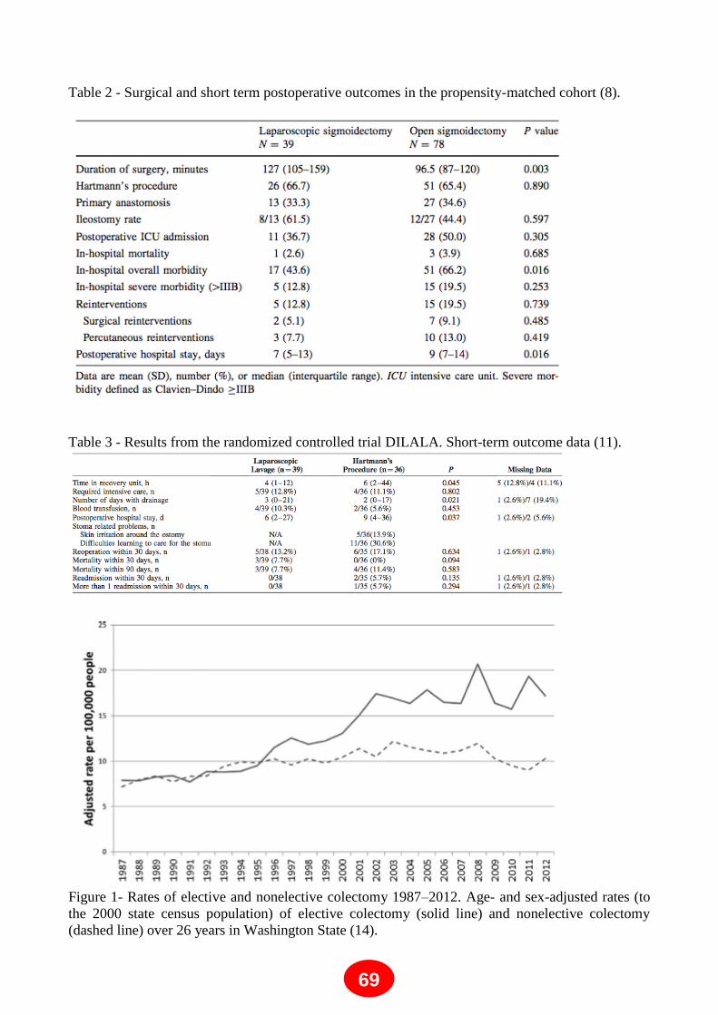
69
Table 2 - Surgical and short term postoperative outcomes in the propensity-matched cohort (8).
Table 3 - Results from the randomized controlled trial DILALA. Short-term outcome data (11).
Figure 1- Rates of elective and nonelective colectomy 1987–2012. Age- and sex-adjusted rates (to
the 2000 state census population) of elective colectomy (solid line) and nonelective colectomy
(dashed line) over 26 years in Washington State (14).
70
LONG-TERM OUTCOME OF ELECTIVE SURGERY
Roberto Persiani, Alberto Biondi, Francesco Santullo, Valeria Fico,
Domenico D’ ’Ugo
Catholic University of Rome, “A. Gemelli” Medical Hospital, Rome - Italy.
Background and Indications
The indication for elective surgery after an acute episode of diverticulitis is currently under
revision. Nowadays a consensus on the indications for elective sigmoid resection in patients with
acute diverticulitis and concomitant abscess initially managed with a non–operative way has not
been reached yet, unlike the surgical management of perforation, fistulae and stricture. Before
2006 elective surgery was considered the best choice after two episodes of diverticulitis (1), while
in 2006, the American Society of Colon and Rectal Surgeons (ASCRS) declared that the decision
of elective sigmoid resection should be taken by a case-by-case evaluation (2).
Recent data about the natural history of diverticulitis has shown that only 5.5% of patients with
recurrent episodes of diverticulitis require emergency surgery, while the remaining part of patients
has a good prognosis without urgent surgical treatment (3). Moreover, the majority of patients with
complicated diverticulitis show such event at the first episode. (4-6). The number of previous
episodes of diverticulitis can‘t be considered an indication for elective surgical treatment because
the risk of perforation seems to be reduced in recurrent diverticulitis, possibly due to adhesion
formation by inflammation (4-7).
There are some characteristics that should be considered to select patients who might benefit from
elective sigmoid resection; surgical treatment is probably the best approach in a selected group of
patients who suffer greatly from their disease.
Many studies have demonstrated that 40-80% of patients show symptoms after conservative
treatment, with reduced health-related quality of life (HRQoL) and increased costs for multiple
specialist consultations, pain medication and productivity losses. Obviously this is the same
situation of patients with recurrent diverticulitis, which often present symptoms also between the
recurrence (8).
Surgery
Patients with complicated diverticulosis can be treated either with an open or a laparoscopic
sigmoid resection. There are two randomized trials which show the advantages of laparoscopic
surgery compared to open surgery. In fact both in the ―Sigma trial‖ (9) and in the study by Gervaz
et al (10) patients underwent to open surgery are characterized by more complications, higher pain
scores and longer hospital stay compared to patients with laparoscopic treatment. Operating time is
significantly longer in the laparoscopic group. Patients treated with open sigmoid resection
have anastomotic leaks more frequently than patients treated by laparoscopic sigmoid resection,
and even if patients subjected to open surgery have more comorbidities, the rate of anastomotic
leaks remains higher in this group even when these differences are accounted for (11).
Proximal and distal margin resection, vascular approach and splenic flexure are the main surgical
steps to consider: the current recommendation about resection margins is to make the proximal
part of the anastomosis in soft pliable colon and to include the colorectal junction in the distal
resection; as regards vascular approach, there are a lot of studies which show that preservation of
inferior mesenteric artery (IMA) could improve the quality of bowel function and could limit the
risk of anastomotic leakage (12); finally the mobilization of splenic flexure is necessary to obtain
tension- free anastomosis (13).
71
Long- term Outcome
Patients with persistent or recurrent symptoms after an episode of diverticulitis show an
improvement of general QoL with elective sigmoid resection, because of a reduction of discomfort
caused by abdominal pain and by abnormal defecation and fatigue. Open and laparoscopic sigmoid
resection are both characterized by excellent long-term results in terms of gastrointestinal function,
quality of life, and patient satisfaction. Laparoscopic surgery has significant benefits only in terms
of cosmetic effects (14), while prospective randomized trials are necessary to demonstrate
additional long-term benefits, such as reduction in incisional hernias, adhesions ad small bowel
obstruction.
Conclusions
Patients with persisting abdominal pains after an episode of diverticulitis and/or recurrent episodes
of diverticulitis can be treated with either a conservative and operative management. However a
randomised controlled trial is necessary to define if the morbidity and mortality associated with
surgery is superior than the risk of complicated recurrence after conservative management.
References
1) D. Feingold, S.R. Steele, S. Lee, A. Kaiser, R. Boushey, W.D. Buie, J.F. Rafferty. American
Society of Colon and Rectum Surgeons. Practice parameters for the treatment of sigmoid
diverticulitis-supporting documentation. Dis Colon Rectum 2014; 57: 284–294
2) Rafferty J, Shellito P, Hyman NH, Buie WD. The Standards Task Force, the American Society of
Colon and Rectum Surgeons. Dis Colon Rectum 2006; 49:939-944
Andeweg CS, Mulder IM, Felt-Bersma RJ, Verbon A, van der Wilt GJ, van Goor H, Lange JF,
Stoker J, Boermeester MA, Bleichrodt RP. Guidelines of Diagnostics and Treatment of Acute
Left-Sided Colonic Diverticulitis. Dig Surg. 2013; 30: 278-292
3) Chapman JR, Dozois EJ, Wolff BG, Gullerud RE, Larson DR. Diverticulitis: a progressive
disease? Do multiple recurrences predict less favorable outcomes? Ann Surg 2006; 243: 876– 880,
discussion 880–883
4) Janes S, Meagher A, Frizelle FA. Elective surgery after acute diverticulitis. Br J Surg 2005; 92:
133–142
5) Collins D, Winter DC. Elective resection for diverticular disease: an evidence-based review.
World J Surg 2008; 32: 2429–2433
6) Broderick-Villa G, Burchette RJ, Collins JC, Abbas MA, Haigh PI. Hospitalization for acute
diverticulitis does not mandate routine elective colectomy. Arch Surg 2005; 140: 576– 581,
discussion 581–583
7) van de Wall B, Draaisma W, Consten E, van der Graaf Y, Otten M,de Wit G, van Stel HF,
Gerhards MF, Wiezer MJ, Cense HA, Stockmann H, Leijtens J, Zimmerman D, Belgers E, van
Wagensveld B, Sonneveld E, Prins HA, Coene P, Karsten T, Klaase J, Muller M, Crolla R,
Broeders I, the Dutch Diverticular Disease (3D) Collaborative Study Group DIRECT trial.
Diverticulitis recurrences or continuing symptoms: Operative versus conservative Treatment. A
multicenter randomised clinical trial. BMC Surgery 2010, 10:25
8) Klarenbeek BR, Veenhof AA, Bergamaschi R, van der Peet DL, van den Broek WT, de Lange
ES, Bemelman WA, Heres P, Lacy AM, Engel AF, Cuesta MA. Laparoscopic sigmoid resection
for diverticulitis decreases major morbidity rates: a randomized control trial. Short term results of
the Sigma Trial. Ann Surg 2009; 249: 39-44
9) Gervaz P, Inan I, Perneger T, Schiffer E, Morel P. A prospective, randomized, single-blind
comparison of laparoscopic versus open colectomy for diverticulitis. Ann Surg 2010; 252: 1-6
10) Levack M, Berger D, Sylla P, Rattner D, Bordeianou L. Laparoscopy Decreases Anastomotic
Leak Rate in Sigmoid Colectomy for Diverticulitis. Arch Surg. 2011; 146: 207-210

72
11) Masoni L, Mari FS, Nigri G, Favi F, Gasparrini M, Dall'Oglio A, Pindozzi F, Pancaldi A,
Brescia A. Preservation of the inferior mesenteric artery via laparoscopic sigmoid colectomy
performed for diverticular disease: real benefit or technical challenge: a randomized controlled
clinical trial. Surg Endosc 2013; 27: 199-206
12) Forgione A, Leroy J, Cahill RA, Bailey C, Simone M, Mutter D, Marescaux J. Prospective
evaluation of functional outcome after laparoscopic sigmoid colectomy. Ann Surg 2009; 249: 218-
224
13) Gervaz P, Mugnier-Konrad B, Morel P, Huber O, Inan I. Laparoscopic versus open sigmoid
resection for diverticulitis: long-term results of a prospective, randomized trial. Surg Endosc. 2011;
25: 3373-8

73
PERITONITIS IN DIVERTICULAR DISEASE. FROM HARTMANN TO
PERITONEAL LAVAGE Ricardo Escalante
Universidad Central de Venezuela, Loira Medical Center, Caracas - Venezuela
Diverticular disease is not a new concept and the presence of diverticula wthin the colon has been
documented as early as 1700 by the French surgeon Alexis Litre. He believed that a diverticulum
may be formed when part only of the wall of the intestine enters the hernial sac and not the whole
diameter, so that only one side of the intestine is pulled out and finally becomes a longer and longer
canal.
In 1896 William Mayo operated on his first case of internal fecal fistula into the bladder, a
and later had resected the colon in cases thought to be cancer which were found to be diverticulitis.
He did the first report of surgical treatment for diverticulitis in 1907.
At the review of the literature, we found that two years before Humphry Rolleton, an English
surgeon, published at Lancet an interesting report. He described a case with an intraperitoneal
abscess circumscribed around the sigmoid. In that time he called Pericolitis Sinistra with Abscess
formation. Now, we can understand that it was a Perforated Diverticulitis. More than one century
ago, Rolleston ―evacuated the pus, washed out the cavity and a large drainage tube was inserted‖.
The first surgical technique, known as the ―three-stage procedure‖, was developed at Mayo Clinic
and the first experience of classic three-stage operation was reported on 1924. The technique consist
in a colostomy at the level of the transverse colon and the positioning of drainage; the resection of
the diseased colon after a period of 3–6 months; and stoma closure after a further 3–6 months. This
technique had a high rate of mortality.
Surgeons didn‘t have antibiotics. Three stage procedure was mainly used in perforation, obstruction
and fistula formation.
The second method, the ―two-stage‖ or Hartmann's procedure, was used for the first time by Henry
Hartmann in 1921 in order to perform sigmoid resection for the treatment of neoplastic disease. It
consists of a segmental resection of the diseased colon without a primary anastomosis but with an
end colostomy; intestinal continuity can be restored during a second operation. Widely used since
the 1950s, Hartmann procedure became the standard of care in the 1980s, but has a significant
complication rate and mortality rates range from 5% to 14%. The continuity restoration was not
possible around 25-50% of cases. During many years many papers reported a controversy between
three and two stage procedure. In 2000, American Society of Colon and Rectal Surgeons (ASCRS)
pointed ―Three-stage operative approach strategy (non resectional surgery) was no longer
recommended for most patients‖. Then, Hartmann‘s procedure was considered a gold standard for
complicated acute diverticulitis.
A ―one-stage‖ procedure [primary resection with anastomosis (PRA)] has been proposed since
1990. The most feared complication of this technique is anastomotic leakage, however, the risk of
anastomotic dehiscence can be reduced by performing a proximal loop ileostomy to be closed
during a second procedure. The performance of a diverting loop ileostomy has been reported to
decrease the rate of symptomatic anastomotic leakage in patients operated for rectal cancer. The
same is found in case of diverticular peritonitis. Improvements in surgical and radiological
intervention techniques and progress in the antibiotics management of peritoneal sepsis led to an
increasing interest in colonic resection with primary anastomosis.
A new controversy appeared. Advantages and disadvantages between Hartman procedure and
resection with primary anastomosis with or without ileostomy. Many papers was done but no
significant differences were found between primary resection with anastomosis and Hartmann‘s
procedure with respect to mortality, morbidity, sepsis, wound complications and duration of
procedure. The key has been in cost.

74
In a bid to reduce the mortality and morbidity associated with emergency surgery, two methods
have been used: percutaneous drainage by ultrasound or computed tomography and peritoneal
lavage with drainage.
The progress of antibiotic development and interventional radiographic techniques has changed the
management of perforated diverticulitis. The high specificity of CT scan has allowed this modality
to become a surrogate to the perioperative assessment made by the Hinchey classification .
Furthermore, CT scan has become an important therapeutic modality. It is now recognized that
patients with small, contained perforations, who are not systemically ill, can be treated initially with
antibiotics alone or by CT-guided percutaneous drainage. It‘s usually done through the anterior or
lateral abdominal wall in order to avoid damaging the inferior epigastric artery and deep circumflex
iliac vessels, although transgluteal, transperineal, transvaginal and transanal approaches can also be
used. The size of the drain used is very important because complete evacuation of the abscess must
be obtained. Percutaneous drainage of abdominal abscesses is the preferred treatment strategy in
contained diverticular perforations.
In 1996, Faranda first described a nonresectional laparoscopic procedure that seemed to be a more
promising alternative. In patients with peritonitis without gross fecal contamination, laparoscopic
peritoneal lavage, inspection of the colon, and the placement of abdominal drains appear to
diminish morbidity and improve outcome. In a series of 100 patients with PPD, Myers showed
excellent results after laparoscopic lavage and drainage of the peritoneal cavity, with morbidity and
mortality rates <5%. This technique is a back to the future, with best technical tools, better medical
support and broad spectrum antibiotics we are doing a similar surgery that Rolleston did more than
100 years ago.
Since 2012 to 2014, we included 17 cases Hinchey III for peritoneal lavage and drainage. None of
them had co morbidities. 12 males and 5 females with an average age of 56.8 years. Main
complications were respiratory. No mortality. Discharged at 4.2 days.
Recently the World Health Organization accepted the laparoscopic peritoneal lavage and drainage
as a secure alternative in patients with Hinchey III.
Probably in a short future the management of Complicated Acute Diverticulitis will be more of
decisions than incisions.
75
DIVERTICULAR DISEASE – HOW TO MAKE THE DIAGNOSIS IN
PRIMARY CARE
Pali Hungin
School of Medicine, Pharmacy and Health, Durham University, United Kingdom
Background
General Practitioners are in the front line of symptoms and problems being presented by patients.
Patients often present with undifferentiated symptoms, with no obvious pointers towards a specific
diagnosis and indeed, there may be no diagnosis possible. Symptoms and clusters of clinical issues
may point towards a likely problem but immediately available investigations may not be of much
value. Against this backdrop of uncertainty GPs find themselves having to reach a probable
diagnosis for conditions that are not clear cut and often co-exist with other medical conditions.
Gastroenterology comprises around 10% of a GPs workload, of which lower gastrointestinal
problems constitute around 50%. The commonest lower GI problems presenting often have no
clear-cut underlying diagnosis: diarrhoea (often due to short duration infections), constipation
(frequently associated with increasing age or drug therapies), functional problems (IBS) and of
course, abdominal pain of differing durations and intensity. Relatively few patients undergo
extensive investigations unless the symptoms are severe, progressive or associated with alarm
symptoms. For example, in a recent study the prevalence of episodic diarrhoea in the community, in
the absence of a pre-existing diagnosis, across eleven countries, was reported as high as 23% (1).
Taking into account other lower GI disorders such as IBS the overall community prevalence of
lower GI problems is likely to be 30% or more.
The diagnostic challenge
Diverticula and diverticular disease commonly affects the left colon, and the prevalence increases
with age. There are global differences as in Asian populations where the condition more often
affects younger patients and the right colon. Approximately 50% of people have diverticula by the
age of 50 years and 70% by the age of 70 years. Around 75% of people with diverticula have
asymptomatic diverticulosis (2).
Therefore, the diagnosis of diverticular disease and diverticulitis in primary care can be challenging.
There are no clear diagnostic symptoms, signs or first line blood tests. There is overlap with other
conditions such as IBS, biliary or other GI conditions and urological and gynecological problems.
Prior knowledge of the presence of diverticula is not always available and the presenting picture can
vary from a non-specific pain and tenderness to acute signs with a mass, requiring urgent attention.
Making the diagnosis
Diverticular disease is characterized by intermittent lower abdominal pain, fever and change in
bowel habit. These may be associated with dysuria, increased urinary frequency and more
significant symptoms such as rectal bleeding, abscess formation and peritonitis. The essential basis
of the diagnosis is the exclusion of other causes and the confirmation of the presence of diverticula.
Where there is no confirmed presence of diverticula urgent action, including hospital admission
may be needed (2).
Positive action is necessary when active diverticular disease or diverticulitis is suspected. In
addition to a history and full examination, particularly to assess the possible acute nature of the
problem, the patient should have a full blood count and if available, the CRP level. There is also
evidence that fecal calprotectin levels are raised in patients with uncomplicated diverticulitis (as
compared with controls and in IBS). Rectal bleeding can result from diverticula, and this finding
requires appropriate investigations. The definitive diagnosis of the presence of diverticula relies on
colonoscopy or CT scanning, sometimes available directly to the primary care physician but this is
not usually an option in the acute situation.

76
An important aspect of the diagnostic process is to assess the severity of the acute condition, as mild
and uncomplicated diverticulitis usually can be managed at home, while in more severe disease, or
with any suspicion of perforation, abscess or fistula, admission to hospital is needed.
The GP needs to be aware of the possibility of diverticular hemorrhage, usually abrupt, painless and
large which occurs in 1% of people with diverticulitis. Other diagnostic but complicating factors
include dysuria and frequency when the inflamed bowel is in contact with the bladder, perforation
and peritonitis, abscess and fistula formation and intestinal obstruction due to recurrent
inflammatory episodes.
Ruling out other diagnoses can be an issue for primary care, and appropriate investigations should
be performed for a satisfactory clarification of the condition.
Differential diagnoses
In patients with lower abdominal pain differential diagnoses include: irritable bowel syndrome,
appendicitis, and colitis and bowel cancer. Gynaecological causes include pelvic inflammatory
disease, ovarian cyst or torsion and ectopic pregnancy. Urological conditions to be excluded include
urinary tract infection or urinary tract obstruction, including ureteric stone. For obvious reasons
patients with rectal bleeding or alteration of bowel habit should be considered for bowel cancer or
colitis (2).
Summary
In summary, the primary care physician should maintain a high index of suspicion for diverticular
problems in people with abdominal pain, especially over the age of 50 years, and in particular,
where a prior investigation may have indicated the presence of diverticula. Whilst the majority of
people with diverticular disease are symptom-free there is the possibility of significant problems,
characteristically lower abdominal pain and fever, and complications such as bleeding, fistula and
abscess formation. These require urgent management and admission to hospital if there is no prior
recorded history of diverticulae. In managing the patient it is important for the GP to assess the
severity of the condition, the probability of diverticular disease being the cause of the patient‘s
symptoms and an evaluation for differential diagnoses, some of which can be life threatening.
References
1) Hungin APS, Paxman L, Koenig K et al. Prevelence, symptom patterns and management of
episodic diarrhoea in the community: a population-based survey in 11 countries. Aliment
Pharmacol Ther 2016; 43: 586-595.
2) Nice Guidelines Diverticular Disease, http://www.evidence.nhs.uk
MANAGEMENT AND REFERRAL OF PATIENTS WITH SYMPTOMATIC
DIVERTICULAR DISEASE IN PRIMARY CARE

77
Rudi De Bastiani, Guido Sanna, Pierluigi Fracasso, Maurizio D’Urso, Edoardo Benedetto,
Antonio Tursi
GIGA-CP Association, Feltre (BL), Italy
Background
Diverticular disease of the colon, in its various forms, is one of the most common diseases of the
large intestine. Although its diagnosis and treatment primarily involves the General Practitioner
(GP), current guidelines are quite conflicting (1,2). These differences can be detected not only
between European countries (1,2), but also between gastroenterologist and surgeons (3). In this
way, we investigate the current opinion of Italian GP on treatment strategies for patients with
diverticulosis and diverticular disease.
Italian Survey on Diverticular Disease
A web-based survey was conducted among Italian GP. After recalling about the definitions of
diverticulosis, Symptomatic Uncomplicated Diverticular Disease (SUDD) and Acute Diverticulitis
of the colon, thirteen questions were aimed at the management options for diverticulosis and
diverticular disease in primary care.
Two-hundred and fifty-five survey were analysed, and 245 surveys were filled out. After reviewing
data supplied, the most important finding were:
a) Diagnosis. Colonoscopy was the most prescribed instrumental tool to pose diagnosis of
diverticulosis and diverticular disease and, when required, in the follow up of SUDD and
diverticulitis; either in SUDD and diverticulitis, follow-up laboratory analysis were frequently
required.
b) Treatment. Fiber supplement was strongly advised in SUDD, more than in diverticulosis; 30% of
the Italian GPs still recommends a no-seeds diet; Rifaximin was the most prescribed drug (in about
three-quarters of patients), followed by probiotics (in about 40% of patients) in managing each form
of the disease. In particular, one forth of patients with simple diverticulosis receive treatment with
rifaximin and/or probiotics, and rifaximin was also the most prescribed drug in the treatment of
SUDD, in the SUDD follow-up, and even in the diverticulitis follow-up.
Question and results are reported in table I.
Conclusions
Review of the surveys supplied by the Italian GPs, clearly emerges that current management of
diverticular disease in primary care still conflict with literature and more recent guidelines.
References
1) Cuomo R, Barbara G, Pace F, Annese V, Bassotti G, Binda GA. Italian consensus conference for
colonic diverticulosis and diverticular disease. United European Gastroenterol J 2014;2: 413-42
2) Kruis W, Germer CT, Leifeld L; German Society for Gastroenterology, Digestive and Metabolic
Diseases and The German Society for General and Visceral Surgery Diverticular disease: guidelines
of the german society for gastroenterology, digestive and metabolic diseases and the german society
for general and visceral surgery. Digestion 2014;90: 190-207
3) Stam MA, Draaisma WA, Consten EC, Broeders IA. Recurrences and ongoing complaints of
diverticulitis; results of a survey among Gastroenterologists and Surgeons. Dig Surg 2016;33: 197-
202
QUESTIONS ANSWERS Percentage distribution of
the answers

78
What is the most frequent symptom
complained by your patients with
Symptomatic Uncomplicated
Diverticular Disease?
a. Abdominal pain in the left lower
quadrant; b. Diffuse abdominal pain;
c. Diarrhea; d. Constipation; e.
Alternate alvus; f. Meteorism
a. 26 %
b. 23 %
c. 8 %
d. 9 %
e. 18 %
f. 16 %
How the diagnosis of Symptomatic
Uncomplicated Diverticular Disease
was made?
a. Rx barium enema b. Colonoscopy
c. Abdomen CT; d. Abdomen
Ultrasonography ; e. Virtual
colonoscopy.
a. 19 %
b .77 %
c. 1 %
d. 1 %
e. 2 %
Do you prescribe dietary advice in
patients with diverticulosis?
a. Low fiber diet; b. High fiber diet;
c. Diet without seeds; d. I do not
prescribe any specific dietary advice
a. 9 %
b. 44 %
c. 30 %
d. 17 %
Do you prescribe dietary advice in
patients with SUDD a. Low fiber diet; b. High fiber diet;
c. Diet without seeds; d. I do not
prescribe any specific dietary advice
a. 15 %
b. 41 %
c. 30 %
d. 14 %
Do you usually prescribe drugs in
patients with asymptomatic
diverticulosis?
a. Rifaximin; b. Mesalamine; c.
Probiotics; d. Ciprofloxacin; e.
Metronidazole; f. Co-trimoxazole; g.
I do not prescribe any medication in
these patients
a. 26 %
b. 6 %
c. 25 %
d. 2 %
f. 1 %
g. 40 %
What medications do you usually
prescribe for symptomatic patients?
a. Rifaximin; b. Mesalamine; c.
Probiotics d. Ciprofloxacin; e.
Metronidazole; f. Co-trimoxazole g.
Spasmolithics; h. Fiber supplement
a. 82.8 %
b. 36.3 %
c. 59.5 %
d. 27.3 %
e. 13.6 %
f. 4.8 %
g. 0 %
h. 10.6 %
Do you usually prescribe drugs in
the follow-up of these patients to
prevent recurrence of Symptomatic
Uncomplicated Diverticular Disease?
a. Rifaximin; b. Mesalamine; c.
Probiotics d. Ciprofloxacin; e.
Metronidazole; f. Co-trimoxazole; g.
Fiber supplement; h. I do not
prescribe any medication in the
follow-up of these patients
a. 66 %
b. 17.1 %
c. 44.4 %
d. 0.2 %
e. 0.2 %
f. 0.0 %
g. 15.5 %
h. 18.7 %
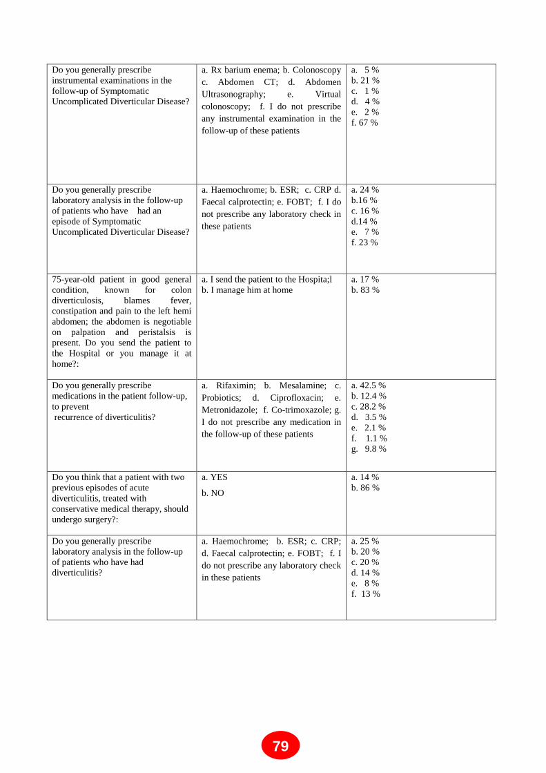
79
Do you generally prescribe
instrumental examinations in the
follow-up of Symptomatic
Uncomplicated Diverticular Disease?
a. Rx barium enema; b. Colonoscopy
c. Abdomen CT; d. Abdomen
Ultrasonography; e. Virtual
colonoscopy; f. I do not prescribe
any instrumental examination in the
follow-up of these patients
a. 5 %
b. 21 %
c. 1 %
d. 4 %
e. 2 %
f. 67 %
Do you generally prescribe
laboratory analysis in the follow-up
of patients who have had an
episode of Symptomatic
Uncomplicated Diverticular Disease?
a. Haemochrome; b. ESR; c. CRP d.
Faecal calprotectin; e. FOBT; f. I do
not prescribe any laboratory check in
these patients
a. 24 %
b.16 %
c. 16 %
d.14 %
e. 7 %
f. 23 %
75-year-old patient in good general
condition, known for colon
diverticulosis, blames fever,
constipation and pain to the left hemi
abdomen; the abdomen is negotiable
on palpation and peristalsis is
present. Do you send the patient to
the Hospital or you manage it at
home?:
a. I send the patient to the Hospita;l
b. I manage him at home
a. 17 %
b. 83 %
Do you generally prescribe
medications in the patient follow-up,
to prevent
recurrence of diverticulitis?
a. Rifaximin; b. Mesalamine; c.
Probiotics; d. Ciprofloxacin; e.
Metronidazole; f. Co-trimoxazole; g.
I do not prescribe any medication in
the follow-up of these patients
a. 42.5 %
b. 12.4 %
c. 28.2 %
d. 3.5 %
e. 2.1 %
f. 1.1 %
g. 9.8 %
Do you think that a patient with two
previous episodes of acute
diverticulitis, treated with
conservative medical therapy, should
undergo surgery?:
a. YES
b. NO
a. 14 %
b. 86 %
Do you generally prescribe
laboratory analysis in the follow-up
of patients who have had
diverticulitis?
a. Haemochrome; b. ESR; c. CRP;
d. Faecal calprotectin; e. FOBT; f. I
do not prescribe any laboratory check
in these patients
a. 25 %
b. 20 %
c. 20 %
d. 14 %
e. 8 %
f. 13 %

80
STATISTIC DANCE ON DIVERTICULAR DISEASE
Walter Elisei
Division of Gastroenterology, ASL RMH, Albano Laziale (Roma) – Italy
Diverticular disease of the colon is the fifth most important gastrointestinal disease in terms of
direct and indirect healthcare costs in western countries.
The main topics on treatment of diverticular disease are:
- Use of Rifaximine
- Use of Mesalazine
- Use of Antibiotics in Diverticulitis.
Rifaximin: This poorly absorbable antibiotic has been successfully used in recent years in the
treatment of SUDD, and also seems to be effective in maintaining SUDD remission. A recent meta-
analysis examined four prospective randomized trials (only one conducted in double-blind placebo-
controlled fashion) including 1660 patients. The pooled rate of difference for symptom relief was
29.0% in favor of rifaximin (rifaximin vs. control; 95% CI 24.5-33.6; P<0.0001) with a clinically
significant Number Needed to Treat (NNT=3)1. The use of rifaximine in prevention of diverticulitis,
data from three open randomized trials (comprising a total of 1492 patients) and four comparing
rifaximin plus glucomannan or fiber supplementation vs. glucomannan or fiber alone, reported that
rifaximin led to a slight benefit in preventing acute diverticulitis, but only the largest study showed
significant results. Cumulative data from placebo controlled and unblinded trials showed that the
rate of acute diverticulitis was significantly less frequent in patients treated with rifaximin plus fiber
supplementation than with fiber alone (11/970 (1.1%) vs. 20/690 (2.9%; P=0.012)2-5
. According to
these results, the number needed to be treated to prevent an attack of acute diverticulitis in 1 year
with the rifaximin plus fiber supplementation regimen reached is 57 (NNT: 57).
Mesalazine: 5-ASA is another option for the treatment of SUDD. Although limited by the open-
label design, the favorable effect of mesalazine on SUDD has been demonstrated by several open-
label studies6,7
. Three double-blind, placebo-controlled studies have also recently assessed the role
of mesalazine in treating those patients8-10
. Data on the role of mesalazine, five randomized open
trials (comprising more than 400 patients) in preventing acute diverticulitis, did not show any
significant effects, however, there were only seven episodes of acute diverticulitis per year (yearly
incidence rate of 2%). More recently, a double-blind, double dummy placebo-controlled trial
assessed the prevention of acute diverticulitis occurrence as secondary endpoint, found mesalazine
significantly better than placebo in preventing acute diverticulitis9.
Antibiotics in Diverticulitis: Conservative treatment of uncomplicated or mild diverticulitis
usually includes antibiotic therapy. It is, however, uncertain whether patients with acute
diverticulitis indeed benefit from antibiotics. In most guidelines issued by professional
organizations antibiotics are considered mandatory in the treatment of mild diverticulitis. The
treatment of acute diverticulitis by intravenous fluid replacement, limiting oral intake, and
broadspectrum antibiotics is common practice but is not supported by a strong evidence base.
People with mild symptoms and no evidence of generalised sepsis can be managed at home with
oral antibiotics. People with CT-proven mild uncomplicated diverticulitis may not benefit from
having intravenous antibiotics. This is in keeping with current thoughts on disease pathophysiology;
however, the evidence for this approach is based on one RCT. People with severe pain or signs of
compromise should be admitted for analgesia, bowel rest, intravenous fluid replacement, and
intravenous antibiotics.
81
Despite these results, further studies are needed as most published trials were uncontrolled, often
small, and lacking of strict enrolment criteria. For these reasons different pharmacological and non-
pharmacological approaches are currently approved in North America or Europe to prevent acute
attacks of diverticulitis or to treat patients with symptomatic disease.
Selecting patients according to the colonic characteristics may be an option to increase therapeutic
efficacy. To this end, an endoscopic classification of DD has been recently developed and
validated11
. This classification, called DICA is able to predict the outcome of the disease according
to the severity of the score. In other words, simple and/or asymptomatic diverticulosis does not
appear to need any maintenance treatment to prevent occurrence of complications, while a colon
with signs of recurrent inflammatory attack may be unresponsive to maintenance treatment to
prevent recurrence of complications.
In conclusion, the DICA Classification may be a useful guide for the treatment of patients.
References
1) Bianchi M, Festa V, Moretti A et al. Meta-analysis: long-term therapy with rifaximin in the
management of uncomplicated diverticular disease. Aliment Pharmacol Ther 2011;33:902-910 2)
Papi C, Ciaco A, Koch M, Capurso L. Efficacy of rifaximin in the treatment of symptomatic
diverticular disease of the colon. A multicentre double-blind placebo-controlled trial. Aliment
Pharmacol Ther 1995;9:33-39
3) Latella G, Pimpo MT, Sottili S et al. Rifaximin improves symptoms of acquired uncomplicated
diverticular disease of the colon. Int J Colorectal Dis 2003;18:55-62
4) Papi C Ciaco A, Koch M, Capurso L. Efficacy of rifaximin on symptoms of uncomplicated
diverticular disease of the colon: a pilot multicentre open trial. Diverticular Disease Study Group.
Ital J Gastroenterol 1992;24:452-456
5) Colecchia A, Vestito A, Pasqui F et al. Efficacy of long term cyclic administration of the poorly
absorbed antibiotic rifaximin in symptomatic, uncomplicated colonic diverticular disease. World J
Gastroenterol 2007;13:264-269
6) Trespi E, Colla C, Panizza P et al. Therapeutic and prophylactic role of mesalazine (5-ASA) in
symptomatic diverticular disease of the large intestine. 4 year follow-up results. Minerva
Gastroenterol Dietol 1999;45:245-252 (Article in Italian)
7) Tursi A, Brandimarte G, Giorgetti GM, Elisei W. Mesalazine and/or Lactobacillus casei in
maintaining long-term remission of symptomatic uncomplicated diverticular disease of the colon.
Hepatogastroenterology 2008;55:916-920
8) Kruis W, Meier E, Schumacher M, Mickisch O, Greinwald R, Mueller R; German SAG-20
Study Group. Randomised clinical trial: mesalazine (Salofalk granules) for uncomplicated
diverticular disease of the colon – a placebo-controlled study. Aliment Pharmacol Ther
2013;37:680-690
9) Tursi A, Brandimarte G, Elisei W et al. Randomised clinical trial: mesalazine and/or probiotics in
maintaining remission of symptomatic uncomplicated diverticular disease – a double-blind,
randomised, placebo-controlled study. Aliment Pharmacol Ther 2013;38:741-751
10) Smith J, Humes D, Garsed K et al. Mechanistic randomised control trial of mesalazine in
symptomatic diverticular disease. Gut 2012;61:A51-A52
11) Tursi A, Brandimarte G, Di Mario, et al. Development and validation of an endoscopic
classification of diverticular disease of the colon: The DICA classification. Dig Dis 2015;33:68-76

82
ITALIAN GUIDELINES ON DIVERTICULAR DISEASE OF THE COLON
Bruno Annibale1, Rosario Cuomo
2
1 Department of Medical and Surgical Sciences and Traslational Medicine, University
Sapienza, Roma 2 Department of Clinical Medicine and Surgery, Federico II University, Napoli
The primary aim was to provide clinical guidelines for appropriate definition, diagnosis and
management of diverticular disease (DD). The promoter of this initiative was the Italian Study
Group of Diverticular Disease (Gruppo Italiano Malattia Diverticolare - GRIMAD). GRIMAD
identified a Scientific Board of Experts), which defined the methodology and targets, and acted as
developer and reviewer.
Topics such as epidemiology, risk factors, diagnosis, medical and surgical treatment of DD in
patients with uncomplicated and complicated DD were reviewed by a scientific board of experts
who proposed 76 statements graded according to level of evidence and strength of
recommendation, and 55 were approved by an independent jury. Each topic was explored focusing
on the more relevant clinical questions. Comparison and discussion of expert opinions, pertinent
statements, and replies to specific questions, were presented and approved based on a systematic
literature search of the available evidence.
The methodology to process guidelines involved six steps:
1) The scientific board selected four main areas of interest in DD : (i) definition and epidemiology,
(ii) pathophysiology, (iii) diagnosis, and (iv) medical and surgical therapy. 2) For each topic, a
working party was created, which included a coordinator and at least two experts. The latter were
chosen on the basis of their recognized scientific expertise in DD . The working party selected,
together with the scientific board, a number of clinically relevant, clear, answerable questions,
focusing on current practice and areas of controversy. The questions were circulated among the
working parties to share relevance, improve clarity, and avoid duplication. A preliminary meeting
of the working parties was held in Bologna (June 2011) in order to share methods, aims, timelines,
and the entire guideline processing. 3) The working parties independently carried out a systematic
search for, and analysis of, the literature relevant to their topics by October 2012, using
Medline/PubMed and the Cochrane data base. Each recommendation was graded according to the
Oxford Centre for Evidence-Based Medicine, according to the level of evidence (EL) (1). 4) By
November 2012 the working parties issued initial statements and attributed to them a grade
(strength) of recommendation (RG), from A to D, consistent with the level of evidence. Each
coordinator drafted provisional statements that were circulated within his/her group. 5)
Subsequently, each four area coordinator evaluated the preliminary statements produced and the
related grades of evidence. A redrafted document containing the statements was then prepared and
submitted to the all participants for an online session for a first round of votes and comments, using
a simplified scale (agreement/disagreement); they voted using a modified Delphi procedure until a
minimum agreement level of at least 67% was achieved for each statement. Statements were then
submitted to the scientific board, which wrote an advanced version, and fed back the new
statements to the working parties. 6) On the 19th
and 20th
of February, 2013 a Consensus Meeting
was held in Bologna. The consensus group consisted of 33 participants, selected taking into account
diverse expertise in various aspects of DD, and geographical distribution. The consensus group was
led by a non-voting chairman and the four non-voting members of the scientific board , and
included experts of working parties and multi-disciplinary professionals/experts such as
gastroenterologists, GI endoscopists, surgeons, radiologists, pathologists, and general practitioners.
Overall, 76 statements were submitted to the global consensus group. Following a plenary
discussion held before voting, twenty-one statements were deleted, and 45 were partially rephrased.
The final 55 statements were then submitted to the global consensus group for anonymous keypad
83
voting (Delphi process) without any explanation or justification. The Delphi process brought to a
change of view from a position previously held, avoiding any uneasiness among participants or
influence on individual votes. The agreement/disagreement level was scored on a six-point Likert
scale as follows: A+: strongly agree; A: agree with minor reservations; A−: agree with major
reservations; D−: disagree with major reservations; D: disagree with minor reservations; D+:
strongly disagree. Level of agreement was expressed as percentage of each point of the scale.
Immediate feedback was given to participants on a screen, who were prompted by the non-voting
chairman and the non-voting members of the scientific board to discuss the statements, and suggest
changes in case of controversy (<67% agreement). The entire work and discussions were tape-
recorded.
The format of the following recommendations comprises the question, statement, its level of
evidence and strength of recommendation, and the percent agreement of the global consensus group
with the final version (2).
The document comprises statements that are accompanied by comments made by each working
party and reviewed by the scientific board taking into account relevant observations and suggestions
made during the plenary discussion. In some areas the evidence level is low, reflecting the lack of
randomized trials and good quality studies. For some topics only the expert opinion was
considered, where appropriate.
These guidelines represent a consensus of best practice based on the available evidence at the time
they were issued. They may not apply to all situations and should be interpreted in the light of
specific clinical situations and resource availability. Further controlled clinical studies may be
needed to clarify some aspects of these statements, and revision may be necessary as new data
become available. These guidelines are intended to be an educational tool to provide information
that may assist gastroenterologist and surgeons in providing care to patients. They are not rules
and should not be constructed as establishing a legal standard of care or as encouraging, advocating,
requiring, or discouraging any particular treatment.
REFERENCES
1) Guyatt GH, Oxman AD, Schünemann HJ, Tugwell P, Knottnerus A. GRADE guidelines:a new
series of articles in the Journal of Clinical Epidemiology. J Clin Epidemiol 2011;64:380-2
2) Cuomo R, Barbara G, Pace F.Italian consensus conference for colonic diverticulosis
and diverticular disease. United European Gastroenterol J 2014;2:413-42
84
SCANDINAVIAN GUIDELINES ON DIVERTICULAR DISEASE
Johannes Kurt Schultz
Dept of Gastrointestinal Surgery, Akershus University Hospital, PB 1000 1478 Lørenskog, Norway
In Scandinavia official guidelines for the management of diverticular disease exist only in Denmark
[1]. However, my impression from several local guidelines, Scandinavian publications and surgical
meetings is that the treatment policy is quite similar in the Scandinavian countries. In my talk I will
concentrate on imaging, treatment options and follow up of acute diverticulitis (complicated and
uncomplicated) and I will discuss indications for elective sigmoid resection in patients with
diverticular disease.
Imaging
In most Scandinavian hospitals computed tomography (CT) is the investigation of choice to
diagnose acute diverticulitis. This is also what is recommended in the Danish guidelines [1]. Also in
several Scandinavian publications CT is used as a standard investigation [2-5]. There is no tradition
in Scandinavia for the use of ultrasound as diagnostic tool for these patients. The main reason for
this seems to be that ultrasound is much more operator dependent than CT.
Acute uncomplicated diverticulitis
Antibiotics
After that results from the AVOD trial [2] have been published, many hospitals in Sweden and
Norway have implemented new treatment policies for acute uncomplicated diverticulitis. In many
hospitals antibiotics are now reserved for high risk patients and patients with complicated
diverticulitis. This is also the recommendation in the Danish guidelines [1].
Follow up after uncomplicated diverticulits
Many hospitals in Scandinavia still practice a routine colon examination after acute uncomplicated
diverticulitis, either as colonoscopy or as CT colonography. This routine has been supported by the
findings of a large population based Swedish study that showed an increased risk of colorectal
cancer within the first 12 month after primary diagnosis of diverticular disease [6]. The Danish
guidelines recommend a colon investigation approximately 6 weeks after an acute attack of
uncomplicated diverticulitis [1]. The recent review articles to this topic [7, 8] have encouraged a
few hospitals to abandoned this practice in patients with CT verified uncomplicated diverticulitis.
Routine colon examination is however standard for all patients with complicated diverticulits.
Perforated diverticulitis with or without abscess formation
Perforated diverticulitis is often diagnosed by extra luminal air or fluid on an abdominal CT which
has become a standard tool in Scandinavia when evaluating patients with an acute abdomen. The
Danish guidelines recommend diagnostic laparoscopy in case of radiological findings suggesting
free perforation. Some hospitals do however reserve operative treatment to patients with clinical
findings of diffuse peritonitis and hemodynamically unstable patients. Many surgeons in
Scandinavia prefer to treat stable patients without clinical sings of peritonitis with antibiotics only.
This practice is supported by recently published cohort studies [9, 10]. Abscesses over the size of 2
cm are drained if possible. For abscesses larger than 5 cm operative drainage is considered if
percutaneous drainage is not feasible.
Operative treatment for patients with perforated diverticulitis
For purulent peritonitis there is currently no consensus among Scandinavian surgeons on which
procedure to perform. Many colleagues agree that primary resection and anastomosis is safe for
many patients but the general condition of the patient should be considered. Two Scandinavian
trials have investigated laparoscopic lavage as an option. The results were somewhat contrary [5,

85
11]. The Danish guidelines were written before publication of the results form the randomized trials
and recommend laparoscopic lavage for patients with purulent diverticulits. In case of feculent
peritonitis (Hinchey grade 4) primary resection with terminal colostomy (Hartmann‘s procedure) is
the treatment of choice for most surgeons in Scandinavia [5, 11]. The Danish guidelines
recommend resection in patients with feculent peritonitis, they make however no recommendation
as to whether or not a primary anastomosis should be performed [1].
Elective sigmoidectomy
After the publication by Haglund in 1979 most of the Scandinavian surgeons have been quite
reluctant to the international trend of extended indications for elective sigmoid resection in patients
with diverticular disease [12]. Haglund showed that elective surgery could not prevent serious
attacks of diverticulitis, as complications are most likely to occur at the first attack. Most hospitals
have reserved elective sigmoid resection for patients with frequent recurrences of diverticulitis and
patients with complications such as stenosis, fistula or persisting abscesses which is also the
recommendation in the Danish guidelines[1]. This practice has now been supported by the latest
international publications and guidelines [13-15].
Symptomatic uncomplicated diverticular disease
Symptomatic uncomplicated diverticular disease (SUDD) is addressed in the Danish guidelines
which make some suggestions for the treatment of this condition[1]. My impression from meetings
and conversations with colleagues is however, that most surgeons consider this condition as a
coincidence of diverticulosis and irritable bowel syndrome. Traditionally Scandinavian surgeons are
reluctant to operate on patients without a history of acute diverticulitis, especially if there is no
morphologic explanation for their symptoms other than the mere existence of diverticulosis.
References
1. Andersen JC, Bundgaard L, Elbrond H, Laurberg S, Walker LR, Stovring J. Danish national
guidelines for treatment of diverticular disease. Dan Med J 2012; 59: C4453.
2. Chabok A, Pahlman L, Hjern F, Haapaniemi S, Smedh K. Randomized clinical trial of
antibiotics in acute uncomplicated diverticulitis. The British journal of surgery 2012; 99:
532-9.
3. Thorisson A, Smedh K, Torkzad MR, Pahlman L, Chabok A. CT imaging for prediction of
complications and recurrence in acute uncomplicated diverticulitis. Int J Colorectal Dis 2015
4. Liljegren G, Chabok A, Wickbom M, Smedh K, Nilsson K. Acute colonic diverticulitis: a
systematic review of diagnostic accuracy. Colorectal Dis 2007; 9: 480-8.
5. Schultz JK, Yaqub S, Wallon C, Blecic L, Forsmo HM, Folkesson J, et al. Laparoscopic
Lavage vs Primary Resection for Acute Perforated Diverticulitis: The SCANDIV
Randomized Clinical Trial. JAMA 2015; 314: 1364-75.
6. Granlund J, Svensson T, Granath F, Hjern F, Ekbom A, Blomqvist P, et al. Diverticular
disease and the risk of colon cancer - a population-based case-control study. Aliment
Pharmacol Ther 2011; 34: 675-81.
7. Sharma PV, Eglinton T, Hider P, Frizelle F. Systematic review and meta-analysis of the role
of routine colonic evaluation after radiologically confirmed acute diverticulitis. Ann Surg
2014; 259: 263-72.
8. de Vries HS, Boerma D, Timmer R, van Ramshorst B, Dieleman LA, van Westreenen HL.
Routine colonoscopy is not required in uncomplicated diverticulitis: a systematic review.
Surg Endosc 2014; 28: 2039-47.
9. Costi R, Cauchy F, Le Bian A, Honart JF, Creuze N, Smadja C. Challenging a classic myth:
pneumoperitoneum associated with acute diverticulitis is not an indication for open or
laparoscopic emergency surgery in hemodynamically stable patients. A 10-year experience
with a nonoperative treatment. Surg Endosc 2012; 26: 2061-71.

86
10. Sallinen VJ, Mentula PJ, Leppaniemi AK. Nonoperative management of perforated
diverticulitis with extraluminal air is safe and effective in selected patients. Dis Colon
Rectum 2014; 57: 875-81.
11. Angenete E, Thornell A, Burcharth J, Pommergaard HC, Skullman S, Bisgaard T, et al.
Laparoscopic Lavage Is Feasible and Safe for the Treatment of Perforated Diverticulitis
With Purulent Peritonitis: The First Results From the Randomized Controlled Trial
DILALA. Ann Surg 2016; 263: 117-22.
12. Haglund U, Hellberg R, Johnsen C, Hulten L. Complicated diverticular disease of the
sigmoid colon. An analysis of short and long term outcome in 392 patients. Ann Chir
Gynaecol 1979; 68: 41-6.
13. Regenbogen SE, Hardiman KM, Hendren S, Morris AM. Surgery for diverticulitis in the
21st century: a systematic review. JAMA Surg 2014; 149: 292-303.
14. Feingold D, Steele SR, Lee S, Kaiser A, Boushey R, Buie WD, et al. Practice parameters for
the treatment of sigmoid diverticulitis. Dis Colon Rectum 2014; 57: 284-94.
15. Schreyer AG, Layer G. S2k Guidlines for Diverticular Disease and Diverticulitis: Diagnosis,
Classification, and Therapy for the Radiologist. Rofo 2015; 187: 676-84.

87
AUSTRALIAN GUIDELINES ON DIVERTICULAR DISEASE
Marjorie M Walker
School of Medicine and Public Health, University of Newcastle, NSW 2308, Australia
In Australia (2008) the proportion of all deaths from digestive disease which did not include cancer,
was 3.4%, and diverticular disease (DD) accounted 0.2% of all deaths (female predominant) (1),
and it is one of the five most costly gastrointestinal disorders affecting the US population. (2)
In a survey conducted in Brisbane, (3), 48% of all comers had diverticular disease found at post
mortem.
For this very common and costly condition, which guidelines are available specifically in Australia?
For physicians and GPs, guidelines most used are: Diverticular Disease and Diverticulitis [revised
2011 Feb]. In: eTG complete [Internet]. Melbourne: Therapeutic Guidelines Limited; 2015 Jul. (4)
For surgeons, the paper by Ooi et al (5) is commonly used which quotes Guidelines issued by the
American Society of Colon and Rectal Surgeons
Consumer information on diverticular disease is available from the Gastroenterological Society of
Australia at their website (6) and dietary advice is available from the Dieticians Association of
Australia (7).
There is a need for a robust epidemiological study in Australia and up to date medical/surgical
guidelines that take the most recent global literature into account.
References
1) http://www.abs.gov.au/
2) Peery AF, Dellon AS, Lund J, et al. Burden of gastrointestinal disease in the United States: 2012
Update. Gastroenterology 2012;143: 1179–1187
3) Hughes LE. Postmortem survey of diverticular disease of the colon. Part I. Diverticulosis and
diverticulitis. Gut 1969;10:336-44
4) Diverticular disease and Diverticulitis [revised 2011 Feb]. In: eTG complete [Internet].
Melbourne: Therapeutic Guidelines Limited; 2015 Jul.
5) Ooi K, Wong SW. Management of symptomatic colonic diverticular disease. Med J Aust
2009;190:37-40 (study from Sydney)
6. http://www.gesa.org.au/
7. http://daa.asn.au/

88
ORAL PRESENTATIONS
89
SYMPTOMATIC UNCOMPLICATED DIVERTICULAR DISEASE:
EVALUATION OF MESALAMINE AND/OR PROBIOTICS TREATMENT
IN FECAL CALPROTECTIN
Mauro Bafutto1,2
, Michelle Bafutto Gomes Costa2, Eduardo Henrique Ferreira Bafutto
2,
Alexandre Augusto Ferreira Bafutto2, Jose Roberto de Almeida
3, Enio Chaves de Oliveira
4
1Departamento de Clínica Médica, Unidade de Gastroenterologia da Faculdade de Medicina da
Universidade Federal de Goiás, Goiânia, Goiás, Brasil; 2Instituto Bafutto de Pesquisa, Goiânia,
Goiás, Brasil; 3Departamento de Clínica Médica, da Faculdade de Medicina da Universidade
Federal de Recife, Recife, Pernambuco, Brasil;4Departamento de Cirurgia Geral da Faculdade de
Medicina da Universidade Federal de Goiás, Goiânia, Goiás, Brasil.
Keywords – Diverticular disease, mesalamine, probiotics
Background - Recent observations suggest that symptomatic uncomplicated diverticular disease
(SUDD) is related to changes in colonc microflora and mild inflammation. Fecal calprotectin (FC)
is a protein related to intestinal inflammation. The use of mesalamine can improve symptoms and
reduce the FC levels to normal levels in many patients but in some cases, these levels remain high.
The persistence of FC in altered levels is related to complications of the diverticular disease. The
management of these patients is still unknown.
Aims - To evaluate the effects of mesalamine and / or probiotics in FC levels in patients with
SUDD. Methods -163 patients with SUDD and FC >150microg/gr were selected after colonoscopy
and CT . Were excluded patients younger than 18 years, with cancer, inflammatory bowel disease,
gastroenteritis, previous intestinal surgery, and use of anti-inflammatory drugs. Patients received
mesalazine 800mg b.i.d. for 60 days and made new dosage FC. 48 patients presented CF >150
microgr g and were divided into 3 groups: 1) MP group: 16 patients using mesalazine 800mg bid
and a mix of probiotics (L. acidophilus, L.casei, L. lactis, B. lactis, B. bifidum) bid for 8 days; 2)
Group P: 16 patients using mix of probiotics (L. acidophilus, L.casei, L. lactis, B. lactis, B. bifidum)
bid for eight days; and 3) M3 group: 16 patients with mesalazine 800 mg tid for 8 days. After
treatment, all of them made new dosage FC.
Results - After 60 days of treatment with mesalazine 800 mg bid, 115 patients (71%) presented FC
<150microgr/g and 48 (29%) FC >150microgr / g. There was a statistically significant decrease
(51.8%) in FC levels after combined treatment: MP group (309.70 ± 121.80 x150.80 ± 104.23, p
<0.002). Statistically significant reduction was also seen in FC levels (42.2%) after treatment with
higher doses of mesalamine : M3 Group (x 455.40±264.17 261.50±209.16, p <0.01 ). No significant
reduction in FC levels were seen in Group P (537± 360, 82 x 284 196.69±50, p=0.08). There was a
greater reduction in levels of CF MP Group (51.8%) than in M3 (42.2%), but there was no
statistically significant difference.
Conclusion - FC levels reduced to baseline in most patients after treatment with mesalazine 1.6g.
The combined use of mesalazine and probiotics or increasing the dose of mesalazine can contribute
to reducing FC levels in SUDD.

90
ROLE OF CT COLONOGRAPHY IN EARLY FOLLOW-UP OF ACUTE
COMPLICATED DIVERTICULITIS
Bassi M, Scagliarini L, Tilli M, Anania G, Agresta F, Rizzati R, Benea G.
Department of Radiology, University of Ferrara, Italy
Keywords: Diverticulitis, CECT, CTC, Hinchey , Outcome.
Introduction: Patient evaluation after an episode of acute complicated diverticulitis (ACD) is
mandatory, and contrast-enhancement CT (CECT) still remains the ―gold-standard‖ [1]. However
the diagnostic follow-up of those patients is still controversial [2, 3] especially because the
indications of a surgical treatment are changing [4, 5], so the re-evaluation after an episode of ACD
represent a possible new indication for other diagnostic techniques [6, 7]. Considering diverticular
disease, CT colonography (CTC) today represents a robust and safe technique that allows a
complete low dose evaluation of the extention of the disease with a precise definition of lenght/wall
thickness of the interested segment, providing a detailed map of colonic/extracolonic findings [8, 9].
Considering ACD early follow-up, we evaluate value of CTC in order to set a correct treatment
strategy.
Material and Methods: From April 2009 to August 2014, 66 patients underwent unenhanced low-
dose CTC follow-up (28 males, 38 females, aged 38-91 yo) 6-8 weeks after ACD, conservatively
treated.
All patients previously performed CECT and were classified using modified Hinchey classification.
CTC exam evaluated colonic/extracolonic findings, assessing short-term course and staging of
diverticular disease. No CTC complications occured.
Results: CTC quality was good in 59/66 patients (89%); in 7 cases we obtain suboptimal distension
due to diverticular disease or colonic stenosis.
At baseline CECT we found: Hinchey I (n=30, 46%), II (n=22, 33%); III (n=14, 21% ); IV (n=0,
0%) stages.
CTC findings follow-up allowed to keep conservative treatment in all cases of Hinchey I (100%), in
8/22 cases of Hinchey II (36%) and in 8/14 cases of Hinchey III (57%).
Twenty patients not understaged (14/22 Hinchey II, 64%, and 6/14 Hinchey III, 43%) underwent
laparoscopic/open surgery. CTC also revealed 4 unknown polyps >6 mm and 28 extracolonic
findings, 4 with major clinical relevance that changed treatment strategy.
Conclusion: CTC is a safe and accurate method to evaluate severity of ACD, especially in short-
term follow-up, allowing an overview of colonic/extracolonic findings and guiding correct
therapeutic planning.
Bibliography: 1.Kaiser AM, Jiang JK, Lake JP, et al. The management of complicated diverticulitis and the role of
computed tomography. Am J Roentgenol 2005;100: 910-7
2. Sai VF, Velayos F, Neuhaus J, et al. Colonoscopy after CT diagnosis of diverticulitis to exclude
colon cancer; a systematic literature review. Radiology 263: 383–90
3. Society for Surgery of the Alimentary Tract (2007). SSAT patient care guidelines: surgical
treatment of diverticulitis.
4. Isacson D, Thorisson A, Andreasson K, et al. Outpatient, non-antibiotic management in acute
uncomplicated diverticulitis: a prospective study. Int J Colorectal Dis 2015;30: 1229-34
5. Hupfeld L, Burcharth J, Pommergaard HC, et al. The Best Choice of Treatment for Acute
Colonic Diverticulitis with Purulent Peritonitis Is Uncertain. BioMed Research International
Volume 2014.
6. Sanford M, Pickhardt PJ. Diagnostic performance of primary 3-dimensional computed
tomography colonography in the setting of colonic diverticular disease. Clin Gastroenterol
Hepatol;4: 1039–47

91
7. Hjern F, Jonas E, Holmström B, et al. CT colonography versus colonoscopy in the follow-up of
patients after diverticulitis. A prospective, comparative study. Clin Radiol;62: 645–50
8. Laghi A. Computed tomography colonography in 2014: an update on technique and indications.
World J Gastroenterol 2014;7;20: 16858-67
9. Flor N, Rigamonti P, Pisani, et al. Diverticular disease severity score based on CT colonography.
Eur Radiol 2013;23: 2723-9
92
EFFECT OF HIGH PERIOPERATIVE OXYGEN FRACTION ON
SURGICAL SITE INFECTION FOLLOWING SURGERY FOR ACUTE
SIGMOID DIVERTICULITIS. A PROSPECTIVE, RANDOMIZED, DOUBLE-
BLIND, CONTROLLED, MONOCENTRIC TRIAL
Sara Colozzi, Mario Schietroma, Beatrice Pessia, Giovanni Cianca, Marco Clementi,
Francesco Carlei, Gianfranco Amicucci.
Department of Surgery, University of L‟Aquila, L‟Aquila, Italy
Key words: controlled trial, hyperoxia, surgical site infection, outcomes.
Background: The clinical role of hyperoxia for preventing surgical site infection (SSI) remains
uncertain because randomized controlled trials on this topic have reported disparate results. One of
the principal reasons for such mixed results may be that prior trials have entered a heterogeneous
population of patients and procedures. The aim of our study was to assess the influence of
hyperoxigenation on SSI by using the most homogeneous study population.
Materials and methods: From January 2009 to December 2015, we studied, in a prospective
randomized study, 85 patients who underwent open intraperitoneal anastomosis for acute sigmoid
diverticulitis. Patients were assigned randomly to an oxygen/air mixture with a fraction of inspired
oxygen (FiO2) of 30% (n=43) or 80% (n=42). Administration was commenced after induction of
anesthesia and maintained for 6 hours after surgery.
Results: The overall wound infection rate was 24.7% (21 out of 85): 14 patients (32.5%) had a
wound infection in the 30% FiO2 group and 7 (16.6%) in the 80% FiO2 group (p< 0.05). the risk of
SSI was 43% lower in the 80% FiO2 group (RR, 0.68; 95% confidence interval, 0.35-0.88) vs 30%
FiO2.
Conclusions: Supplemental 80% FiO2 during and for 6 hours after open surgery for acute sigmoid
diverticulitis, reducing post-operative SSI, should be considered part of ongoing quality
improvement activities releted to surgical care, with few risk to the patients and little associates
cost.
ASSESSMENT OF FECAL MICROBIOTA AND FECAL METABOLOME IN
SYMPTOMATIC UNCOMPLICATED DIVERTICULAR DISEASE OF THE
COLON
93
Paola Mastromarino1, Daniela Capobianco
1, Walter Elisei
2, Alfredo Miccheli
3, Giorgio
Capuani3, Marcello Picchio
4, GianMarco Giorgetti
5, Federica Fabiocchi
5, Michela Di Fonzo
6,
Giovanni Brandimarte6, Antonio Tursi
7
1Department of Public Health, Section of Microbiology, “Sapienza” University, Rome – Italy;
2Division of Gastroenterology, ASL RMH, Albano Laziale (Rome) – Italy;
3 Department of
Chemistry, “Sapienza” University, Rome – Italy; 4Division of Surgery, “P. Colombo” Hospital,
ASL RMH, Velletri (Rome) – Italy; 5Digestive Endoscopy and Nutrition Unit, “S. Eugenio”
Hospital, Rome – Italy; 6Division of Gastroenterology, “Cristo Re” Hospital, Rome – Italy;
7Gastroenterology Service, ASL BAT, Andria (BT) - Italy
Key words: fecal metabolome; fecal microbiota; symptomatic uncomplicated diverticular disease.
Background: Pathogenesis of Symptomatic Uncomplicated Diverticular Disease (SUDD) is not
completely understood. Imbalance of colonic microbiota is considered a milestone in occurrence of
symptoms (1), but whether intestinal microbiota is really altered in those patients is unknown.
Metabolic profiling is a powerful exploratory tool for understanding interactions between nutrients,
the intestinal metabolism and the microbiota composition in health and disease, and metabolomics
technologies have been applied for the screening of different pathological conditions that are linked
with a metabolic imbalance (2).
Aims: We performed a prospective study assessing fecal microbiota and metabolome in
symptomatic uncomplicated diverticular disease (SUDD).
Methods: Stool samples from 52 consecutive female patients (17 with SUDD, 16 with
asymptomatic diverticulosis (AD), and 19 healthy), born and living in the same geographic area,
were analysed. Real-time PCR was used to quantify targeted microorganisms. High-resolution
proton nuclear magnetic resonance spectroscopy in combination with Multivariate Analysisl were
assessed in determining fecal metabolome.
Results: The total amount of bacteria in fecal samples of the three groups was not significantly
different. In AD group a reduction in the percentage of bifidobacteria and enterobacteria and an
increase of Clostridia and lactobacilli compared to total bacteria was observed. In both AD and
SUDD groups an increase of Akkermansia muciniphila was also observed in comparison to healthy
group. These differences were however not significant. NMR-based metabolomics of fecal waters
did not show any significant change between healthy and SUDD subjects. However, significant
differences in N-Acetyl-compounds were found between AD and SUDD patients. Partial Least
Square Analysis showed that SUDD patients, with higher N-Acetyl-compound levels as compared
to AD patients, showed higher levels in bifidobacteria and enterobacteria, and lower levels in
clostridia and lactobacillus spp.
Conclusions: SUDD does not show any significant quantitative and qualitative alteration of the
analysed fecal microbiota. However, increasing expression of some metabolites as expression of
different SUDD metabolic activity was found.
References
1) Strate LL, Modi R, Cohen E, Spiegel BM. Diverticular disease as a chronic illness: evolving
epidemiologic and clinical insights. Am J Gastroenterol 2012;107: 1486-93
2) De Preter V, Verbeke K. Metabolomics as a diagnostic tool in gastroenterology. World J
Gastrointest Pharmacol Ther 2013; 4: 97-107
THE EFFECT OF A LIQUID MULTI-STRAIN PROBIOTIC IN
SYMPTOMATIC DIVERTICULAR DISEASE – A RANDOMISED DOUBLE-
BLIND CONTROLLED-TRIAL

94
C.L. Kvasnovsky, S. Papagrigoriadis, N. Donaldson, I. Bjarnason
Departments of Surgery & Gastroenterology, King‟s College Hospital, London, United Kingdom
Background
Diverticular disease is a significant burden on healthcare systems without standardized treatment.
While some patients may benefit from elective surgery, other patients have less acute complaints
and/or are poor surgical candidates. Probiotics are a plausible treatment option, as they may alter
intestinal bacterial composition and have other local metabolic effects. We hypothesized that a
probiotic would reduce abdominal pain in patients with symptomatic diverticular disease.
Methods
We conducted a single-center, randomized, double-blind, placebo-controlled trial of probiotic
treatment in adult patients with symptomatic diverticular disease. We randomly assigned 143
patients to receive 1 mL/kg/day of probiotic liquid (72 patients) or placebo (71 patients) daily for 3
months. The primary endpoint was a decrease in abdominal pain. Secondary endpoints included a
decrease in the frequency of 9 abdominal symptoms.
Results
143 patients were enrolled and 120 patients completed the trial. Overall, patients initiated treatment
with a mean pain score 8.5 +/- 7.4, with a reduction to 6.0 +/- 6.6 following treatment. There was no
difference between patients on probiotic and on placebo (P=0.12).
The odds of experiencing constipation at least a few times per week was significantly less for
patients on Symprove™ (OR=0.25 [95% CI 0.08-0.79], P=0.02). The odds of reporting frequent
diarrhea were also significantly less for patients on Symprove™ (OR 0.33 [95% CI 0.13-0.84],
P=0.02).
Conclusions Multi-strain liquid probiotic did not improve abdominal pain scores, but did significantly decrease
the frequency of constipation and diarrhea. Patients with altered bowel habits and diverticular
disease may benefit from probiotic treatment.

95
POSTERS
ASSOCIATION BETWEEN COLONIC DIVERTICULA AND
COLORECTAL POLYPS AND CANCER

96
Fabiana Ciccone1, Giorgio Valerii1, Dolores Gabrieli1, Sonia Calcina1, Annalisa
Capannolo1, Angelo Viscido1, Dina Melideo1, Stefano Necozione2, Giuseppe Frieri1,
Giovanni Latella1.
1Gastroenterology Unit, Department of Life, Health and Environmental Sciences, University of
L‟Aquila; 2Epidemiology Unit, Department of Life, Health and Environmental Sciences, University
of L‟Aquila - Italy
Keywords: Diverticula, Colorectal cancer, Polyps, Adenoma, Dysplasia.
Introduction: colonic diverticula and neoplastic colorectal lesions are found in similar ranges of
age and populations, but it is unclear whether there is a shared pathway in their development. Their
frequency increases with age and seems to be associated with a lack of dietary fibres, increased
dietary saturated fats, obesity and a slow colonic transit time. The association of diverticula and
colorectal polyps and cancer has been previously evaluated, reporting conflicting results. Despite
common epidemiologic predisposing factors, the association between colonic diverticula and
colorectal polyps and cancer remains unclear and needs to be better defined, as it could have
important implications for the screening of colorectal cancer.
The aim of this study is to evaluate the association between colonic diverticula and colorectal
polyps and cancer.
Materials and methods: A one-year prospective study including all consecutive patients
undergoing to routine colonoscopy at our GI Unit from September 2014 to September 2015. The
presence and location of diverticula, polyps, and cancers was recorded using colonoscopy reports.
Types of colorectal neoplastic lesions were defined by histopathological examination. Polyps were
classified into adenoma (with low or high dysplasia), hyperplastic or inflammatory polyps. A
multiple logistic regression analysis was done to evaluate the association between diverticular
disease and colonic lesions.
Results: During the study period 1490 colonoscopies were performed. 447 patients were included
(245 M, 202 F, mean age 66 years): 166 (37.1%) patients presented only diverticula, 155 (34.7%)
patients presented only polyps, and 126 (28.2%) patients presented both the diseases associated. 14
patients presented colorectal cancer, 5 of which had also diverticula.
There was no significant association between colonic diverticula and adenoma, as well as between
diverticula and colorectal cancer. On the other hand, colorectal inflammatory polyps showed a
significant association with colonic diverticula.
Conclusions: The results of the study show no association between colonic diverticula and
colorectal adenoma nor between diverticula and cancer. Therefore, patients with colonic diverticula
do not require a different follow-up for the prevention of colorectal cancer than the general
population. Inflammatory polyps are frequently associated to colonic diverticular disease probably
due to the same pathogenic factors.
Bibliography
1. Anne F. Peery , Christopher F et al. Colonic Diverticula Are Not Associated With an
Increased Risk of Colorectal Adenomas. Am J Gastroenterol advance online publication, 3
November 2015.
2. Singh S , Singh PP , Murad MH et al. Prevalence, risk factors, and outcomes of interval
colorectal cancers: a systematic review and meta-analysis . Am J Gastroenterol 2014.
3. Morini S , Hassan C , Zullo A et al. Diverticular disease as a risk factor for sigmoid colon
adenomas. Dig Liver Dis 2002.

97
4. Morini S , de Angelis P , Manurita L et al. Association of colonic diverticula with adenomas
and carcinomas. A colonoscopic experience . Dis Colon Rectum 1988.
COMPLICATED DIVERTICULAR DISEASE: OUR EXPERIENCE AND
CURRENT MANAGEMENT

98
Sara Colozzi, Mario Schietroma, Giovanni Cianca, Marco Clementi, Francesco Carlei,
Gianfranco Amicucci.
Department of Surgery, University of L‟Aquila, Italy
Key words: complicated diverticular disease, Hartmann procedure, outcomes, surgery.
Background: On Literature there is no agreement about treatment of complicated diverticular
disease. We analyzed our experience and we compared our results with data from Literature.
Materials and methods: From January 2009 to December 2015 we observed 72 patients (40 male,
32 female) with complicated diverticular disease. Mean age was 72 years (range 51-86). All
patients presented bowel perforation. We evaluated patients using Hinchey criteria: 15 patients
stage 0, 8 patients stage I, 20 patients stage II, 17 patients stage III and 12 patients stage IV. We
performed at stage 0 resection and anastomosis; at stage I resection and anastomosis in 6 cases and
resection and anastomosis with ―protection‖ ileostomy in 2 cases; at stage II resection and
anastomosis in 15 cases, resection and anastomosis with ―protection‖ ileostomy in 4 cases and
Hartmann intervention in one case; at stage III resection and anastomosis in 3 cases, resection and
anastomosis with ―protection‖ ileostomy in 6 cases and Hartmann intervention in 8 cases. At stage
IV we performed in all cases Hartmann intervention.
Results: 8/72 patients have some complications. After Hartmann intervention we observed
complications on 5/21 patients; 3 of them were surgical complications: hernia of stoma in 2 cases,
fistula of stoma in one case. Medical complications were respiratory distress and renal failure.
After resection and primary anastomosis we observed complication on 3/51 patients (anastomotic
dehiscence). Review of Literature didn‘t shown gold standard on treatment of complicated
diverticular disease. In our experience we observed more complications in patients treated with
Hartmann intervention.
Conclusion: Correct staging of complicated diverticular disease can determine correct treatment.
PREVALENCE OF COLONIC DIVERTICULOSIS IN PATIENTS
AFFECTED BY ULCERATIVE COLITIS: A PROSPECTIVE STUDY

99
Claudio Cassieri1, Roberta Pica
1, Eleonora Veronica Avallone
1, Maddalena Zippi
1, Pietro
Crispino1, Daniela De Nitto
1, Paolo Paoluzi
1, Giovanni Brandimarte
2, Piera Giuseppina
Lecca2, Luigi Di Cesare
2, Walter Elisei
3, Marcello Picchio
4, Antonio Tursi
5.
1 Department of Internal Medicine and Medical Specialties, "Sapienza" University, Rome – Italy;
2
Division of Internal Medicine and Gastroenterology, “Cristo Re” Hospital, Rome – Italy; 3
Division
of Gastroenterology, ASL Roma H, Albano Laziale (Rome) – Italy; 4
Division of Surgery, “P.
Colombo” Hospital, Velletri (Rome) – Italy; 5
Gastroenterology Service, ASL BAT, Andria (BT) –
Italy
Key words: colonoscopy, diverticulosis, epidemiology, ulcerative colitis.
Background: Diverticulosis of the colon is an acquired condition that results from herniation of the
mucosa and submucosa through defects in the muscular layer [1]. The true prevalence of colonic
diverticulosis is difficult to measure because most individuals are asymptomatic. In particularly, in
literature, there are few study about the prevalence of colonic diverticulosis in patients affected by
ulcerative colitis (UC)[2-4].
Aims: To investigate the prevalence of colonic diverticulosis in UC in an adult population referred
to a single centre.
Methods: Computerized data of consecutive patients, referred to our Institution to undergo a
colonoscopy for colorectal cancer screening and/or for UC assessment, between January 1, 2009
and December 31, 2009, were retrospectively studied.
Results: Six hundred and five consecutive patients were retrospectively studied. Of these patients,
438 (72.4%) underwent colonoscopy for colorectal cancer screening (Group A) and 167 (27.6%) for
UC assessment (Group B). In group A 224 patients (51.1%) were male (average age of 62.7 ± 14.2
SD years, range 35-86 years), in group B 102 (61.1%) were male (average age of 57.6 ± 12.1 SD
years, range 25-84 years). Prevalence of colonic diverticulosis was significantly higher in group A
(122 patients, 27.8%) than group B (18 patients, 10.8%) (p<0.0001, Fisher's exact test). Female
gender in patients with colonic diverticulosis was significantly higher in group A than group B (68
patients, 55.7% versus 4 patients, 22.2%, p=0.0106, Fisher's exact test). In group A sigma and left
colon was involved in 119 (97.6%) patients versus 12 (66.7%) of Group B, with a statistically
significant difference (p=0.0001, Fisher's exact test), in Group B the right colon was involved in 4
(22.2%) patients versus 1 (0.8%) of Group A, a statistically significant difference (p=0.0009,
Fisher's exact test).
Conclusions: Results of our study demonstrated that prevalence of colonic diverticulosis was
significantly lower in patients with UC than in adult population, emphasizing the relevance of the
coexistence of UC and colonic diverticulosis because could make very difficult the clinical
management of these patients.
References
1) Tursi A, Papa A, Danese S. Review article: the pathophysiology and medical management of
diverticulosis and diverticular disease of the colon. Aliment Pharmacol Ther 2015;42: 664-84
2) Lahat A, Avidan B, Bar-Meir S, Chowers Y. Long-standing colonic inflammation is associated
with a low prevalence of diverticuli in inflammatory bowel disease patients. Inflamm Bowel Dis
2007;13: 733-6
3) Rispo A, Pasquale L, Cozzolino A et al. Lower prevalence of diverticulosis in patients with
ulcerative colitis. Dis Colon Rectum 2007;50:1164-8
100
4) Kinnucan J, Tomal J, Rubin DT. U.S. patients with ulcerative colitis do not have a decreased risk
of diverticulosis. Inflamm Bowel Dis 2015;21: 2154-7
THE EFFICACY OF LACTIBIANE IKI
® (Bifidobacterium Lactis LA 304,
Lactobacillus Salivarius LA 302, Lactobacillus Acidophilus LA 201) IN
101
REDUCING ABDOMINAL SYMPTOMS AND INFLAMMATORY
BIOMARKERS IN ACUTE UNCOMPLICATED DIVERTICULITIS
V. Ojetti, C. Petruzziello, D. Sinatti, F. Franceschi.
Department of Emergency Medicine, Catholic University, Rome - Italy
Key words: Acute Uncomplicated Diverticulitis, probiotics, lactibiane iki, antibiotic, abdominal
pain, C-Reactive Protein
INTRODUCTION: Diverticular Disease (DD) is the most frequent abnormality in the digestive
tract mainly in developed countries.Most of people suffering from DD are asymptomatic, while
20% experience abdominal symptoms and eventually complications, episodes of diverticulitis or
bleeding.
Acute Uncomplicated Diverticulitis (AUD) is defined as the inflammation of a colon diverticulum.
Conventional treatment of AUD includes antibiotic therapy, usually Ciprofloxacin and
Metronidazole, fasting and fluid therapy.
Although several studies have been performed aimed at evaluating the clinical efficacy of probiotics
in AUD, no definitive results have been achieved yet.
Aim of our pilot study is to test the efficacy of Bifidobacterium Lactis LA 304, Lactobacillus
Salivarius LA 302, Lactobacillus Acidophilus LA 201 (Lactibiane IKI®, Biocure), in association
with conventional antibiotics in treating AUD compared to conventional antibiotic therapy.
PATIENTS AND METHODS: We enrolled 20 (7M/13F mean age 61,5 +- 11,5 years) consecutive
patients who came to the Emergency Department of Foundation Policlinico A. Gemelli Hospital
with a diagnosis of AUD. All patients performed routine blood test, dosage of C-Reactive Protein
value and they were then randomly divided into two groups:
Group A (10 patients, 2M/8F mean age 59,7 +- 10,3 years) was treated with ciprofloxacin 400mg
twice a day and metronidazole 500mg three times a day for one week, with a supplementation of
Lactibiane IKI®
twice a day for 10 days; Group B (10 patients, 5M/5F mean age 62,6 +- 10,4 years)
was treated with ciprofloxacin 400mg twice a day and metronidazole 500mg three times a day for
one week.
All patients filled a daily Visual Analog Scale (VAS) for abdominal pain, with a range value from 0
(asymptomatic) to 10, and C-RP value was determined on admission and at discharge. Primary
outcome of the study is the reduction of abdominal pain and inflammatory markers (C-RP) in the
group treated with Lactibiane IKI®
supplementation.
RESULTS: All patients completed the study. No side effect were observed.
As regards the VAS values, we have found that the improvement between day 1 and day 10 for
group A has an average of -7.8, compared to -7.0 observed in group B (p=0.45) (-97% vs -86%).
The VAS difference between days 1 and 5, is of -6.9 for group A, compared to -5. for group B,
with a statistical significance (p = 0.004) (-87% vs -63%). Finally, the difference between days 1
and 3 is also significant (p = 0.011), with a reduction of pain of -4.5 in Group A, against -2.8 in
group B (-56% vs -35%).
Regarding C-RP value, the difference between the admittance value and the demission value was -
56.1 in patients treated with lactibiane iki compared to -15.5 in the control group, (p = 0.0086).
CONCLUSION: Our pilot study showed that the supplementation with Lactibiane IKI® in the
standard AUD therapy significantly reduce abdominal pain and inflammatory markers compared to
control group. These interesting results could be due to its anti-inflammatory activity, already well
documented in the IBD therapy. Larger studies are needed to validate its use in the clinical practice.
AGE, BMI AND SEVERITY OF ACUTE DIVERTICULITIS: MYTHS
OR FACTS?

102
Dario Tartaglia, Virginia Coli, Francesco Arces, Sandomenico Raffaele, Andrea Bertolucci,
Matteo Modesti, Luigi Cobuccio, Ismail Cengeli, Marsia Pucciarelli, Christian Galatioto,
Massimo Chiarugi
Emergency Surgery Unit, University of Pisa, Azienda Ospedaliera Universitaria Pisana, Pisa -
Italy
KEY WORDS: Modified Hinchey's classification, complicated acute diverticulitis, BMI,
retrospective study.
INTRODUCTION: Although it is commonly believed that patients presenting with severe acute
diverticulitis are more often obese and elderly, only few evidences are reported in literature about
these clinical observations1,2
. In this study, we tried to determine if body mass index (BMI) and old
age are associated with a higher incidence of complicated acute diverticulitis.
MATERIAL AND METHODS: A retrospective review of patients hospitalized with acute
diverticulitis between 2013 and 2015 was conducted. Severity of Acute Diverticulitis was graded in
according to modified Hinchey's classification3. The mean BMI and mean age for each group of
patients were calculated. Statistical analysis was performed by one way anova test with significance
set at p < 0.05.
RESULTS: Charts of 90 patients hospitalized with acute diverticulitis were reviewed. Five groups
were identified: 32 patients (36%) were admitted with Hinchey's stage Ia; 21 (23%) with stage Ib;
20 (22%) with stage II; 12 (13%) with stage III and 5 (6%) with stage IV. Mean BMI and mean age
were respectively: 25,45 Kg/m2 (range 40,40-19,10) and 58ys (range 35-87) in group 1; 26,78
Kg/m2 (range 3,33-20,23) and 58ys (range 34-83) in group 2; 26,14 Kg/m2 (range 30,48-22,73) and
63ys (range 49-83) in group 3; 26,68 Kg/m2 (range 34,28-21,25) and 58ys (range 38-87) in group
4; 24,44 Kg/m2 (range 28,3-18,13) and 66ys (range 26-90) in group 5. There was no significant
difference among these groups by either age (p = 0.762) or BMI (p = 0.334).
DISCUSSION: Numerous studies have shown a correlation between acute diverticulitis and
obesity, particularly in people of advanced age4. This retrospective study was undertaken to identify
a possible link between BMI, age and complicated acute diverticulitis. No significant differences
were recognized, among the groups with different grades of acute diverticulitis, in terms of BMI
and age. Despite this result, the aetiological relationship between obesity, old age and diverticular
complications still remain unclear.
REFERENCES
1. Zaidi E, Daly B. CT and clinical features of acute diverticulitis in an urban US population: rising
frequency in young, obese adults. AJR Am J Roentgenol 2006;187:689–694
2. Sorser SA, Hazan TB, Piper M et al. Obesity and complicated diverticular disease: is there an
association? South Med J. 2009;102(4):350-3
3. Wasvary H, Turfah F, Kadro O, et al. Same hospitalization resection for acute diverticulitis. Am
Surg.1999;65:632–635
4. Wolf C. Diverticulitis: the relationship between body mass index and disease location,
recurrence, and complications. Gastroenterol Nurs 2012;35(1):46-51
EVALUATION OF NUTRITIONAL STATUS IN PATIENTS WITH
SYMPTOMATIC UNCOMPLICATED DIVERTICULAR DISEASE VERSUS

103
PATIENTS WITH IRRITABLE BOWEL SYNDROME
Vecchiarelli V, Cingolani A, Usai P.
Department of Internal Medicine, Gastroenterology Clinic, “Monserrato” University Hospital,
Cagliari - Italy
KEYWORDS: nutritional status, diverticulosis,uncomplicated diverticular disease, irritable bowel
syndrome.
INTRODUCTION: Symptomatic uncomplicated diverticular disease (SUDD) and Irritable Bowel
Syndrome (IBS) can lead to different food habits, which could produce different nutritional
conditions.1-2
IBS With Diarrhea (IBS-D) patients are reported to self restrict their diet more than
the other subgroups, maybe because usually these patients are more symptomatic.3 Our aim was to
study the nutritional status of patients affected by IBS versus SUDD patients‘ one.
MATERIALS AND METHODS: Between November 2015 and January 2016, 46 outpatients
were enrolled. Nineteen of them (4 males, 15 females), aged between 37 and 84 (mean age 64.6),
were diagnosed with Symptomatic Uncomplicated Diverticular Disease (SUDD) (17 DICA I, 2
DICA II)4,5
. The other 27 patients (5 males, 22 females) aged between 18 and 61 (mean age 37),
were diagnosed with IBS according to the Rome III criteria6 (IBS-D 12, IBS-C 4, IBS- U 2, IBS-M
9). For each patient anthropometric indices (height, weight, BMI, waist circumference) were
measured, plus abdominal pain and bloating grade were evaluated via visual analogic scale (VAS).
RESULTS: Mean BMI and waist circumference of patients affected by SUDD are higher than the
IBS patients‘ ones (25,34 vs 22,29 p=0,009; 94,55 cm vs 73,5 cm, p=0,0000001). Differently,
mean VAS pain of IBS patients is higher than the SUDD patients‘ one (7,8 mm vs 1,5 mm,
p=0,00000000001). We found no significant difference between mean VAS bloating scores of the
two groups. Comparing IBS-D to SUDD patients, we found that mean BMI and waist
circumference of SUDD patients are higher than IBS-D ones (25,34 vs 21,97, p=0,01; 94,55 cm vs
76,04 cm, p=0,00004), mean VAS pain of IBS D patients is higher the SUDD patients‘ one (7,58
mm vs 1,5 mm, p=0,0000007) and the difference between the VAS bloating scores of the two
groups is not significative. All data analysis was performed with T Student Test.
CONCLUSION: Patients with SUDD appear to have higher BMI and waist circumference, maybe
because of a low-symptomatic disease, or because they tend to exclude only fiber from their diet.
The IBS-D vs SUDD analysis related results are less significant than the IBS vs SUDD ones,
contrarily to what we expected. Probably this result is due to the low number of the examined
group. It would be advisable to extend and continue this study, in order to elaborate some criteria to
provide these patients some proper diet suggestions.
REFERENCES
1. Monsbakken KW, Vandvik PO, Farup PG. Perceived food intolerance in subjects with irritable
bowel syndrome - Etiology, prevalence and consequences. Eur J Cl Nutr 2006;60: 667-72
2. Ohta M, Ishiguro S, Iwane S et al. An epidemiological study on relationship between intake of
dietary fiber and colonic diseases. Nihon Shokakibyo Gakkai Zasshi 1985; 82: 51–57.
3. Bohn L, Störsrud S, Törnblom H, Bengtsson U, Simrén M. Self-reported food related
gastrointestinal symptoms in IBS are common and associated with more severe symptoms and
reduced quality of life. Am J Gastroenterol 2013;108: 634-41
4. Tursi A, Brandimarte G, Di Mario F et al. Development and validation of an endoscopic
classification of diverticular disease of the colon: the DICA classification. Dig Dis. 2015;33: 68-76
5. Cuomo R, Barbara G, Pace F et al. Italian consensus conference for colonic diverticulosis and
diverticular disease. United European Gastroenterol J 2014;2: 413–42
6. Longstreth GF, Thompson WG, Chey WD, Houghton LA, Mearin F, Spiller RC. Functional
Bowel Disorders. Gastroenterology 2006;130: 1480-91

104
SURGICAL TREATMENT OF COMPLICATED ACUTE

105
DIVERTICULITIS (HINCHEY III). RESULTS OF 9 YEARS
OF EXPERIENCE
Enrico Molinari, Alberto Talamini, Pietro Tedesco, Filippo Tollini, Paola
Pirani, Francesca Panzeri, Riccardo Rossato, Ernesto Laterza.
Department of surgery, "Mater Salutis" Hospital, ULSS21 Legnago, Verona, Italy
Key words: complicated diverticulitis, laparoscopy, outcomes, surgery.
Introduction: surgical treatment of acute diverticulitis complicated (ADC) is
debated. In a recent study (1) of 1046 patients admitted with ADC with a median
folow-up of 10 years, 23 % underwent emergency surgery; 27% of whom underwent a
colostomy with or without resection, 33% had surgical complications, 4.5% mortality. Another
study suggests that the minimally invasive procedure (peritoneal lavage and drainage of the
abdominal cavity) has good results in short and long term (2).
Materials and methods : in the present study we considered 200 patients
hospitalized in the surgical Department of the hospital in Legnago (Verona, Italy)
from 2007 to 2015 with diagnosis of acute diverticulitis. 140 patients (70%) were
treated conservatively and 60 (30%) underwent surgery. All cases were
approached with laparoscopy. In 48 cases (80%) with Hinchey III we performed a
laparoscopic peritoneal lavage, 12 (20%) with a Hinchey IV had an open Hartmann
resection. The patients were followed-up.
Results: of 48 patients subjected to laparoscopic peritoneal lavage in 2 procedure
(4%) was necessary a re-intervention (Hartmann resection) for fluid collections
with peritonitis respecttively after 10 and 15 days since the first procedure.
The mean hospital stay after laparoscopic peritoneal lavage was 6 days (range 4-9) and, in the case
of resection 20 days.
Patients undergoing laparoscopic peritoneal lavage were followed in follow up with mean of 60
months (range 2-90): 6 cases (12%) underwent an elective resection: 3 for recurrent episodes of
acute diverticulitis not responsive to medical therapy and 3 for stenosis of the sigma. Laparoscopic
peritoneal lavage in ADC with Hinchey III was successful in 96% of cases.
Conclusions : Laparoscopic peritoneal lavage in ADC is a valid, safe procedure with a
good out-come in the short term, solving cases with Hinchey III in 96% of cases,
decreasing postoperative complications. This procedure was failure in only in 4%
of cases and was neccessary a reintervention.
References
1) Binda GA, Arezzo A, Serventi A et al. Multicentre observational study of the natural history of
left-sided acute diverticulitis. Br J Surg 2012;99: 276-85
2) Sorrentino M, Brizzolari M, Scarpa E et al Laparoscopic peritoneal lavage for perforated colonic
diverticulitis: a definitive treatment? Retrospective analysis of 63 cases. Tech Coloproctol 2015;19:
105-10

106
BUDESONIDE FOR THE TREATMENT OF SEGMENTAL COLITIS
ASSOCIATED WITH DIVERTICULOSIS (SCAD): PRELIMINARY
EXPERIENCE.
Penna A*, Tursi A**, Luisi T°, Licci C°°, Fornaro R^
*Division of Gastroenterology, “S.Paolo” Hospital, ASL Bari, Bari; **Gastroenteorlogy Service,
ASL BAT, Andria; °Gastroenterology Service, CVM, Bari; °°Gastroenterologist, Monopoli (Ba);
^IRCCS “De Bellis” Hospital, Castellana Grotte (Ba).
Key Words: budesonide; follow-up; segmental colitis associated with diverticulosis; treatment
Background: Drugs currently used in the treatment of complicated diverticular disease do not
always have specific indication for those patients, and are therefore used as ―off label‖. The
endoscopic classification DICA (Diverticular Inflammation and Complication Assessment) has
recently developed for patients suffering from diverticulosis and diverticular disease [1]. This
classification seems to be useful to identify subclass of patients to treat with specific treatments [2].
Aims: To define whether budesonide may be effective in treating segmental colitis associated with
diverticulosis (SCAD) with DICA 3 score.
Material and Methods: We enrolled 20 patients (12 F, 8M, mean age 68.5 yrs, range 55-70 yrs)
suffering from SCAD. Patients took mesalazine 2.4 g/day plus rifaximin 1.2 g/day or ciprofloxacin
1 g/day for 7 days/month. They suffered from abdominal pain with bloody diarrhea, and showed
leukocytosis and significant increasing of ESR, CRP and fecal calprotectin. Diagnosis was posed
according to endoscopic [1,3] and histological [3] criteria. All patients were treated with budesonide
9 mg/day for 1 month, followed by 6 mg/day for 7 days/month for 1 year.
Results: All symptoms disappeared except for diarrhea withn 7 days, diarrhea disappeared with 2
weeks; leukocytosis was normal within 2 weeks, and ESR, CRP and fecal calprotectin fell normal
within 40 days. During the follow-up, only 2 patients (10%) showed recurrence of symptoms, and
were successfully retreated with the same therapeutic regimen of the induction. No significant
adverse events were recorded during the follow-up.
Conclusions: SCAD may be considered as a IBD-like inflammation occurring in the colon
harbouring diverticula [4]. This permits to use successfully also drugs specifically designed for the
treatment of IBD. In this way, this preliminary experience confirms that the treatment of
budesonide may be a therapeutic option for the treatment of SCAD too.
References
1) Tursi A, Brandimarte G, Di Mario F et al. Development and validation of an endoscopic
classification of diverticular disease of the colon: the DICA classification. Dig Dis 2015;33: 68-76
2) Tursi A, Brandimarte G, Di Mario F et al. Predictive outcome of the ―DICA‖ endoscopic
classification on the outcome of diverticular disease of the colon: an international study. United Eur
Gastroenterol J (in press)
3) Tursi A, Elisei W, Brandimarte G et al. The endoscopic spectrum of segmental colitis associated
with diverticulosis. Colorectal Dis 2010;12: 464-70
4) Tursi A, Inchingolo CD, Picchio M et al. Histopathology of segmental colitis associated with
diverticulosis resembles inflammatory bowel diseases. J Clin Gastroenterol 2015;49: 350-1

107
MESALAZINE FOR TREATMENT OF SYMPTOMATIC
UNCOMPLICATED DIVERTICULAR DISEASE OF THE COLON AND FOR
PRIMARY PREVENTION OF DIVERTICULITIS: A SYSTEMATIC REVIEW
OF RANDOMIZED CLINICAL TRIALS
Marcello Picchio1, Antonio Tursi
2, , Walter Elisei
3, Giovanni Brandimarte
4, Francesco Di
Mario5, Peter Malfertheiner
6, Carmelo Scarpignato
7
1Division of Surgery, “P. Colombo” Hospital, ASL RMH, Velletri (Rome), Italy;
2Gastroenterology
Service, ASL BAT, Andria, Italy; 3Division of Gastroenterology, ASL RMH, Albano Laziale (Rome),
Italy; 4Division of Internal Medicine and Gastroenterology, “Cristo Re” Hospital, Rome, Italy;
5Department of Clinical & Experimental Medicine, Gastroenterology Unit, University of Parma,
Parma, Italy; 6Department of Gastroenterology, Hepatology and Infectious Diseases, University of
Magdeburg, Magdeburg, Germany; 7Department of Clinical & Experimental Medicine, Clinical
Pharmacology and Physiopathology Unit, University of Parma, Parma, Italy
Key words: Controlled trials - Diverticular disease – Diverticulitis – Mesalazine -Systematic
review
Background: Symptomatic Uncomplicated Diverticular disease (SUDD) is a common
gastrointestinal disease, since it affects about one fourth of patient harboring colonic diverticula.
Mesalazine has been proposed for the treatment of this disease, but no clear data are currently
available.
Aims: To assess the effectiveness of mesalazine in improving symptoms (namely abdominal pain)
and in preventing diverticulitis occurrence in patients with SUDD.
Methods: Only randomized clinical trials (RCTs) (irrespective of language, blinding, or publication
status), which compared mesalazine with placebo or any other therapy in SUDD, were evaluated.
The selected end points were symptom relief and diverticulitis occurrence at maximal follow-up.
Absolute Risk Reduction (ARR, with 95% CIs) and the Number Needed to Treat (NNT) were used
as measure of the therapeutic effect.
Results: Six RCTs enrolled 1021 patients: 526 patients were treated with mesalazine and 495 with
placebo or other therapies. Symptom relief (evaluated in all the studies) with mesalazine was
always larger compared to placebo and other therapies. However, ARR was significant only when
mesalazine was compared with placebo (30.4, C.I.s 19.4-41.3), high-fiber diet (18.5 %, C.I.s 7.23-
29.76) and low-dose rifaximin (23.4 %, C.I.s 14.9-31.8). The incidence of diverticulitis (reported by
4 trials) with mesalazine was lower than that observed with placebo and other treatments, being
significant only when compared with placebo (ARR 12 %, C.I.s 2.9-21.0).
Conclusions: Mesalazine is effective in achieving symptom relief and primary prevention of
diverticulitis in patients with SUDD.

108
NUTRITIONAL RISK SCREENING (NRS2002) SYSTEM IN
HOSPITALIZED PATIENTS WITH ACUTE DIVERTICULAR DISEASE IN
A MEDICINE DEPARTMENT: IMPLEMENTATION OF A LARGE SCALE
GianMarco Giorgetti*, Federica Fabiocchi*,Giovanni Brandimarte**, Antonio Tursi°.
*Digestive Endoscopy and Nutrition Unit, “S. Eugenio” Hospital, Rome; **Division of Internal
Medicine and Gastroenterology, “Cristo Re” Hospital, Rome, ° Gastroenterology Service, ASL
BAT, Andria - Italy
Key Words: Nutritional Risk Security (NRS2002) System; acute diverticular disease; nutritional
treatment; hospitalization.
Background: The Nutritional Risk Security (NRS2002) System [1] is recommended for
hospitalized patients in order to assess their nutritional status. However, studies assessing large
scale systematic screening policies are lacking.
Aims: The aim of this study, which was endorsed by Italian Ministry of Health, was to assess the
feasibility of implementing a screening strategy concerning all admissions for diverticular disease
of the colon in the Department of Medicine of a Tertiary Hospital.
Material and Methods: All patients suffering from diverticular disease, and admitted to the
Medicine Department from 1 January to 31 December 2015, were pre-screened by NRS2002
System by the nursing staff of the Nutritional team at the day of the admission. If the pre-screening
was positive, the patients were referred to a supplementary assessment performed by a dietician.
Results: The global number of admission in our Institution was 18,377 patients: 4,667 patients
were admitted to the Medicine Department, and 133 suffered from acute diverticular disease. The
NRS2002 was applied to all of them. A positive pre-screening test was recorded in 97 (72,9%)
patients, and all of those patients underwent to a dietician and nutrition team assessment. A NRS>3
score, describing a severe impaired nutritional status, was found in 61 patients (62,9%). All 97
patients with a NRS2002 positive screening received initial nutritional support by oral supplements
(17 patients, 17,52%) or enteral nutrition (22 patients, 22,68%) or Total Parenteral Nutrition ( 58
patients, 59,8%).
The mean lenght in hospital stay for all 133 patients usually was 6,9 day. However, the lenght in
hospital stay was significantly longer for patients with a positive NRS2002, with a mean of 18 days
(p=0.01)
Conclusions: A large number of hospitalized patients due to acute diverticular disease are at
nutritional risk and have a significant longer stay. Although systematic screening by the nursing
staff seems feasible, dietitian complementary evaluation and management is still advised.
References
1) Kondrup J, Allison SP, Elia M, Vellas B, Plauth M; Educational and Clinical Practice
Committee, European Society of Parenteral and Enteral Nutrition (ESPEN). ESPEN guidelines for
nutrition screening 2002. Clin Nutr 2003;22(3): 321-36