Embed Size (px)
Citation preview

174
IntroductionRole of microorganisms in the development and maintenance of pulpal and periapical inflammation have been well documented. Primary root canal infections are polymicrobial, typically dominated by obligate anaerobic bacteria [1]. The success of endodontic therapy largely depends on the elimination of these microbial contaminations from the root canal system. The objectives of root canal instrumentation are thorough debridement, creation of an optimum space for delivery of antimicrobial substances and to facilitate 3D obturation of the root canal system to prevent recolonization by oral microbiota [2]. Mechanical instrumentation in combination with a chemically inert irrigating solution cannot adequately reduce viable microorganisms in the infected root canal system [3]. With the current nickel titanium and traditional stainless-steel instruments, almost half of the root canal walls were shown to be left unprepared [4]. Hence the use of intracanal medicaments has been widely advocated to help eliminate these remaining bacteria. Intra canal medicaments can be classified on the basis of their chemical composition into phenolic compounds (e.g. eugenol and camphorated monochlorophenol), aldehydes (Formocresol), halides (e.g. iodine potassium iodide), calcium hydroxide, antibiotics, and various combinations [5]. The majority of these preparations are not used in contemporary endodontic practice due to reported toxicity, development of resistant strains, and suppression of the immune system [6].
Intracanal medicaments that exert their antibacterial action in a vapour form are formocresol, camphorated mono-chlorophenol, merthiolate, metacresylacetate, beechwood creosote, and glutaraldehyde [6,7]. Although, they are effective against certain microorganism implicated in periradicular
disease, alpha hemolytic streptococci and enterococci have been found to be resistant to the vapors of these medicaments. Moreover they have a limited role because they are extremely toxic, antigenic and its effect is lost after a few days.
The medicaments reduce periapical inflammation, pain and induce healing [7]. It also aids in controlling inflammatory root resorption and prevent contamination between appointments by acting as a physicochemical barrier, precluding the proliferation of residual intracanal microorganisms and preventing reinfection of the root canal by bacteria from the oral cavity [8].
Ideal requirements for an intra canal medicament are that they should be biocompatible, easily retrievable, non-staining and have no effect on obturating materials.
Ocimum sanctum (Holy basil, Tulsi) is a plant native to India with known medicinal properties since the Vedic period. It is classified as a “rasayana”, - a herb that nourishes a person’s growth to perfect health and promotes long life. It has known antibacterial [9], antifungal [10] and antiviral properties [11]. Extracts of Ocimum sanctum inhibits acute as well as chronic inflammation and is used in the treatment of arthritis [12]. In addition, this oil has a strong analgesic effect [13]. The dried leaves of the plant can be powdered, mixed with mustard oil to make a dentrifice. It can prevent dental caries and apthous ulcers in the mouth [14]. There are few studies documenting the use of herbal extracts as intracanal medicaments [15,16], however no study has been reported on essential oil extract of Ocimum sanctum as an intracanal medicament The aim of the present preliminary study was to evaluate the essential oil extract of Ocimum sanctum for its antibacterial, anti inflammatory property for its proposed use as an intracanal medicament.
Preliminary Ex-vivo and an Animal Model Evaluation of Ocimum sanctum’s Essential Oil Extract for its Antibacterial and Anti- Inflammatory Properties
Navin Mishra1, Ajay Logani1, Naseem Shah1, Seema Sood2, Surendra Singh3, Isha Narang4
1Department of Conservative Dentistry and Endodontics, Centre for Dental Education and Research, All India Institute of Medical Sciences, New Delhi, India. 2Department of Microbiology, All India Institute of Medical Sciences, New Delhi, India. 3Department of Pharmacology, All India Institute of Medical Sciences, New Delhi, India. 4Consultant Endodontist, New Delhi, India.
AbstractAim: To evaluate the essential oil extract of the au Ocimum sanctum for its antibacterial and anti inflammatory efficacy.Methods: The essential oil extract was prepared in the Clevenger’s apparatus. Antibacterial efficacy was tested against Enterococcus faecalis at two concentrations i.e. 100 & 50%. Bacterial growth was measured spectrophotometrically and percentage inhibition was calculated. Anti- inflammatory efficacy was tested on an established adult albino rat model. Autoclaved cotton pellets were used for inducing the chronic inflammatory conditions for 7 days. On day 8, the cotton pellets induced granuloma were excised, dried and weighed. The percentage inhibition of the granuloma was calculated.Results: The essential oil extract of Ocimum sanctum showed antibacterial efficacy, which improved with an increase in concentration and contact period. It also had a significant (p=0.034) anti- inflammatory action.Conclusion: This ex-vivo and an animal model study documented the antibacterial and anti- inflammatory properties of the essential oil extract of Ocimum sanctum, for its proposed use as an intracanal medicament.
Key Words: E. faecalis, Herbal Medicine, Endodontics, Ocimum sanctum
Corresponding author: Navin Mishra, Department of Conservative Dentistry and Endodontics, Centre for Dental Education and Research, All India Institute of Medical Sciences, New Delhi, India, Tel: +91-9873686829; e-mail: [email protected]
OHDM - Vol. 12 - No. 3 - September, 2013
175
Subjects and Methods
AimTo evaluate the essential oil extract of Ocimum sanctum for its antibacterial and anti granulomatous efficacy.
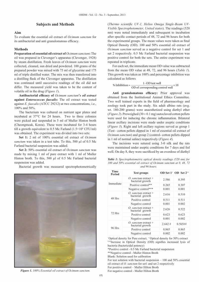
MethodsPreparation of essential oil extract of Ocimum sanctum: The oil was prepared in Clevenger’s apparatus (Clevenger, 1928) by steam distillation. Fresh leaves of Ocimum sanctum were collected, cleaned, sun dried and powdered. 100 grams of the prepared powder was mixed with 75 ml of glycerine and 175 ml of triple distilled water. The mix was then transferred into a distilling flask of the Clevenger apparatus. The distillation was continued until successive readings of the oil did not differ. The measured yield was taken to be the content of volatile oil in the drug (Figure 1).
Antibacterial efficacy of Ocimum sanctum’s oil extract against Enterococcus faecalis: The oil extract was tested against E. faecalis (ATCC 29212) at two concentrations, i.e., 100% and 50%.
The bacterium was cultured on nutrient agar plates and incubated at 37°C for 24 hours. Two to three colonies were picked and suspended in 5 ml of Muller Hinton broth (Cheongmeak, Korea). These were incubated for 3-4 hours till a growth equivalent to 0.5 Mc Farland (1.5×10⁸ CFU/ml) was obtained. The experiment was divided into two sets:
Set 1: 2 ml of 100% essential oil extract of Ocimum sanctum was taken in a test tube. To this, 500 µl of 0.5 Mc Farland bacterial suspension was added.
Set 2: 50% essential oil extract of Ocimum sanctum was made by mixing 1 ml of pure extract with 1 ml of Muller Hinton broth. To this, 500 µl of 0.5 Mc Farland bacterial suspension was added.
Bacterial growth was measured spectrophotometrically
(Thermo scientific UV-1, Helios Omega Single-Beam UV-Visible Spectrophotometer, United states). The readings (520 nm) were noted immediately and subsequent to incubation after specific contact periods of 48, 72 and 96 hours for both the experimental groups. The mean values were taken as final Optical Density (OD). 100 and 50% essential oil extract of Ocimum sanctum served as a negative control for set 1 and set 2 respectively. 0.5 Mc Farland bacterial suspension was positive control for both the sets. The entire experiment was repeated in triplicate.
For each set, the immediate mean OD value was subtracted from the mean OD value at 48, 72, and 96 hours (Table 1). This growth was taken as 100% and percentage inhibition was calculated as follows
×1- OD test well % Inhibition = 10
OD of corresponding control well
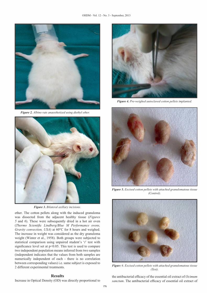
Anti granulomatous efficacy: Prior approval was obtained from the Institutional Animal Ethics Committee. Two well trained experts in the field of pharmacology and zoology took part in the study. Six adult albino rats (avg. wt. 180-200 grams) were anaesthetized using diethyl ether (Figure 2). Preweighed (30 ± 0.1 mg) autoclaved cotton pellets were used for inducing the chronic inflammation. Bilateral linear axillary incisions were made under aseptic conditions (Figure 3). Right and left axillary regions served as group 1 (Test - cotton pellets dipped in 1 ml of essential oil extract of Ocimum sanctum) and group 2 (control- cotton pellets dipped in 1 ml of normal saline) respectively (Figure 4).
The incisions were sutured using 3-0 silk and the rats were maintained under aseptic conditions for 7 days and fed well. On day 8, they were sacrificed by an overdose of diethyl
Figure 1. 100% Essential oil extract of Ocimum sanctum.
Time Period Test groups OD Set 1+ OD Set 2++
Immediate
O. sanctum extract + bacterial growth 2.586 0.395
Positive control** 0.265 0.307Negative control*** 0.001 0.001
48 Hrs
O. sanctum extract + bacterial growth 2.616 0.510
Positive control 0.511 0.511Negative control 0.001 0.002
72 Hrs
O. sanctum extract + bacterial growth 2.626 0.522
Positive control 0.623 0.623Negative control 0.001 0.002
96 Hrs
O. sanctum extract + bacterial growth 2.642 # 0.565##
Positive control 0.865 0.865Negative control 0.002 0.002
+Optical density for Pure extract, ++Optical density for 50% extract #,##Increase in Optical Density (OD) signifies increased lysis of bacteria (bactericidal potency)*Positive control - 0.5 Mc Farland bacterial suspension**Negative control - Muller Hinton BrothBlank: Solution used for calibrationFor test solution with bacterial suspension – 100 and 50% essential oil extract of O. sanctum for set1 and set 2 respectivelyFor positive control – Muller Hilton Broth For negative control - Muller Hilton Broth
Table 1: Spectrophotometric optical density readings (520 nm) for 100 and 50% essential oil extract of Ocimum sanctum at 0, 48, 72
and 96 hours.
OHDM - Vol. 12 - No. 3 - September, 2013
176
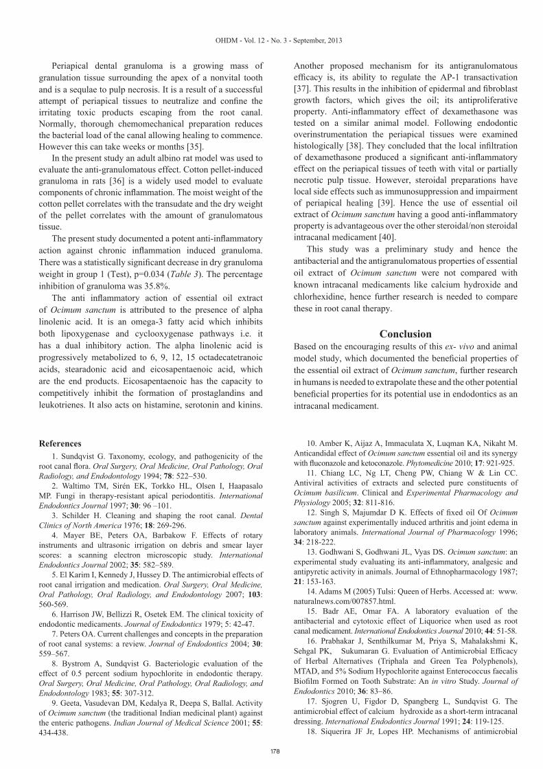
ether. The cotton pellets along with the induced granuloma was dissected from the adjacent healthy tissue (Figures 5 and 6). These were subsequently dried in a hot air oven (Thermo Scientific Lindberg/Blue M Performance ovens, Gravity convection, USA) at 60°C for 8 hours and weighed. The increase in weight was considered as the dry granuloma weight (Winter et al., 1958). Both groups were subjected to statistical comparison using unpaired student’s ‘t’ test with significance level set at p<0.05. This test is used to compare two independent population means inferred from two samples (independent indicates that the values from both samples are numerically independent of each - there is no correlation between corresponding values) i.e. same subject is exposed to 2 different experimental treatments.
ResultsIncrease in Optical Density (OD) was directly proportional to
Figure 2. Albino rats anaesthetized using diethyl ether.
Figure 3. Bilateral axillary incisions.
Figure 4. Pre-weighed autoclaved cotton pellets implanted.
Figure 5. Excised cotton pellets with attached granulomatous tissue (Control).
Figure 6. Excised cotton pellets with attached granulomatous tissue (Test).
the antibacterial efficacy of the essential oil extract of Ocimum sanctum. The antibacterial efficacy of essential oil extract of

OHDM - Vol. 12 - No. 3 - September, 2013
177
Ocimum sanctum improved with an increase in concentration and contact period (Table 2).
Increase or decrease in pre and post implantation cotton pellets weight (mgs) implied a larger or a smaller granuloma formation (Table 3). Both groups were subjected to statistical comparison using unpaired student’s ‘t’ test with significance level set at p<0.05. There was a statistically significant decrease in dry granuloma weight in group 1 (Test), p= 0.034, as compared to group 2 (control). Percentage inhibition of granuloma formation was calculated as follows
Average weight of implanted cotton pellet with Test solution 1- ×100Average Weight of implanted cotton pellet with saline
The percentage inhibition of granuloma was 35.8%.
DiscussionThe use of an intracanal medicament after canal preparation is generally recommended in multiple sitting endodontics [17]. Calcium hydroxide is the gold standard with a high degree of success [18]. However, specific microorganisms such as Enterococcus faecalis have shown resistance against this [19] and other commonly used intracanal medicaments [18].
Increase in antibiotic resistant strains and side effects caused by various synthetic drugs have prompted the researchers to look for herbal/botanical therapy. One such product with wide usage in Indian context is Ocimum sanctum (Tulsi). It belongs to the Labiateae family and has three varieties: Sri, Krishna and Vana Tulsi. It has the following common names: Tulsi (Hindi), Manjari (Sanskrit) and Holy Basil (English). It is a branched, erect, stout and aromatic herb. It grows up to the height of about 75 to 90 cm and is hairy all over. The chief source of the essential oil is leaves, followed by inflorescence and stem. The oil extracted from seeds is called ‘fixed oil’ and mainly contains fatty acids and sitosterol [20].
The oil can be obtained by steam, petroleum ether or benzene distillation [21,22]. In the present study steam
distilled essential oil extract of O. sanctum was prepared. This is a volatile oil.
For intracanal medicaments to be effective against bacteria, they should be able to diffuse into the dentinal tubules at sufficient concentrations and exceed the dentin buffering ability [18]. It has been suggested that use of the medicament's vapor might be the best method to regulate the dose and still get the drug into all the irregularities within the root canal system for successful disinfection [3].
Intracanal medicaments that exert their antibacterial action in a vapour form are formocresol, camphorated mono-chlorophenol, merthiolate, metacresylacetate, beechwood creosote, and glutaraldehyde [23-25]. Although, they are effective against certain microorganism implicated in periradicular disease, alpha hemolytic streptococci and enterococci have been found to be resistant to the vapors of these medicaments. Moreover they have a limited role because they are extremely toxic, antigenic and its effect is lost after a few days [26].
Enterococcus faecalis is the most commonly isolated or detected species from failed root canal therapy [17]. Its high resistance to antibacterial substances is widely documented [27] and is attributed to its ability to enter into a viable but nonculturable state during environmental stresses [28,29]. Furthermore, it can penetrate deeply into the dentinal tubules and resist bactericidal substances commonly used in endodontic procedures [30]. Recognizing the potential role of E. faecalis in the failure of root canal therapy and its resistance for various drugs makes it a good test model for evaluating the antibacterial efficacy of essential oil extract of Ocimum sanctum.
The present study utilized Spectrophotometric system for percentage inhibition assay because it is less subjective, cheaper and has a greater sensitivity than the standard well and disc diffusion methods. The antibacterial effect of Ocimum sanctum is due to the presence of linoleic acid, linolenic acid, eugenol (1-hydroxy-2-methoxy-4-allylbenzene), and carvacrol [31]. The mechanism of antibacterial action of this oil is due to the formation of Malondialdehyde, an aldehyde formed as a breakdown product of linoleic acid and linolenic acid, which are peroxidized polyunsaturated lipids. Malondialdehyde is a cross linker and initiates oxidation reactions in which undesirable bond formation occurs in the bacterial DNA and RNA [32] resulting in inhibition of replication of DNA and subsequent bacterial death. Another hypothesized mechanism of action of the extract is that, Malondialdehyde crosslink with different amino groups of various bacterial enzymes jeopardizing the metabolic processes in the bacteria and culminating in their death [33].
A herb Aloe vera, in a gel form was used as an intracanal medicament against E. faecalis. It was found that only (22.5%) of the teeth medicated with this herbal gel yielded negative cultures. It was concluded that the gel is ineffective against the organism and hence cannot be used as intracanal medicament [34]. However, the result of the present study showed that the antibacterial efficacy of essential oil extract of Ocimum sanctum was directly proportional to an increase in concentration and contact period. 100% essential oil had greater antibacterial efficacy tested at all the contact periods (Table 2).
Contact Period Set 1* Set 2** 48 hrs 87.8% 43.7% 72 hrs 88.83% 59.9% 96 hrs 90.67% 69.54%
* Pure essential oil extract of Ocimum sanctum** 50% essential oil extract of Ocimum sanctum
Table 2: Comparative percentage inhibition of E. faecalis.
Group 1 ** Group 2***31.2 32.521.0 42.718.1 51.432.0 32.126.5 34.525.0 41.5
* Increase in weight denotes larger granuloma formation and vice-versa**Test- preweighed cotton pellet (30 ± 0.1) mg dipped in 1 ml of essential oil extract of Ocimum sanctum***Control- preweighed cotton pellet (30 ± 0.1) mg dipped in 1 ml of saline
Table 3: Change in cotton pellets weight (in mgs)* before and after implantation.
OHDM - Vol. 12 - No. 3 - September, 2013
178
Periapical dental granuloma is a growing mass of granulation tissue surrounding the apex of a nonvital tooth and is a sequlae to pulp necrosis. It is a result of a successful attempt of periapical tissues to neutralize and confine the irritating toxic products escaping from the root canal. Normally, thorough chemomechanical preparation reduces the bacterial load of the canal allowing healing to commence. However this can take weeks or months [35].
In the present study an adult albino rat model was used to evaluate the anti-granulomatous effect. Cotton pellet-induced granuloma in rats [36] is a widely used model to evaluate components of chronic inflammation. The moist weight of the cotton pellet correlates with the transudate and the dry weight of the pellet correlates with the amount of granulomatous tissue.
The present study documented a potent anti-inflammatory action against chronic inflammation induced granuloma. There was a statistically significant decrease in dry granuloma weight in group 1 (Test), p=0.034 (Table 3). The percentage inhibition of granuloma was 35.8%.
The anti inflammatory action of essential oil extract of Ocimum sanctum is attributed to the presence of alpha linolenic acid. It is an omega-3 fatty acid which inhibits both lipoxygenase and cyclooxygenase pathways i.e. it has a dual inhibitory action. The alpha linolenic acid is progressively metabolized to 6, 9, 12, 15 octadecatetranoic acids, stearadonic acid and eicosapentaenoic acid, which are the end products. Eicosapentaenoic has the capacity to competitively inhibit the formation of prostaglandins and leukotrienes. It also acts on histamine, serotonin and kinins.
Another proposed mechanism for its antigranulomatous efficacy is, its ability to regulate the AP-1 transactivation [37]. This results in the inhibition of epidermal and fibroblast growth factors, which gives the oil; its antiproliferative property. Anti-inflammatory effect of dexamethasone was tested on a similar animal model. Following endodontic overinstrumentation the periapical tissues were examined histologically [38]. They concluded that the local infiltration of dexamethasone produced a significant anti-inflammatory effect on the periapical tissues of teeth with vital or partially necrotic pulp tissue. However, steroidal preparations have local side effects such as immunosuppression and impairment of periapical healing [39]. Hence the use of essential oil extract of Ocimum sanctum having a good anti-inflammatory property is advantageous over the other steroidal/non steroidal intracanal medicament [40].
This study was a preliminary study and hence the antibacterial and the antigranulomatous properties of essential oil extract of Ocimum sanctum were not compared with known intracanal medicaments like calcium hydroxide and chlorhexidine, hence further research is needed to compare these in root canal therapy.
ConclusionBased on the encouraging results of this ex- vivo and animal model study, which documented the beneficial properties of the essential oil extract of Ocimum sanctum, further research in humans is needed to extrapolate these and the other potential beneficial properties for its potential use in endodontics as an intracanal medicament.
References1. Sundqvist G. Taxonomy, ecology, and pathogenicity of the
root canal flora. Oral Surgery, Oral Medicine, Oral Pathology, Oral Radiology, and Endodontology 1994; 78: 522–530.
2. Waltimo TM, Sirén EK, Torkko HL, Olsen I, Haapasalo MP. Fungi in therapy-resistant apical periodontitis. International Endodontics Journal 1997; 30: 96 –101.
3. Schilder H. Cleaning and shaping the root canal. Dental Clinics of North America 1976; 18: 269-296.
4. Mayer BE, Peters OA, Barbakow F. Effects of rotary instruments and ultrasonic irrigation on debris and smear layer scores: a scanning electron microscopic study. International Endodontics Journal 2002; 35: 582–589.
5. El Karim I, Kennedy J, Hussey D. The antimicrobial effects of root canal irrigation and medication. Oral Surgery, Oral Medicine, Oral Pathology, Oral Radiology, and Endodontology 2007; 103: 560-569.
6. Harrison JW, Bellizzi R, Osetek EM. The clinical toxicity of endodontic medicaments. Journal of Endodontics 1979; 5: 42-47.
7. Peters OA. Current challenges and concepts in the preparation of root canal systems: a review. Journal of Endodontics 2004; 30: 559–567.
8. Bystrom A, Sundqvist G. Bacteriologic evaluation of the effect of 0.5 percent sodium hypochlorite in endodontic therapy. Oral Surgery, Oral Medicine, Oral Pathology, Oral Radiology, and Endodontology 1983; 55: 307-312.
9. Geeta, Vasudevan DM, Kedalya R, Deepa S, Ballal. Activity of Ocimum sanctum (the traditional Indian medicinal plant) against the enteric pathogens. Indian Journal of Medical Science 2001; 55: 434-438.
10. Amber K, Aijaz A, Immaculata X, Luqman KA, Nikaht M. Anticandidal effect of Ocimum sanctum essential oil and its synergy with fluconazole and ketoconazole. Phytomedicine 2010; 17: 921-925.
11. Chiang LC, Ng LT, Cheng PW, Chiang W & Lin CC. Antiviral activities of extracts and selected pure constituents of Ocimum basilicum. Clinical and Experimental Pharmacology and Physiology 2005; 32: 811-816.
12. Singh S, Majumdar D K. Effects of fixed oil Of Ocimum sanctum against experimentally induced arthritis and joint edema in laboratory animals. International Journal of Pharmacology 1996; 34: 218-222.
13. Godhwani S, Godhwani JL, Vyas DS. Ocimum sanctum: an experimental study evaluating its anti-inflammatory, analgesic and antipyretic activity in animals. Journal of Ethnopharmacology 1987; 21: 153-163.
14. Adams M (2005) Tulsi: Queen of Herbs. Accessed at: www.naturalnews.com/007857.html.
15. Badr AE, Omar FA. A laboratory evaluation of the antibacterial and cytotoxic effect of Liquorice when used as root canal medicament. International Endodontics Journal 2010; 44: 51-58.
16. Prabhakar J, Senthilkumar M, Priya S, Mahalakshmi K, Sehgal PK, Sukumaran G. Evaluation of Antimicrobial Efficacy of Herbal Alternatives (Triphala and Green Tea Polyphenols), MTAD, and 5% Sodium Hypochlorite against Enterococcus faecalis Biofilm Formed on Tooth Substrate: An in vitro Study. Journal of Endodontics 2010; 36: 83–86.
17. Sjogren U, Figdor D, Spangberg L, Sundqvist G. The antimicrobial effect of calcium hydroxide as a short-term intracanal dressing. International Endodontics Journal 1991; 24: 119-125.
18. Siquerira JF Jr, Lopes HP. Mechanisms of antimicrobial

OHDM - Vol. 12 - No. 3 - September, 2013
179
activity of calcium hydroxide: a critical review. International Endodontics Journal 1999; 32: 361-369.
19. Evans M, Davies JK, Sundqvist G, Figdor D. Mechanisms involved in the resistance of Enterococcus faecalis to calcium hydroxide. International Endodontics Journal 2002 ; 35: 221–228.
20. Portenier I, Haapasalo H, Rye A, Waltimo T, Orstavik D, Haapasalo MPP. Inactivation of root canal medicaments by dentine, hydroxyapatite, and bovine serum. International Endodontics Journal 2001 ; 34: 184–188.
21. Mondal S, Mirdha BR, Mahapatra SC. The science behind sacredness of Tulsi (Ocimum sanctum Linn.). Indian Journal of Physiology and Pharmacology 2009; 53: 291-306.
22. Sen P. Therapeutic potentials of Tulsi: from experience to facts. Drugs News & Views 1993; 1: 15–21.
23. Khanna N, Bhatia J. Action of Ocimum sanctum (Tulsi) in mice: possible mechanism involved. Journal of Ethnopharmacology 2003; 88: 293–296.
24. Cwikla JR. The vaporization and capillarity effect of endodontic medicaments. Oral Surgery, Oral Medicine, Oral Pathology, Oral Radiology, and Endodontology 1972; 34: 117-121.
25. Pear JR .Bactericidal effects of some drugs used in pulp canal therapy. Journal of American Dental Association 1970; 29: 244-247.
26. Bender IB, Seltzer S, Turkenk S. To culture or not to culture. Oral Surgery, Oral Medicine, Oral Pathology, Oral Radiology, and Endodontology 1964; 18: 527-540.
27. Wall GL, Dowson J, Shipman C Jr. Antibacterial efficacy and cytotoxicity of three drugs. Oral Surgery, Oral Medicine, Oral Pathology, Oral Radiology, and Endodontology 1971; 33: 230-241.
28. Morrison D, Woodford N, Cookson B. Enterococci as emerging pathogens of humans. Society for Applied Bacteriology Symposium Series 1997; 83: 89S-99S.
29. Lleo MM, Bonato B, Tafi MC, Signoretto C, Boaretti M, Canepari P. Resuscitation rate in different enterococcal species in the viable but non-culturable state. Journal of Applied Microbiology 2001; 91: 1095–1102.
30. Spangberg L, Engstrom B, Langeland K. Biologic effects of
dental materials and toxicity and antimicrobial effect of endodontic antiseptics in vitro. Oral Surgery, Oral Medicine, Oral Pathology, Oral Radiology, and Endodontology 1973; 36: 856-871.
31. Sjogren U, Figdor D, Persson S, Sundqvist G. Influence of infection at the time of root filling on the outcome of endodontic treatment of teeth with apical periodontitis. International Endodontics Journal 1997; 30: 297–306.
32. Singh S, Malhotra M, Majumdar D K. Antibacterial activity of Ocimum sanctum L. fixed oil. Indian Journal of Experimental Biology 2005; 43: 835-37.
33. Hall ED, Oostveen JA, Andrus PK, Anderson DK, Thomas CE. Immunocytochemical method for investigating in vivo neuronal oxygen radical induced lipid peroxidation. Journal of Neuroscience Methods 1997; 76: 115-122.
34. Łuczaj W, Skrzydlewska E. DNA damage caused by lipid peroxidation products. Cellular and Molecular Biology Letters 2003; 8: 391-413.
35. Maguire H, Torabinejad M, Kettering JD. The Use of Aloe Vera Gel as an Intracanal Medicament. Journal of Endodontics 1996; 22: 193-195.
36. Nair PN (1999) Cholesterol as an aetiological agent in endodontic failures –a review. Australian Endodontic Journal 1999; 25: 19–26.
37. Vogel H. Anti-Inflammatory Activity. Drug Discovery and Evaluation 1996; 725-771.
38. Liu G, Bibus D, Bode A, Ma W, Holman R, Dong Z. Omega -3 but not omega-6 fatty acids inhibits AP-1 activity and cell transformation in JB6 cells. Proceedings of the National Academy of sciences of United States of America 2003; 98: 7510-7515.
39. Nobuhara WK, Carnes DL, Gilles JA. Anti-Inflammatory effects of dexamethasone on periapical tissues following endodontic overinstrumentation. Journal of Endodontics 1997; 19: 501-507.
40. Abbott PV, Heithersay GS, Hume WR. Release and diffusion through human tooth roots in vitro of corticosteroid and tetracycline trace molecules from Ledermix paste. Endodontics & dental traumatology 1988; 4: 55–62.


![Ex vivo Rheology of Spider Silk @ - [email protected]](https://img.pdfslide.us/doc/110x75/6206405f8c2f7b173005e063/ex-vivo-rheology-of-spider-silk-emailprotected.jpg)