Embed Size (px)
Citation preview
1
Immunomodulatory potential and clinical efficacy of Allostatine against
Type 1 and 2 Herpes Simplex Virus (HSV) lesions
Imene Ben Toumia1,2, Dmitry Tulin3, Andrey Yakovlev3, Anna K. Savva3, Marco Ponassi1,
Franz Heinrich Kohnke4, Leila Chekir-Ghedira2, Alberto Izzotti 1,5, Virgil Schijns6,7,#, Camillo
Rosano 1,#,§
1 IRCCS Policlinico San Martino Genova, Italy
2 Unit of Bioactive Natural Substances and Biotechnology UR17ES47, Faculty of Dental
Medicine of Monastir, University of Monastir, Avicenne Street, 5000 Monastir, Tunisia
3 Laboratory of Insect Biopharmacology and Immunology, Faculty of Biology, St. Petersburg
State University, Russia
4 Department CHIBIOFARAM, University of Messina, Messina, Italy.
5 Department of Experimental Medicine, School of Medicine; University of Genoa, Italy
6 Epitopoietic Research Corporation (ERC), Landerd campus, Nistelrooisebaan 3, 5374 RE
Schaijk, The Netherlands,
7 Cell Biology and Immunology, Wageningen, University, The Netherlands. (The
Netherlands).
# Senior co-authors
§ corresponding author
It takes a fool to remain sane
- The Ark
In memory of Professor Sergey Ivanovich Chernysh (1951-2020), inventor of Allostatine, great
scientist and good friend. A life spent to fight Viruses and Bacteria, killed by human idiocy
while crossing the road.
2
Abstract
Allostatine is a peptide deriving from the Alloferon peptides, a “family of first line defense
molecules” used by insects to fight infections. Allostatine has been reported to be particularly
attractive as a potent antiviral and as a possible immunomodulatory adjuvant in vaccines, such
as therapeutic cancer vaccines. To evaluate the ability of Allostatine to facilitate the killing a
wide range of virus-infected target cells, as a result of triggering human natural killer (NK) and
T lymphocyte (CTL), we assessed the effects of this peptide on both murine and human immune
responses. In addition, in order to investigate Allostatine’s capabilities to modulate the immune
system during HSV pathogenesis, clinical tests were carried on patients affected by skin and
genital herpes resulting from HSV type 1 and type 2 infections. Some of these patients were
experiencing relapsing herpes lesions for more than 4 years before the study. The efficacy in
the treatment of cold sores and the elimination of genital herpes symptoms were evaluated by
comparing to Acyclovir treatment.
For the first time we report that Allostatine significantly enhances both NK and CTL activities
as well as macrophage functions by increasing nitric oxide (NO) release in mice. We also
demonstrate that this peptide is selectively localized in the leukocyte cytoplasm and that it
increases the expression of human NK markers NKp80, CD244, CD226 and CD25.
Interestingly, we also show the peptide’s ability to increase IL-2–triggered and IFN-γ synthesis
of CD3+/CD28+ activated T cells.
Our clinical test results provide evidence that Allostatine influences human T-lymphocytes
activation markers. Allostatine also facilitates the full lesion recovery of HSV affected patients.
A clear improvement of relapsing herpes lesions was observed at different anatomical locations
in the majority of patients treated with the Allostatine-based product Allomedin. No adverse or
toxic effect was observed.
Thus, we observed in murine and human in vitro, ex-vivo and clinical studies that Allostatine
may be useful for modulating NK and T cell immune functions and promoting macrophage
activities. In a clinical study a formulation based on Allostatine beneficially counteracted HSV-
induced pathological features and relapses.
3
Introduction
Herpes Simplex Virus (HSV) is a commonly found human pathogen. An estimated 4.0 billion
people are infected with Herpes Simplex Virus globally. The incidence rate of cold sores caused
by the HSV1 virus, is the second largest worldwide, trailing only behind common cold. About
90% of the world population is infected with HSV1, with about 25-35% of the adults enduring
recurrent spate of cold sores [Wald and Corey, 2007]. Genital herpes, an infection caused by
HSV2, is one of the most common sexually transmitted diseases in the US [McQuillan et al.,
2018]. The rate of occurrence of genital herpes varies across different countries in developing
regions. Certain African nations dealing with the HIV epidemic are experiencing a high
prevalence of HSV2 infection. The majority of people (approximately 80%) display no or very
mild HSV2 symptoms that are easy to miss. In fact, the most common way to catch either strain
of HSV is from someone who sheds the virus without any overt evidence of infection. This
infection, however, can be dangerous under certain circumstances. If passed from a mother to
a newborn, HSV2 can be fatal. In rare cases HSV1 can infect the brain, causing encephalitis,
which is usually fatal; a growing body of facts shows that chronic brain infection with HSV1
makes a great contribution to Alzheimer’s disease [Carter, C.J., 2008]. Furthermore, genital
herpes also increases the risk of HIV infection by 40% [Looker et al, 2020]. According to WHO
assessment, genital herpes is a cause of 10% of all suicides worldwide [Nissen et al., 2019].
HSV infection is lifelong and daily medication can be burdensome.
There is no drug presently available in the market that can permanently cure herpes infection,
however, various anti-viral therapies are available to alleviate painful symptoms of the
infection. Present therapies for Herpes infection include nucleoside analogs such as acyclovir,
and other related drugs. These drugs act by stalling the synthesis of viral DNA. Major drugs
available in the market include Zovirax, Famvir, Abreva, Denavir, and Vistide. However, these
drugs are known to cause drug-related toxicity as a result of their interference with the natural
metabolism of the human body. Also, with the virus subsequently developing resistance against
the existing array of anti-virals, the need for innovative therapies for the treatment of herpes
simplex infection, and the search for novel antiviral drugs continues. Therefore, there is a large
potential for the herpes simplex therapeutics market in future.
NKs, CTL and macrophages are the major protective weapons used by the immune system to
fight HSV1 and HSV2 virus infections; NK cells produce interferon-γ (IFN-γ) and promote
IFN-γ production via T cells in response to many viral infections.
4
Alloferons were isolated from bacteria-challenged larvae of the blow fly Calliphora vicina and
constitute a group of natural occurring peptides with strong antiviral and antitumoral abilities
[Chernysh et al., 2002; Chernysh et al., 2012; Chernysh et al., 2013]. Along with other surgical
maggots, Calliphora v. has a long history of medical use in wound and ulcer healing
[Kruglikova A.A: & Chernysh S.I., 2013], with a first documented application back in the XVI
Century by the French surgeon Abroise Paré (1510-1590) [Whitaker et al., 2007]. Calliphora
is well known to produce a series of potent antimicrobial substances after being challenged by
bacterial infection. These antimicrobial peptides (AMPs) have primary structures similar to
those described for other insects, such as insect defensin, diptericins, cecropins, and proline-
rich peptides [Chernysh et al., 2002]. Calliphora’s hemolymph possess a cytotoxic activity
similar to the one of human NKs [Chernysh et al., 2004]. Particularly, two peptides of 13 and
12 amino acids have been identified in Calliphora’s hemolymph which have been named
Alloferon1 and Alloferon2, respectively [Chernish et al, 2002]. Administration of a picomolar
concentration of Alloferon1 to mice turned out into the stimulation of natural cytotoxicity as
well as into antiviral and antitumoral activities in vivo [Chernysh et al., 2002; Chernysh et al.,
2012].
Allostatine (also known as Alloferon3) is an “humanized” peptide which originated from
rationally designed modifications of Alloferon1. By substituting two amino acids within the
Alloferon1 sequence a pattern similar to human immunoglobulins was found. Allostatine-
induced antiviral and antitumoral abilities have been demonstrated in vitro and in vivo.
Particularly, the properties of Allostatine to act as a therapeutic cancer vaccine adjuvant has
been demonstrated in vivo in DBA/2 mice vaccinated together with tumor antigen [Chernysh
and Kozuharova, 2013].
Here, we present results on Allostatine efficacy against human HSV (cold sores and genital
herpes) in patients.
Matherials and Methods
5
Peptide
Diapharm Co, St. Petersburg has synthesized Allostatine, using the synthesis of solid phase and
according to Fmoc/But strategy and purified by reverse phase HPLC with more than 98% as
peptide final purity. It is a 13 amino acid linear peptide sequence His–Gly–Val–Ser–Gly–Trp–
Gly–Gln–His–Gly–Thr–His–Gly and its empiric formula is С56Н77N21O17 and its molecular
mass is 1316 Da.
Allomedin® is a gel formulation product based on Allostatine, developed by the Company
Allopharm ltd., created by Prof. Sergey Ivanovich Chernysh. This compound is composed of
Allostatine and water, carbopol, allantoin, phenyethanol, ethylhexyglyceol, sodium hydroxide
as excipients, and it was developed to protect skin and mucous tunic against viral infections and
registered in Russia and Countries belonging to the former USSR, as a skin care product.
Ex-vivo experiments
Animals
All experiments on Balb/c mice (22–26 g) were performed in accordance with the Council of
the European Communities (86/609/EEC; November 24th 1986). Directives regulating the
welfare of experimental animals, and experiments were approved by the Life Sciences and
Health Research Ethics Committee (cer-svs) of the Institute of Biotechnology (University of
Monastir, Tunisia; ethical approval no. 2019/02/I/CER-SVS/ISBM; 9 January, 2019). In an
accredited pathogen-free facility mice were housed under standard conditions. All the animals
were provided ad libitum access to filtered water and standard rodent chow.
Isolation and culture of macrophages and splenocytes
Mice were sacrificed by cervical dislocation and primary splenocytes were extracted under
sterile conditions, immersed in phosphate buffered saline (PBS, pH 7.4)
(Gibco, BRL) and gently dissected with forceps. After exposure to lysis buffer (144 mM
NH4Cl, 1.7 mM Tris Base) to remove red blood cells, the cells were resuspended in complete
RPMI-1640 medium (Gibco, BRL) containing 10% fetal bovine serum (FBS; Gibco), 1%
penicillin (10,000 U/ml) and streptomycin (10,000 µg/ml) (GibcoBRL, Paisley, UK) after two
washing steps. The cells were incubated at 37 °C at 5 % enriched CO2 atmosphere. Other
Balb/c mice were used in order to obtain peritoneal macrophages after PBS intraperitoneal
6
lavage. The extracted cells were resuspended in complete RPMI 1640 medium after washing
steps. Cell viability was determined using the trypan blue exclusion technique.
Activity of natural killer (NK) and the cell-mediated cytotoxicity assay
Spleens prepared as described above were used as the source of effector cells; isolated
splenocytes were plated at a density of 5×10⁵ cells for NK activity at a concentration of 5×10⁶
cells/ml per well in 100 µl aliquot/well. These cells were exposed and stimulated by different
concentrations of Allostatine (5,10,20 and 40 ng/ml) for 24 h. Subsequently, 100 µl of target
K562 cells and B16-F10 cells (5×10⁴ cells/ml; yielding a 100:1 expected effector-target ratio)
were added to the wells respectively for NK and CTL activity. Medium with 1% DMSO was
used as a negative control. The plates were then incubated for 4 h and 24 h at 37°C in 5% CO2
atmosphere respectively for NK and CTL activity. Forty micro-liters of MTT solution was
added to each well. After incubation, the supernatants were removed by centrifugation and 100
µl of DMSO was added to each well. The absorbance was measured at 540 nm using a
microplate reader.
Target cell control, blank control and effector cell control were performed. NK cell activity was
calculated as follows: NK activity or CTL activity (%) =100 ×(ODT- (ODS -ODE))/ODT,
where ODT = optical density value of target cells control, ODS= optical density value of test
samples, and ODE = optical density value of effector cells control
In vitro phagocytic assay on cellular lysosomal enzyme activity and NO measurement
production
The cellular lysosomal enzyme activity has been determined by acid phosphatase activity.
Briefly, a volume of 100 µl of allostatine at different concentrations (5,10,20 and 40 ng/ml)
were added to 100 µl of a macrophage suspension (3×10⁶ cells/ml) in flat-bottom, 96-well plates
at 37 °C in humidified 5% CO2 for 48 h. The medium was discarded and the macrophage
monolayer in each well was solubilized with 20 µl of 0.1% Triton X-100 (Biomatik
Corporation, Cambridge, UK). Next 10 µl of 100 mM p-nitrophenylphosphate (p-NPP)
(AppliChem, GmbH, Darmstadt, Germany) solution and 50 µl of 0.1 M citrate buffer (pH 5.0)
were added. The plate was further incubated for 30 min, 150 µl of 0.2 M borate buffer (pH 9.8)
was used to terminate the reaction and the absorbance at 405 nm was measured. The percentage
activity was calculated according to the following equation:
7
Lysosomal enzyme activity (%) = (OD sample-OD negative control)/ OD negative control )×
100.
Nitrite concentration (NO2) released by macrophages was determined using Griess reaction in
cell culture supernatants. Briefly, 100 µl of supernatant from each sample was mixed with 100
µl of Griess reagent (equal volumes of 1 % sulfanilamide and 0.1 % naphthylethylenediamine
dihydrochloride in 2.5 % phosphoric acid), and incubated at room temperature for 15 min.
Absorbance was then measured at 540 nm using a microplate reader and compared with a
standard curve of sodium nitrite
Purification of human peripheral blood mononuclear cells (PBMCs)
Venous peripheral blood samples were obtained after informed consent from healthy human
volunteers between the ages of 18 and 60 years. Mononuclear cells were obtained using BD
Vacutainer CPT (Cat. No. 362780) according to the protocol recommended by the
manufacturer, and then transferred to the medium.
Evaluating of NK cell phenotype changing during cytotoxicity test
The mononuclear fraction was obtained from the venous blood of each donor using a cell
preparation tube (362753 BD, USA) in accordance with the manufacturer's instructions. NK
cells (CD3– / CD16 + / CD56 +) were isolated from the mononuclear fraction using magnetic
separation (ThermoFisher 11349D Dynabeads™ Untouched™ Human NK Cells Kit)
according to the manufacturer’s protocol, resulting NK cell concentration was adjusted to >
90%. Using an automatic cell counter (BioRad), the number of NK cells obtained was
determined. The isolated cells were cultured in RPMI 1640 medium containing 5% inactivated
autologous serum and antibiotics (Sigma-Aldrich A5955 Antibiotic Antimycotic Solution
100×). A portion of NK cells was preincubated overnight in the allostatine-containing medium.
NK cells were further mixed with target cells (K562), at a E:T ration of 1:10. In accordance
with the experimental scheme, the resulting mixture was separated by volume and placed in the
well of a U-shaped plate for the 18 hour incubation in a CO2 thermostat at +37 °C. After
incubation, changes in the expression of the following markers were evaluated: CD25 (Becton
Dickinson: anti-Human CD25, 562442), NKp80 (Miltenyi Biotec: anti-Human NKp80-FITC,
8
130-094-843), CD 226 (BioLegend: anti-Human CD226_FITC 338303) and CD 244 (Miltenyi
Biotec: anti-Human CD244 (2B4)-PE, 130-099-071).
Visualization of allostatine interaction with human leucocytes in vitro
10 μg / ml fluorescein-labeled allostatine was added to fresh human blood. After an hour of
incubation at +37 °C, erythrocytes were lysed using BD FACS Lysing Solution (Cat. No.
349202) according to the manufacturer's recommendation. The remaining fraction leukocytes
was washed with PBS, fixed with 2% paraformaldehyde in PBS, placed on glass and enclosed
in Diamond Antifade Mountant with DAPI (Invitrogen ™ ProLong ™, Cat. No. P36966). The
resulting preparations were analyzed with a Leica TCS SP5 confocal scanning microscope.
The effect of Allostatine on the production of cytokines by activated human T cells in vitro
Polyclonal activation of T cells was performed using beads coated with CD3 / CD28 antibodies
(Thermo Fisher Scientific, Dynabeads Human T-Activator CD3 / CD28, Cat. No. 11131D),
according to the manufacturer's recommendations. The response was evaluated by the
production of IFN-γ and IL-2 cytokines by cells. For detection of cytokine producing cells, the
protocol recommended by Becton Dickinson (BD) was used, IFN-γ was stained using BD Fast
Immune Anti-Human IFN-γ-FITC, Cat. No. 340449, IL-2 - BD FastImmune Anti-Human IL-
2-PE, Cat. No. 340,450.
Allostatine-mediated immunostimulation and study design for clinical trials
Ethic statement
The Permission for carring out the Allostatine clinical trials No. 544 was received from Russian
Ministry of Health on November 18, 2008, and also the excerpt from the protocol No. 12 of the
meeting of the Ethics Committee was received in July 15, 2008 (Moscow, Russia). Patients
were screened before the study. If the Patient met the inclusion / exclusion criteria, he was
included in the study, given unique blind-coded patient identification number (UINP) and
randomized. All the selected patients signed an informed consent to agree to participate in the
experience of clinical use.
9
The effect of subcutaneous injection of Allostatine on human T-lymphocytes
In order to determine the response of the whole T-cell population, healthy volunteers, after
informed consent, received a subcutaneous injection of 1 mg Allostatine. Immediately prior to
injection, 12 hours and 5 days after injection, the proportion of T cells (Miltenyi Biotec VioBlue
labeled anti-Human CD3, Cat. No. 130-098-165) in peripheral blood expressing CD25 (with
BD BV605 labeled anti-Human CD25, Cat. No. 562661) and HLA-DR (with BD APC labeled
anti-Human HLA-DR, Cat. No. 560744) activation markers were determined.
Clincal study on patients suffering from Herpes simplex virus
Allostatine (Allomedin gel) safety and therapeutic efficacy in patients experienced relapsing
herpes lesions
The clinical study characterizing the safety and therapeutic efficacy of Allomedin (a gel
formulation based on Allostatine for topical use) was evaluated in 104 patients suffering from
Herpes simplex virus (81 females, 23 males). The clinical effect of Allomedin was compared
to treatment effect of acyclovir for cold sores and the elimination of genital herpes symptoms.
All these patients experienced relapsing herpes lesions at different anatomical locations for
more than 4 years before the study. The age of the patients was varied from 18 to 60 years.
Efficacy and safe use of Allostatin (Allomedin gel) in patients with chronic recurrent herpes
simplex lesions.
We evaluated the efficacy (therapeutic equivalence) of topical application of Allomedin
cosmetic gel for skin care. The degree of reduction of objective and subjective symptoms was
studied in patients suffering from recurrent Herpes Simplex Virus lesions compared to the
effectiveness of therapy with Zovirax, cream and Fenistil Pentsivir cream.
Also, we examined the safety, side effects and tolerability of local application of the
“Allomedin” cosmetic gel in relieving Herpes Simplex Virus relapses by registering adverse
reactions (AR) and serious adverse reactions (SAR).
This evaluation included 156 patients of both sexes (80 men and 76 women) from 18 to 80
years old (mean age 40.7 ± 2.1 years) with a clinically established diagnosis of chronic recurrent
herpes simplex lesions.
10
Inclusion criteria
In a first study, patients of both sexes, ranging from18-60 years of age, with a medical diagnosis
of recurrent (>3 relapses in the past 4 years) herpes simplex lesions, clinically and in laboratory
confirmed, were selected for the study.
For the second clinical evaluation, 156 patients aged from 18 to 80 years old, 80 men and 76
women, were selected. These patients were clinically diagnosed with chronic recurrent herpes
simplex.
Exclusion criteria
Patients who refused to participate in the study and/or the ones who violated the offered
protocol were excluded from the study. Excluded were also patients with somatic diseases in
the stage of decompensation (Cardiovascular diseases, chronic pulmonary, renal and hepatic
failure). Individuals reporting putative intolerance to the components of drugs were also not
admitted (e.g., fly puncture allergies).
Alcoholism and drug addiction in the anamnesis, mental illness that does not allow an
assessment of the effectiveness of therapy were considered as cause of exclusion as well as the
presence of active tuberculosis, systemic connective tissue diseases, HIV infection, acute
respiratory viral infections, acute and chronic hepatitis, acute bronchitis and pneumonia.
Patients who previously used drugs with antiviral and immunomodulatory activity and women
during pregnancy and lactation, as well as women with a wish to become pregnant were
excluded. Also, patients who participated in another clinical trial in the last 3 months were not
admitted.
Acceptable Concomitant Therapy:
Concomitant therapy for acute or chronic diseases could be continued in accordance with the
doctor’s appointments. Information about concomitant drugs (trade name, dosage or dosage
changes, indication, start date, termination date) was noted in the Patient’s Individual
Registration Card (IRC).
Non-acceptable Concomitant Therapy:
Any other antiviral drugs (except Allomedin, Fenistil Pentsivir, Zovirax) were not allowed for
use during the study.
11
At the start of the trial treatment (1st day) and on the 3rd, 5th, 8th, 10th day of treatment,
complaints and the clinical picture of the disease (presence of subjective symptoms, localization
of the process, size of lesions, presence of vesicles, hyperemia, edema, erosions, crusts), were
evaluated. The data obtained were fixed in the Individual Registration Card (IRC) of the patient.
The severity of the sign (symptom) were evaluated on a 3-point verbal analogue scale as:
0 - no symptom,
1 - weak manifestation
2 – moderate, acceptable
3 - pronounced manifestation.
The treatment consisted of Allomedin application onto the surface of herpetic lesions 2 to 3
times a day for 3 to 5 days.
At the 5th (final) visit (on day 10), the Investigator and the Patient carried out a subjective
assessment of the effectiveness of the treatment (good effect, satisfactory or unsatisfactory).
Evaluation of the effectiveness of the use of gel "Allomedin" and reference products was
determined as follows:
• good effect - marked improvement: arresting the manifestations of the disease, the lack of new
manifestations and relapses;
• satisfactory effect - incomplete relief of disease manifestations;
• unsatisfactory effect - lack of effect from Treatment or worsening of subjective and objective
symptoms.
Safety assessment
During and at the end of the Trial, adverse reaction (AR) scores were collected, and estimated
for the possible connection with Allomedin, and estimated for the possible connection with
other drugs.
12
Assessment of severity of adverse events:
• Light - not progressive, causing minimal discomfort;
• Medium - significantly, but not completely violating the usual daily activity;
• Severe - reactions that completely or very significantly disrupt daily activity
The degree of possible reliability of the association of adverse reaction (AR) with the studied
drugs was determined as follows:
• No connection - there is clear evidence of a lack of association of adverse events (AE) with
the Allomedin
• Doubtful connection - there is no clear temporary connection with the use of the product, there
are other factors (drugs, diseases, chemicals) that may cause them
• Possible connection - related in time with the use of the product, can be explained by the
presence of associated diseases or taking other drugs or chemical compounds, reactions to
withdrawal of the product and repeated use are unknown
• Probable connection — associated in time with the using drug, hardly ever to be associated
with comorbidities or other factors, decreases withdrawal of the drug, the response to repeated
using is not known.
Statistical analysis
Assays were performed three times as expressed in the procedure. All data are represented as
mean ± standard deviation. Graphs were drawn using GraphPad Prism® software (Graph- Pad
Prism® 6.07, GraphPad Software, Inc., CA, USA). One-way analysis of variance was used to
examine the differences between sets of data. A P-value less than 0.05 were considered as
significant.
Results
Allostatine improves murine NK and CTL cells activities
Natural Killer cells (NKs) represent the first innate line of defense against many pathogens,
while MHC-resticted cytolytic T cells (CTL) are required for specific pathogen eradication as
an adaptive cellular response. The examination of cytotoxic activity of splenic NK and CTL
cells against NK sensitive tumor cells (K562 line) and melanoma cells (B16-F10) respectively,
showed that, compared with the control cells, Allostatine treatment was able to significantly
enhance NK and CTL cells activities at a concentration of 10 and 20 ng/mL. Our results showed
a maximum increase at 20 ng/mL (81.69%) of the Allostatine dose (Fig. 1A). Also, cells treated
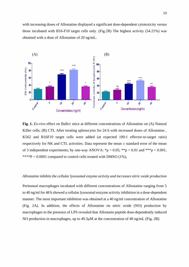
13
with increasing doses of Allostatine displayed a significant dose-dependent cytotoxicity versus
those incubated with B16-F10 target cells only. (Fig.2B) The highest activity (54.21%) was
obtained with a dose of Allostatine of 20 ng/mL.
Fig. 1. Ex-vivo effect on Balb/c mice at different concentrations of Allostatine on (A) Natural
Killer cells; (B) CTL After treating splenocytes for 24 h with increased doses of Allostatine ,
K562 and B16F10 target cells were added (at expected 100:1 effector-to-target ratio)
respectively for NK and CTL activities. Data represent the mean ± standard error of the mean
of 3 independent experiments; by one-way ANOVA: *p < 0.05; **p < 0.01 and ***p < 0.001,
****P < 0.0001 compared to control cells treated with DMSO (1%),
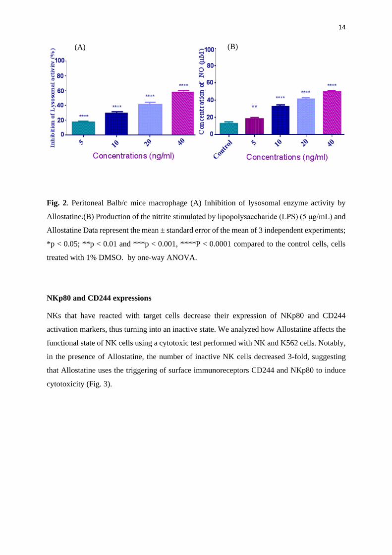
Allostatine inhibits the cellular lysosomal enzyme activity and increases nitric oxide production
Peritoneal macrophages incubated with different concentrations of Allostatine ranging from 5
to 40 ng/ml for 48 h showed a cellular lysosomal enzyme activity inhibition in a dose-dependent
manner. The most important inhibition was obtained at a 40 ng/ml concentration of Allostatine
(Fig. 2A). In addition, the effects of Allostatine on nitric oxide (NO) production by
macrophages in the presence of LPS revealed that Allostatin peptide dose-dependently induced
NO production in macrophages, up to 49.3µM at the concentration of 40 ng/mL (Fig. 2B)
(A) (B)
14
Fig. 2. Peritoneal Balb/c mice macrophage (A) Inhibition of lysosomal enzyme activity by
Allostatine.(B) Production of the nitrite stimulated by lipopolysaccharide (LPS) (5 μg/mL) and
Allostatine Data represent the mean ± standard error of the mean of 3 independent experiments;
*p < 0.05; **p < 0.01 and ***p < 0.001, ****P < 0.0001 compared to the control cells, cells
treated with 1% DMSO. by one-way ANOVA.
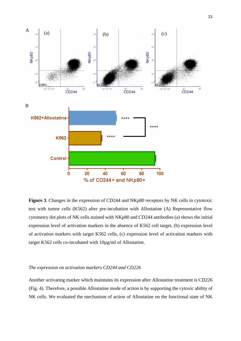
NKp80 and CD244 expressions
NKs that have reacted with target cells decrease their expression of NKp80 and CD244
activation markers, thus turning into an inactive state. We analyzed how Allostatine affects the
functional state of NK cells using a cytotoxic test performed with NK and K562 cells. Notably,
in the presence of Allostatine, the number of inactive NK cells decreased 3-fold, suggesting
that Allostatine uses the triggering of surface immunoreceptors CD244 and NKp80 to induce
cytotoxicity (Fig. 3).
(A) (B)
15
Figure 3. Changes in the expression of CD244 and NKp80 receptors by NK cells in cytotoxic
test with tumor cells (K562) after pre-incubation with Allostatine (A) Representative flow
cytometry dot plots of NK cells stained with NKp80 and CD244 antibodies (a) shows the initial
expression level of activation markers in the absence of K562 cell target, (b) expression level
of activation markers with target K562 cells, (c) expression level of activation markers with
target K562 cells co-incubated with 10µg/ml of Allostatine.
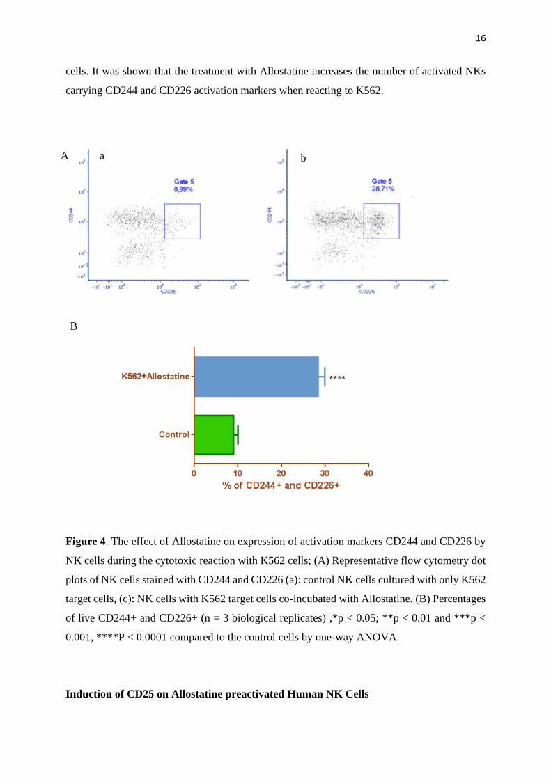
The expression on activation markers CD244 and CD226
Another activating marker which maintains its expression after Allostatine treatment is CD226
(Fig. 4). Therefore, a possible Allostatine mode of action is by supporting the cytoxic ability of
NK cells. We evaluated the mechanism of action of Allostatine on the functional state of NK
A
B
(c) (b) (a)
16
cells. It was shown that the treatment with Allostatine increases the number of activated NKs
carrying CD244 and CD226 activation markers when reacting to K562.
Figure 4. The effect of Allostatine on expression of activation markers CD244 and CD226 by
NK cells during the cytotoxic reaction with K562 cells; (A) Representative flow cytometry dot
plots of NK cells stained with CD244 and CD226 (a): control NK cells cultured with only K562
target cells, (c): NK cells with K562 target cells co-incubated with Allostatine. (B) Percentages
of live CD244+ and CD226+ (n = 3 biological replicates) ,*p < 0.05; **p < 0.01 and ***p <
0.001, ****P < 0.0001 compared to the control cells by one-way ANOVA.
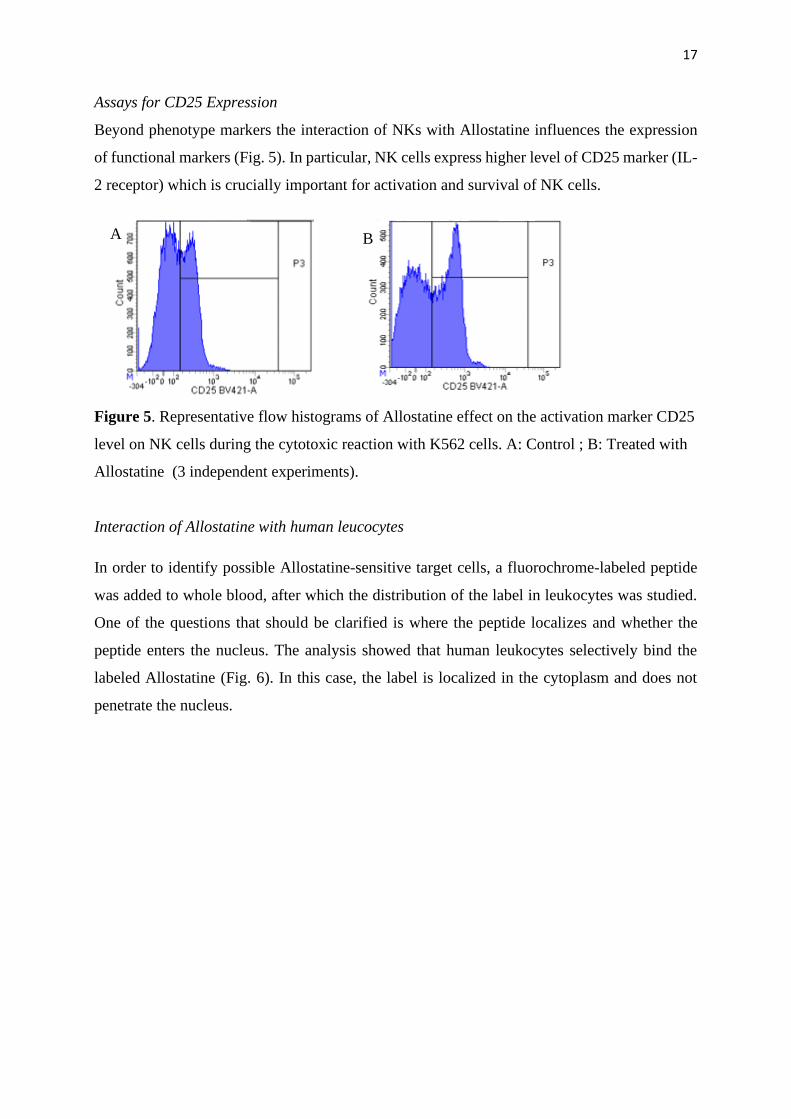
Induction of CD25 on Allostatine preactivated Human NK Cells
A
B
a b
17
Assays for CD25 Expression
Beyond phenotype markers the interaction of NKs with Allostatine influences the expression
of functional markers (Fig. 5). In particular, NK cells express higher level of CD25 marker (IL-
2 receptor) which is crucially important for activation and survival of NK cells.
Figure 5. Representative flow histograms of Allostatine effect on the activation marker CD25
level on NK cells during the cytotoxic reaction with K562 cells. A: Control ; B: Treated with
Allostatine (3 independent experiments).
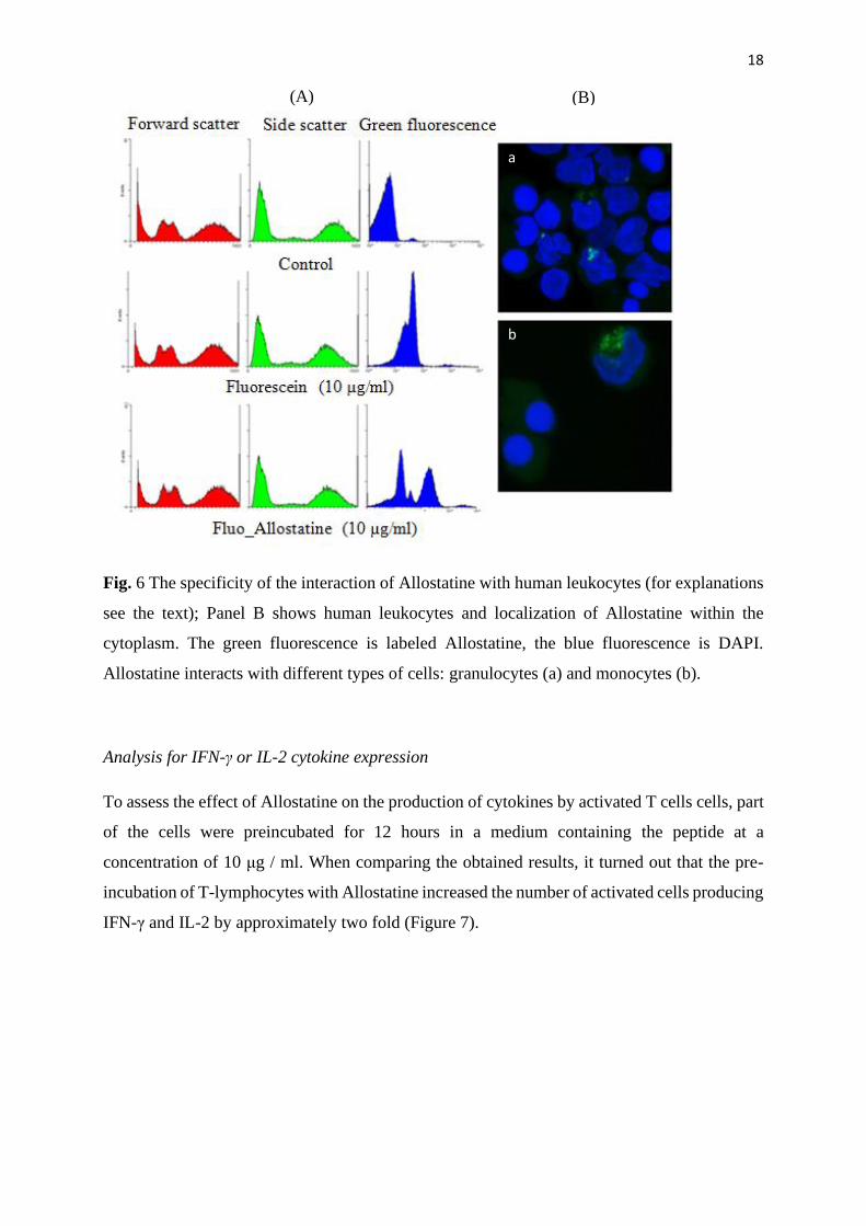
Interaction of Allostatine with human leucocytes
In order to identify possible Allostatine-sensitive target cells, a fluorochrome-labeled peptide
was added to whole blood, after which the distribution of the label in leukocytes was studied.
One of the questions that should be clarified is where the peptide localizes and whether the
peptide enters the nucleus. The analysis showed that human leukocytes selectively bind the
labeled Allostatine (Fig. 6). In this case, the label is localized in the cytoplasm and does not
penetrate the nucleus.
A B
18
Fig. 6 The specificity of the interaction of Allostatine with human leukocytes (for explanations
see the text); Panel B shows human leukocytes and localization of Allostatine within the
cytoplasm. The green fluorescence is labeled Allostatine, the blue fluorescence is DAPI.
Allostatine interacts with different types of cells: granulocytes (a) and monocytes (b).
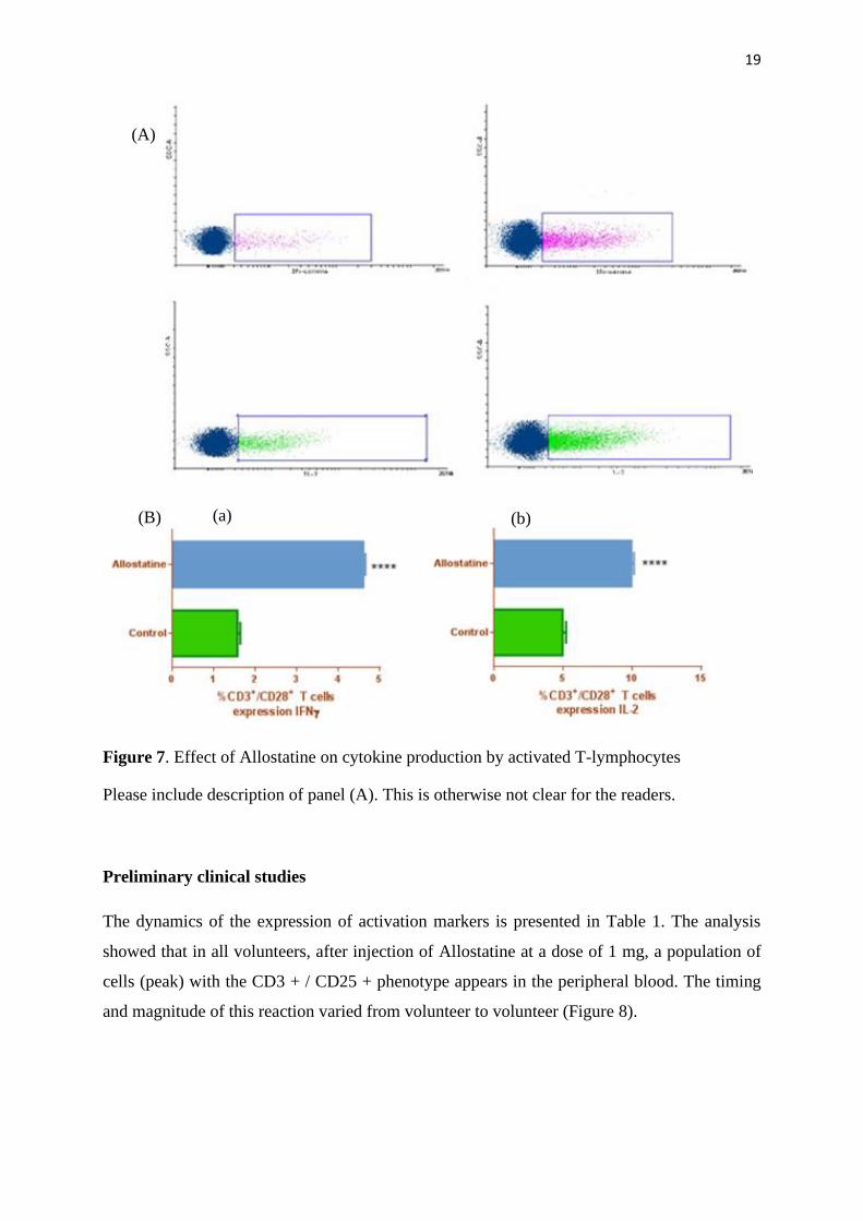
Analysis for IFN-γ or IL-2 cytokine expression
To assess the effect of Allostatine on the production of cytokines by activated T cells cells, part
of the cells were preincubated for 12 hours in a medium containing the peptide at a
concentration of 10 μg / ml. When comparing the obtained results, it turned out that the pre-
incubation of T-lymphocytes with Allostatine increased the number of activated cells producing
IFN-γ and IL-2 by approximately two fold (Figure 7).
(A) (B)
a
b
19
Figure 7. Effect of Allostatine on cytokine production by activated T-lymphocytes
Please include description of panel (A). This is otherwise not clear for the readers.
Preliminary clinical studies
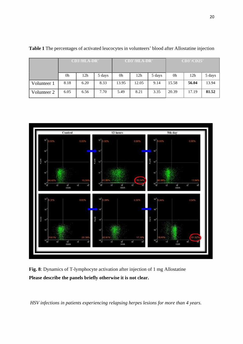
The dynamics of the expression of activation markers is presented in Table 1. The analysis
showed that in all volunteers, after injection of Allostatine at a dose of 1 mg, a population of
cells (peak) with the CD3 + / CD25 + phenotype appears in the peripheral blood. The timing
and magnitude of this reaction varied from volunteer to volunteer (Figure 8).
(A)
(B) (a) (b)
20
Table 1 The percentages of activated leucocytes in volunteers’ blood after Allostatine injection
CD3-/HLA-DR+ CD3+/HLA-DR+ CD3+/CD25+
0h 12h 5 days 0h 12h 5 days 0h 12h 5 days
Volunteer 1 8.18 6.20 8.33 13.95 12.05 9.14 15.58 56.04 13.94
Volunteer 2 6.05 6.56 7.70 5.49 8.21 3.35 20.39 17.19 81.52
Fig. 8: Dynamics of T-lymphocyte activation after injection of 1 mg Allostatine
Please describe the panels briefly otherwise it is not clear.
HSV infections in patients experiencing relapsing herpes lesions for more than 4 years.
21
The preliminary clinical data characterizing Allomedin’s therapeutic efficacy in patients
infected with Herpes simplex virus involved results from 104 patients (81 females, 23 males)
experiencing relapsing herpes of different localizations for more than 4 years before the study.
The age of the patients varied from 18 to 60 years. Allomedin was applied onto the surface of
herpetic lesions 2 to 3 times a day during 3 to 5 days. Results of the studies are summarized in
Table 2.
Table 2. Allomedin clinical efficacy in the therapy of herpes lesion relapses with reference to
the lesions localization (summary of a multicenter clinical study).
Localization Therapeutic efficacy evaluation
Patients number Positive responses
N %
Skin 27 26 96,3
Vagina or urethra mucous tunic 44 41 93,1
Mouth mucous tunic 33 20 66,6
Data of Table 2 demonstrate that the majority of patients with skin herpes and genital mucous
tunic herpes (labial and genital herpes, correspondingly) can be effectively treated by
Allomedin. Clinical studies of Allomedin efficacy in labial and genital herpes demonstrated
also an extraordinary fast relief of herpes symptoms, including reduced burning duration and
reduction in size of oedema zone, when compared to Acyclovir which was the comparative
standard antiviral. Tables 3 and 4 illustrate this fact. Allomedin rapidly eliminated inflammation
symptoms like itch, burning, oedema such that the duration of the symptoms decreases about
10-fold when compared to acyclovir treatment.
Table 3. Allomedin and acyclovir comparative efficacy in the treatment of cold sores (labial
herpes) symptoms.
Index The index rate (duration in hours)
Acyclovir Allomedin
Itch and burning duration 60 – 84 4-8
50% decrease in the size of
oedema zone
84 -108 8-12
22
Total relapse duration 168 – 192 72 – 96
Table 4. Allomedin and acyclovir comparative efficacy in the elimination of genital herpes
symptoms
Indices and stages Symptoms duration, hours
Acyclovir Allomedin
Itch 84,3 ± 9,4 13,6 ± 3,7
Burning 93,2 ± 12,8 14,0 ± 2,2
Vesicular-erosive stage 96,2 ± 12,8 54,6 ± 10,2
Scab stage 114,1 ± 20,8 98,0 ± 12,8
Treatment of HSV infections in patients with chronic recurrent herpes simplex.
In this second study 156 patients, 80 men and 76 women with a clinical diagnosis of chronic
recurrent herpes simplex were included for the treatment. Genital herpes was diagnosed in 53
patients (33.97%), orofacial herpes in 98 (62.82%), herpes in other sites in 5 (3.21%) (belly -
1, brush - 1, nates - 3 patients). All patients participating in the study had exacerbation of herpes
infection (no later than 3 days from the onset of relapse).
For the division of patients into 3 groups, depending on the drug used, the method of adaptive
randomization was used, while gender characteristics and localization of the herpetic process
were not taken into account (genital and labial herpes were taken as equivalent pathology).
Patients of group 1 received for external treatment for 10 days gel “Allomedin” (every 8 hours
- 3 times a day for the lesions), patients of group 2 were given - Zovirax cream (every 4 hours
- 5 times a day), the third group was treated with "Fenistil Pentsivir" –cream (every 2 hours - 8
times a day). The patient received the drug in the form of monotherapy. If the lesions regressed
earlier, the use of the drug was interrupted. For “Allomedin” treatment in group 1, itching, pain
and burning sensation regressed towards the 2nd visit (on the 3rd day). Also, all complaints for
patient in group 1 treated with Allomedin regressed almost simultaneously: itching - for 3.8 ±
0.4 days, pain and burning - for 3.7 ± 0.4 days. Pain regressed in 3.3 ± 0.3 and 4.3 ± 0.5 in
groups 2 and 3 recepectively. Burning sensation was registered over 3.9 ± 0, 5 and 4.3 ± 0.7
days, in groups 2 and 3 respectively. Herpetic lesions and itching were regressed somewhat
slower compared with group 1 (4.3 ± 0.5, p> 0.2 and 5.3 ± 0.7 , p> 0.05 in group 2 and 3
recepectively).
23
Objective symptoms of genital infection (GI) in patients of group 1 regressed in an average of
8.76 ± 0.4 days (for 210.4 ± 10.2 hours), for the group 2 the regression was registered 9.7 ±
0.3 days (for 232.9 ± 7.1 hours, p> 0.05), while patients in group 3 present regration after 9.5 ±
0.3 days (for 227.3 ± 7.7 hours, p> 0.1). These data can be described as the average duration of
relapse.
All the researchers involved noted a potential epithelial effect of Allomedin compared with
other drugs. When compared with the control drugs treatment 58.8% of patients in group 1
presented eruptions in the exposed vesicular eruptions site. However, 88.2% * (p <0.05) and
94.1% * patients (p <0.01) presented eruptions in group 2 and 3, recepectively, when compared
with group 1. Furthermore, we registered a significantly faster epithelialization in 5.0 ± 0.6 days
in group 1, when compared with other groups. The process of epithelialization in group 2 took
7.1 ± 0.6 * days ( p <0.02 compared with group 1), for the third group this indicator was 6.6 ±
0.4 * days (p <0.05 compared with group 1). Consequently, the formation and drop of crusts on
the sites of the former eruptions also occurred faster in the Allomedin group at 8.4 ± 0.4 days
(in group 2 at 9.5 ± 0.3 * days, p <0.05; in group 3 at 9.2 ± 0.5 days, p> 0.2, when compared
with group 1).
A decrease in the level of herpetic eruptions was noted by a factor of 2 (in group 1, it was 4.8
± 0.4 days (114.4 ± 10.8 hours). In the 2nd group, 6.0 ± 0.4 days (in 144.0 * ± 10.2 hours, p
<0.05 compared with group 1), in the 3rd group day - by 5.7 ± 0.5 days (for 136.9 ± 11.0 hours,
p> 0 , 1 compared to group 1). We registered for 1 patient of the 3rd group with genital herpes
at the second visit, the appearance of 2 small fresh elements along the periphery of the main
nidus (against the background of regular use of Fenistil Pentsivir gel).
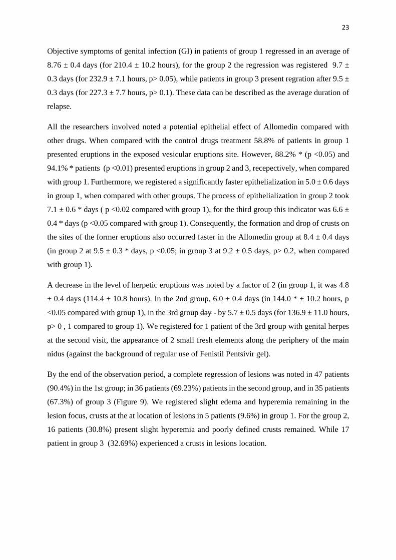
By the end of the observation period, a complete regression of lesions was noted in 47 patients
(90.4%) in the 1st group; in 36 patients (69.23%) patients in the second group, and in 35 patients
(67.3%) of group 3 (Figure 9). We registered slight edema and hyperemia remaining in the
lesion focus, crusts at the at location of lesions in 5 patients (9.6%) in group 1. For the group 2,
16 patients (30.8%) present slight hyperemia and poorly defined crusts remained. While 17
patient in group 3 (32.69%) experienced a crusts in lesions location.
24
Figure 9. Complete regression of herpes clinical manifestations after Allomedin, Fenistil
Pentsivir and Zovirax treatments
Based on the total summation of all indicators (regress of complaints, clinical symptoms,
tolerance of the prescribed drug), patients and Medical Researchers evaluated the effectiveness
of the treatment of Herpes Infection exacerbation. A good effect after treatments was indicated
by 92.3% of patients in group 1, 75% in group 2 and 69.23% of patients in group 3. We noted
as a “satisfactory” treatment result in 7.7% of the patients for group 1, and 25% of the patients
in group 2, and 28.85% of the patients in group 3. The ‘unsatisfactory “effect of the treatment
was noted by 1 patient of the 3 groups. The effect of the treatment in this case was regarded by
the doctor as satisfactory.
The subjective assessment of the treatment performed by the patient and the doctor in most
cases coincided in all groups. In group 1, the opinions of the patient and the doctor coincided
in 98.08% of cases, only in 1 case the patient assessed the result of treatment as satisfactory,
and the doctor as good. In group 2 in 5 patients (9.61%) the opinions of the doctor and the
patient differed in the assessment of treatment: in 3 cases the patient rated the result as
satisfactory, and the doctor as good, in 2 cases the opposite. In group 3, the discrepancy was
noted for 4 patients - 7.69%: in 1 case the patient rated the treatment effect as good, and the
doctor as satisfactory, in 2 cases - on the contrary, in the 4th case the patient considered the
effect of treatment unsatisfactory and the doctor is satisfactory (table).
25
The safety of the use of the gel "Allomedin" is proven by the absence of adverse events
associated with the use of the drug, as well as the absence of local and common adverse and
allergic reactions. In this regard, the drug can be recommended as a choice for widespread use
in clinical practice. Based on the total summation of all indicators (regression of complaints,
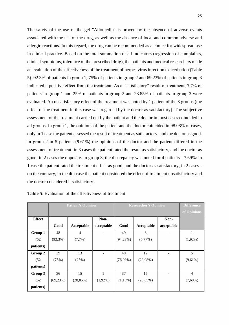
clinical symptoms, tolerance of the prescribed drug), the patients and medical researchers made
an evaluation of the effectiveness of the treatment of herpes virus infection exacerbation (Table
5). 92.3% of patients in group 1, 75% of patients in group 2 and 69.23% of patients in group 3
indicated a positive effect from the treatment. As a “satisfactory” result of treatment, 7.7% of
patients in group 1 and 25% of patients in group 2 and 28.85% of patients in group 3 were
evaluated. An unsatisfactory effect of the treatment was noted by 1 patient of the 3 groups (the
effect of the treatment in this case was regarded by the doctor as satisfactory). The subjective
assessment of the treatment carried out by the patient and the doctor in most cases coincided in
all groups. In group 1, the opinions of the patient and the doctor coincided in 98.08% of cases,
only in 1 case the patient assessed the result of treatment as satisfactory, and the doctor as good.
In group 2 in 5 patients (9.61%) the opinions of the doctor and the patient differed in the
assessment of treatment: in 3 cases the patient rated the result as satisfactory, and the doctor as
good, in 2 cases the opposite. In group 3, the discrepancy was noted for 4 patients - 7.69%: in
1 case the patient rated the treatment effect as good, and the doctor as satisfactory, in 2 cases -
on the contrary, in the 4th case the patient considered the effect of treatment unsatisfactory and
the doctor considered it satisfactory.
Table 5: Evaluation of the effectiveness of treatment
Patient’s Opinion
Researcher’s Opinion Difference
of Opinions
Effect
Good
Acceptable
Non-
acceptable
Good
Acceptable
Non-
acceptable
Group 1
(52
patients)
48
(92,3%)
4
(7,7%)
- 49
(94,23%)
3
(5,77%)
- 1
(1,92%)
Group 2
(52
patients)
39
(75%)
13
(25%)
- 40
(76,92%)
12
(23,08%)
- 5
(9,61%)
Group 3
(52
patients)
36
(69,23%)
15
(28,85%)
1
(1,92%)
37
(71,15%)
15
(28,85%)
- 4
(7,69%)
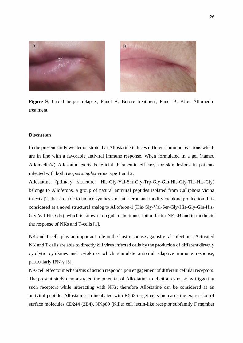
26
Figure 9. Labial herpes relapse.; Panel A: Before treatment, Panel B: After Allomedin
treatment
Discussion
In the present study we demonstrate that Allostatine induces different immune reactions which
are in line with a favorable antiviral immune response. When formulated in a gel (named
Allomedin) Allostatin exerts beneficial therapeutic efficacy for skin lesions in patients
infected with both Herpes simplex virus type 1 and 2.
Allostatine (primary structure: His-Gly-Val-Ser-Gly-Trp-Gly-Gln-His-Gly-Thr-His-Gly)
belongs to Alloferons, a group of natural antiviral peptides isolated from Calliphora vicina
insects [2] that are able to induce synthesis of interferon and modify cytokine production. It is
considered as a novel structural analog to Alloferon-1 (His-Gly-Val-Ser-Gly-His-Gly-Gln-His-
Gly-Val-His-Gly), which is known to regulate the transcription factor NF-kB and to modulate
the response of NKs and T-cells [1].
NK and T cells play an important role in the host response against viral infections. Activated
NK and T cells are able to directly kill virus infected cells by the producion of different directly
cytolytic cytokines and cytokines which stimulate antiviral adaptive immune response,
particularly IFN-γ [3].
NK-cell effector mechanisms of action respond upon engagement of different cellular receptors.
The present study demonstrated the potential of Allostatine to elicit a response by triggering
such receptors while interacting with NKs; therefore Allostatine can be considered as an
antiviral peptide. Allostatine co-incubated with K562 target cells increases the expression of
surface molecules CD244 (2B4), NKp80 (Killer cell lectin-like receptor subfamily F member
A B
27
1 - KLRF1) and CD226. CD244 signaling pathway involves the interaction with the small
intracellular SH2 domain–containing protein 1, SH2D1A and with the Src homology 2 (SH2)-
domain of the Src tyrosine-family member Fyn, a non-receptor tyrosine-protein kinase
expressed in NK, T and B-cells (Engel et al., 2003) [5]. NKp80 is known to behave as an
activator of C the -type lectin-like immunoreceptor (CTLR) in human NK cells [6]. It has also
been shown that Allostatine may activate CD226 (also known as DNAM-1 or DNAX accessory
molecule-1), a 65 kDa glycoprotein expressed on the surface of NKs, platelets, monocytes and
a subset of T cells [7].
Interestingly, CD244, Nkp80 and CD226 have been previously described as inducers of IFNγ
production [8]. CD244 is a member of SLAM (signaling lymphocytic activation molecule)
protein family with the ability to induce IFNγ secretion and cytotoxicity. This stimulatory effect
is due to the presence in the intracytoplasmic tail of the extra tyrosine motif (Watzl et al., 2000)
[9]. Cytokine production and NK cytotoxicity can be explained as a consequence of NKp80 up-
regulation and its binding to the cognate ligand ‘activation-induced C-type lectin’ (AICL) [10].
Furthermore, CD226 induces the stimulation of NK cell–mediated cytotoxicity and enhance
secretion of IFN-γ [11].
After incubation of Allostatine with T cells, activated by CD3 and CD28, we observed an
increased secretion of both IL2 and IFNγ.
These findings are in line with data that have been demonstrated previously, indicating that
peptides of the Alloferons family induce NK cells cytotoxicity against cancer cells by up-
regulating the NK-activating receptors CD244 and with the production of IFNγ, TNF- and the
promotion of granule exocytosis [13].
In this study, we highlighted the potential generation of immune responses against viral
infections following Allostatine treatment by demonstrating activation of the CD25 marker of
NK cells in vitro. Also, we evaluated the increase of CD3 and CD25 expression by T
lymphocytes in Allostatine-treated patients. CD25 is the α-chain receptor of interleukin 2 (IL-
2Rα) (Waldmann, 1989) [14]. Our data demonstrated that Allostatine stimultes IL-2Rα receptor
on human NK and activated T cells, followed by cytokine pre-activation.
Many studies demonstrated that the initial stages of an HSV infection are influenced by the
activity of IFNs , NK cells and T cells which serve to limit the spread of virus to the nervous
system [5, 6. 15] Other experiments report that macrophages are considered as potent early
inhibitors of HSV infection [16]. Activated macrophages inhibit pathogen replication by
releasing nitric oxide [17].
28
The observed allostatine-related immune response data are coherent with the results of the
clinical study performed with allostatine as a the treatment of recurrent herpes simplex lesions.
Our data proved the effectiveness and safety of Allostatine against both HSV1 and HSV2. Its
high efficacy and good tolerability as a topical treatment, even as a monotherapy for these
diseases.
The allostatine-containing gel "Allomedin" quickly suppressed subjective sensations and
objective manifestations of exacerbation of herpes infection, especially with early treatment
(please explain). Allomedin treatment showed a superior and significantly different clinical
benefit when compared to Zovirax and Fenistil Petsivir cream treatments. The clinical data
obtained also showed high degree of compliance of the treatment. In addition, Allomedin
evoked a more pronounced epithelial effect, reducing the period of formation and falling away
of the crusts. The application of the drug 2-3 times a day showed excellent cosmetic properties
suggesting the use of Allomedin gel to stop exacerbations of labial or genital herpes of any
severity. This was also confirmed by a subjective assessment of the treatment, given by the
patients and the doctor. These finding support Allomedin as a means of choice for widespread
use in clinical practice.
The tolerability of all the drugs for external use was rated as Good, with no registered adverse
local neither general reactions, nor allergic. Notably, patients of the group treated with
Allomedin noted the convenience of using Allomedin gel only 2-3 times a day. Allomedin is
reported also for its good organoleptic property such us the transparency and no-shining was
reported after Allomedin application.
Conclusion
According to our clinical data, Allostatine and it gel formulation Allomedin, demonstrate
excellent therapeutic efficacy and safety as a therapy for skin and mucous tunic HSV1 and
HSV2 infections. In fact, the clinical data from the present study confirm that 90 to 95% of
colds sores and genital herpes cases can be effectively controlled by Allomedin treatment. To
the best of our knowledge, there is no other drug or cosmeceutical that is currently as effective
as Allomedin for this indication. Nowadays about 90% of the human population is infected with
HSV. HSV causes lifelong infections manifesting in different forms varying from mild
disorders (cold sores) to severe chronic forms such as relapsing genital herpes. According to
European STD guidelines approved by the WHO [Patel et al., 2017], a group of drugs is
29
recommended for systemic administration (per os or iv) since their topical formulations have
very limited efficacy. We demonstrated that Allomedin eliminates most of the unpalatable
sympoms of HSV infections such as itch, burning, oedema, much faster than acyclovir. Worthy
to note is that long term treatment with acyclovir give rise to acyclovir resistant strains that,
obviously, worsen herpes therapy perspectives. Since the Allostatine mode of action is
completely different from that of acyclovir, Allomedin can be effectively used against acyclovir
resistant HSV as well.
References
Carter C.J. Interactions between the products of the Herpes simplex genome and Alzheimer's
disease susceptibility genes: relevance to pathological-signalling cascades. Neurochem Int.
2008 May;52(6):920-34.
Chernysh S., Kim S.I., Bekker G., Pleskach V.A., Filatova N.A., Anikin V.B., Platonov V.G.,
Bulet P. Antiviral and antitumor peptides from insects. Proc Natl Acad Sci U S A. 2002 Oct
1;99(20):12628-32.
Chernysh S.I., Filatova N.A., Chernysh N.S., Nesin A.P. Cytotoxic activity of blowfly
Calliphora vicina hemocytes J Insect Physiol. 2004 Sep;50(9):777-81.
Chernysh S., Irina K., Irina A. Anti-tumor activity of immunomodulatory peptide alloferon-1
in mouse tumor transplantation model. Int Immunopharmacol. 2012 Jan;12(1):312-4.
Chernysh S., Kozuharova I. Anti-tumor activity of a peptide combining patterns of insect
alloferons and mammalian immunoglobulins in naïve and tumor antigen vaccinated mice. Int
Immunopharmacol. 2013 Dec;17(4):1090-3
Kruglikova A.A: & Chernysh S.I. Surgical Maggots and the history of their medical use.
Entomological Review. 2013, v.93(6), 667-674
30
Looker K.J., Welton N.J., Sabin K.M., Dalal S., Vickerman P., Turner K.M.E., Boily M.C.,
Gottlieb S.L. Global and regional estimates of the contribution of herpes simplex virus type 2
infection to HIV incidence: a population attributable fraction analysis using published
epidemiological data. Lancet Infect Dis. 2020 Feb;20(2):240-249.
McQuillan G., Kruszon-Moran D., Flagg E.W., Paulose-Ram R. Prevalence of herpes simplex
virus type 1 and type 2 in persons aged 14–49: United States, 2015–2016. NCHS Data Brief,
no 304. Hyattsville, MD: National Center for Health Statistics. 2018.
Nissen J., Trabjerg B., Pedersen M.G., Banasik K., Pedersen O.B., Sørensen E., Nielsen K.R.,
Erikstrup C., Petersen M.S., Paarup H.M., Bruun-Rasmussen P., Westergaard D., Hansen T.F.,
Pedersen C.B., Werge T., Torrey F., Hjalgrim H., Mortensen P.B., Yolken R., Brunak S., Ullum
H., Burgdorf K.S. Herpes Simplex Virus Type 1 infection is associated with suicidal behavior
and first registered psychiatric diagnosis in a healthy population. Psychoneuroendocrinology.
2019 Oct;108:150-154.
Patel R1, Kennedy OJ2, Clarke E1, Geretti A3, Nilsen A4, Lautenschlager S5, Green J6,
Donders G7, van der Meijden W8, Gomberg M9, Moi H10,11, Foley E1. 2017 European
guidelines for the management of genital herpes. Int J STD AIDS. 2017 Dec;28(14):1366-1379.
doi: 10.1177/0956462417727194. Epub 2017 Aug 24.
Wald A. & Corey, L. (2007). "Chapter 36: Persistence in the population: epidemiology,
transmission". Human Herpesviruses: Biology, Therapy, and Immunoprophylaxis. Cambridge
University Press. ISBN 978-0-521-82714-0.
Whitaker I.S., Twine C., Whitaker M.J., Welck M., Brown C.S., Shandall A. Larval therapy
from antiquity to the present day: mechanisms of action, clinical applications and future
potential. Postgrad Med J. 2007 Jun;83(980):409-13

31