Embed Size (px)
Citation preview

Poly--caprolactone scaffold and reduced in vitro cellcultureBrugmans, M.C.P.; Driessen - Mol, A.; Rubbens, M.P.; Cox, M.A.J.; Baaijens, F.P.T.
Published in:Journal of Tissue Engineering and Regenerative Medicine
DOI:10.1002/term.1753
Published: 01/12/2015
Document VersionAccepted manuscript including changes made at the peer-review stage
Please check the document version of this publication:
• A submitted manuscript is the author's version of the article upon submission and before peer-review. There can be important differencesbetween the submitted version and the official published version of record. People interested in the research are advised to contact theauthor for the final version of the publication, or visit the DOI to the publisher's website.• The final author version and the galley proof are versions of the publication after peer review.• The final published version features the final layout of the paper including the volume, issue and page numbers.
Link to publication
Citation for published version (APA):Brugmans, M. C. P., Driessen - Mol, A., Rubbens, M. P., Cox, M. A. J., & Baaijens, F. P. T. (2015). Poly--caprolactone scaffold and reduced in vitro cell culture: beneficial effect on compaction and improved valvulartissue formation. Journal of Tissue Engineering and Regenerative Medicine, 9(12), E289-E301. DOI:10.1002/term.1753
General rightsCopyright and moral rights for the publications made accessible in the public portal are retained by the authors and/or other copyright ownersand it is a condition of accessing publications that users recognise and abide by the legal requirements associated with these rights.
• Users may download and print one copy of any publication from the public portal for the purpose of private study or research. • You may not further distribute the material or use it for any profit-making activity or commercial gain • You may freely distribute the URL identifying the publication in the public portal ?
Take down policyIf you believe that this document breaches copyright please contact us providing details, and we will remove access to the work immediatelyand investigate your claim.
Download date: 21. Apr. 2018

Poly-e-caprolactone scaffold and reduced in vitrocell culture: beneficial effect on compaction andimproved valvular tissue formationMarieke M. C. P. Brugmans1,2*, Anita Driessen-Mol2, Mirjam P. Rubbens1, Martijn A. J. Cox1
and Frank P. T. Baaijens21Xeltis, Eindhoven, The Netherlands2Department of Biomedical Engineering, Eindhoven University of Technology, The Netherlands
Abstract
Tissue-engineered heart valves (TEHVs), based on polyglycolic acid (PGA) scaffolds coated with poly-4-hydroxybutyrate (P4HB), have shown promising in vivo results in terms of tissue formation. However,a major drawback of these TEHVs is compaction and retraction of the leaflets, causing regurgitation. Toovercome this problem, the aimof this studywas to investigate: (a) the use of the slowly degrading poly-«-caprolactone (PCL) scaffold for prolongedmechanical integrity; and (b) the use of lower passage cellsfor enhanced tissue formation. Passage 3, 5 and 7 (P3, P5 and P7) human and ovine vascular-derived cells were seeded onto both PGA–P4HB and PCL scaffold strips. After 4 weeks of culture,compaction, tissue formation, mechanical properties and cell phenotypes were compared. TEHVswere cultured to observe retraction of the leaflets in the native-like geometry. After culture, tissuesbased on PGA–P4HB scaffold showed 50–60% compaction, while PCL-based tissues showed compactionof 0–10%. Tissue formation, stiffness and strength were increased with decreasing passage number; how-ever, this did not influence compaction. Ovine PCL-based tissues did render less strong tissues comparedto PGA–P4HB-based tissues. No differences in cell phenotype between the scaffold materials, species orcell passage numberswere observed. This study shows that PCL scaffoldsmay serve as alternative scaffoldmaterials for humanTEHVswithminimal compaction andwithout compromising tissue composition andproperties, while further optimization of ovine TEHVs is needed. Reducing cell expansion time will resultin faster generation of TEHVs, providingmore rapid treatment for patients. Copyright © 2013 JohnWiley& Sons, Ltd.
Received 23 October 2012; Revised 5 March 2013; Accepted 21 March 2013
Keywords heart valve; tissue engineering; scaffold; compaction; cell passage; extracellular matrix;mechanical properties
1. Introduction
With increasing number and ageing of the world popula-tion, valvular heart disease is an expanding health problem.Approximately 290 000 heart valve replacements areperformed annually worldwide and this number is esti-mated to increase to 850 000 by 2050 (Yacoub andTakkenberg, 2005). Bioprosthetic and mechanical heart
valves, which have been successfully used for decades,improve quality of life and life prolongation for mostpatients (Mendelson and Schoen, 2006; Shahbudin,2003). However, these valves have some restrictions, asthey consist of non-living and non-autologous materials.Therefore, they are not able to grow, adapt or remodelto changing physiological environments, resulting indecreased durability (Yacoub and Takkenberg, 2005).Furthermore, bioprosthetic valves are susceptible to calcifica-tion, whilemechanical valves require lifelong anticoagulationtherapy to prevent thromboembolism (Mendelson andSchoen, 2006; Yacoub and Takkenberg, 2005). Toovercome these problems, researchers are studying the
*Correspondence to: M. M. C. P. Brugmans, Xeltis and Depart-ment of Biomedical Engineering TU/e, Catalyst Building 2.07,PO Box 80, 5612 AR Eindhoven, The Netherlands. E-mail:[email protected]
Copyright © 2013 John Wiley & Sons, Ltd.
JOURNAL OF TISSUE ENGINEERING AND REGENERATIVE MEDICINE RESEARCH ARTICLEJ Tissue Eng Regen Med (2013)Published online in Wiley Online Library (wileyonlinelibrary.com) DOI: 10.1002/term.1753

possibility of creating tissue-engineered heart valves(TEHVs) (Mendelson and Schoen, 2006). Patients’ owncells are incorporated, resulting in valves of autologousliving tissue that are able to grow, remodel and adapt tothe changing environment after implantation (Mendelsonand Schoen, 2006). Our approach to create such TEHVs isto isolate patients’ cells from the vena saphena magna,expand them in vitro to the desired amount of cells andsubsequently seed them onto a biodegradable syntheticscaffold in the shape of a heart valve. After a culture periodin a bioreactor of 4 weeks, where the valves are exposed tomechanical stimuli in order to stimulate tissue formation,the valves are able to withstand systemic pressures inin vitro tests (Mol et al., 2006), aiming ultimately atimplanting them into patients.
Different types of synthetic scaffolds are used forcardiovascular tissue-engineering applications. In particu-lar, a polyglycolic acid (PGA) scaffold, coated with poly-4-hydroxybutyrate (P4HB) or combined with anotherscaffold material, was shown to be a promising candidatein terms of tissue formation, as demonstrated in vasculargraft studies (Cummings et al., 2011; Hoerstrup et al.,2006) and in vivo TEHV studies (Gottlieb et al., 2010;Hoerstrup et al., 2000; Schmidt et al., 2010; Weberet al., 2011). Hoerstrup et al. (2000) demonstrated in anovine model that after 20 weeks in vivo, the valves yieldedan organized, layered structure with many architecturalfeatures and extracellular matrix characteristics thatare present in native valves. In vivo, PGA and P4HB aredegraded completely within 4 and 8 weeks, respectivelyThe down side of using this rapid degrading PGA scaffoldis compaction (flattening of the leaflets) and retraction(shrinkage of the leaflets), causing regurgitation (Gottliebet al., 2010; Mol et al., 2005a; Weber et al., 2011). This isa result of traction forces exerted by the cells, likely incombination with an imbalance of the newly formedtissue and loss of mechanical integrity of the scaffolddue to degradation (Mol et al., 2006; van Vlimmerenet al., 2011, 2012). Rabkin-Aikawa et al. (2004) demon-strated TEHVs containing aSMA-positive cells duringin vitro culture, while after 20 weeks in vivo there was astrong decrease of aSMA-positive cells. As aSMA is relatedto traction forces of the cells (Chen et al., 2007), we assumethat after 20 weeks, these traction forces will be decreasedin vivo. Therefore, a scaffold with proper mechanicalintegrity during in vitro culture and the first months afterimplantation is desired to withstand the cell traction forcesduring this phase. The use of a slower degrading scaffoldmaterial, such as poly-e-caprolactone (PCL), may representa promising alternative, as TEHVs can be produced that aremechanically reliable for months, thereby offering sufficientmechanical integrity to prevent tissue compaction andretraction (Klouda et al., 2008). As PCL can be processedby electrospinning, it is possible to create complex geome-tries and mould the scaffold directly into the desiredthree-dimensional (3D) shape of a heart valve (Kloudaet al., 2008). This direct 3D moulding is not feasible forPGA scaffolds, which are only available in sheets. Anotherbenefit of PCL is the possibility of creating thin leaflets
(thickness 300 mm), while the PGA meshes are producedwith a thickness of 1000 mm. As PGA–P4HB scaffolds aremore rapidly degrading, the cells might be exposed to largermagnitudes of mechanical loading than the cells in PCLscaffolds, which might on their turn be partly protectedfrom loads by the long-term presence of the scaffold. Asthe stress level exerted on the vascular cells is known tochange the phenotype of the cells towards activatedmyofibroblasts (Hinz, 2010), the tissue formation capacityof cells in the two scaffold types might differ, along withdifferent phenotypes (Beamish et al., 2010; Hinz et al.,2007; Lacolley et al., 2012; Rensen et al., 2007). Therefore,it is important to compare cell phenotype, tissue formationcapacity and compaction in tissues based on both scaffoldtypes when considering the use of PCL as a scaffoldmaterialto produce TEHVs. Based on the above, we hypothesizethat the cells in PGA–P4HB might have a more activatedphenotype, accompanied by increased tissue formationcapacity, than cells in PCL scaffolds.
Another alternative to tackling the compaction andretraction of TEHVs might be by using cells of a lowpassage number. Ageing cells, due to in vitro expansion, losetheir potential to proliferate (Ragnauth et al., 2010; Safwaniet al., 2012). Currently, in our laboratory, cells are expandedto passage 6–7 (P6–7) to ensure enough cells for seedingmultiple TEHVs (Mol et al., 2006). Whether the amountof tissue formation or cell phenotype in 3D cultures isinfluenced by the use of cells of a low passage number is stillunclear, as to the best of our knowledge previous work onthe effect of cell ageing by expansion has been performedon 2D cultures only. Therefore, the role of cell ageing in3D tissue formation capacity needs to be investigatedfurther. We hypothesize that cells of a low passage number(P3) are more productive, resulting in more tissue forma-tion and of a higher quality, compared to cells of a highpassage number (P7). This improved tissue-formationcapacity in its turn may result in less compaction and retrac-tion, as it is influencing the balance between matrix qualityand the mechanical integrity of the scaffold towardsincreased matrix quality. We assume that the increasedmatrix formation will increase the resistance to the tractionforces exerted by the cells. An additional benefit of usingcells of a lower passage number is the reduction in cellexpansion time, which will result in faster generationof TEHVs and, thereby, provide more rapid treatmentto patients.
To summarize, the aim of this study was to evaluatealternative approaches to overcoming the compaction andretraction of TEHVs, as observed with the use of rapidlydegrading PGA–P4HB scaffolds, without compromisingtissue composition and properties. The alternativeapproaches studied here were: (a) the use of a slowlydegrading PCL scaffold for prolonged mechanical integ-rity; and (b) the use of lower passage vascular cells forenhanced tissue formation. Compaction, tissue forma-tion, cell phenotype and mechanical properties ofengineered tissues based on P3, P5 and P7 vascular cellsin both PCL and PGA–P4HB scaffolds were compared.TEHVs aim to be designed for humans, but since the
M. M. C. P. Brugmans et al.
Copyright © 2013 John Wiley & Sons, Ltd. J Tissue Eng Regen Med (2013)DOI: 10.1002/term

ovine model is commonly used to show proof of principle,both human and ovine cells were used.
2. Materials and methods
2.1. Cell culture
Human vascular-derived cells were harvested fromsegments of a vena saphena magna from a 60 year-oldpatient who underwent bypass surgery, and were obtainedaccording to the Dutch guidelines for secondarily usedmaterials. Ovine vascular-derived cells were obtained fromthe vena jugularis of adult sheep ca. 2 years old (n=2;Swifter). The cells were isolated via the outgrowth method.In short, endothelial cells of the vessels were removed byincubation with a collagenase solution. The remainingendothelial cells were removed from the lumen sideusing a cell scraper. After removal of the endothelialcells, the vessels were minced into small pieces(ca. 1 mm2) and the fragments were plated into six-wellplates. The outgrowing cells were expanded usingstandard culture methods in a humidified atmospherecontaining 5% CO2 at 37�C, and passaged at 90–100%confluency. Plating densities were 3.3–4.6�103 cells/cm2
for human and 1.6–2.3�104 cells/cm2 for ovine cells,based on differences in cell size. The isolation andexpansion medium consisted of advanced Dulbecco’smodified Eagle’s medium (DMEM; Invitrogen, Breda,The Netherlands), supplemented with 1% GlutaMax(Invitrogen), 1% penicillin–streptomycin (P–S; Lonza,Basel, Switzerland) and 10% fetal bovine serum(FBS; Greiner BioOne, Frickenhausen, Germany) for humancells or 10% lamb serum (Invitrogen) for ovine cells.During culture, cells of all passage numbers grew in thecharacteristic ‘hill and valley’ morphology, indicatingsmooth muscle cells.
2.2. Scaffold preparation and sterilization
Rectangular strips (25� 5 mm) were cut out of PGAmeshes (PGA, Cellon, Bascharage, Luxemburg) andconventionally electrospun PCL meshes, with thicknessesof 1000 and 300 mm, respectively. As heart valves containa more complex geometry compared to strips, whichmight result in differences in terms of compaction,trileaflet heart valve scaffolds were fabricated usingscaffold meshes of the same thickness. PGA scaffolds wereadditionally coated with poly-4-hydroxybutyrate (P4HB,received via a collaboration with Professor Hoerstrup ofthe University Hospital, Zurich) to provide structuralintegrity to the mesh. The outer 3–4 mm of both PGAand PCL scaffold strips were attached onto stainless steelrings (RVS Paleis, Geleen, The Netherlands) using 15%polyurethane–tetrahydrofuran (PU, Desmopan) glue,leaving an 18� 5 mm area for cell seeding. The solventwas allowed to evaporate overnight in a vacuumoven. The PCL scaffolds were sterilized by g-irradiation
(Isotron, Ede, The Netherlands). PGA–P4HB scaffold steril-ization was achieved by immersion in 70% sterile ethanolfor 30 min. To facilitate cell attachment, the scaffolds wereincubated overnight with tissue-engineered (TE) medium,consisting of expansion medium supplemented with0.25 mg/ml L-ascorbic acid 2-phosphate (Sigma). Lambserum (0.1%) and FBS (10%) were added to ovine andhuman TE medium, respectively.
2.3. Cell seeding and tissue culture
P3, P5 and P7 cells were seeded onto both PGA and PCLscaffolds (n= 6/passage and scaffold for each cell type)at a seeding density of 20 million cells/cm3, using fibrinas a cell carrier (Mol et al., 2005b). In short, the cells weresuspended in TE medium containing thrombin (10 U/ml,Sigma). This cell suspension was mixed with an equalvolume of TE medium containing fibrinogen (10 mg/ml,Sigma) and dripped onto one side of the scaffolds beforepolymerization of the gel was accomplished. As controlstrips, three PGA and PCL scaffolds were seeded withfibrin only. After seeding, the scaffolds were placed in anincubator at 37�C for 30 min to allow polymerization ofthe fibrin gel. Thereafter, 6 ml TE medium was added toeach scaffold. The strips were cultured for 4 weeks andthe TE medium was changed twice a week. For the heartvalve cultures, P7 cells were used and seeded according tosimilar protocols as for the strips. After seeding, the valveswere placed in a bioreactor system and cultured for4 weeks (Dijkman et al., 2012b) .
2.4. Compaction
Compaction was assessed from upper view photographsof the strips that were taken once a week. The valves werephotographed after 4 weeks only. Compaction of thestrips was defined as the reduction of width comparedto the width at the start of culture. The photographs wereanalysed using the program ImageJ (v. 1.43u).
2.5. Biochemical assays
For the quantification of tissue formation after 4 weeks ofculture, TE strips were lyophilized after mechanicaltesting (n= 4–5/group) and used for biochemical assays.The total amount of DNA was determined as an indicatorof cell number, the amount of hydroxyproline as an indica-tor for collagen content and the amount of sulphatedglycosaminoglycans (sGAG). Measurements were averagedper group. Lyophilized tissue samples were weighed anddigested in papain solution (100 mM phosphate buffer, pH6.5, 5 mM L-cysteine, 5 mM ethylene-diamine-tetra-aceticacid (EDTA) and 125–140 mg papain/ml; all from Sigma)at 60�C for 16 h. After centrifuging the samples, the digestsupernatant was collected and used for the DNA, sGAGand collagen assays. The amount of DNA in the TEstrips was determined using the Hoechst dye method
PCL scaffold and reduced in vitro cell culture for improved TE valves
Copyright © 2013 John Wiley & Sons, Ltd. J Tissue Eng Regen Med (2013)DOI: 10.1002/term

(Cesarone et al., 1979) and a standard curve prepared ofcalf thymus DNA (Sigma). Using the assumption that allcells contain 6.5 pg DNA (Dolezel et al., 2003), the amountof cells per TE construct was calculated. sGAG content wasdetermined using a modification of the protocol describedby Farndale et al. (1986). In short, 40 ml diluted samplewas pipetted into a 96-well plate in duplicate, followedby the addition of 150 ml/well dimethyl-methylene blue.Absorbance was measured at 540 nm. Subsequently, theamount of sGAGs in the TE strips was determined from areference curve prepared from shark cartilage chondroitinsulphate (Sigma). Collagen content was determined by anassay as described by Huszar et al. (1980) and a standardcurve was prepared from trans-4-hydroxyproline (Sigma).
2.6. Mechanical testing
After 4 weeks of culture, the mechanical properties ofthe TE strips (n= 4–5/group) were assessed by uniaxialtensile tests in the longitudinal direction, with a uniaxialtensile stage (Kammrath and Weis, Dortmund, Germany)equipped with a 20 N load cell. Mechanical test data wasaveraged per group. Thickness of the strips was deter-mined from representative histology sections. Sampleswere measured at three spots and mean thickness wasused. The standard deviation (SD) of the measurementswas in the range 1.5–10%. Stress–strain curves wereobtained and, as a measure of tissue strength, the ulti-mate tensile strength (UTS) was defined as the peakstress value. The elasticity modulus (E-modulus) wasdetermined as the slope of the linear (end) part of thecurve, as a measure for tissue stiffness.
2.7. Histology
To analyse tissue formation qualitatively, TE strips wereprocessed for histology (n= 1/group). Representativetissue samples were embedded in tissue freezing medium(Tissue Tek, Sakura, Torrance, USA) and cryosections of10 mm were cut. The sections were formalin-fixed andstudied by Masson’s Trichrome (MT) staining (MTC kit,Sigma, Venlo, The Netherlands) for collagen depositionand by Picrosirius Red (PR) staining to assess the maturityof the collagen matrix (Junqueira et al., 1979). The MTstaining was analysed using light microscopy and the PRstaining by polarized light microscopy (Axio Observer,Zeiss, Göttingen, Germany). In this study, maturity ofthe collagen fibres was assessed by the amount and den-sity of the collagen fibres visible with polarized light mi-croscopy. Mature fibres with a high density werecoloured orange/red, while immature or less dense fibreswere green.
Cell phenotype within the TE strips was analysed byimmunofluorescence. After acetone fixation for 10 min,the sections were incubated with 5% bovine serumalbumin (BSA) in phosphate-buffered saline (PBS) for30 min at room temperature. After blocking, the sections
were incubated with a primary antibody overnight at4�C. The antibodies used were mouse anti a-smoothmuscle actin (aSMA) to stain smooth muscle cells andmyofibroblasts (a2547, clone 1A4; 1:400; Sigma), mouseanti-smoothelin to stain contractile smooth muscle cells(clone R4A, 1:4; kindly provided by GJ van Eys, Universityof Maastricht) or rabbit anti-S100A4, which belongs tothe S100 superfamily of cytoplasmic calcium-bindingproteins, to stain fibroblasts and myofibroblasts (ab27957,1:200; Abcam). After primary antibody incubation,the sections were washed with PBS and incubated withAlexa 488-labelled secondary antibodies (1:300; Sigmaand Molecular Probes) to visualize the specific stainingsand DAPI (1:500; Sigma) to stain all cell nuclei for 30 minat room temperature. After staining, the sections weremounted with Mowiol 4–88 (Calbiochem, San Diego, CA,USA) and visualized by fluorescent microscopy (Axiovert200M, Zeiss, Göttingen, Germany).
2.8. Statistical analyses
All data are presented as mean� standard error of themean (SEM). Data of all experiments were normalizedto human P3 PGA–P4HB strips in each experiment, in or-der to be able to compare experiments and perform statis-tical analyses. Pearson correlation coefficients werecalculated to determine correlations between tissue pa-rameters and cell passage numbers for both species andscaffold groups. Unpaired t-tests were used to comparethe tissue properties between the scaffold materialswithin one cell passage and species, and to compare thetissue properties between species, within the same scaf-fold material and cell passage number. Statistics wereperformed using Graphpad Prism v. 5.04 and differenceswere considered significant at p< 0.05.
3. Results
3.1. Compaction after 4 weeks
The remaining width of the strips of all groups after4 weeks of culture is shown in Figure 1A. A remainingwidth of strips of 100% is the initial width of the stripsand represents no compaction. Tissues based on PCLscaffold and PCL and PGA–P4HB control strips showedcompaction of 0–10%. Tissues based on PGA–P4HBscaffold resulted in significantly more compaction(ca. 50%) after 4 weeks (p< 0.001).
In ovine strips, no significant correlations were foundbetween passage number and both types of scaffold. Anegative correlation was found between human cellpassage numbers and PGA–P4HB strips (p< 0.01), whilethere was a positive correlation between the human cellpassage numbers and PCL strips (p< 0.05). This indicatesthat passage number and species did not consistentlyinfluence compaction. TEHVs based on PGA–P4HBscaffolds showed severe compaction and retraction of
M. M. C. P. Brugmans et al.
Copyright © 2013 John Wiley & Sons, Ltd. J Tissue Eng Regen Med (2013)DOI: 10.1002/term

the leaflets after 4 weeks of culture in both species,while no compaction or retraction was observed in thePCL-based valves (Figure 1B–E), confirming the resultsfound using the engineered strips.
3.2. Biochemical assays
Normalized collagen and sGAG per DNA of all groups arepresented in Figure 2. Significant negative correlationsbetween cell passage numbers and collagen amountper DNA were found in both human and ovine tissues ofboth scaffold materials (p< 0.001), demonstrating thatincreasing passage number resulted in decreased collagenper DNA. Low amounts of collagen per DNA weredetectable in ovine PCL p7 strips. In general, ovine tissuestrips demonstrated an increased amount of collagenwhen compared to human (p< 0.001). Collagen contentper DNA of both human and ovine P7 cells seeded onPCL scaffolds was decreased, compared to humanand ovine cells that were seeded on PGA–P4HB scaffolds(p< 0.05 for human cells; p< 0.001 for ovine cells).Although we showed that collagen and sGAG per DNAwere increased with decreasing passage number, nodifferences in compaction of the tissues could be observed.
Biochemical parameters are related, as observed bycorrelation matrices, showing that collagen per DNA wasincreased when sGAG per DNA was increased. Overall, theamount of sGAG per DNA decreased with increasing cellpassage (p< 0.05 for ovine PGA–P4HB strips; p< 0.001for human PCL strips), although this effect was lesspronounced with collagen per DNA. Except for ovine P7PCL strips, ovine cells resulted in a higher amount of sGAGper DNA compared to human cells (p< 0.05 for ovine p3PCL strips; p< 0.001 for all other ovine strips). No consis-tent differences in sGAG content by the cells were observeddue to different scaffold materials.
3.3. Mechanical testing
Figures 3A and 3B, show averaged stress–strain curves ofhuman and ovine P3 strips, which are representative forthe other passage numbers, and the PGA–P4HB and PCLcontrol strips. Bare PCL strips were able to bear higherstresses than bare PGA–P4HB strips, which was due tothe differences in degradation time of the two scaffoldmaterials. The PGA–P4HB cultured tissues of both humanand ovine cells showed typical non-linear mechanicalbehaviour representing tissue behaviour. When PCL
Figure 1. (A) Compaction of strips after 4 weeks of culture. Initial width of strips was set at 100% (dotted line). PGA–P4HB showedaround 50% compaction of the strips, while the use of PCL strips demonstrated reduced compaction, as the final reduction in widthwas only 0–10%. **Difference between the scaffold materials, p<0.001; #, ##significant differences of p<0.05 and p<0.001, respec-tively, compared to human tissues. Negative or positive Pearson r correlations between the cell passage numbers are presented byarrows, combined with their p-values. Species and cell passage number did not consistently influence compaction of the TE strips.(B-E) Top-view photos of human PGA–P4HB (B); human PCL (C); ovine PGA–P4HB (D); and ovine PCL (E) TEHVs after 4 weeks ofculture. Valves based on PGA–P4HB scaffold resulted in severe retraction of the leaflets after 4 weeks, while PCL valves did not showthis. These results were consistent for both human and ovine cells
PCL scaffold and reduced in vitro cell culture for improved TE valves
Copyright © 2013 John Wiley & Sons, Ltd. J Tissue Eng Regen Med (2013)DOI: 10.1002/term

scaffold was used, the human tissues showed linearmechanical behaviour, while the ovine tissues werefollowing the curve of the control PCL strips. Thus, PCLscaffolds are still influencing the mechanical propertiesof the engineered tissues after 4 weeks of culture, whilePGA–P4HB scaffolds do not.
With a decrease in cell passage numbers, the parame-ters stiffness and strength were increasing in both speciesand scaffold materials, as significant negative correlationswere observed between increasing cell passage numbersand both the stiffness (p< 0.05 for human PGA–P4HBstrips; p< 0.001 for human PCL and ovine PGA–P4HBstrips) and strength (p< 0.05 for human PGA–P4HB and
ovine PCL strips; p< 0.001 for human PCL and ovinePGA–P4HB strips) in human and ovine tissues based onboth scaffold materials (Figure 3C, D). In human tissuesamples, stiffness was higher in PCL samples comparedto PGA–P4HB samples (p< 0.05 in P3 and P7 tissues),while in ovine tissue samples a higher stiffness wasobtained in tissues based on PGA–P4HB scaffoldscompared to PCL scaffolds (p< 0.05). Furthermore, tissuestrength was increased in human PCL samples of allpassage numbers and ovine PCL samples at P5 andP7 compared to PGA–P4HB tissue samples (p< 0.05),which was probably due to the influence of the PCLscaffold not yet degraded. When PCL scaffold was used,
Figure 2. Collagen per DNA (A) and sGAG per DNA (B). #, ##Significant differences compared to human tissues; *,**significances ofdifferences between scaffold materials, p<0.05 and p<0.001, respectively. Pearson r correlations between the cell passage numbersare presented by arrows combined with their p- values. Collagen per DNA decreases with increasing passage number in both humanand ovine tissues and both scaffold materials (A). sGAG per DNA show the same trends, although less distinct (B). The scaffold doesnot influence the amount of collagen and sGAG formed, while more collagen and sGAG per DNA are formed within ovine tissuescompared to human tissues
Figure 3. Mechanical data of engineered strips. Averaged stress–strain curves of human (A) and ovine (B) p3 strips are given as mean�SEM. PGA–P4HB-based tissues demonstrate non-linear curves in both human and ovine strips, representing tissue behaviour. Thestress–strain curve of human PCL strips is linear, while ovine strips follow the curve of the control scaffolds. Control PCL scaffoldsare still influencing mechanical properties after 4 weeks of culture, while PGA–P4HB scaffolds are not. Tissue stiffness (C) andstrength (D) increase with decreasing passage number. #,##Significant differences compared to human tissues; *,**significances ofdifferences between scaffold materials, p<0.05 and p<0.001, respectively. Pearson r correlations between the cell passage numbersare presented by arrows combined with their p-values. In human samples, the highest values are obtained in PCL strips, while in ovinesamples this is observed in PGA–P4HB scaffold strips
M. M. C. P. Brugmans et al.
Copyright © 2013 John Wiley & Sons, Ltd. J Tissue Eng Regen Med (2013)DOI: 10.1002/term
the values of the mechanical properties of the ovinetissues were equally or just slightly increased comparedto the PCL control strips, while the values of the humantissues were higher than the control strips (data not
shown). This indicated that tissues newly formed by ovinecells were not of the same quality as their human counter-parts, as the added value of tissue to the mechanicalproperties of the ovine strips was relatively low.
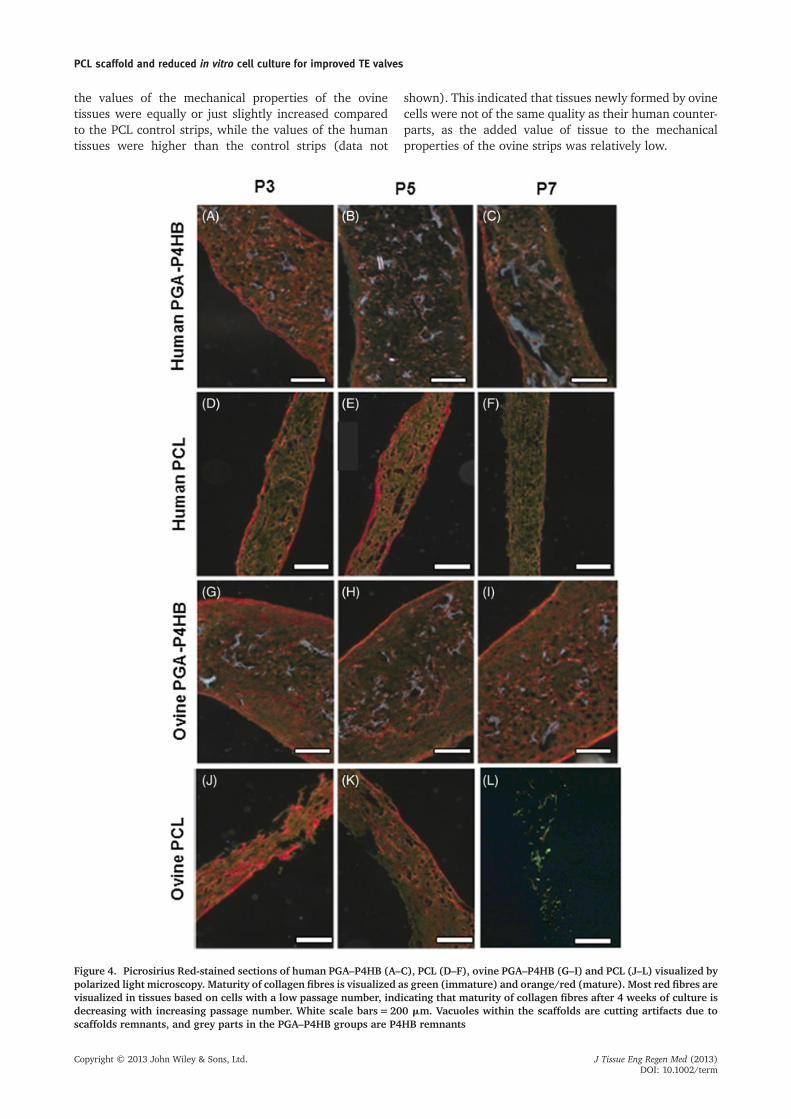
Figure 4. Picrosirius Red-stained sections of human PGA–P4HB (A–C), PCL (D–F), ovine PGA–P4HB (G–I) and PCL (J–L) visualized bypolarized light microscopy. Maturity of collagen fibres is visualized as green (immature) and orange/red (mature). Most red fibres arevisualized in tissues based on cells with a low passage number, indicating that maturity of collagen fibres after 4 weeks of culture isdecreasing with increasing passage number. White scale bars=200 mm. Vacuoles within the scaffolds are cutting artifacts due toscaffolds remnants, and grey parts in the PGA–P4HB groups are P4HB remnants
PCL scaffold and reduced in vitro cell culture for improved TE valves
Copyright © 2013 John Wiley & Sons, Ltd. J Tissue Eng Regen Med (2013)DOI: 10.1002/term

Correlation matrices demonstrated that the mechan-ical parameters were related to one another, resultingin increased tissue strength when tissue stiffnessobtained higher values, while the mechanical parame-ters were not related to the matrix properties ofthe tissues.
3.4. Histology
Histology of the TE strips revealed cellular tissue withdense surface layers. Picrosirius Red and Masson’sTrichrome stainings (Figures 4, 5) showed collagen fibresin strips of all groups after 4 weeks of culture. A highernumber of red fibres was seen in most tissues with cellsof a low passage number (Figure 4). This indicated that tis-sues based on a low cell passage number resulted in more
mature collagen fibre formation. Histology furthermoreindicated that the total amount of collagen fibresdecreased with increasing passage numbers in bothPGA–P4HB and PCL strips (Figure 5). Ovine PGA–P4HBtissues showed a higher amount of collagen compared tothe human tissues. However, ovine PCL-based tissues showedsmaller amounts of collagen than human PCL-based tissues.The total amount of collagen was higher in PGA–P4HBstrips than PCL strips, which can be explained by triplethe amount of cells seeded onto the PGA–P4HB stripscompared to the PCL strips, due to differences in thicknessof the scaffold materials.
Immunofluorescent stainings indicated no differencesin cell phenotype with cell passage number, scaffoldmaterial or species, as tissues of all groups contained cellsthat were aSMA- and S100A4-positive and smoothelin-negative (Figure 6), indicative of synthetic myofibroblasts.
Figure 5. Masson’s Trichrome staining of human PGA–P4HB (A–C), PCL (D–F), ovine PGA–P4HB (G–I) and PCL (J–L) sections.Blue scale bars=200 mm. Collagen is shown in blue and cytoplasm and muscle tissue in red. Vacuoles within the scaffolds are cuttingartifacts due to scaffolds remnants. The total amount of collagen fibres seem to decrease with increasing passage number in bothscaffold materials. Ovine PGA–P4HB strips show more collagen than human strips, while in PCL strips most collagen is visualizedin human samples
M. M. C. P. Brugmans et al.
Copyright © 2013 John Wiley & Sons, Ltd. J Tissue Eng Regen Med (2013)DOI: 10.1002/term
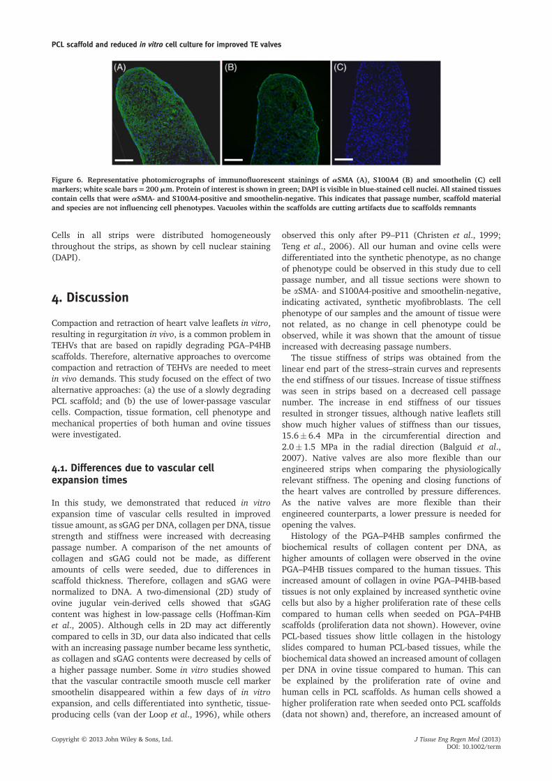
Cells in all strips were distributed homogeneouslythroughout the strips, as shown by cell nuclear staining(DAPI).
4. Discussion
Compaction and retraction of heart valve leaflets in vitro,resulting in regurgitation in vivo, is a common problem inTEHVs that are based on rapidly degrading PGA–P4HBscaffolds. Therefore, alternative approaches to overcomecompaction and retraction of TEHVs are needed to meetin vivo demands. This study focused on the effect of twoalternative approaches: (a) the use of a slowly degradingPCL scaffold; and (b) the use of lower-passage vascularcells. Compaction, tissue formation, cell phenotype andmechanical properties of both human and ovine tissueswere investigated.
4.1. Differences due to vascular cellexpansion times
In this study, we demonstrated that reduced in vitroexpansion time of vascular cells resulted in improvedtissue amount, as sGAG per DNA, collagen per DNA, tissuestrength and stiffness were increased with decreasingpassage number. A comparison of the net amounts ofcollagen and sGAG could not be made, as differentamounts of cells were seeded, due to differences inscaffold thickness. Therefore, collagen and sGAG werenormalized to DNA. A two-dimensional (2D) study ofovine jugular vein-derived cells showed that sGAGcontent was highest in low-passage cells (Hoffman-Kimet al., 2005). Although cells in 2D may act differentlycompared to cells in 3D, our data also indicated that cellswith an increasing passage number became less synthetic,as collagen and sGAG contents were decreased by cells ofa higher passage number. Some in vitro studies showedthat the vascular contractile smooth muscle cell markersmoothelin disappeared within a few days of in vitroexpansion, and cells differentiated into synthetic, tissue-producing cells (van der Loop et al., 1996), while others
observed this only after P9–P11 (Christen et al., 1999;Teng et al., 2006). All our human and ovine cells weredifferentiated into the synthetic phenotype, as no changeof phenotype could be observed in this study due to cellpassage number, and all tissue sections were shown tobe aSMA- and S100A4-positive and smoothelin-negative,indicating activated, synthetic myofibroblasts. The cellphenotype of our samples and the amount of tissue werenot related, as no change in cell phenotype could beobserved, while it was shown that the amount of tissueincreased with decreasing passage numbers.
The tissue stiffness of strips was obtained from thelinear end part of the stress–strain curves and representsthe end stiffness of our tissues. Increase of tissue stiffnesswas seen in strips based on a decreased cell passagenumber. The increase in end stiffness of our tissuesresulted in stronger tissues, although native leaflets stillshow much higher values of stiffness than our tissues,15.6� 6.4 MPa in the circumferential direction and2.0� 1.5 MPa in the radial direction (Balguid et al.,2007). Native valves are also more flexible than ourengineered strips when comparing the physiologicallyrelevant stiffness. The opening and closing functions ofthe heart valves are controlled by pressure differences.As the native valves are more flexible than theirengineered counterparts, a lower pressure is needed foropening the valves.
Histology of the PGA–P4HB samples confirmed thebiochemical results of collagen content per DNA, ashigher amounts of collagen were observed in the ovinePGA–P4HB tissues compared to the human tissues. Thisincreased amount of collagen in ovine PGA–P4HB-basedtissues is not only explained by increased synthetic ovinecells but also by a higher proliferation rate of these cellscompared to human cells when seeded on PGA–P4HBscaffolds (proliferation data not shown). However, ovinePCL-based tissues show little collagen in the histologyslides compared to human PCL-based tissues, while thebiochemical data showed an increased amount of collagenper DNA in ovine tissue compared to human. This canbe explained by the proliferation rate of ovine andhuman cells in PCL scaffolds. As human cells showed ahigher proliferation rate when seeded onto PCL scaffolds(data not shown) and, therefore, an increased amount of
Figure 6. Representative photomicrographs of immunofluorescent stainings of aSMA (A), S100A4 (B) and smoothelin (C) cellmarkers; white scale bars=200 mm. Protein of interest is shown in green; DAPI is visible in blue-stained cell nuclei. All stained tissuescontain cells that were aSMA- and S100A4-positive and smoothelin-negative. This indicates that passage number, scaffold materialand species are not influencing cell phenotypes. Vacuoles within the scaffolds are cutting artifacts due to scaffolds remnants
PCL scaffold and reduced in vitro cell culture for improved TE valves
Copyright © 2013 John Wiley & Sons, Ltd. J Tissue Eng Regen Med (2013)DOI: 10.1002/term

total DNA per strip in PCL scaffolds compared to ovinecells, the amount of collagen per DNA is lower in human,while the total amount of collagen per strip might behigher, due to the presence of more collagen-producingcells. More research is needed to investigate why differ-ences in proliferation rates of ovine and human cells arepresent when different types of scaffolds are used.
Mechanical results also correlated with the histologicalfindings. Strips that showed more, and increased maturityof, collagen fibres also resulted in an increased tissuestiffness and strength. This is in line with previousfindings, where a dominant role for collagen maturity bycross-linking of the collagen over collagen content wasfound with respect to the mechanical properties of thetissues (Balguid et al., 2007).
It is remarkable that ovine p7 PCL strips resulted in onlya few cells being present after 4 weeks. The collagencontent of these cells was also low, resulting in weakstrips, as observed in the tensile tests. We hypothesizethat this might be due to the combination of severalfactors. One might be the use of a low amount of serum(0.1% in ovine 3D medium). This could have resulted innon-synthetic and non-dividing cells. In combination withthe high passage number, which also showed to result inless activated or synthetic cells, this could have been thereason for the low amount of cells present after 4 weeksand the reduced amount of collagen. Furthermore,the use of PCL scaffolds is likely to have influencedthe amount of collagen, as ovine P7 cells seeded onPGA–P4HB scaffolds did show higher amounts ofcollagen. We hypothesized that the use of PCL scaffoldwith ovine cells resulted in non-synthetic cells, as themechanical integrity of this scaffold was present for alonger time span, resulting in no urgent need for the cellsto create tissue. However, culturing TEHVs with ovineP7 cells did result in proper tissue formation. This mightbe explained by the different culture protocols ofengineered strips and TEHVs. TEHVs undergo mechanicalloading in a bioreactor during culture, while strips arecultured statically. Furthermore, interspecies differencesmight have played a role, as cells of a different sheep wereused to culture the TEHVs.
Concerns might arise about the clinical applicability ofusing cells with a low passage number, mainly in children,as a relatively large number of cells need to be obtained.However, in the case of children, fewer cells are neededto be able to produce a TEHV compared to adults, as theannulus of the pulmonary valve in children is 10–17mm, while this is around 25 mm in adults. The size ofthe leaflets in young patients is also smaller. Furthermore,when PCL-based TEHVs are produced instead ofPGA–P4HB-based TEHVs, fewer cells are needed, due todifferences in scaffold thickness. To produce a PCL-basedTEHV scaffold for adults, 20�106 cells are needed, whilethis would be 2–10�106 cells in the case of children. Theseamounts of cells can be obtained by the outgrowthmethod, as the saphenous vein segments need to beonly 1 cm. In conclusion, cells from a lower passagenumber were demonstrated to increase the amount of
tissue formation and tissue strength without influencingcell phenotype. Despite the improved tissue formation,compaction of the tissues was not influenced by a lowercell passage number.
4.2. PGA–P4HB vs PCL scaffold
In this study, we demonstrated that human and ovinetissues cultured for 4 weeks using PCL scaffoldstrips showed almost no compaction (0–10%), whilePGA–P4HB-based tissues showed compaction up to 50%.Furthermore, we showed that PGA–P4HB-based TEHVsresulted in severe retraction of the leaflets in both species,while this was not seen in the PCL-based TEHVs.This demonstrates that PCL is a promising scaffold mate-rial to reduce compaction and retraction in TEHVs.Dijkman et al. (2012b) described another approach toprevent compaction and retraction of PGA–P4HB-basedTEHVs. Trileaflet heart valves of PGA–P4HB scaffoldswere seeded with ovine myofibroblasts and subsequentlydecellularized to prevent retraction. Decellularizationrepresented to be a powerful tool to reduce tissueretraction, as it was shown that cell-induced retractionaccounted for 85% of total tissue retraction. Residualmatrix stresses are known to still account for 15% of thetotal retraction (van Vlimmeren et al., 2012). These resid-ual matrix stresses minimized the coaptation area in thestudy of Dijkman et al. (2012b) and it has to be elucidatedin future studies whether this will influence in vivo valvebehaviour. We believe that by using a slowly degradingscaffold, retraction can be even more effectively reducedby resisting residual matrix stresses while maintainingtissue viability.
Results of the mechanical tests demonstrated thatin PCL strips the mechanical properties were not deter-mined only by the formed tissue, but also by the remainingPCL scaffold, as it was not yet degraded. PGA–P4HB isknown to start to degrade after 2 weeks, and thereforewas not influencing the mechanical properties of thetissues. As amounts of sGAG and collagen per DNA werenot influenced by the scaffold materials, the increasedtissue strength of the human PCL strips compared to thePGA–P4HB strips are likely due to the remaining PCLscaffold. Ovine strips did not show the same results, whichmight be due to the low amount of DNA and, therefore, alower amount of total tissue, in ovine PCL strips. Themechanical properties of the ovine PCL strips were mainlyinfluenced by the remaining scaffold and not by the formedtissue, while in PGA–P4HB strips the measured mechanicalproperties represented tissue only. Furthermore, ovinetissues based on PCL scaffolds did not influence mechanicalproperties as much as human PCL tissues, as tissue strengthand stiffness values were equally or just slightly increasedcompared to the PCL control strips. This indicated thatthe newly formed tissues based on ovine cells were not ofthe same quality as their human equivalents.
Differences in scaffold thickness could possibly haveresulted in differences in tissue formation, due to variation
M. M. C. P. Brugmans et al.
Copyright © 2013 John Wiley & Sons, Ltd. J Tissue Eng Regen Med (2013)DOI: 10.1002/term

in nutrient and oxygen levels within the tissues. This ismainly seen in ovine strips, as human strips show morehomogeneously distributed tissue. Our ovine strips possessa denser layer of collagen and cells on the surface in bothscaffold materials. However, cells were not present only atthe surface layer but also distributed throughout the centresof both scaffold materials. Not only the cells at the surfacelayer but also the cells in the centre produced collagenand expressed the synthetic smooth muscle cell markers,as visualized by histology. Furthermore, biochemical assaysdemonstrated no influence of the scaffold materials on thecollagen and sGAG formation per DNA, and differences inmechanical properties of the tissues are most likely due toPCL scaffold remnants instead of differences betweenmate-rial thicknesses. Directly after seeding, the high porosity ofthe scaffold strips allowed oxygen and nutrient supply tothe cells that were situated on the scaffold fibres in themiddle part of the strip. When tissue was produced, poros-ity decreased and oxygen and nutrient supply might havebeen decreased, resulting in the formation of surface layers.
Native human heart valve leaflets are avascular, as theyare thin enough to receive nutrients and oxygen throughdiffusion and haemodynamic convection (Butcher et al.,2011). As PCL scaffolds are 300 mm, we do not expectproblems when placing PCL TEHVs in vivo. TEHVs basedon PGA–P4HB did show increased thickness in the ovinemodel (Schmidt et al., 2010), which might lead toreduced oxygen and nutrient supply to the cells in thecentre. This problem might be less pronounced in humans,as these tissues are also compacting in the vertical direction,and therefore decreasing in thickness.
We conclude that the use of PCL scaffold seems to be analternative scaffold material for the culture of humanTEHVs to reduce compaction, while further optimizationis needed when ovine cells are used, to ensure propertissue formation.
4.3.. Interspecies differences
Tissue properties were different between species. In ourstudy, ovine cells were shown to be more synthetic thanhuman cells, as they contained more sGAG and collagenper DNA, while a study by van Geemen et al. (2012)demonstrated the opposite effect. Van Geemen et al.showed that human P7 cells contained double the amountof sGAG per DNA (4.8� 0.8 mg/mg DNA in ovine and8.2� 1.4 mg/mg DNA in human cells) and five timesthe amount of collagen per DNA (1.1� 0.3 mg/mg DNAin ovine and 5.9� 2.5 mg/mg DNA in human cells)compared to ovine P7 cells. Tissues based on P7 cells inour experiments obtained values for sGAG per DNA of6.5� 0.2 mg/mg ovine DNA and 5.5� 0.3 mg/mg humanDNA. Collagen per DNA was 3.2� 0.1 mg/mg DNA, and3.7� 0.3 mg/mg DNA in ovine and human, respectively. Thissuggests that ovine cells in our study weremore synthetic orless proliferative, which might be due to the amount ofserum used in the culture medium. Van Geemen used2.5% lamb serum, while in this study only 0.1% serum
was used, as an in vitro TEHVs study by Dijkman et al.(2012a) demonstrated more homogeneous tissue forma-tion throughout the wall and leaflets when 0.1% lambserum was used. A review by Mol et al. (2009) describedthat the outcome of ovine TEHVswas dramatically differentfrom their human equivalents when using the same cultureconditions, and lower amounts of serum resulted in tissueoutcome comparable to that of humans. This shows thedifficulties in the translation step from animal studiestowards the clinic and vice versa. Furthermore, previousstudies showed that not only interspecies but also intraspe-cies variations of tissue properties are large (Balguid et al.,2007; Kortsmit et al., 2009; van Geemen et al., 2012).Within this study we investigated the tissue properties ofthe strips seeded with cells of one sheep and one patientonly. While it would be preferred to have more data on sev-eral human and ovine cell sources, we assume that withinspecies the effects of, for eaxmple, cell passage numberare comparable. Furthermore, the first goal of this studywas to compare different types of scaffold to preventcompaction. This was investigated on cells of two species(human and ovine) and different cell passage numbers ofthose species. While two species and cell passage numberswere used and differences in terms of tissue productionwere observed between these species and cells passagenumbers, the outcome of compaction was similar in allresearch groups. This indicates that the influence of thescaffold type is larger than the influence of the tissueproduction of several cell sources, in terms of compaction.
A limitation of our study is that the ovine cellsoriginated from a young, healthy sheep, while the humanvascular-derived cells were obtained from an older personwho had undergone bypass surgery. This might haveinfluenced the outcome of the tissue properties, as notonly cell passage number but also patient age may havean effect on cell functioning, doubling time and ability oftissue production in different cell types (Han et al.,2010; Klinger et al., 2006; Ragnauth et al., 2010).
In conclusion, differences in absolute values betweenovine and human samples were seen within this experi-ment, although the general effects of reducing cellpassage numbers and the use of PCL scaffold on compactionand the amount of tissue formation were comparable.
5. Conclusion
This study showed that PCL may serve as an alternativescaffold material for human TEHVs, with minimal com-paction and without compromising tissue compositionand properties, while further optimization of ovine TEHVsbased on PCL scaffolds is needed, to ensure not onlyreduced compaction but also strong tissues of a highquality. Cells from lower passages demonstrated to im-prove tissue formation, without influencing compactionand cell phenotype. In addition, reducing cell expansionwill result in faster generation of TEHVs, providing morerapid treatment to patients.
PCL scaffold and reduced in vitro cell culture for improved TE valves
Copyright © 2013 John Wiley & Sons, Ltd. J Tissue Eng Regen Med (2013)DOI: 10.1002/term

Acknowledgements
This work was supported by a grant from the Dutch governmentto the Netherlands Institute for Regenerative Medicine (NIRM;Grant No. FES0908). The authors wish to thank Tom Lavrijsen,Leonie Grootzwagers and Anita van de Loo for their help withthe mechanical tests and culturing the TEHVs, and Marc Simonetis acknowledged for the production of PCL scaffolds. Thesmoothelin antibody was kindly provided by Dr G. J. Van Eys,
Department of Molecular Genetics, Cardiovascular ResearchInstitute Maastricht, University of Maastricht, The Netherlands.
Conflicts of interest
Anita Driessen-Mol and Frank P. T. Baaijens are share-holders of Xeltis.
References
Balguid A, Rubbens MP, Mol A, et al.2007; The role of collagen cross-linksin biomechanical behavior of humanaortic heart valve leaflets-relevance fortissue engineering, Tissue Eng 13:1501–1511.
Beamish JA, He P, Kottke-Marchant K, et al.2010; Molecular regulation of contractilesmooth muscle cell phenotype: implica-tions for vascular tissue engineering, Tis-sue Eng Part B Rev 16: 467–491.
Butcher JT, Mahler GJ, Hockaday LA. 2011;Aortic valve disease and treatment: theneed for naturally engineered solutions,Adv Drug Deliv Rev 63: 242–268.
Cesarone CF, Bolognesi C, Santi L. 1979;Improved microfluorometric DNA deter-mination in biological material using33258 Hoechst, Anal Biochem 100:188–197.
Chen J, Li H, SundarRaj N, et al. 2007, Alpha-smooth muscle actin expression enhancescell traction force. Cell Motil Cytoskeleton64: 248–257.
Christen T, Bochaton-Piallat ML, Neuville P,et al. 1999, Cultured porcine coronary ar-tery smooth muscle cells. A new modelwith advanced differentiation. Circ Res85: 99–107.
Cummings I, George S, Kelm J, et al. 2011;Tissue-engineered vascular graft remodel-ing in a growing lamb model: expressionof matrix metalloproteinases. Eur JCardiothorac Surg 41: 167–172.
Dijkman PE, Driessen-Mol A, de Heer LM,et al. 2012a; Trans-apical versus surgicalimplantation of autologous ovine tissue-engineered heart valves. J Heart Valve Dis21: 670–678.
Dijkman PE, Driessen-Mol A, Frese L, et al.2012b; Decellularized homologous tissue-engineered heart valves as off-the-shelf al-ternatives to xeno- and homografts.Biomater 33: 4545–4554.
Dolezel J, Bartos J, Voglmayr H, et al. 2003,Nuclear DNA content and genome sizeof trout and human. Cytometry A 51:127–128
Farndale RW, Buttle DJ, and Barrett AJ.1986; Improved quantitation and discrimi-nation of sulphated glycosaminoglycans byuse of dimethylmethylene blue. BiochimBiophys Acta 883: 173–177
Gottlieb D, Kunal T, Emani S, et al. 2010; Invivo monitoring of function of autologousengineered pulmonary valve. J ThoracCardiovasc Surg 139: 723–731.
Han J, Liu JY, Swartz DD, et al. 2010; Molec-ular and functional effects of organismalageing on smooth muscle cells derived
from bone marrow mesenchymal stemcells. Cardiovasc Res 87: 147–155.
Hinz B. 2010; The myofibroblast: paradigmfor a mechanically active cell. J Biomech43: 146–155.
Hinz B, Phan SH, Thannickal VJ, et al.2007; The myofibroblast: one function,multiple origins. Am J Pathol 170:1807–1816.
Hoerstrup SP, Cummings MI, Lachat M, et al.2006; Functional growth in tissue-engineered living, vascular grafts: follow-up at 100weeks in a large animal model.Circulation 114: I159–I166.
Hoerstrup SP, Sodian R, Daebritz S, et al.2000; Functional living trileaflet heartvalves grown in vitro. Circulation 102:III44–III49.
Hoffman-Kim D, Maish MS, Krueger PM,et al. 2005; Comparison of threemyofibroblast cell sources for the tissueengineering of cardiac valves. TissueEng 11: 288–301.
Huszar G, Maiocco J, Naftolin F. 1980; Mon-itoring of collagen and collagen fragmentsin chromatography of protein mixtures.Anal Biochem 105: 424–429.
Junqueira LC, Bignolas G, Brentani RR. 1979;Picrosirius staining plus polarization mi-croscopy, a specific method for collagendetection in tissue sections. Histochem J11: 447–455.
Klinger RY, Blum JL, Hearn B, et al. 2006; Rel-evance and safety of telomerase for humantissue engineering. Proc Nati Acad Sci U S A103: 2500–2505.
Klouda L, Vaz C, Mol A, et al. 2008; Effect ofbiomimetic conditions on mechanical andstructural integrity of PGA/P4HB andelectrospun PCL scaffolds. J Mater SciMater Med 19: 1137–1144.
Kortsmit J, Driessen NJ, Rutten MC, et al.2009; Real time, non-invasive assessmentof leaflet deformation in heart valvetissue engineering. Ann Biomed Eng 37:532–541.
Lacolley P, Regnault V, Nicoletti A, et al.2012; The vascular smooth muscle cellin arterial pathology: a cell that can takeon multiple roles. Cardiovasc Res 95:194–204.
Mendelson K, Schoen F. 2006; Heart ValveTissue Engineering: Concepts, Approaches,Progress, and Challenges. Ann Biomed Eng34: 1799–1819.
Mol A, Driessen NJ, Rutten MC, et al. 2005a;Tissue engineering of human heart valveleaflets: a novel bioreactor for a strain-based conditioning approach. Ann BiomedEng 33: 1778–1788.
Mol A, Rutten MC, Driessen NJ, et al.2006; Autologous human tissue-engineered heart valves: prospects forsystemic application. Circulation 114:I152–I158.
Mol A, Smits AI, Bouten CV, et al. 2009;Tissue engineering of heart valves: advancesand current challenges. Expert Rev MedDevices 6: 259–275.
Mol A, van Lieshout MI, Dam-de VeenCG, et al. 2005b; Fibrin as a cellcarrier in cardiovascular tissue engi-neering applications. Biomaterials 26:3113–3121.
Rabkin-Aikawa E, Farber M, Aikawa M, et al.2004; Dynamic and reversible changes ofinterstitial cell phenotype during remodel-ing of cardiac valves. J Heart Valve Dis 13:841–847.
Ragnauth CD, Warren DT, Liu Y, et al. 2010;Prelamin A acts to accelerate smoothmuscle cell senescence and is a novel bio-marker of human vascular aging. Circula-tion 121: 2200–2210.
Rensen SS, Doevendans PA, van Eys GJ.2007; Regulation and characteristicsof vascular smooth muscle cell phe-notypic diversity. Neth Heart J 15:100–108.
Safwani WK, Makpol S, Sathapan S, et al.2012; The impact of long-term in vitroexpansion on the senescence-associatedmarkers of human adipose-derived stemcells. Appl Biochem Biotechnol 166:2101–2113.
Schmidt D, Dijkman PE, Driessen-Mol A,et al. 2010; Minimally-invasive im-plantation of living tissue engineeredheart valves: a comprehensive ap-proach from autologous vascular cellsto stem cells. J Am Coll Cardiol 56:510–520.
Shahbudin H. 2003; Choice of prostheticheart valve for adult patients. J Am CollCardiol 41: 893–904.
Teng B, Ansari HR, Oldenburg PJ, et al.2006; Isolation and characterizationof coronary endothelial and smoothmuscle cells from A1 adenosine recep-tor-knockout mice. Am J Physiol HeartCirc Physiol, 290: H1713–H1720.
van der Loop FT, Schaart G, TimmerED, et al. 1996; Smoothelin, a novelcytoskeletal protein specific forsmooth muscle cells. J Cell Biol 134:401–411.
van Geemen D, Driessen-Mol A,Grootzwagers LG, et al. 2012; Variationin tissue outcome of ovine and humanengineered heart valve constructs:
M. M. C. P. Brugmans et al.
Copyright © 2013 John Wiley & Sons, Ltd. J Tissue Eng Regen Med (2013)DOI: 10.1002/term

relevance for tissue engineering. RegenMed 7: 59–70.
van Vlimmeren MA, Driessen-Mol A,Oomens CW, et al. 2011; An in vitro modelsystem to quantify stress generation, com-paction, and retraction in engineered heartvalve tissue. Tissue Eng Part C Methods 17:983–991.
van Vlimmeren MA, Driessen-Mol A,Oomens CW, et al. 2012; Passive andactive contributions to generated forceand retraction in heart valve tissue engi-neering. Biomech Model Mechanobiol 11:1015–1027.
Weber B, Scherman J, Emmert MY, et al.2011; Injectable living marrow stromal
cell-based autologous tissue engineeredheart valves: first experiences with a one-step intervention in primates. Eur Heart J32: 2830–2840.
Yacoub MH, Takkenberg JJ. 2005; Will heartvalve tissue engineering change theworld? Nat Clin Pract Cardiovasc Med 2:60–61.
PCL scaffold and reduced in vitro cell culture for improved TE valves
Copyright © 2013 John Wiley & Sons, Ltd. J Tissue Eng Regen Med (2013)DOI: 10.1002/term


![Biomaterials - Ghent University · modulated [11,12]. Various scaffold materials have been analyzed in experiments for autotransplantation as well as for in vitro or in vivo growth](https://img.pdfslide.us/doc/110x75/5e1eef70c7cb2853550fc593/biomaterials-ghent-university-modulated-1112-various-scaffold-materials-have.jpg)


![Copolymerization of [epsiv]-caprolactone and morpholine-2 ... · Makromol. Chem. 193, 1927-1942 (1992) 1927 Copolymerization of &-caprolactone and morpholine-2,5-dione derivatives](https://img.pdfslide.us/doc/110x75/5ad096377f8b9ae2138dec54/copolymerization-of-epsiv-caprolactone-and-morpholine-2-chem-193-1927-1942.jpg)