Embed Size (px)
Citation preview
Pituitary Adenylate Cyclase-Activating Polypeptide (PACAP) Inhibitsthe Slow Afterhyperpolarizing Current sIAHP in CA1 Pyramidal
Neurons by Activating Multiple Signaling Pathways
Ruth D.T. Taylor,1 Marita Gr�nning Madsen,1 Michael Krause,2
Marisol Sampedro-Casta~neda,1 Martin Stocker,1* and Paola Pedarzani1*
ABSTRACT: The slow afterhyperpolarizing current (sIAHP) is a calcium-dependent potassium current that underlies the late phase of spike frequencyadaptation in hippocampal and neocortical neurons. sIAHP is a well-known tar-get of modulation by several neurotransmitters acting via the cyclic AMP(cAMP) and protein kinase A (PKA)-dependent pathway. The neuropeptidepituitary adenylate cyclase activating peptide (PACAP) and its receptors arepresent in the hippocampal formation. In this study we have investigated theeffect of PACAP on the sIAHP and the signal transduction pathway used to mod-ulate intrinsic excitability of hippocampal pyramidal neurons. We show thatPACAP inhibits the sIAHP, resulting in a decrease of spike frequency adaptation,in rat CA1 pyramidal cells. The suppression of sIAHP by PACAP is mediated byPAC1 and VPAC1 receptors. Inhibition of PKA reduced the effect of PACAP onsIAHP, suggesting that PACAP exerts part of its inhibitory effect on sIAHP byincreasing cAMP and activating PKA. The suppression of sIAHP by PACAP wasalso strongly hindered by the inhibition of p38 MAP kinase (p38 MAPK). Con-comitant inhibition of PKA and p38 MAPK indicates that these two kinases actin a sequential manner in the same pathway leading to the suppression of sIAHP.Conversely, protein kinase C is not part of the signal transduction pathway usedby PACAP to inhibit sIAHP in CA1 neurons. Our results show that PACAP enhan-ces the excitability of CA1 pyramidal neurons by inhibiting the sIAHP through
the activation of multiple signaling pathways, most promi-nently cAMP/PKA and p38 MAPK. Our findings disclose anovel modulatory action of p38 MAPK on intrinsic excit-ability and the sIAHP, underscoring the role of this currentas a neuromodulatory hub regulated by multiple proteinkinases in cortical neurons. VC 2013 The Authors. Hippo-campus Published by Wiley Periodicals, Inc.
KEY WORDS: hippocampus; slow afterhyperpolari-zation; neuropeptide; protein kinase A; p38 MAPkinase
INTRODUCTION
Action potentials in hippocampal pyramidal neuronsare followed by an afterhyperpolarization (AHP) that hasthree kinetically distinct phases: fast (fAHP), medium(mAHP), and slow AHP (sAHP) (reviewed in Sah andFaber, 2002; Stocker et al., 2004). The sAHP has a time-course of seconds (1–5 s), is evident after bursts of actionpotentials, and its amplitude is enhanced with increasingnumber of spikes in the burst (Alger and Nicoll, 1980;Hotson and Prince, 1980; Schwartzkroin and Stafstrom,1980; Lancaster and Adams, 1986). The sAHP is medi-ated by a slow calcium-dependent potassium current,sIAHP, that contributes to the late phase of spike fre-quency adaptation in hippocampal pyramidal neurons(Madison and Nicoll, 1982; Lancaster and Adams,1986; Lancaster and Nicoll, 1987). A trademark featureof the sIAHP is its modulation by several neurotrans-mitters activating various signal transduction pathways.The modulation of sIAHP by monoamine transmittersthrough the cyclic AMP (cAMP)-protein kinase A(PKA) pathway has been extensively studied (reviewed inStocker et al., 2004). Beside monoamine transmitters,some neuropeptides expressed in interneurons andreleased in the hippocampal formation inhibit the sIAHP
and reduce spike frequency adaptation in pyramidal neu-rons. These include the vasoactive intestinal peptide(VIP, Haas and Gahwiler, 1992), the corticotropin releas-ing factor (CRF, Aldenhoff et al., 1983) and the calcito-nin gene-related peptide (CGRP, Haug and Storm,2000), all exerting their inhibitory action on sIAHP viathe cAMP-PKA pathway (Haug and Storm, 2000).
The neuropeptide pituitary adenylate cyclase-activating polypeptide (PACAP) belongs to a
This is an open access article under the terms of the Creative CommonsAttribution License, which permits use, distribution and reproduction inany medium, provided the original work is properly cited.1Research Department of Neuroscience, Physiology and Pharmacology,University College London, London, United Kingdom; 2 Department ofMolecular Biology of Neuronal Signals, Max Planck Institute for Experi-mental Medicine, 37075, G€ottingen, GermanyGrant sponsor: MRC Career Establishment Grant; Grant number:G0100066; Grant sponsor: Wellcome Trust Senior Research Fellowship;Grant number: 061198/Z/00A; Grant sponsor: Human Frontier ScienceProgram Organization; Grant number: RGP0013/2010; Grant sponsors:UK Medical Research Council (MRC); Mechanisms of Neuronal SignalTransduction, University of Freiburg, Germany; European NeuroscienceInstitutes Network (ENI-Net).Ruth D.T. Taylor is currently at MRC Center for Developmental Neurobi-ology, King’s College London, London, UK.Marita Gr�nning Madsen is currently at Lund Stem Cell Center, LundUniversity Hospital, SE-221 84 Lund, Sweden.Michael Krause is currently at Gilead Sciences, Fremont, California94555, USA.R. D. T. Taylor and M. G. Madsen contributed equally to this work.
*Correspondence to: Martin Stocker, Research Department of Neuro-science, Physiology and Pharmacology, University College London,Gower Street, London WC1E 6BT, United Kingdom. E-mail: [email protected] or Paola Pedarzani, Research Department of Neuroscience,Physiology and Pharmacology, University College London, Gower Street,London WC1E 6BT, United Kingdom. E-mail: [email protected] for publication 20 August 2013.DOI 10.1002/hipo.22201Published online 30 September 2013 in Wiley Online Library(wileyonlinelibrary.com).
VC 2013 THE AUTHORS. HIPPOCAMPUS PUBLISHED BY WILEY PERIODICALS, INC.
HIPPOCAMPUS 24:32–43 (2014)
superfamily of structurally related peptide hormones thatinclude glucagon, glucagon-like peptides, secretin and growthhormone-releasing hormone (Harmar, 2001). PACAP consistsof two isoforms, PACAP-27 with 27 and PACAP-38 with 38amino acids, sharing identical sequences for the initial 27 aminoacids. Both isoforms were first identified and isolated from ovinehypothalamic tissue (Miyata et al., 1989, 1990) and subse-quently found to be preserved from protochordates to mammals(Vaudry et al., 2009). PACAP exhibits a wide range of functionsand acts as a neurotransmitter and trophic factor in the CNS(Vaudry et al., 2009). The first 28 amino acids of PACAP-38show 68% sequence identity with VIP (Miyata et al., 1989,1990), suggesting that VIP and PACAP might mediate their bio-logical effects through shared receptors. Two classes of PACAPreceptors have been characterized based on their relative affinityfor PACAP and VIP. PACAP has a potency >100-fold higherthan VIP at the recombinant PAC1 receptor, whereas it displaysa potency similar to VIP at the recombinant VPAC receptors(VPAC1 and VPAC2) in radioligand binding assays (reviewed inVaudry et al., 2009; Harmar et al., 2012). PAC1 and VPACreceptors are coupled to the heterotrimeric G-protein Gas andincrease cAMP levels (reviewed in Harmar, 2001).
PACAP and its receptors are widely expressed in the CNS.PACAP mRNA (Skoglosa et al., 1999; Jaworski and Proctor,2000; Hannibal, 2002) and immunoreactivity (Koves et al., 1991;Piggins et al., 1996; Hannibal, 2002) have been observed in therat hippocampus, in the cell body layers of the CA1-CA3 fields, indentate gyrus granule cells, mossy cells and pyramidal basket cells.Additionally, PACAP immunoreactive fibers have been detectedboth in the CA1-CA3 fields and in the dentate gyrus. In the hip-pocampus, PACAP might therefore be released by neurons(pyramidal cells or interneurons) within the hippocampal forma-tion and/or by afferents coming from PACAP-rich regions, suchas the hypothalamus (Piggins et al., 1996). Northern blot analysisand in situ hybridization revealed the presence of PAC1 (Hashi-moto et al., 1993; Spengler et al., 1993), VPAC1 (Ishihara et al.,1992) and VPAC2 (Lutz et al., 1993) receptor mRNA in the hip-pocampal formation. Moreover, PACAP binding sites and recep-tors were detected in the CA1-CA3 regions and dentate gyrus ofthe hippocampus in autoradiographic studies (Cauvin et al., 1991;Masuo et al., 1992). Immunohistochemistry showed that VPAC2
receptors were highly expressed in dentate gyrus granule cells andin CA1-CA3 pyramidal cells, whilst PAC1 and VPAC1 wereexpressed in the same cells but at lower levels (Joo et al., 2004).
The presence of PACAP and PACAP receptors in the hippo-campus prompted us to hypothesize that PACAP might modu-late the sIAHP in hippocampal pyramidal neurons, therebyaffecting their excitability and firing pattern. In this study wehave investigated the effect of PACAP on the sIAHP and the sig-nal transduction pathway used in rat CA1 pyramidal neurons.
MATERIALS AND METHODS
Male Sprague Dawley rats, 21- to 24-days old, were used toprepare transverse hippocampal brain slices. Rats were anesthe-
tized with isoflurane and decapitated according to the UKHome Office Animal Scientific Procedures Act (1986), andprotocols were reviewed and approved by the University Col-lege London Animal Ethical Committee.
Whole-cell patch clamp recordings were performed fromCA1 pyramidal neurons using the blind patching technique(Blanton et al., 1989). All whole-cell patch clamp recordingswere conducted with an EPC10 amplifier (HEKA, Germany)and the software Pulse for data acquisition. Slices were perfusedwith ACSF (in mM: 125 NaCl, 1.25 KCl, 2.5 CaCl2, 1.5MgCl2, 1.25 KH2PO4, 25 NaHCO3, and 16 glucose) and thesIAHP was recorded at room temperature with patch pipettesmade of borosilicate glass and containing a gluconate-basedintracellular solution (in mM: 135 K-gluconate, 10 KCl, 10HEPES, 1 MgCl2, 2 ATP-Na, and 0.4 GTP-Na). The liquidjunction potential for the intracellular solution is 211 mV.Voltage values reported here are not corrected for the liquidjunction potential. Only cells with a resting membrane poten-tial �250 mV and a series resistance �30 MX were includedin this study.
The sIAHP was elicited by stepping to 110–30 mV for 100–150 ms from a holding potential of 250 mV. Recordings wereperformed in the presence of 0.5 mM tetrodotoxin (TTX) toblock voltage-gated sodium channels, and 1 mM tetraethylam-monium (TEA) to block a subset of voltage-gated potassiumchannels to increase the calcium influx and thereby the calciumdependent sIAHP. Traces were filtered at 0.25 kHz and sampledat 1.25 kHz and the stimulus was repeated with a 30 s interval.Bovine serum albumin (BSA) was added to the ACSF (25 mgml21) when peptides were used extracellularly to minimizeunspecific binding. Peptides were applied by bath perfusion. Itis worth noticing that the actual concentration of the peptide/sat the relevant receptors might differ from that nominallyapplied, due to the size and diffusion of the peptide/s to neu-rons located at different depths in the brain slice and, possibly,to the action of extracellular peptidases. Protein kinase inhibi-tors were applied intracellularly through the patch pipette.DMSO up to 0.13% did not affect either the sIAHP amplitudeor charge transfer, or the effect of PACAP-27 on the sIAHP
amplitude or charge transfer when applied intracellularlythrough the patch pipette as a vehicle to dissolve some proteinkinase inhibitors.
Cells were excluded if the series resistance changed signifi-cantly (>25%) in the course of experiments. Modulation ofsIAHP was determined as changes in amplitude and chargetransfer of sIAHP. The sIAHP amplitude was measured by calcu-lating the mean current between two cursors placed 50 msapart, 1 s after the initiation of the voltage step to avoid con-tamination by other, faster potassium currents. Charge transferwas determined as the area under the curve starting from thesIAHP peak until full decay had occurred.
Isoproterenol hydrochloride and tetraethylammonium chlo-ride (TEA) were obtained from Sigma (UK); PACAP-27amide, SB 203580, phorbol-12,13-dibutyrate (PDBu), bisindo-lylmaleimide I (BIM-1), and chelerythrine chloride from Cal-biochem (now Merck Serono Ltd, Middlesex, UK); PACAP-38
PACAP INHIBITION OF sIAHP VIA MULTIPLE PROTEIN KINASES 33
Hippocampus

from Peninsula Laboratories Europe (St. Helens, UK); PACAP-(6–38) from Bachem (Bubendorf, Switzerland); tetrodotoxin(TTX) from Latoxan (Valence, France); and Rp-cAMPS and 8-(4-chloro-phenylthio)22’-O-methyl-cAMP (8CPT-O-Me-cAMP)from BioLog Life Science Institute (Bremen, Germany). Maxadilanand max.d.4 were a generous gift from Dr. Ethan Lerner atHarvard Medical School.
Statistical analysis was performed with GraphPad InStat.Data are presented as mean with error bars indicating thestandard error of the mean (S.E.M.), when not otherwisespecified.
RESULTS
The sIAHP was recorded in the whole-cell patch-clamp con-figuration from 152 CA1 pyramidal neurons. CA1 neurons hadan average resting membrane potential of 259.8 6 3.5 mV(n 5 84; mean 6 SD) and an input resistance of 175.4 6 51.1MX (n 5 80; mean 6 SD). The sIAHP amplitude was on aver-age 40.6 6 24.2 pA and the charge transfer 101.4 6 63.8 pC(n 5 84; mean 6 SD).
PACAP Suppresses sIAHP in CA1 PyramidalNeurons
The modulatory effects on the sIAHP of VIP and other neu-ropeptides that activate the cAMP-PKA pathway in hippocam-pal pyramidal neurons are well studied (Aldenhoff et al., 1983;Haas and Gahwiler, 1992; Haug and Storm, 2000). Given thestructural similarity of PACAP and VIP (Miyata et al., 1989;Miyata et al., 1990) and the fact that they share a subset ofreceptors (VPAC1 and VPAC2), coupled to the cAMP-PKApathway (reviewed in Vaudry et al., 2009), we hypothesizedthat PACAP might suppress sIAHP in hippocampal pyramidalneurons. We found that PACAP-38 (500 nM) reduced thesIAHP amplitude (Figs. 1A,C) by 67% 6 5.9% (Fig. 1E, n 5 7)and the charge transfer by 77.3% 6 5.2% (Fig. 1E, n 5 7) inCA1 pyramidal neurons.
The region responsible for receptor activation of PACAP-38resides in its N-terminal 27-amino acid sequence, which corre-sponds to the naturally occurring isoform PACAP-27 (Miyataet al., 1990). We therefore tested the effect of PACAP-27 (500nM) on sIAHP. PACAP-27 suppressed sIAHP within 15–20 minof bath application (Fig. 1B,D), and no reversibility wasobserved within 15–35 min of wash-out. PACAP-27 sup-pressed the sIAHP amplitude by 86.1% 6 4.7% and the chargetransfer by 85.8% 6 5.1% (Fig. 1E, n 5 4). No significant dif-ference between PACAP-27 and PACAP-38 was found withregard to the reduction in sIAHP amplitude (P> 0.06, unpairedt test) and charge transfer (P> 0.3, unpaired t test). We there-fore decided to use PACAP-27 in the following experiments.
PACAP-27 suppressed the sIAHP in a concentration depend-ent manner (Fig. 1F). While 100 nM PACAP-27 only slightlyreduced the sIAHP amplitude (29.9% 6 10.6%, n 5 4), the
FIGURE 1. PACAP-38 and PACAP-27 suppress sIAHP in hip-pocampal neurons. Averaged current traces (n 5 3) of the sIAHP
before (control) and after application of 500 nM PACAP-38 (A)or 500 nM PACAP-27 (B). Overlaid traces are shown in the right-most panels (A and B), control (black) and PACAP (grey). Thedashed line corresponds to the baseline current. Time course ofthe effect of PACAP-38 (C) and PACAP-27 (D) on the sIAHP
amplitude from the experiments shown in A and B. Bars indicatethe presence of 500 nM PACAP in the bath solution. (E) Effect of500 nM PACAP-38 (black) and PACAP-27 (white) on sIAHP
amplitude and charge transfer. No significant difference wasobserved between the two PACAP isoforms. (F) The effect ofPACAP-27 on sIAHP was concentration-dependent. PACAP-27 sig-nificantly suppressed sIAHP amplitude at both 250 nM (n 5 18,P < 0.0001) and 500 nM (n 5 4, P < 0.01), while at 100 nMPACAP-27 did not cause a significant inhibition of sIAHP (n 5 4,P > 0.05). The difference between the sIAHP reduction caused by250 and 500 nM PACAP-27 was not significant (P > 0.05, Dunn’smultiple comparisons test). (G) PACAP-27 (250 nM) decreasedspike frequency adaptation in a representative CA1 neuron inresponse to a current injection of 140 pA, resting membranepotential 5 259 mV. Similar effects were observed in seven cells.
34 TAYLOR ET AL.
Hippocampus

reduction with 250 nM was more substantial (77.4% 6 6.7%,n 5 18) and comparable to the effect of 500 nM PACAP-27(86.1% 6 4.7%, n 5 4). PACAP-27 at 250 nM caused a sup-pression of the sIAHP amplitude by more than 90% in 50% ofthe neurons (9 out of 18 cells; Fig. 1F). 2 out of 18 cells dis-played a sIAHP inhibition comprised between 85 and 90%; 2out of 18 cells between 70 and 75%; 4 out of 18 cells between40 and 55% (Fig. 1F). In one case, corresponding to 5.6% ofall cells tested, the sIAHP was not affected by 250 nM PACAP-27 (2.4% inhibition; Fig. 1F). All cells treated with 500 nMPACAP-27 displayed reductions of the sIAHP amplitude in therange of 80–100% (n 5 4; Fig. 1F).
Beside inhibiting sIAHP, PACAP produced an inward shift inthe holding current at the holding potential of 250 mV, corre-sponding to a depolarizing effect. PACAP-38 at 500 nM eli-cited an inward current of 16.0 6 2.1 pA in 7 out of 7 cells(n 5 7; P 5 0.0002). An inward current of 14 6 3.3 pA(n 5 18; P 5 0.0006) was observed in response to 250 nMPACAP-27 in 16 out of 18 cells. The inward current might bedue to the modulatory action of PACAP on background (leak)channels, HCN channels, and/or other nonspecific cationicchannels, in analogy to what has been observed for other neu-rotransmitters inhibiting the sIAHP in hippocampal neurons.
PACAP-27 at 250 nM did not significantly affect the inputresistance of CA1 pyramidal neurons, which was 164.5 6 9.7 MXbefore and 163.9 6 8.1 MX after PACAP-27 application (n 5 18;P 5 0.96, Wilcoxon matched-pairs signed-ranks test).
The inhibition of the sIAHP reduces late spike frequencyadaptation in hippocampal pyramidal neurons (Madison andNicoll, 1982; Madison and Nicoll, 1984). Application of 250nM PACAP-27 reduced spike frequency adaptation in CA1pyramidal neurons (Fig. 1G, n 5 7), in agreement with itsinhibitory effect on the sIAHP.
Activation of PAC-1 Receptors Partially Mimicsthe Effect of PACAP on sIAHP
Neuronal PACAP receptors comprise PAC1, selective forPACAP and coupled to both adenylyl cyclases and phospholipaseC, and VPAC1 and VPAC2 receptors, activated by both PACAPand VIP and coupled to adenylyl cyclases (Vaudry et al., 2009).All three PACAP receptor subtypes are expressed in CA1 pyram-idal cells (Hashimoto et al., 1996; Shioda et al., 1997; Jooet al., 2004). To elucidate the specific contribution of PAC1
receptors to the PACAP-mediated sIAHP inhibition in CA1pyramidal cells, we used the PAC1 selective agonist maxadilan, apeptide structurally unrelated to PACAP (Lerner et al., 1991),which has no activity at VPAC1 and VPAC2 receptors (Moroand Lerner, 1997). Maxadilan and PACAP activate PAC1 recep-tors with a similar potency in the rat brain (Moro et al., 1996).When tested at 250 nM, the same concentration as used forPACAP-27, maxadilan reduced the sIAHP amplitude (Figs 2A,B)by 32.6% 6 3.9% (n 5 5; P 5 0.001, t test) and charge transferby 24.4% 6 4.7% (n 5 5; P 5 0.007, t test) (Fig. 2D). Thisindicates that PAC1 receptors contribute to the PACAP-inducedsuppression of sIAHP in CA1 pyramidal neurons.
To investigate whether the reduction of sIAHP induced bymaxadilan could be prevented by a PACAP receptor antagonist,we used PACAP-(6–38), a truncated PACAP-38 lacking sixamino terminal amino acids. PACAP-(6–38) is a selectiveantagonist at PAC1 and VPAC2 receptors (Dickinson et al.,1997; Moro et al., 1999). In the presence of PACAP-(6–38)(500 nM), application of maxadilan (250 nM) did not signifi-cantly reduce the sIAHP amplitude (5.7% 6 10.7%, n 5 4,P> 0.5, t test) or charge transfer (25.3% 6 13.6%, n 5 4,P> 0.5 t test) (Fig. 2C,E). The lack of reduction of the sIAHP
FIGURE 2. Selective activation of PAC1 receptors partlymimics the inhibitory effect of PACAP on the sIAHP. Averaged cur-rent traces (n 5 3) of the sIAHP before (control) and after applica-tion of the selective PAC1 receptor agonist maxadilan at 250 nM(A). In the rightmost panel, the traces in the absence (black) andpresence (grey) of maxadilan are superimposed. The dashed linecorresponds to the baseline current. (B) Time-course of the effectof 250 nM maxadilan on the amplitude of sIAHP. Each point isthe mean 6 SEM of five experiments. Application of maxadilan isindicated by the arrow. (C) 500 nM PACAP-(6–38), a PAC1 recep-tor antagonist, prevented the effect of 250 nM maxadilan asshown on averaged current traces (n 5 3) of the sIAHP. The dashedline corresponds to the baseline current. Overlaid traces are shownin the rightmost panel. Bar charts summarizing the reduction ofsIAHP amplitude and charge transfer by 250 nM maxadilan (D)and the absence of maxadilan effect in the presence of 500 nMPACAP-(6–38) (E) in five cells. Error bars indicate S.E.M.
PACAP INHIBITION OF sIAHP VIA MULTIPLE PROTEIN KINASES 35
Hippocampus
by a specific PAC1 receptor agonist (maxadilan) in the presenceof a PAC1 receptor antagonist further supports a contributionby PAC1 receptors.
Surprisingly, the reduction of the sIAHP as a consequence ofPAC1 receptor activation is not supported by the resultsobtained upon 250 nM PACAP-27 application in the presenceof PACAP-(6–38). Under this condition, PACAP-27 reducedthe sIAHP amplitude (79.2% 6 11.2%, n 5 5, P 5 0.002, t test)and the charge transfer (86.8% 6 8.5%, n 5 5, P 5 0.0005, ttest) to a comparable extent (amplitude: P> 0.5, charge trans-fer: P> 0.3) as observed in the absence of PACAP-(6–38) (Fig.1). The suppression of sIAHP by maxadilan but the lack ofantagonism of PACAP-27 by PACAP-(6–38) suggests the acti-vation of more than one receptor subtype in the PACAP-mediated inhibition of this current.
Max.d.4 is a derivative of maxadilan, in which nineteenamino acids between positions 24 and 42 are deleted. It is aselective PAC1 receptor antagonist that is more potent thanPACAP-(6–38) (Moro et al., 1999). In the presence of 500nM max.d.4, 250 nM PACAP-27 inhibited the sIAHP ampli-tude (69.8% 6 10.8%, n 5 4, P< 0.01, t test) and chargetransfer (74.5% 6 7.3%, n 5 4, P< 0.01, t test) to an extentthat was comparable (amplitude and charge transfer: P> 0.5)to the effect of PACAP-27 alone (Fig. 1). This further indicatesthat antagonism of PAC1 receptors alone is not sufficient toprevent the PACAP-27 mediated inhibition of sIAHP, which isinstead due to the activation of more than just PAC1 receptors.
PACAP-27 Suppresses sIAHP Partly ThroughPKA Activation
In hippocampal neurons monoaminergic neurotransmitterssuch as noradrenaline, histamine, serotonin and dopamineinhibit the sIAHP through PKA activation (Pedarzani andStorm, 1993, 1995). Moreover, in CA1 pyramidal neuronsCRF, VIP and CGRP have been shown to suppress sIAHP in aPKA-dependent manner (Haug and Storm, 2000). We there-fore asked whether PKA was required to mediate the inhibitoryeffect of PACAP on sIAHP in hippocampal pyramidal neurons.
To address this question, we used the PKA inhibitor Rp-cAMPS added to the intracellular solution (Pedarzani andStorm, 1993). Application of PACAP-27 (250 nM) in thepresence of Rp-cAMPS (500 mM) caused an inhibition of thesIAHP that was attenuated when compared to the effect ofPACAP-27 alone (Fig. 3A,B). The inhibition of the sIAHP
amplitude was reduced from 77.4% 6 6.7% in the absence ofRp-cAMPS (Figs. 1F and 3C) to 56.8% 6 5.9% in the pres-ence of Rp-cAMPS (Fig. 3C). Similarly, the sIAHP inhibitionby 500 nM PACAP-27 was reduced from 86.1% 6 4.7% inthe absence (Fig. 1F and 3C) to 42.7% 6 11.1% in the pres-ence of Rp-cAMPS (Fig. 3C). An inward shift in the holdingcurrent of 16.6 6 6.8 pA (n 5 15; P 5 0.002) was observedupon application of PACAP-27 (250 nM) in the presence ofRp-cAMPS.
To ensure that Rp-cAMPS inhibited PKA under ourexperimental conditions, we used isoproterenol (1 mM), a
b-adrenergic receptor agonist that inhibits the sIAHP by activa-tion of the cAMP/PKA pathway (Pedarzani and Storm, 1993).The sIAHP inhibiton caused by isoproterenol (100% 6 0.02%,n 5 4) was significantly reduced in the presence of Rp-cAMPS(41.2% 6 8.4%, n 5 5, P 5 0.0005, unpaired t test). Whenisoproterenol was applied after PACAP-27 in the presence ofRp-cAMPS, the sIAHP was inhibited to 43.1% 6 7% (n 5 5) ofthe amplitude left after PACAP-27 inhibition, showing thatRp-cAMPS inhibited PKA. These results show that PKA con-tributes to the PACAP-27-mediated inhibition of the sIAHP.
Are MAP Kinases Involved in the PACAP-27-Mediated Inhibition of sIAHP?
In a Drosophila mutant PACAP modulates a potassium cur-rent through the activation of a mitogen-activated protein(MAP) kinase pathway (Zhong, 1995). PACAP has also beenshown to activate the MAP kinase kinase (MEK)/extracellular-signal-regulated kinase (ERK) pathway in cultured cerebellar
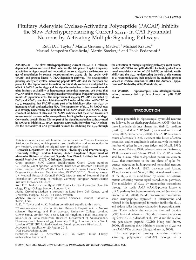
FIGURE 3. PKA activation contributes to the inhibition sIAHP
by PACAP-27. Averaged current traces (n 5 3) of the sIAHP beforeand after bath application of 250 nM PACAP-27 in the absence(upper traces) and in the presence (lower traces) of the PKA inhib-itor Rp-cAMPS (500 mM) (A). In the rightmost panels, the tracesin the absence (black) and presence (grey) of PACAP-27 are super-imposed. The dashed line corresponds to the baseline current. (B)Time course of the normalized sIAHP amplitude from the experi-ments shown in A before, during, and after PACAP-27 applicationin the presence (black squares) and absence (white squares) ofintracellular Rp-cAMPS. Bar indicates the presence of 250 nMPACAP-27. (C) Bar chart summarizing the effect on the sIAHP
amplitude of PACAP-27 at 250 and 500 nM in the absence (whitebars) and presence (black bars) of Rp-cAMPS. Rp-cAMPS signifi-cantly reduced the suppression of sIAHP amplitude by PACAP-27at 250 nM (P 5 0.04, n 5 15, Mann–Whitney test), and at 500nM (P 5 0.01, n 5 5, unpaired t test). The values for sIAHP inhibi-tion by PACAP-27 at 250 nM (n 5 18) and 500 nM (n 5 4) in theabsence of Rp-cAMPS are the same as in Figure 1E,F and arereported here for comparison. Error bars indicate S.E.M. * indi-cates statistical significance.
36 TAYLOR ET AL.
Hippocampus
granule cells (Villalba et al., 1997). Furthermore, PACAP-38activates p38 MAP kinase (p38 MAPK) in mouse cultured cer-ebellar granule cells (Ster et al., 2007). We therefore askedwhether the effect of PACAP on the sIAHP could be mediatedby the activation of the ERK or p38 MAPK pathway.
To first test for a potential contribution of the MEK-ERK1/2 pathway, we applied intracellularly the MEK inhibitorUO126 (40 mM) (Favata et al., 1998). In the presence ofUO126, PACAP-27 (250 nM) inhibited the sIAHP
(58.5% 6 8.7%, n 5 8) to the same extent as in intercalatedcontrol experiments performed with 0.13% DMSO in the pip-ette solution (63.1% 6 7.1%, n 5 6, P> 0.6, Mann–Whitneytest). Similarly, inhibition of MEK by UO126 did not affectthe suppression of sIAHP by 500 nM PACAP-27(90.2% 6 4.4%, n 5 6, P> 0.1, Mann–Whitney test).
Next, we tested the hypothesis of an involvement of p38MAPK in the PACAP-mediated inhibition of sIAHP. In thepresence of SB 203580 (20 mM), a p38 MAPK inhibitor(Eyers et al., 1998), PACAP-27 (250 nM) partly inhibited thesIAHP (Fig. 4A). Figure 4B shows the time course of the reduc-tion in sIAHP amplitude from the experiment in Figure 4Abefore, during, and after PACAP-27 application. When com-pared with experiments performed in the absence of SB203580, the inhibition of the sIAHP amplitude by PACAP-27was reduced in the presence of the p38 MAPK inhibitor(31.3% 6 11.2%, n 5 7, P 5 0.006, Mann–Whitney test, Fig.4C). Similar experiments were performed with an increasedconcentration of PACAP-27 (500 nM). In this case SB 203580did not reduce the PACAP-27-induced inhibition of the sIAHP
amplitude (70.4% 6 11.6%) when compared with the sIAHP
inhibition induced in the absence of the p38 MAPK inhibitor(n 5 5, P> 0.2, unpaired t test). SB 203580 largely preventedthe inward shift in the holding current (4.8 6 2.9 pA; n 5 7;P 5 0.14) caused by PACAP-27 (250 nM).
Taken together, these results indicate that p38 MAPK isinvolved in mediating the inhibitory effect of PACAP on thesIAHP in CA1 pyramidal neurons, while a contribution by theMEK/ERK1/2 pathway seems unlikely.
Does PACAP Activate p38 MAP Kinase andPKA in the Same or in Parallel PathwaysResulting in sIAHP Inhibition?
Both the inhibition of PKA by Rp-cAMPS (Fig. 3) and ofp38 MAPK by SB 203580 (Fig. 4) result in a decrease of thePACAP-27-induced sIAHP suppression. This prompts the ques-tion as to whether PACAP-27 activates p38 MAPK and PKA aspart of the same or two parallel pathways leading to sIAHP sup-pression. If PKA and p38 MAPK operate sequentially as part ofthe same pathway, their concomitant inhibition should not havea stronger impact on the effect of PACAP-27 than inhibition ofeach single kinase. Conversely, if PKA and p38 MAPK actthrough parallel, independent pathways their combined inhibi-tion should have a stronger impact on the effect of PACAP-27.
The PKA inhibitor Rp-cAMPS and the p38 MAPK inhibi-tor SB 203580 were included together in the intracellular solu-
tion, and 250 nM PACAP-27 was applied for �15 min,causing a partial inhibition of the sIAHP (Fig. 5B). ThePACAP-27-induced inhibition of sIAHP was attenuated in thepresence of Rp-cAMPS and SB 203580 (Fig. 5A). The reduc-tion of the PACAP-27 effect on the sIAHP upon combinedinhibition of PKA and p38 MAPK was significant comparedwith control conditions (51.8% 6 4.9%; n 5 7; P 5 0.02,Mann–Whitney test; Fig. 5C). However, the PACAP-27-mediated suppression of sIAHP amplitude observed in thepresence of Rp-cAMPS and SB 203580 was not significantlydifferent from that observed in the presence of Rp-cAMPSalone (compare Figs. 5C and 3C) or SB 203580 alone (com-pare Figs. 5C and 4C) (Kruskal–Wallis Test—nonparametricANOVA: P 5 0.105). The observation that the combined inhi-bition of PKA and p38 MAPK does not prevent the PACAP-27 reduction of the sIAHP to a larger extent than the inhibitionof each single kinase suggests that they act sequentially as partof the same signal transduction pathway.
cAMP activates not only PKA but also “exchange proteinsdirectly activated by cAMP” (EPAC) (Rehmann et al., 2003),which can lead to the activation of small G-proteins and p38MAPK (Shi et al., 2006; Ster et al., 2007). Activation of p38
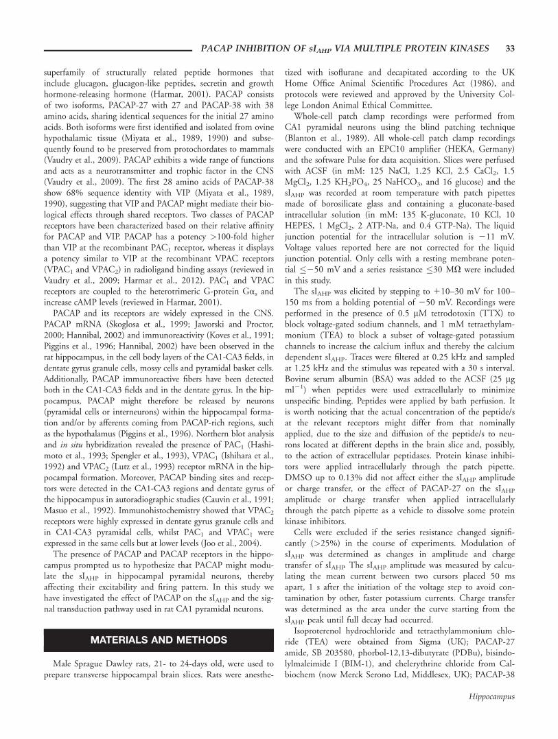
FIGURE 4. p38 MAPK activation is involved in the PACAP-27-induced inhibition of sIAHP. Averaged current traces (n 5 3) ofthe sIAHP before and after application of 250 nM PACAP-27 inthe absence (upper traces) and in the presence (lower traces) ofthe p38 MAPK inhibitor SB 203580 (20 mM) in the intracellularsolution (A). The rightmost panels show superimposed traces inthe absence (black) and presence (grey) of PACAP-27. The dashedline corresponds to the baseline current. (B) Time course of theeffect of PACAP-27 on the normalized sIAHP amplitude from theexperiments in A, in the absence (white squares) and in the pres-ence (black squares) of SB 203580. Bar indicates the presence of250 nM PACAP-27. (C) Bar chart summarizing the relative inhibi-tion of the sIAHP amplitude by 250 nM PACAP-27 under controlconditions (n 5 18; white bar) and in the presence of SB 203580(n 5 7; black bar). SB 203580 markedly inhibited the suppressionof sIAHP mediated by PACAP-27. Error bars indicate S.E.M.* indicates statistical significance.
PACAP INHIBITION OF sIAHP VIA MULTIPLE PROTEIN KINASES 37
Hippocampus

MAPK by EPACs would be in parallel to the one by PKA(Fig. 8). If indeed PKA and p38 MAPK act sequentially in thesame pathway to mediate the inhibition of sIAHP by PACAP-27, as suggested by the combined inhibition of PKA and p38MAPK (Fig. 5), we would expect the EPAC contribution tothis pathway to be minor or negligible. To distinguish betweenPKA and EPAC-mediated effects, we applied the highly specificEPAC superactivator 8CPT-O-Me-cAMP (Rehmann et al.,2003). 8CPT-O-Me-cAMP (5 mM) reduced the sIAHP ampli-tude by 10.3% 6 5.2% (Fig. 6A–C, n 5 7). This reduction wasnot significant (P> 0.05). To assess the integrity of the PKAmeditated modulation of the sIAHP we applied to the samecells the membrane permeable PKA activator 8CPT-cAMP(Fig. 6A,B). PKA activation by 8CPT-cAMP led to a suppres-sion of the sIAHP by 92.8% 6 3.4% (n 5 7, Fig. 6C), showingthe functionality of the PKA pathway. The lack of current inhi-bition by EPAC activation excludes a contribution by EPACsto the modulation of the sIAHP. This supports the finding that
PKA and p38 MAPK are arranged sequentially to mediate theinhibition of the sIAHP by PACAP (Fig. 8).
PKC is not Involved in the sIAHP InhibitionMediated by PACAP-27
Besides their coupling to the cAMP/PKA and MAP kinasepathways, PACAP receptors have been shown to activate phos-pholipase C (PLC) linked signaling cascades (Spengler et al.,1993; Harmar, 2001; reviewed in Vaudry et al., 2009). Proteinkinase C (PKC) is generally thought to act downstream ofPLC (Nishizuka, 1992) and its activation suppresses the sIAHP
in hippocampal pyramidal neurons (Malenka et al., 1986). Wetherefore investigated whether, beside PKA and p38 MAPK,also PKC is involved in the sIAHP suppression induced byPACAP-27.
To obtain maximal PKC inhibition, a combination of twospecific inhibitors acting at different sites on PKC, 20 mM che-lerythrine (Herbert et al., 1990) and 500 nM bisindolylmalei-mide I (BIM-1) (Toullec et al., 1991), was appliedintracellularly. Inhibition of sIAHP amplitude (89.3% 6 7.7%,n 5 5) by PACAP-27 (250 nM) was not significantly differentcompared to recordings performed in the absence of chelerythr-ine and BIM-1 (Fig. 7A,B, and D; P> 0.3, Mann–Whitneytest). An inward shift in the holding current of 29.7 6 9.1 pA
FIGURE 5. Simultaneous inhibition of p38 MAPK and PKApartly prevents the suppression of sIAHP mediated by PACAP-27.Averaged current traces (n 5 3) of the sIAHP in the absence andpresence of 250 nM PACAP-27 recorded without (upper traces)and with (lower traces) the PKA inhibitor Rp-cAMPS (500 mM)and the p38 MAP kinase inhibitor SB 203580 (20 mM) in thepatch pipette (A). The rightmost panels display the superimposedtraces in the absence (black) and presence (grey) of PACAP-27.The dashed line corresponds to the baseline current. (B) Timecourse of the effect of PACAP-27 on the sIAHP amplitude from theexperiments in A, with (black squares) and without (white squares)Rp-cAMPS 1 SB 203580. Bar indicates the application of 250 nMPACAP-27. (C) Bar chart summarizing the relative inhibition ofsIAHP by 250 nM PACAP-27 under control conditions (n 5 18;white bar) and in the presence of Rp-cAMPS 1 SB 203580 (n 5 7;black bar). Coapplication of Rp-cAMPS and SB 203580 partlyprevented the suppression of sIAHP amplitude mediated by 250nM PACAP-27. Error bars indicate S.E.M. * indicates statisticalsignificance.
FIGURE 6. Activation of EPACs does not suppress sIAHP inCA1 neurons. Averaged sIAHP traces (n 5 3) recorded in theabsence (control) and presence of the EPAC superactivator 8CPT-O-Me-cAMP (5 mM) and of the PKA activator 8CPT-cAMP (5mM) (A). The rightmost panel shows the same traces superim-posed. (B) Time course of action of 8CPT-O-Me-cAMP (5 mM)and 8CPT-cAMP (5 mM) on the sIAHP amplitude in the same cellas in A. (C) Bar diagram summarizing the effects of 8CPT-O-Me-cAMP (5 mM; black bar) and subsequently applied 8CPT-cAMP(5–50 mM; white bar) on the sIAHP amplitude in seven cells. The8CPT-cAMP was used at a concentration of 50 mM in 6 out of 7cells. The suppression of sIAHP caused by 8CPT-cAMP was signifi-cantly larger than that observed in response to 8CPT-O-Me-cAMP(P < 0.0001, n 5 7, unpaired t test). Error bars indicate S.E.M.* indicates statistical significance.
38 TAYLOR ET AL.
Hippocampus

(n 5 5; P 5 0.03) was observed upon application of PACAP-27(250 nM) in the presence of the PKC inhibitors. To validatePKC inhibition under our experimental conditions, we acti-vated PKC by phorbol-12,13-dibutyrate (PDBu). Under con-trol conditions, PDBu (500 nM) caused a strong inhibition ofthe sIAHP amplitude (74.4% 6 3.3%; n 5 5), which was signifi-cantly reduced (34.7% 6 15.4%, n 5 6, P 5 0.04, unpaired ttest) in the presence of chelerythrine (20 mM) and BIM-1(500 nM). This indicates that PKC was indeed inhibited underthese experimental conditions.
These experiments show that the PKC signaling cascadedoes not significantly contribute to the inhibition of the sIAHP
by PACAP.
Is a Crosstalk Between the PKA and PKCPathways Involved in the Inhibition of sIAHP byPACAP?
The lack of PKC involvement in the suppression of the sIAHP
by PACAP inferred from the experiments where only PKC wasinhibited does not entirely exclude the possibility of a functionof PKC in the context of a crosstalk with the PKA/p38 MAPKpathway. To elucidate this hypothesis we applied PACAP-27 inthe presence of both PKA and PKC inhibitors. The inhibitionof sIAHP by PACAP-27 was reduced to 56.4% 6 5.0% in thepresence of Rp-cAMPS, chelerythrine and BIM-1 (Fig. 7C,D;n 5 10, P 5 0.03, Mann–Whitney test). The inhibition of thesIAHP amplitude by PACAP-27 was similar in the presence ofRp-cAMPS alone and the combination of Rp-cAMPS, cheler-ythrine and BIM-1 (Fig. 7D; P> 0.8, Mann–Whitney test). Bycontrast, inhibition of PKC alone by chelerythrine and BIM-1with no Rp-cAMPS did not reduce the PACAP effect to a simi-lar extent (Fig. 7D; P 5 0.003 Mann–Whitney test).
These results suggest that the involvement of PKC in thePACAP-mediated inhibition of the sIAHP is unlikely also aspart of a crosstalk.
DISCUSSION
This study shows that PACAP increases the excitability ofCA1 pyramidal neurons by inhibiting the slow Ca21-activatedK1 current sIAHP through the activation of multiple signalingpathways, most prominently cAMP/PKA and p38 MAPK. Ourfindings reveal a novel modulation of the sIAHP by p38 MAPKin CA1 pyramidal neurons, adding to the role of this currentas a convergency point for modulatory inputs mediated bymultiple protein kinases in central neurons.
Previous studies have identified the slow afterhyperpolariza-tion in hippocampal neurons as a target for the neuromodula-tory actions of several neuropeptides, including CRF(Aldenhoff et al., 1983; Haug and Storm, 2000), VIP (Haasand Gahwiler, 1992; Haug and Storm, 2000), and CGRP(Haug and Storm, 2000). Similar effects on the sIAHP havebeen reported for CRF, VIP and PACAP in layer II/III and Vneocortical pyramidal neurons (Hu et al., 2011). Our studyshows that in CA1 pyramidal neurons both PACAP isoforms,PACAP-38 and PACAP-27, suppress the sIAHP, leading to anenhancement of intrinsic excitability and attenuation of spikefrequency adaptation. The suppression of the sIAHP by PACAPshown in our study provides a cellular mechanism for theincrease in spontaneous firing of CA1 neurons observed in vivoupon application of PACAP-38 (Di Mauro et al., 2003).
CA1 pyramidal neurons express all three types of PACAPreceptors, PAC1, VPAC1 and VPAC2 (Hashimoto et al., 1996;
FIGURE 7. PKC is not involved in the sIAHP suppressionmediated by PACAP-27. Superimposed averaged sIAHP traces(n 5 3) recorded in the absence and presence of 250 nM PACAP-27 without (top panel) and with (lower panel) the PKC inhibitorschelerythrine (20 mM) and BIM-1 (500 nM) in the patch pipette(A). The dashed line corresponds to the baseline current. (B) Timecourse of the effect of PACAP-27 on the normalized sIAHP ampli-tude from the experiments in A, with white squares indicating theabsence and black squares the presence of Chelerythrine 1 BIM-1.Bar indicates the application of 250 nM PACAP-27. (C) AveragedsIAHP traces (n 5 3) recorded in the absence and presence of 250nM PACAP-27 with chelerythrine (20 mM), BIM-1 (500 nM) andRp-cAMPS (500 mM) in the patch pipette. The lower panel showsthe traces in the absence (black) and presence (grey) of PACAP-27superimposed. The dashed line represents the baseline current. (D)Bar chart summarizing the relative inhibition of sIAHP by 250 nMPACAP-27 under control conditions (n 5 18; white bar; same dataas in Fig. 1F, reported here for comparison) and in the presence ofChelerythrine 1 BIM-1 (n 5 5; black bar), Rp-cAMPS (n 5 15;grey bar; same data as in Fig. 3C, reported here for comparison),and Chelerythrine 1 BIM-1 1 Rp-cAMPS (n 5 10; stripy bar).Chelerythrine and BIM-1 did not prevent the suppression of sIAHP
by PACAP-27 and did not affect the partial inhibition of thePACAP-27 effect by Rp-cAMPS. Error bars indicate S.E.M. * indi-cates statistical significance.
PACAP INHIBITION OF sIAHP VIA MULTIPLE PROTEIN KINASES 39
Hippocampus
Shioda et al., 1997; Joo et al., 2004). The �30% reduction ofthe sIAHP amplitude we observed upon application of the selec-tive PAC1 agonist maxadilan suggests a potential contributionof PAC1 receptors. The presence of the PAC1/VPAC2 antago-nist PACAP-(6–38) (Dickinson et al., 1997; Moro et al.,1999) indeed prevented the effect of maxadilan, supporting apotential role for PAC1 receptors. Additionally the activationof VPAC1, but not VPAC2 receptors, leads to the suppressionof the sIAHP amplitude, because the activation of PAC1 recep-tors alone by maxadilan is not sufficient to inhibit the sIAHP toa similar extent as caused by PACAP. The involvement ofVPAC1 receptors was further inferred by the observation thatthe PAC1/VPAC2 antagonist PACAP-(6–38) did not preventthe inhibition of the sIAHP by PACAP. This is different fromwhat has been observed in the neocortex, where PACAP hasbeen reported to activate the cAMP/PKA pathway by actingmainly through PAC1 receptors (Hu et al., 2011). Our studyconcludes that the sIAHP suppression by PACAP can be elicitedthrough the activation of PAC1 and most likely VPAC1 recep-tors in CA1 pyramidal neurons.
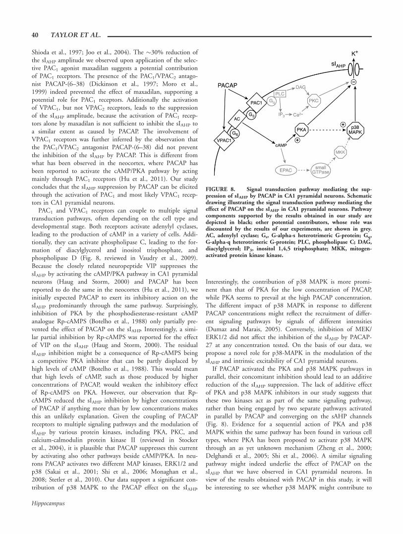
PAC1 and VPAC1 receptors can couple to multiple signaltransduction pathways, often depending on the cell type anddevelopmental stage. Both receptors activate adenylyl cyclases,leading to the production of cAMP in a variety of cells. Addi-tionally, they can activate phospholipase C, leading to the for-mation of diacylglycerol and inositol trisphosphate, andphospholipase D (Fig. 8, reviewed in Vaudry et al., 2009).Because the closely related neuropeptide VIP suppresses thesIAHP by activating the cAMP/PKA pathway in CA1 pyramidalneurons (Haug and Storm, 2000) and PACAP has beenreported to do the same in the neocortex (Hu et al., 2011), weinitially expected PACAP to exert its inhibitory action on thesIAHP predominantly through the same pathway. Surprisingly,inhibition of PKA by the phosphodiesterase-resistant cAMPanalogue Rp-cAMPS (Botelho et al., 1988) only partially pre-vented the effect of PACAP on the sIAHP. Interestingly, a simi-lar partial inhibition by Rp-cAMPS was reported for the effectof VIP on the sIAHP (Haug and Storm, 2000). The residualsIAHP inhibition might be a consequence of Rp-cAMPS beinga competitive PKA inhibitor that can be partly displaced byhigh levels of cAMP (Botelho et al., 1988). This would meanthat high levels of cAMP, such as those produced by higherconcentrations of PACAP, would weaken the inhibitory effectof Rp-cAMPS on PKA. However, our observation that Rp-cAMPS reduced the sIAHP inhibition by higher concentrationsof PACAP if anything more than by low concentrations makesthis an unlikely explanation. Given the coupling of PACAPreceptors to multiple signaling pathways and the modulation ofsIAHP by various protein kinases, including PKA, PKC, andcalcium-calmodulin protein kinase II (reviewed in Stockeret al., 2004), it is plausible that PACAP suppresses this currentby activating also other pathways beside cAMP/PKA. In neu-rons PACAP activates two different MAP kinases, ERK1/2 andp38 (Sakai et al., 2001; Shi et al., 2006; Monaghan et al.,2008; Stetler et al., 2010). Our data support a significant con-tribution of p38 MAPK to the PACAP effect on the sIAHP.
Interestingly, the contribution of p38 MAPK is more promi-nent than that of PKA for the low concentration of PACAP,while PKA seems to prevail at the high PACAP concentration.The different impact of p38 MAPK in response to differentPACAP concentrations might reflect the recruitment of differ-ent signaling pathways by signals of different intensities(Dumaz and Marais, 2005). Conversely, inhibition of MEK/ERK1/2 did not affect the inhibition of the sIAHP by PACAP-27 at any concentration tested. On the basis of our data, wepropose a novel role for p38-MAPK in the modulation of thesIAHP and intrinsic excitability of CA1 pyramidal neurons.
If PACAP activated the PKA and p38 MAPK pathways inparallel, their concomitant inhibition should lead to an additivereduction of the sIAHP suppression. The lack of additive effectof PKA and p38 MAPK inhibitors in our study suggests thatthese two kinases act as part of the same signaling pathway,rather than being engaged by two separate pathways activatedin parallel by PACAP and converging on the sAHP channels(Fig. 8). Evidence for a sequential action of PKA and p38MAPK within the same pathway has been found in various celltypes, where PKA has been proposed to activate p38 MAPKthrough an as yet unknown mechanism (Zheng et al., 2000;Delghandi et al., 2005; Shi et al., 2006). A similar signalingpathway might indeed underlie the effect of PACAP on thesIAHP that we have observed in CA1 pyramidal neurons. Inview of the results obtained with PACAP in this study, it willbe interesting to see whether p38 MAPK might contribute to
FIGURE 8. Signal transduction pathway mediating the sup-pression of sIAHP by PACAP in CA1 pyramidal neurons. Schematicdrawing illustrating the signal transduction pathway mediating theeffect of PACAP on the sIAHP in CA1 pyramidal neurons. Pathwaycomponents supported by the results obtained in our study aredepicted in black; other potential contributors, whose role wasdiscounted by the results of our experiments, are shown in grey.AC, adenylyl cyclase; Gs, G-alpha-s heterotrimeric G-protein; Gq,G-alpha-q heterotrimeric G-protein; PLC, phospholipase C; DAG,diacylglycerol; IP3, inositol 1,4,5 trisphosphate; MKK, mitogen-activated protein kinase kinase.
40 TAYLOR ET AL.
Hippocampus

the neuromodulatory effects also of other neuropeptides andneurotransmitters. Indeed, p38 MAPK has been shown tomediate the persistent sAHP suppression caused by prolongedstimulation of type 5 metabotropic glutamate receptors in CA3pyramidal neurons (Young et al., 2008).
In other studies, p38 MAPK is a downstream substrate ofthe cAMP signaling pathway independent of PKA, through theactivation of exchange proteins directly activated by cAMP(EPACs) and small G-proteins (Fig. 8, Shi et al., 2006; Steret al., 2007). Our results, obtained by using a highly specificEPAC superactivator (Rehmann et al., 2003), do not supportan involvement of EPACs in the modulation of sIAHP.
PAC1 and VPAC1 receptors are coupled to the phospholi-pase C (PLC) pathway, leading to the activation of PKC (Fig.8, reviewed in Vaudry et al., 2009). PKC suppresses the sIAHP
in hippocampal pyramidal neurons (Malenka et al., 1986).However, our data show that PACAP does not suppress sIAHP
by activating PKC in CA1 pyramidal neurons. This argues infavor of selective coupling between PACAP receptors and spe-cific signal transduction pathways in hippocampal neurons.The basis for this selectivity might lie in the existence of signal-ing microdomains defining the spatio-temporal dynamics ofPACAP signaling, a topic that will be addressed in futurestudies.
In conclusion, our study shows that PACAP effectively mod-ulates the intrinsic excitability and firing behavior of CA1pyramidal neurons through the suppression of sIAHP. It is wellestablished that a reduction in the sAHP in CA1 and CA3neurons accompanies acquisition of hippocampus-dependentlearning, whereas increases in the sAHP are correlated withcognitive impairment (reviewed in Disterhoft et al., 2004).The impact of the sIAHP on learning processes reflects its multi-ple roles in controlling firing and intrinsic excitability (Zhangand Linden, 2003) and integration of synaptic signals (Sah andBekkers, 1996; Borde et al., 1999; Lancaster et al., 2001; Wuet al., 2004; Fernandez de Sevilla et al., 2007). Drosophila har-boring a mutation in the PACAP-related gene amnesiac displaydeficits in associative learning (Quinn et al., 1979; Feany andQuinn, 1995). In the rat, PACAP intracerebroventricularadministration enhances learning, possibly by affecting memoryconsolidation (Sacchetti et al., 2001). Mice lacking PAC1
receptors display impaired long-term potentiation (LTP) ofsynaptic transmission at several synapses in the hippocampalformation (Otto et al., 2001; Matsuyama et al., 2003), corre-lating with a deficit in contextual fear conditioning, ahippocampus-dependent learning task (Sauvage et al., 2000;Otto et al., 2001; Matsuyama et al., 2003). PACAP regulatesbidirectional synaptic plasticity at the Schaffer collateral CA1synapse in a concentration-dependent manner, whereby sub-nanomolar concentrations of the peptide induce long-lastingfacilitation of excitatory synaptic potentials (Roberto et al.,2001), while concentrations similar to those used in this study(250–500 nM) cause either an initial decrease followed by anenhancement (Roberto et al., 2001), or a long-lasting depres-sion of excitatory synaptic transmission (Kondo et al., 1997;Ster et al., 2009) that depends on EPAC and p38 MAPK acti-
vation (Ster et al., 2009). Taken together, our results suggestthat modulation of the sIAHP via PAC1 and VPAC1 receptorsmight contribute to the complex effects of PACAP on synapticplasticity in the hippocampal formation and ultimately onlearning and memory consolidation.
Acknowledgments
The authors gratefully acknowledge Prof. Ethan Lerner, Har-vard Medical School, for the kind gift of maxadilan andmax.d.4 peptides. We thank Dr Jason Rothman, UCL, forhelp and assistance with Neuromatic. The authors have noconflict of interest to declare.
REFERENCES
Aldenhoff JB, Gruol DL, Rivier J, Vale W, Siggins GR. 1983. Corti-cotropin releasing factor decreases postburst hyperpolarizations andexcites hippocampal neurons. Science 221:875–877.
Alger BE, Nicoll RA. 1980. Epileptiform burst afterhyperolarization:Calcium-dependent potassium potential in hippocampal CA1pyramidal cells. Science 210:1122–1124.
Blanton MG, Lo Turco JJ, Kriegstein AR. 1989. Whole cell recordingfrom neurons in slices of reptilian and mammalian cerebral cortex.J Neurosci Methods 30:203–210.
Borde M, Bonansco C, Buno W. 1999. The activity-dependent poten-tiation of the slow Ca21-activated K1 current regulates synapticefficacy in rat CA1 pyramidal neurons. Pflugers Arch 437:261–266.
Botelho LH, Rothermel JD, Coombs RV, Jastorff B. 1988. cAMP ana-log antagonists of cAMP action. Methods Enzymol 159:159–172.
Cauvin A, Robberecht P, De Neef P, Gourlet P, Vandermeers A,Vandermeers-Piret MC, Christophe J. 1991. Properties and distri-bution of receptors for pituitary adenylate cyclase activatingpeptide (PACAP) in rat brain and spinal cord. Regul Pept 35:161–173.
Delghandi MP, Johannessen M, Moens U. 2005. The cAMP signallingpathway activates CREB through PKA, p38 and MSK1 in NIH3T3 cells. Cell Signal 17:1343–1351.
Di Mauro M, Cavallaro S, Ciranna L. 2003. Pituitary adenylatecyclase-activating polypeptide modifies the electrical activity ofCA1 hippocampal neurons in the rat. Neurosci Lett 337:97–100.
Dickinson T, Fleetwood-Walker SM, Mitchell R, Lutz EM. 1997. Evi-dence for roles of vasoactive intestinal polypeptide (VIP) and pitui-tary adenylate cyclase activating polypeptide (PACAP) receptors inmodulating the responses of rat dorsal horn neurons to sensoryinputs. Neuropeptides 31:175–185.
Disterhoft JF, Wu WW, Ohno M. 2004. Biophysical alterations ofhippocampal pyramidal neurons in learning, ageing and Alzhei-mer’s disease. Ageing Res Rev 3:383–406.
Dumaz N, Marais R. 2005. Integrating signals between cAMP andthe RAS/RAF/MEK/ERK signalling pathways. FEBS J 272:3491–3504.
Eyers PA, Craxton M, Morrice N, Cohen P, Goedert M. 1998. Con-version of SB 203580-insensitive MAP kinase family members todrug-sensitive forms by a single amino-acid substitution. ChemBiol 5:321–328.
Favata MF, Horiuchi KY, Manos EJ, Daulerio AJ, Stradley DA, FeeserWS, Van Dyk DE, Pitts WJ, Earl RA, Hobbs F, Copeland RA,Magolda RL, Scherle PA, Trzaskos JM. 1998. Identification of a
PACAP INHIBITION OF sIAHP VIA MULTIPLE PROTEIN KINASES 41
Hippocampus
novel inhibitor of mitogen-activated protein kinase kinase. J BiolChem 273:18623–18632.
Feany MB, Quinn WG. 1995. A neuropeptide gene defined by theDrosophila memory mutant amnesiac. Science 268:869–873.
Fernandez de Sevilla D, Fuenzalida M, Porto Pazos AB, Buno W.2007. Selective shunting of the NMDA EPSP component by theslow afterhyperpolarization in rat CA1 pyramidal neurons. J Neu-rophysiol 97:3242–3255.
Haas HL, Gahwiler BH. 1992. Vasoactive intestinal polypeptidemodulates neuronal excitability in hippocampal slices of the rat.Neuroscience 47:273–277.
Hannibal J. 2002. Pituitary adenylate cyclase-activating peptide in therat central nervous system: An immunohistochemical and in situhybridization study. J Comp Neurol 453:389–417.
Harmar AJ. 2001. Family-B G-protein-coupled receptors. GenomeBiol 2:reviews3013.1–3013.10.
Harmar AJ, Fahrenkrug J, Gozes I, Laburthe M, May V, Pisegna JR,Vaudry D, Vaudry H, Waschek JA, Said SI. 2012. Pharmacologyand functions of receptors for vasoactive intestinal peptide andpituitary adenylate cyclase-activating polypeptide. Br J Pharmacol166:4–17.
Hashimoto H, Ishihara T, Shigemoto R, Mori K, Nagata S. 1993.Molecular cloning and tissue distribution of a receptor for pituitaryadenylate cyclase-activating polypeptide. Neuron 11:333–342.
Hashimoto H, Nogi H, Mori K, Ohishi H, Shigemoto R, YamamotoK, Matsuda T, Mizuno N, Nagata S, Baba A. 1996. Distributionof the mRNA for a pituitary adenylate cyclase-activating polypep-tide receptor in the rat brain: An in situ hybridization study.J Comp Neurol 371:567–577.
Haug T, Storm JF. 2000. Protein kinase A mediates the modulation ofthe slow Ca21-dependent K1 current, IsAHP, by the neuropeptidesCRF, VIP, and CGRP in hippocampal pyramidal neurons.J Neurophysiol 83:2071–2079.
Herbert JM, Augereau JM, Gleye J, Maffrand JP. 1990. Chelerythrineis a potent and specific inhibitor of protein kinase C. BiochemBiophys Res Commun 172:993–999.
Hotson JR, Prince DA. 1980. A calcium-activated hyperpolarizationfollows repetitive firing in hippocampal neurons. J Neurophysiol43:409–419.
Hu E, Demmou L, Cauli B, Gallopin T, Geoffroy H, Harris-WarrickRM, Paupardin-Tritsch D, Lambolez B, Vincent P, Hepp R. 2011.VIP, CRF, and PACAP act at distinct receptors to elicit differentcAMP/PKA dynamics in the neocortex. Cereb Cortex 21:708–718.
Ishihara T, Shigemoto R, Mori K, Takahashi K, Nagata S. 1992.Functional expression and tissue distribution of a novel receptorfor vasoactive intestinal polypeptide. Neuron 8:811–819.
Jaworski DM, Proctor MD. 2000. Developmental regulation of pitui-tary adenylate cyclase-activating polypeptide and PAC1 receptormRNA expression in the rat central nervous system. Brain ResDev Brain Res 120:27–39.
Joo KM, Chung YH, Kim MK, Nam RH, Lee BL, Lee KH, Cha CI.2004. Distribution of vasoactive intestinal peptide and pituitaryadenylate cyclase-activating polypeptide receptors (VPAC1, VPAC2,and PAC1 receptor) in the rat brain. J Comp Neurol 476:388–413.
Kondo T, Tominaga T, Ichikawa M, Iijima T. 1997. Differentialalteration of hippocampal synaptic strength induced by pituitaryadenylate cyclase activating polypeptide-38 (PACAP-38). NeurosciLett 221:189–192.
Koves K, Arimura A, Gorcs TG, Somogyvari-Vigh A. 1991. Compar-ative distribution of immunoreactive pituitary adenylate cyclaseactivating polypeptide and vasoactive intestinal polypeptide in ratforebrain. Neuroendocrinology 54:159–169.
Lancaster B, Adams PR. 1986. Calcium-dependent current generatingthe afterhyperpolarization of hippocampal neurons. J Neurophysiol55:1268–1282.
Lancaster B, Hu H, Ramakers GM, Storm JF. 2001. Interactionbetween synaptic excitation and slow afterhyperpolarization currentin rat hippocampal pyramidal cells. J Physiol 536:809–823.
Lancaster B, Nicoll RA. 1987. Properties of two calcium-activatedhyperpolarizations in rat hippocampal neurones. J Physiol 389:187–203.
Lerner EA, Ribeiro JM, Nelson RJ, Lerner MR. 1991. Isolation ofmaxadilan, a potent vasodilatory peptide from the salivary glandsof the sand fly Lutzomyia longipalpis. J Biol Chem 266:11234–11236.
Lutz EM, Sheward WJ, West KM, Morrow JA, Fink G, Harmar AJ.1993. The VIP2 receptor: Molecular characterisation of a cDNAencoding a novel receptor for vasoactive intestinal peptide. FEBSLett 334:3–8.
Madison DV, Nicoll RA. 1982. Noradrenaline blocks accommodationof pyramidal cell discharge in the hippocampus. Nature 299:636–638.
Madison DV, Nicoll RA. 1984. Control of the repetitive discharge ofrat CA 1 pyramidal neurones in vitro. J Physiol 354:319–331.
Malenka RC, Madison DV, Andrade R, Nicoll RA. 1986. Phorbolesters mimic some cholinergic actions in hippocampal pyramidalneurons. J Neurosci 6:475–480.
Masuo Y, Ohtaki T, Masuda Y, Tsuda M, Fujino M. 1992. Bindingsites for pituitary adenylate cyclase activating polypeptide(PACAP): Comparison with vasoactive intestinal polypeptide (VIP)binding site localization in rat brain sections. Brain Res 575:113–123.
Matsuyama S, Matsumoto A, Hashimoto H, Shintani N, Baba A.2003. Impaired long-term potentiation in vivo in the dentate gyrusof pituitary adenylate cyclase-activating polypeptide (PACAP) orPACAP type 1 receptor-mutant mice. Neuroreport 14:2095–2098.
Miyata A, Arimura A, Dahl RR, Minamino N, Uehara A, Jiang L,Culler MD, Coy DH. 1989. Isolation of a novel 38 residue-hypothalamic polypeptide which stimulates adenylate cyclase inpituitary cells. Biochem Biophys Res Commun 164:567–574.
Miyata A, Jiang L, Dahl RD, Kitada C, Kubo K, Fujino M,Minamino N, Arimura A. 1990. Isolation of a neuropeptide corre-sponding to the N-terminal 27 residues of the pituitary adenylatecyclase activating polypeptide with 38 residues (PACAP38). Bio-chem Biophys Res Commun 170:643–648.
Monaghan TK, Mackenzie CJ, Plevin R, Lutz EM. 2008. PACAP-38induces neuronal differentiation of human SH-SY5Y neuroblas-toma cells via cAMP-mediated activation of ERK and p38 MAPkinases. J Neurochem 104:74–88.
Moro O, Lerner EA. 1997. Maxadilan, the vasodilator from sand flies,is a specific pituitary adenylate cyclase activating peptide type Ireceptor agonist. J Biol Chem 272:966–970.
Moro O, Tajima M, Lerner EA. 1996. Receptors for the vasodilatormaxadilan are expressed on selected neural crest and smoothmuscle-derived cells. Insect Biochem Mol Biol 26:1019–1025.
Moro O, Wakita K, Ohnuma M, Denda S, Lerner EA, Tajima M.1999. Functional characterization of structural alterations in thesequence of the vasodilatory peptide maxadilan yields a pituitaryadenylate cyclase-activating peptide type 1 receptor-specificantagonist. J Biol Chem 274:23103–23110.
Nishizuka Y. 1992. Intracellular signaling by hydrolysis of phospholi-pids and activation of protein kinase C. Science 258:607–614.
Otto C, Kovalchuk Y, Wolfer DP, Gass P, Martin M, Zuschratter W,Grone HJ, Kellendonk C, Tronche F, Maldonado R, Lipp H-P,Konnerth A, Sch€utz G. 2001. Impairment of mossy fiber long-term potentiation and associative learning in pituitary adenylatecyclase activating polypeptide type I receptor-deficient mice. J Neu-rosci 21:5520–5527.
Pedarzani P, Storm JF. 1993. PKA mediates the effects of monoaminetransmitters on the K1 current underlying the slow spike frequencyadaptation in hippocampal neurons. Neuron 11:1023–1035.
42 TAYLOR ET AL.
Hippocampus
Pedarzani P, Storm JF. 1995. Dopamine modulates the slow Ca21-activated K1 current IAHP via cyclic AMP-dependent proteinkinase in hippocampal neurons. J Neurophysiol 74:2749–2753.
Piggins HD, Stamp JA, Burns J, Rusak B, Semba K. 1996. Distribu-tion of pituitary adenylate cyclase activating polypeptide (PACAP)immunoreactivity in the hypothalamus and extended amygdala ofthe rat. J Comp Neurol 376:278–294.
Quinn WG, Sziber PP, Booker R. 1979. The Drosophila memorymutant amnesiac. Nature 277:212–214.
Rehmann H, Schwede F, Doskeland SO, Wittinghofer A, Bos JL.2003. Ligand-mediated activation of the cAMP-responsive guaninenucleotide exchange factor Epac. J Biol Chem 278:38548–38556.
Roberto M, Scuri R, Brunelli M. 2001. Differential effects ofPACAP-38 on synaptic responses in rat hippocampal CA1 region.Learn Mem 8:265–271.
Sacchetti B, Lorenzini CA, Baldi E, Bucherelli C, Roberto M, TassoniG, Brunelli M. 2001. Pituitary adenylate cyclase-activating poly-peptide hormone (PACAP) at very low dosages improves memoryin the rat. Neurobiol Learn Mem 76:1–6.
Sah P, Bekkers JM. 1996. Apical dendritic location of slow afterhyper-polarization current in hippocampal pyramidal neurons: Implica-tions for the integration of long-term potentiation. J Neurosci 16:4537–4542.
Sah P, Faber ES. 2002. Channels underlying neuronal calcium-activated potassium currents. Prog Neurobiol 66:345–353.
Sakai Y, Hashimoto H, Shintani N, Tomimoto S, Tanaka K, IchiboriA, Hirose M, Baba A. 2001. Involvement of p38 MAP kinasepathway in the synergistic activation of PACAP mRNA expressionby NGF and PACAP in PC12h cells. Biochem Biophys Res Com-mun 285:656–661.
Sauvage M, Brabet P, Holsboer F, Bockaert J, Steckler T. 2000. Milddeficits in mice lacking pituitary adenylate cyclase-activating poly-peptide receptor type 1 (PAC1) performing on memory tasks.Brain Res Mol Brain Res 84:79–89.
Schwartzkroin PA, Stafstrom CE. 1980. Effects of EGTA on thecalcium-activated afterhyperpolarization in hippocampal CA3pyramidal cells. Science 210:1125–1126.
Shi GX, Rehmann H, Andres DA. 2006. A novel cyclic AMP-dependent Epac-Rit signaling pathway contributes to PACAP38-mediated neuronal differentiation. Mol Cell Biol 26:9136–9147.
Shioda S, Shuto Y, Somogyvari-Vigh A, Legradi G, Onda H, CoyDH, Nakajo S, Arimura A. 1997. Localization and gene expressionof the receptor for pituitary adenylate cyclase-activating polypep-tide in the rat brain. Neurosci Res 28:345–354.
Skoglosa Y, Takei N, Lindholm D. 1999. Distribution of pituitaryadenylate cyclase activating polypeptide mRNA in the developingrat brain. Brain Res Mol Brain Res 65:1–13.
Spengler D, Waeber C, Pantaloni C, Holsboer F, Bockaert J, SeeburgPH, Journot L. 1993. Differential signal transduction by five splicevariants of the PACAP receptor. Nature 365:170–175.
Ster J, de Bock F, Bertaso F, Abitbol K, Daniel H, Bockaert J, FagniL. 2009. Epac mediates PACAP-dependent long-term depressionin the hippocampus. J Physiol 587:101–113.
Ster J, De Bock F, Guerineau NC, Janossy A, Barrere-Lemaire S, BosJL, Bockaert J, Fagni L. 2007. Exchange protein activated bycAMP (Epac) mediates cAMP activation of p38 MAPK and modu-lation of Ca21-dependent K1 channels in cerebellar neurons. ProcNatl Acad Sci USA 104:2519–2524.
Stetler RA, Gao Y, Zukin RS, Vosler PS, Zhang L, Zhang F, Cao G,Bennett MV, Chen J. 2010. Apurinic/apyrimidinic endonucleaseAPE1 is required for PACAP-induced neuroprotection againstglobal cerebral ischemia. Proc Natl Acad Sci USA 107:3204–3209.
Stocker M, Hirzel K, D’Hoedt D, Pedarzani P. 2004. Matching mole-cules to function: Neuronal Ca21-activated K1 channels andafterhyperpolarizations. Toxicon 43:933–949.
Toullec D, Pianetti P, Coste H, Bellevergue P, Grand-Perret T,Ajakane M, Baudet V, Boissin P, Boursier E, Loriolle F, DuhamelL, Charon D, Kirilovsky J. 1991. The bisindolylmaleimide GF109203X is a potent and selective inhibitor of protein kinase C. JBiol Chem 266:15771–15781.
Vaudry D, Falluel-Morel A, Bourgault S, Basille M, Burel D, WurtzO, Fournier A, Chow BK, Hashimoto H, Galas L, Vaudry H.2009. Pituitary adenylate cyclase-activating polypeptide and itsreceptors: 20 years after the discovery. Pharmacol Rev 61:283–357.
Villalba M, Bockaert J, Journot L. 1997. Pituitary adenylate cyclase-activating polypeptide (PACAP-38) protects cerebellar granule neu-rons from apoptosis by activating the mitogen-activated proteinkinase (MAP kinase) pathway. J Neurosci 17:83–90.
Wu WW, Chan CS, Disterhoft JF. 2004. Slow afterhyperpolarizationgoverns the development of NMDA receptor-dependent afterdepo-larization in CA1 pyramidal neurons during synaptic stimulation. JNeurophysiol 92:2346–2356.
Young SR, Bianchi R, Wong RK. 2008. Signaling mechanisms under-lying group I mGluR-induced persistent AHP suppression in CA3hippocampal neurons. J Neurophysiol 99:1105–1118.
Zhang W, Linden DJ. 2003. The other side of the engram:Experience-driven changes in neuronal intrinsic excitability. NatRev Neurosci 4:885–900.
Zheng M, Zhang SJ, Zhu WZ, Ziman B, Kobilka BK, Xiao RP.2000. beta 2-adrenergic receptor-induced p38 MAPK activation ismediated by protein kinase A rather than by Gi or gbeta gammain adult mouse cardiomyocytes. J Biol Chem 275:40635–40640.
Zhong Y. 1995. Mediation of PACAP-like neuropeptide transmissionby coactivation of Ras/Raf and cAMP signal transduction pathwaysin Drosophila. Nature 375:588–592.
PACAP INHIBITION OF sIAHP VIA MULTIPLE PROTEIN KINASES 43
Hippocampus