Embed Size (px)
Citation preview

Ps
VF
a
ARRAA
KEBSSPD
1
aftHaccCga
cvwea
ea�
h0
Journal of Electron Spectroscopy and Related Phenomena 197 (2014) 43–49
Contents lists available at ScienceDirect
Journal of Electron Spectroscopy andRelated Phenomena
jou rn al h om epage: www.elsev ier .com/ locate /e lspec
hotoelectron spectra and electronic structure of somepiroborate complexes
.I. Vovna, S.A. Tikhonov, I.B. Lvov ∗, I.S. Osmushko, I.V. Svistunova, O.L. Shchekaar Eastern Federal University, 8 Sukhanova Str., Vladivostok 690600, Russian Federation
r t i c l e i n f o
rticle history:eceived 13 May 2014eceived in revised form 28 August 2014ccepted 30 August 2014vailable online 8 September 2014
a b s t r a c t
The electronic structure of the valence and core levels of three spiroborate complexes – boron 1,2-dioxyphenylene �-diketonates – has been investigated by methods of UV and X-ray photoelectronspectroscopy and quantum chemical density functional theory. The ionization energy of �- and n-orbitalsof the dioxyphenylene fragment and �-diketonate ligand were measured from UV photoelectron spectra.This made it possible to determine the effect of substitution of one or two methyl groups by the phenylin diketone on the electronic structure of complexes. The binding energy of nonequivalent carbon and
eywords:lectronic structureoron �-diketonatespiroborate complexespiroconjugationhotoelectron spectroscopy
oxygen atoms were measured from X-ray photoelectron spectra. The results of calculations of the energyof the valence orbitals of complexes allowed us to refer bands observed in the spectra of the valenceelectrons to the 2s-type levels of carbon and oxygen.
© 2014 Elsevier B.V. All rights reserved.
ensity functional theory
. Introduction
Modern chemistry of boron organic compounds is primarilyssociated with borane chemistry, the study of which is explainedrom both the theoretical (multi-centre bonds, clusters) and prac-ical (fuel, neutron capture, radiotherapy) points of view [1].owever, other classes of boron compounds have attracted thettention of researchers. These include, in particular, spiroborateomplexes, which are relatively easily synthesized [2,3] and suffi-iently stable and have a crystal structure and high melting points.hromophoric fragments of spiroborate complexes are not conju-ated to each other. In this regard, study of the electronic structurend optical properties of these compounds is also interesting [4,5].
The determination of the relationship between the functionalharacteristics of the substances and their electronic structure pro-ides opportunities for the directed synthesis of new compoundsith desired properties. The most complete information about the
lectronic structure of the complexes can be obtained by the jointpplication of experimental and theoretical methods.
Previously, we have reported the results of research on the
lectronic structure of boron difluoride �-diketonates containingromatic substituents with one [6] and two [7] benzene rings in-positions by methods of ultraviolet photoelectron spectroscopy∗ Corresponding author. Tel.: +7 9243389961.E-mail addresses: [email protected], [email protected] (I.B. Lvov).
ttp://dx.doi.org/10.1016/j.elspec.2014.08.009368-2048/© 2014 Elsevier B.V. All rights reserved.
(UPS) of vapour and density functional theory (DFT). Boron difluo-ride dibenzoylmethanate (F2BDbm) has been studied by UPS,X-ray photoelectron spectroscopy (XPS), and X-ray emission spec-troscopy [8,9]. As noted in [6–8], the DFT method make it possible tocalculate the energy of the ionized states of the boron �-diketonatecomplexes with high accuracy. This is a good theoretical basis forthe unambiguous interpretation of the photoelectron spectra andanalysis of the structure of the valence electron levels.
This paper presents the results of a study of the electronicstructure of acetylacetonate (I), benzoylacetonate (II), and diben-zoylmethanate (III) of boron 1,2-dihydroxyphenylene, as obtainedby UPS, XPS, and DFT.
(I) R1 = R2 = Me(II) R1 = Me; R2 = Ph(III) R1 = R2 = Ph
2. Experimental and calculation methods
Spiroborate complexes I–III were obtained by the reaction of2-n-butoxy-1,2,3-benzodioxaborol with �-diketones according to[1]. UV-photoelectron spectra were registered by serial electronspectrometer ES-3201 with a monochromatic radiation source,

4 scopy and Related Phenomena 197 (2014) 43–49
Hwtiuwwibobalaaoem
pfOehcsopTt
ee
I
wof
iegic
ltiaiotae
3
Ir
papc
Fig. 1. Ultraviolet photoelectron spectra of the vapours of compounds I–III: exper-imental spectrum (heavy line), Gaussian components (thin envelope line), andcalculated energies of molecular orbitals shifted by the value of Koopmans’ defect
4 V.I. Vovna et al. / Journal of Electron Spectro
e-I (h� = 21.2 eV). The determination accuracy of band maximaas under 0.02 eV. The temperature in the ionization cell was 200
o 240 ◦C. The X-ray photoelectron spectra of compounds I and IIIn condensed state were measured using an MXPS XP high vac-um double-chamber system (manufacturer—Omicron, Germany)ith a hemispherical electrostatic analyser. The radiation energyas 1253.6 eV (Mg K�). The spread function of the spectrometer
n the mode of characteristic atomic level registration, which hadeen determined from the Ag3d5/2 band shape, had a half-widthf 1.2 eV. Electron binding energy calibration (Eb) was performedased on the intrinsic standard method with the C1s carbon levels a mark. Purification of samples was done by repeated recrystal-ization. While estimating the atomic concentration of elements in
sample, we took into account relative ionization cross-sectionsnd electron escape depth. The relative concentration of elementsbtained from the intensities of the bands corresponding to 1s-lectrons coincided with that calculated within the error of theethod (10%).All calculations were performed using the Firefly 7.1.G program
ackage [10]. In the DFT method, a hybrid exchange-correlationunctional B3LYP5 and basis set def2-TZVPP have been used [11,12].ur choice of the basis set and functional for calculation of thelectronic structure of �-diketonate complexes, including boron,as been justified by test calculation in [13] and successful appli-ation of theoretical models for the interpretation of the electronpectra [6–8,14,15]. In order to check the correspondence betweenptimized structures and local minimum points on a surface ofotential energy, calculation of a Hessian matrix was performed.he absence of imaginary frequencies in vibration spectra pointsoward real minima on the potential energy surface.
For comparison of the experimental values of vertical ionizationnergies (IE) with energies of Kohn–Sham orbitals, εi, we used anxpanded version of Koopmans’ theorem:
Ei = −εi + ıi,
here IEi is the calculated ionization energy; −εi is the Kohn–Shamne-electron energy; ıi is the Koopmans defect, which is constantor a given type of MO energy correction.
The IEi value corresponds to the energy of the Gaussian max-mum, IEg. The Gaussian maximum position for three higherlectron levels is consistent with experimental ionization ener-ies. UV bands corresponding to several orbitals were decomposednto Gaussians, taking into account the energy intervals betweenalculated electron levels.
The Koopmans defect ıi is a measure of the deviation of calcu-ated one-electron energies εi from the experimental vertical IEi,hat is, a measure of the violation of Koopmans’ theorem. Thats why Koopmans defect analysis is a useful tool as a theoreticalpproximation of quantum chemistry to describe the energy ofonic states of molecules. When taking into account the dependencef the amendment to the orbital energy ıi on the nature of the elec-ronic level, Koopmans’ theorem makes it possible to obtain a goodgreement between the experimental energy IEi and the calculatednergy εi of the Kohn–Sham molecular orbital (MO) [13,14,16].
. Results and discussion
According to the calculation results, the geometry of compound corresponds to the symmetry group C2v (the planes of the chelateing and fragment PheO2B are perpendicular to each other).
Under substitution of one methyl group in compound I by
henyl (compound II), axis C2 of fragment PheO2B deviates by anngle of 15◦ relative to the plane of the chelate ligand, and in com-lex III this angle increases to 19◦. Coplanarity of the planes ofhelate and benzene rings is absent for compounds II and III, and(vertical lines).
the dihedral angles are 12◦ and 14◦, respectively. According to thecalculated results, compound III has symmetry Cs, but for conve-nience of comparison among MOs I–III we use the notation of thesymmetry group C2v in Table 3, Figs. 2 and 6. The usage of C2v sym-metry is justified by the inconspicuous difference between the totalenergies of the structures with Cs and C2v symmetries (84 cm−1).
For compounds I–III, the interatomic distances B O3 and B O4(1.44 A) are much smaller than the distances B O1 and O2 B(1.52 A). Also, for compounds I–III, small differences in the rele-vant interatomic distances (≤0.02 A) and angles between bondswithin the chelate ligand and dioxyphenylene fragment (≤2◦) wereobserved.
Fig. 1 shows the UV-photoelectron spectra of the vapours ofthe compounds studied. Under the decomposition of the spectralbands into Gaussian components, we took into account the num-ber of calculated electronic levels, the energy intervals betweenthem, and the closeness values of the ionization cross-sections ofthe electronic levels.
The images of the molecular orbitals of complexes I and III whichare the most important for interpretation of UV-photoelectronspectra are shown in Fig. 2. Tables 1–3 contain the results of calcula-tion of the relative contributions of the 1,2-dioxifenylene fragmentPheO2B, chelate ring, and substituents R in the molecular orbitals.
The predominant localization of the MO is designated by super-scripts: “x”—on a PheO2B fragment, “ˇ”—on a �-diketonate ligand,and “R”—on the substituents CH3 and C6H5.
V.I. Vovna et al. / Journal of Electron Spectroscopy and Related Phenomena 197 (2014) 43–49 45
Fig. 2. Some molecular orbitals of
Table 1Electron density localization (%), experimental and calculated energy levels (eV),and Koopmans defect of the compound I.
MO Electron density localization −εi IEg ıi
PheO2B/2O HC(CO)2/2O 2R
10b2 (�5x) 99/22 1/1 0 5.23 7.56 2.33
4a2 (�4x) 99/11 1/0 0 6.07 8.47 2.40
10b1 (�3ˇ) 6/4 92/30 2 7.61 9.76 2.15
9b2 (�3x) 88/20 10/8 2 8.42 10.63 2.21
17a1 (�x − n+х) 100/25 0/0 0 8.55 10.95 2.40
8b2 (n–ˇ) 15/1 73/54 12 8.97 11.29 2.32
9b1 (n–x) 97/78 3/0 0 8.99 11.34 2.35
3a2 (�2x) 92/70 6/4 2 9.10 11.45 2.35
8b1 (�x) 99/13 1/0 0 9.51 11.81 2.3016a1 (�x + n+
x) 96/22 4/3 0 10.22 (12.5)15a1 (n+
ˇ) 15/2 75/51 10 10.61 (12.9)
oi
(
TEa
*
2a2 (�2ˇ) 9/6 57/50 34 10.80 (13.1)
7b2 (�1x) 80/31 8/5 12 11.12 (13.4)
A correlation diagram of the upper �-, n-, and �-MOs was built
n the basis of calculations for compounds I–III and two previouslynvestigated boron difluoride complexes [6,8] (Fig. 3).Each of the first three bands in the UV-photoelectron spectraFig. 1) is caused by one-electron ionization processes. The first two
able 2lectron density localization (%), experimental and calculated energy levels (eV),nd Koopmans defect of the compound II.
MO number,type*
Electron density localization −εi IEg ıi
PheO2B/2O HC(CO)2/2O 2R
73, b2 (�5x) 99/22 1/1 0 5.19 7.52 2.33
72, a2 (�4x) 100/12 0/0 0 6.03 8.45 2.42
71, ((3ˇ–(3
R) 4/2 62/16 34 7.25 9.29 2.0470, ((2
R) 0/0 1/1 99 7.69 9.81 2.1269, ((3
R + (3ˇ) 9/5 35/20 56 8.04 10.12 2.08
68, b2 (�3x) 87/20 10/8 3 8.39 10.46 2.07
67, a1 (�x − n+x) 99/25 1/1 0 8.51 10.67 2.16
66, (n–ˇ) 20/6 67/50 13 8.90 10.94 2.04
65, b1 (n–x) 90/71 8/4 2 8.95 10.99 2.04
64, a2 (�2x) 91/69 5/3 4 9.09 11.24 2.15
63, b1 (�x) 98/12 1/0 1 9.49 (11.6)62, (�R − �ˇ) 6/1 14/9 80 10.01 (12.1)61, a1 (�x + n+
x) 92/21 1/1 7 10.20 (12.3)60, (�R + �ˇ) 5/1 22/12 73 10.33 (12.4)56, b2 (�1
x) 80/32 10/7 10 11.14 (13.1)
For MO, localized predominantly on PheO2B fragment, the C2v symmetry is used.
compounds I (a) and III (b).
peaks in the spectra of compounds I–III are due to the two upperMOs of fragment PheO2B—�5
x and �4x, representing a combina-
tion of the top two antibonding �-orbitals of the phenylene rings(1e1g in benzene) and 2p �-orbitals of the oxygen. The third bandcorresponds to MO 10b1 (�3
ˇ) of the chelate ring (compound I) oran antibonding combination of MOs �3
ˇ − �3R (compounds II and
III) (Fig. 2). Unambiguous interpretation of the other bands is notpossible without the results of quantum chemical modelling.
According to calculations that agree with the UV-photoelectronspectra, only two of the nine upper electronic levels of compoundI with IE < 12 eV are located on the �-diketonate cycle (Fig. 3). Anintense band with a maximum at 11.3 eV and a shoulder at 10.6 eVwas decomposed into six Gaussian MOs in accordance with thecalculated results. Among them, except for, as noted above, 8b2(n–
ˇ), are two �-orbitals, 9b2 (�3x) and 3a2 (�2
x), and three MOs ofn- and �-type localized predominantly on PheO2B (Fig. 2, Table 1).The overlap population of the two B O bonds in PheO2 B (1.08),which exceeds the 35% overlap population of B O bonds in theacetylacetonate ring (Table 4), is mainly realized by �-orbitals 17a1and 9b1 (Fig. 2).
Substitution of the methyl group by the phenyl one (compoundII) leads to mixing of the orbitals (3
ˇ and �3R. The theoretical
energy interval between orbital 71 ((3ˇ − (3
R) and bonding orbital69 ((3
R + (3ˇ) is 0.79 eV (Table 2). This is close to the experimental
Table 3Electron density localization (%), experimental and calculated energy levels (eV),and Koopmans defect of the compound III.
MO Electron density localization −εi IEg ıi
PheO2B/2O HC(CO)2/2O� 2R
19b2 (�5x) 99/22 1/1 0 5.16 7.45 2.35
6a2 (�4x) 100/12 0/0 0 6.01 8.34 2.35
12b1 ((3ˇ − �3
R) 1/1 53/10 46 6.99 8.85 1.865a2 (�2
R) 0/0 1/1 99 7.62 9.64 2.0211b1 (�2
R) 0/0 1/1 99 7.62 9.65 2.034a2 (�3
R) 3/1 13/10 84 7.79 9.82 2.0310b1 (�3
R + (3ˇ) 12/7 39/23 49 8.06 9.91 1.85
18b2 (�3x) 87/20 11/8 2 8.37 10.14 1.77
26a1 (�x − n+x) 99/26 1/1 0 8.48 10.34 1.86
17b2 (n–ˇ) 16/2 70/53 14 8.88 (10.8)
9b1 (n–x) 93/76 5/1 2 8.93 (10.9)
3a2 (�2x) 91/68 5/2 4 9.09 (11.0)
8b1 (�x) 99/11 1/0 0 9.47 (11.4)24a1 (�x + n+
x) 93/22 1/1 6 10.17 (12.1)14b2 (�1
x) 86/35 6/4 8 11.13 (13.1)

46 V.I. Vovna et al. / Journal of Electron Spectroscopy and Related Phenomena 197 (2014) 43–49
Table 4Effective charges (a.u.) of the fragments in NBO approximation and B O, B F bond orders.
Compound Effective charge Bond order
B PheO2/2O 2F HC(CO)2/2O 2R B O1 B O3 B F
PheO2Baa (I) +1.13 −0.94/−1.30 −0.29/−1.12 +0.10 0.80 1.08PheO2Bba (II) +1.13 −0.95/−1.30 −0.31/−1.13 +0.13 0.79 1.07PheO2Bdbm (III) +1.13 −0.95/−1.30 −0.33/−1.14 +0.15 0.80 1.08
0.31/0.33/0.35/
v(Aa(I
sotco1m
mvttFa0
FIm
F2Baa +1.21 −1.00 −F2Bba +1.21 −1.00 −F2Bdbm +1.21 −1.00 −
alue (0.83 eV). The orbital (2R, which is degenerate with the upper
3-МO (e1g) of benzene, is between delocalized orbitals 71 and 69.ccording to the calculation results, the next six MOs’ localizationsnd energies are close to the molecular orbitals 9b2 (�3
x), 17a1�x–n+
х), 8b2 (n–ˇ), 9b1 (n–
x), 3a2 (�2x), and 8b1 (�x) of compound
(Table 1).A broad band with a maximum at 9.7 eV in the UV-photoelectron
pectrum of compound III is due to the photoionization processesf the six electronic levels. According to the calculation, the firsthree bands under the decomposition into Gaussian componentsorrespond to three �-orbitals of the phenyl groups, and the fourthne—corresponds to the orbital 10b1 ((3
R + (3ˇ). A shoulder at
0.3 eV is due to two MOs, 18b2 (�3x) and 26a1 (�x–n+
x), of frag-ent PheO2B (Fig. 1, Table 3).For orbitals �5
x and �4x, localized on a fragment PheO2, Koop-
ans’ defect ıi in the order I–II–III varies slightly (the averagealue for the three compounds is 2.36 eV). For other levels, dueo increased relaxation correction in the final state under substitu-ion of the CH3 group by C6H5, the average value of ıi decreases.
or compounds I, II, and III, the average values ıi are 2.30, 2.09,nd 1.92 eV with an average deviation of levels of 0.07, 0.04, and.09 eV, respectively. On matching the calculated energies to theig. 3. The correlation diagram of the upper �- and �-MOs of the compounds–III, boron difluoride benzoylacetonate (F2Bba), and boron difluoride dibenzoyl-
ethanate (F2Bdbm).
−1.14 +0.10 0.77 1.15−1.15 +0.12 0.77 1.15−1.16 +0.14 0.77 1.17
experimental spectra the scale −εi was shifted by 2.36 eV for the toptwo MOs. For the next levels, the shift was assumed to be equal toıi for every compound. Thus, the Kohn–Sham MO calculated ener-gies reproduce the order of the orbital IE and the energy intervalsbetween levels within the error of 0.1 eV. For deeper MOs, the IEvalues were estimated with an accuracy of 0.1 eV from εi and ıi
(Tables 1–3).For compound I, IE3 = 9.76 eV (�3
ˇ) and IE6 = 11.29 eV (n-ˇ)
(Table 1), coinciding with the corresponding values of IE in the UV-photoelectron spectrum of boron difluoride acetylacetonate F2Baa(first two bands) with an accuracy of 0.1 eV [6]. A similar coinci-dence of the values of IE was also found for the top three levels of�-diketonate ligand in compound II (Table 2) and boron difluoridebenzoylacetonate F2Bba [6]. Minor changes of εi values for �-MOunder substitution of two fluorine atoms by PheO2 group are seenon the correlation diagram of calculated orbital energies of com-pounds I–III, boron difluoride benzoylacetonate F2Bba, and borondifluoride dibenzoylmethanate F2Bdbm (Fig. 3). Low sensitivity ofthe calculated and experimental energy of levels �3
ˇ, n–ˇ, and �R to
substitution of two acceptor atoms F by the aromatic group PheO2 isexplained by similar values of effective charges in the �-diketonateligand. The total effective charges of atoms of �-diketonate ligandin complexes of boron difluoride and compounds I–III differ by 0.02a.u. according to the calculations in the approximation of natu-ral bonding orbitals (NBOs) (Table 4). The correlation diagram alsoshows the weak energy sensitivity of seven higher levels of PheO2Bfragment in the order I–II–III under substitution of methyl groupsby the phenyl ones.
More than 80% of the electron density lost by the boron atomthrough the formation of chemical bonds is localized on the PheO2group, which affects the energies of the core electrons (Table 4).
The calculated energies determining the half-width and positionof the band maxima in the scale of binding energies (Eb) are plot-ted on the X-ray photoelectron spectra of C1s- and O1s-electronsof compounds I and III (Fig. 4). For C1s-electrons in compound I,
Fig. 4. X-ray photoelectron spectra of C1s- and O1s-levels of compounds I and III.
V.I. Vovna et al. / Journal of Electron Spectroscopy and Related Phenomena 197 (2014) 43–49 47
Table 5Binding energies Eb and half-widths (eV) of bands in X-ray photoelectron spectra of1s-electrons of compounds I, III, and F2Bdbm.
Compound PheO2Baa PheO2Bdbm F2Bdbm [8]
Eb Half-width Eb Half-width Eb Half-width
C1s 285.0 2.2 285.3 1.9 285.5 1.6C1s(C�) 287.7 1.8 287.7 2.1 288.0 1.8O1s 533.0 2.2 533.2 2.1 533.7 1.9
toatg2AowEg9(t[caO
sTtctf
i2a
(r
sAott
e(om
ea6
balm
t
B1s 193.1 1.7 193.3 1.7 194.6 1.5
he low-energy group of levels is conditioned by the atoms C6–C9f the phenylene ring. The levels of the two carbonyl carbon atomsre shifted by 4 eV, while the central group of levels correspondso the remaining five carbon atoms. The overlapping bands of tworoups of levels in the Eb range of 284–286 eV caused an FWHM of.2 eV for the total band with its maximum at 285.0 eV (Table 5).toms of two C6H5 groups in compound III increased the numberf levels in the second group up to 11, thereby reducing the band-idth at half maximum to 1.9 eV. When determining the value of
b (C1s) = 285.3 eV we considered, as well as for F2Bdbm [8], therowth of the values of IE of �-electrons of the phenyl groups from.24 eV in the benzene molecule [19] to 9.64 eV for compound IIITable 3). On the basis of the shift of the valence �-electrons theable value of Eb (C1s) for benzene in the condensed state 284.9 eV20] increased by 0.4 eV. The FWHM for O1s-electrons (2.2 eV) isaused by the splitting (according to calculation) of the energies oftoms O1 and O2 of the beta-diketonate group and atoms O3 and4 of the PheO2 group (Fig. 4).
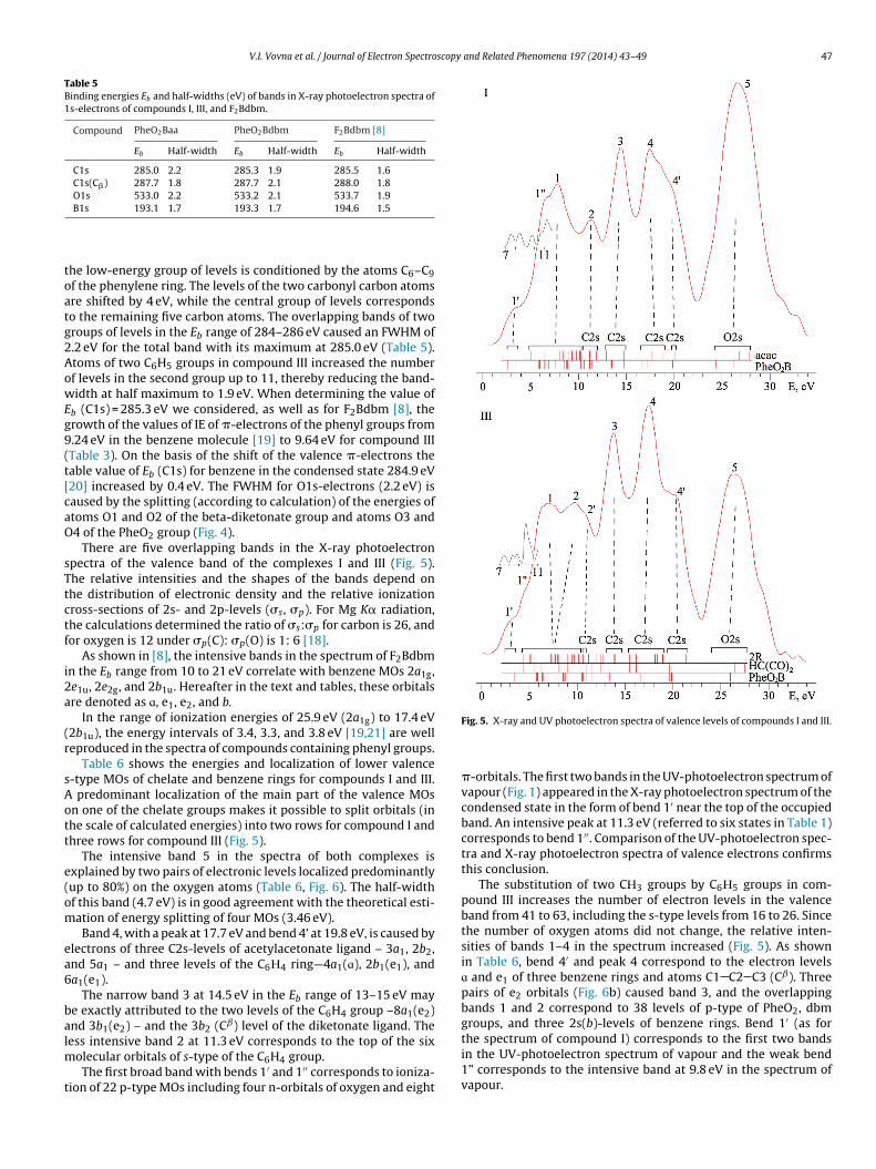
There are five overlapping bands in the X-ray photoelectronpectra of the valence band of the complexes I and III (Fig. 5).he relative intensities and the shapes of the bands depend onhe distribution of electronic density and the relative ionizationross-sections of 2s- and 2p-levels (�s, �p). For Mg K� radiation,he calculations determined the ratio of �s:�p for carbon is 26, andor oxygen is 12 under �p(C): �p(O) is 1: 6 [18].
As shown in [8], the intensive bands in the spectrum of F2Bdbmn the Eb range from 10 to 21 eV correlate with benzene MOs 2a1g,e1u, 2e2g, and 2b1u. Hereafter in the text and tables, these orbitalsre denoted as ɑ, e1, e2, and b.
In the range of ionization energies of 25.9 eV (2a1g) to 17.4 eV2b1u), the energy intervals of 3.4, 3.3, and 3.8 eV [19,21] are welleproduced in the spectra of compounds containing phenyl groups.
Table 6 shows the energies and localization of lower valence-type MOs of chelate and benzene rings for compounds I and III.
predominant localization of the main part of the valence MOsn one of the chelate groups makes it possible to split orbitals (inhe scale of calculated energies) into two rows for compound I andhree rows for compound III (Fig. 5).
The intensive band 5 in the spectra of both complexes isxplained by two pairs of electronic levels localized predominantlyup to 80%) on the oxygen atoms (Table 6, Fig. 6). The half-widthf this band (4.7 eV) is in good agreement with the theoretical esti-ation of energy splitting of four MOs (3.46 eV).Band 4, with a peak at 17.7 eV and bend 4’ at 19.8 eV, is caused by
lectrons of three C2s-levels of acetylacetonate ligand – 3a1, 2b2,nd 5a1 – and three levels of the C6Н4 ring—4a1(ɑ), 2b1(e1), anda1(e1).
The narrow band 3 at 14.5 eV in the Eb range of 13–15 eV maye exactly attributed to the two levels of the C6H4 group –8a1(e2)nd 3b1(e2) – and the 3b2 (Cˇ) level of the diketonate ligand. Theess intensive band 2 at 11.3 eV corresponds to the top of the six
olecular orbitals of s-type of the C6H4 group.The first broad band with bends 1′ and 1′′ corresponds to ioniza-
ion of 22 p-type MOs including four n-orbitals of oxygen and eight
Fig. 5. X-ray and UV photoelectron spectra of valence levels of compounds I and III.
�-orbitals. The first two bands in the UV-photoelectron spectrum ofvapour (Fig. 1) appeared in the X-ray photoelectron spectrum of thecondensed state in the form of bend 1′ near the top of the occupiedband. An intensive peak at 11.3 eV (referred to six states in Table 1)corresponds to bend 1′′. Comparison of the UV-photoelectron spec-tra and X-ray photoelectron spectra of valence electrons confirmsthis conclusion.
The substitution of two CН3 groups by C6Н5 groups in com-pound III increases the number of electron levels in the valenceband from 41 to 63, including the s-type levels from 16 to 26. Sincethe number of oxygen atoms did not change, the relative inten-sities of bands 1–4 in the spectrum increased (Fig. 5). As shownin Table 6, bend 4′ and peak 4 correspond to the electron levelsɑ and e1 of three benzene rings and atoms C1 C2 C3 (Cˇ). Threepairs of e2 orbitals (Fig. 6b) caused band 3, and the overlappingbands 1 and 2 correspond to 38 levels of p-type of PheO2, dbmgroups, and three 2s(b)-levels of benzene rings. Bend 1′ (as forthe spectrum of compound I) corresponds to the first two bandsin the UV-photoelectron spectrum of vapour and the weak bend
1” corresponds to the intensive band at 9.8 eV in the spectrum ofvapour.
48 V.I. Vovna et al. / Journal of Electron Spectroscopy and Related Phenomena 197 (2014) 43–49
Table 6Localization (%) and calculated energies (eV) of 22 lower valence MOs of compounds I and III.
Compound I Compound III
Band MO Localization −εi Band MO Localization −εi
HC3O2 2R PheO2B HC3O2 2R PheO2B
5 1a1 90 0 10 30.40 5 1a1 87 0 13 30.181b2 97 1 2 29.37 1b2 96 2 2 29.112a1 7 0 93 28.78 2a1 9 0 91 28.681b1 0 0 100 26.94 1b1 0 0 100 26.86
4′ 3a1(Cˇ) 60 16 24 22.72 4′ 3a1(a) 5 95 0 24.114a1(a) 17 4 79 22.42 2b2(a) 0 100 0 24.06
4 2b2(Cˇ) 37 62 1 21.45 4a1(a) 29 16 55 22.525a1(Cˇ) 44 55 1 20.30 5a1(Cˇ) 35 18 47 22.312b1(e1) 0 0 100 19.73 4 3b2(e1) 19 80 1 21.586a1(e1) 4 2 94 19.26 6a1(e1) 6 94 0 21.06
3 7a1 53 12 35 17.33 4b2(e1) 1 99 0 21.013b2(Cˇ) 59 35 6 17.30 7a1(e1) 27 73 0 20.798a1(e2) 1 0 99 16.09 2b1(e1) 0 0 100 19.673b1(e2) 3 0 97 15.92 8a1(e1) 7 2 91 19.225b2 79 10 11 15.49 5b2(C�) 44 54 2 18.80
2 9a1 81 16 3 14.46 9a1(C�) 34 49 17 18.0910a1 16 8 76 14.02 3 6b2(e2) 6 93 1 17.244b1(b) 38 11 51 13.75 10a1(e2) 8 90 2 17.1011a1 0 0 100 13.18 11a1(e2) 25 54 21 16.62
1 5b1 21 34 45 12.85 7b2(e2) 22 74 4 16.461a2 38 62 0 12.74 12a1(e2) 2 2 96 16.026b2 49 46 5 12.67 3b1(e2) 8 1 91 15.84
me lo
4
dolSpusrooboi
t(
Fig. 6. The distribution of electronic density on so
. Conclusion
For three spiroborate complexes (boron 1,2-dioxyphenylene �-iketonates), it was determined by UV-photoelectron spectroscopyf vapour and density functional theory that the two top electronevels were localized on the 1,2-dioxyphenylene fragment PheO2.ubstitution of CН3 groups by C6Н5 ones in the �-diketonate com-lexes did not strongly effect the energy and localization of the twopper as well as the deeper �- and �-orbitals of PheO2. Besides, theubstitution of two fluorine atoms by PheO2 groups in boron difluo-ide �-diketonates F2Bba and F2Bdbm did not affect on the energyr the composition of �-levels of �-diketonate ligands. An absencef mutual disturbance of the electron levels of the two chelate ringsinding with the boron atom (determined by experimental and the-retical methods) is explained by the orthogonality of their planes
n spiroborate complexes.The structure of X-ray photoelectron spectra of the valence elec-rons is in good agreement with the energies and compositioncontribution of O2s and C2s) of Kohn–Sham orbitals.
wer valence levels of compounds I (a) and III (b).
Acknowledgements
The project was supported by the Ministry of Education and Sci-ence of Russia (state agreement no. 1137) and by the Scientific Fundof the Far Eastern National University (Grant no. 12-03-13008-16/13).
References
[1] B.M. Mikhaılov, U.N. Bubnov, Organoboron Compounds in Organic Synthesis,Harwood Academic, New York, NY, 1984.
[2] A.T. Balaban, C.T. Renjea, M. Mocanu-Parasciv, E. Romas, Rev. Roum. Chim. 10(1965) 849–864.
[3] A.T. Balaban, C. Párkányi, I. Ghiviriga, J.-J. Aaron, Z. Zajícková, O.R. Martíne,ARKIVOC XIII (2008) 1–9.
[4] Y.L. Chow, Y.H. Zhang, M.X. Zheng, A. Rassat, Chem. Phys. Lett. 272 (1997)
471–477.[5] H. Lim, S. Yap, T. Tou, S. Ng, Opt. Mater. 27 (2005) 1815–1818.[6] V.I. Vovna, S.A. Tikhonov, I.B. Lvov, Rus. J. Phys. Chem. A 85 (2011)
1942–1948.[7] V.I. Vovna, S.A. Tikhonov, I.B. Lvov, Rus. J. Phys. Chem. A 85 (2013) 688–693.

scopy
[
[
[
[
[
[[[
[Nauka, Moscow, 1991 (in Russian).
[20] V.I. Nefedov, X-Ray Photoelectron Spectroscopy of Chemical Compounds,
V.I. Vovna et al. / Journal of Electron Spectro
[8] V.I. Vovna, S.A. Tikhonov, M.V. Kazachek, I.B. Lvov, V.V. Korochentsev, E.V.Fedorenko, A.G. Mirochnik, J. Electron. Spectrosc. Relat. Phenom. 189 (2013)116–121.
[9] O.L. Shcheka, A.V. Borisenko, V.I. Vovna, Russ. J. Gen. Chem. 62 (1992) 489–494.10] A.A. Granovsky, Firefly version 7.1.G. 〈http://classic.chem.msu.su/gran/firefly/
index.html.〉.11] K.L. Schuchardt, B.T. Didier, T. Elsethagen, L. Sun, V. Gurumoorthi, J.
Chase, J. Li, T.L. Windus, Basis Set Exchange: A Community Database forComputational Sciences J, Chem. Inf. Model. 47 (3) (2007) 1045–1052,
http://dx.doi.org/10.1021/ci600510j.12] K. Eichkorn, F. Weigend, O. Treutler, R. Ahlrichs, Theor. Chem. Acc. 97 (1997)119–124.
13] V.I. Vovna, V.V. Korochentsev, A.A. Dotsenko, Rus. J. Coord. Chem. 38 (2012)36–43.
[
and Related Phenomena 197 (2014) 43–49 49
14] V.I. Vovna, V.V. Korochentsev, A.A. Komissarov, I.B. L’vov, Russ. J. Phys. Chem.B 7 (2013) 220–224.
15] V.I. Vovna, M.V. Kazachek, I.B. L’vov, Opt. Spectrosc. 112 (2012) 497–505.16] I.V. Krauklis, Yu.V. Chizhov, Opt. Spectrosc. 96 (2004) 47.18] V.I. Nefedov, V.I. Vovna, Electronic Structure of Chemical Compounds, Nauka,
Moscow, 1987 (in Russian).19] V.I. Vovna, Electronic Structure of Organic Compounds: Photoelectron Data,
Khimiya, Moscow, 1984 (in Russian).21] W. von Niessen, L.S. Cederbaum, W.P. Kraemer, J. Chem. Phys. 65 (1976)
1378–1386.