Embed Size (px)
Citation preview

Photoelectron spectra and structures of three cyclic dipeptides: PhePhe,TyrPro, and HisGlyAnoja P. Wickrama Arachchilage, Feng Wang, Vitaliy Feyer, Oksana Plekan, and Kevin C. Prince Citation: J. Chem. Phys. 136, 124301 (2012); doi: 10.1063/1.3693763 View online: http://dx.doi.org/10.1063/1.3693763 View Table of Contents: http://jcp.aip.org/resource/1/JCPSA6/v136/i12 Published by the American Institute of Physics. Additional information on J. Chem. Phys.Journal Homepage: http://jcp.aip.org/ Journal Information: http://jcp.aip.org/about/about_the_journal Top downloads: http://jcp.aip.org/features/most_downloaded Information for Authors: http://jcp.aip.org/authors
Downloaded 26 Mar 2012 to 136.186.1.81. Redistribution subject to AIP license or copyright; see http://jcp.aip.org/about/rights_and_permissions
THE JOURNAL OF CHEMICAL PHYSICS 136, 124301 (2012)
Photoelectron spectra and structures of three cyclic dipeptides:PhePhe, TyrPro, and HisGly
Anoja P. Wickrama Arachchilage,1 Feng Wang,1 Vitaliy Feyer,2,3 Oksana Plekan,2,a)
and Kevin C. Prince2,4
1eChemistry Laboratory, Faculty of Life and Social Sciences, Swinburne University of Technology, Hawthorn,Melbourne, Victoria 3122, Australia2Sincrotrone Trieste, in Area Science Park, I-34149 Basovizza, Trieste, Italy3Forschungszentrum Jülich GmbH - IFF - IEE, 52425 Jülich, Germany4Istituto Officina dei Materiali, Consiglio Nazionale delle Ricerche, Area Science Park, I-34149 Trieste, Italy
(Received 4 January 2012; accepted 25 February 2012; published online 22 March 2012)
We have investigated the electronic structure of three cyclic dipeptides: cyclo(Histidyl-Glycyl) (cHis-Gly), cyclo(Tyrosyl-Prolyl) (cTyrPro), and cyclo(Phenylalanyl-Phenylalanyl) (cPhePhe) in the vaporphase, by means of photoemission spectroscopy and theoretical modeling. The last compound wasevaporated from the solid linear dipeptide, but cyclised, losing water to form cPhePhe in the gasphase. The results are compared with our previous studies of three other cyclopeptides. Experimen-tal valence and core level spectra have been interpreted in the light of calculations to identify thebasic chemical properties associated with the central diketopiperazine ring, and with the additionalfunctional groups. The valence spectra are generally characterized by a restricted set of outer va-lence orbitals separated by a gap from most other valence orbitals. The theoretically simulated coreand valence spectra of all three cyclic dipeptides agree reasonably well with the experimental spec-tra. The central ring and the side chains act as independent chromophores whose spectra do notinfluence one another, except for prolyl dipeptides, where the pyrrole ring is fused with the cen-tral ring. In this case, significant changes in the valence and core level spectra were observed, andexplained by stronger hybridization of the valence orbitals. © 2012 American Institute of Physics.[http://dx.doi.org/10.1063/1.3693763]
I. INTRODUCTION
We have previously reported a theoretical and experimen-tal photoemission investigation of three cyclic dipeptides,1
namely the smallest dipeptide, cyclo(Glycyl-Glycyl) (dike-topiperazine, or DKP hereafter), cyclo(Leucyl-Prolyl), andcyclo(Phenylalanyl-Prolyl). Cyclic dipeptides are a subset ofthe much larger class of peptides, and many are biologicallyactive. These dipeptides consist of a ring containing two pep-tide bonds and this ring structure confers high stability2 andresistance to human digestion. This last property allows thedipeptides to be used as scaffolds for drugs, and they alsopossess a number of interesting biological properties includ-ing antiviral, antibiotic, and anti-tumour activity. Some ma-rine organisms produce cyclopeptides,3 and the compoundshave shown promise for several pharmaceutical and otherapplications.4–7 The rigid central ring limits conformationalfreedom,7 making them tractable models of larger peptides.3
We note that the adsorption of peptides on surfaces is a sub-ject of wide current interest, and this includes dipeptides:8–10
to investigate them in the condensed state, it is of great helpto have reference data for the isolated molecules.
Structure dictates function, but intrinsic properties of bi-ologically active molecules may be modified or masked bytheir aqueous environment or by their interactions with it.
a)Present address: Aarhus University, Department of Physics and Astronomy,Ny Munkegade 120, 8000 Aarhus C, Denmark.
Investigation of free molecules in the gas phase permits theidentification of intrinsic properties without perturbations dueto interaction with solvents. The aim of this study is to deter-mine the intrinsic structure-property relationship of a numberof cyclic dipeptides, using the experimental11–14 and theoreti-cal methods15–17 previously applied to modelling both valenceand inner-shell electronic states of biomolecules.
The compounds selected for study, shown in Figure 1,are cyclo(Histidyl-Glycyl) (cHisGly), cyclo(Tyrosyl-Prolyl)(cTyrPro), and cyclo(Phenylalanyl-Phenylalanyl) (cPhePhe).These all consist of a core of the six-member DKP ring, anddiffer by the side chains that the constituent amino acids con-tain: hydrogen and imidazole in cHisGly, phenol and pyrroli-dine in cTyrPro, and two phenyl groups in cPhePhe. Luciettoet al.18 reported the results of an NMR and structure mod-elling study of cHisGly, as well as data on its pharmacologicalproperties. They concluded that the energetically favourableconformers adopt a boat configuration in solution, with re-spect to the DKP ring. Zhu et al.19 calculated the structures ofall of the cyclic peptides consisting of the same two aminoacids, including cPhePhe, and found that they adopted theboat conformation. We recently summarised other previousstudies of cyclic dipeptides.1
The amino acids constituting the dipeptides under studyhere are glycine, proline, phenylalanine, tyrosine, and his-tidine, and the core level spectra and accompanying the-oretical calculations of the first four of these have beenreported recently,13, 14 as well as the spectra of the linear
0021-9606/2012/136(12)/124301/8/$30.00 © 2012 American Institute of Physics136, 124301-1
Downloaded 26 Mar 2012 to 136.186.1.81. Redistribution subject to AIP license or copyright; see http://jcp.aip.org/about/rights_and_permissions
124301-2 Wickrama Arachchilage et al. J. Chem. Phys. 136, 124301 (2012)
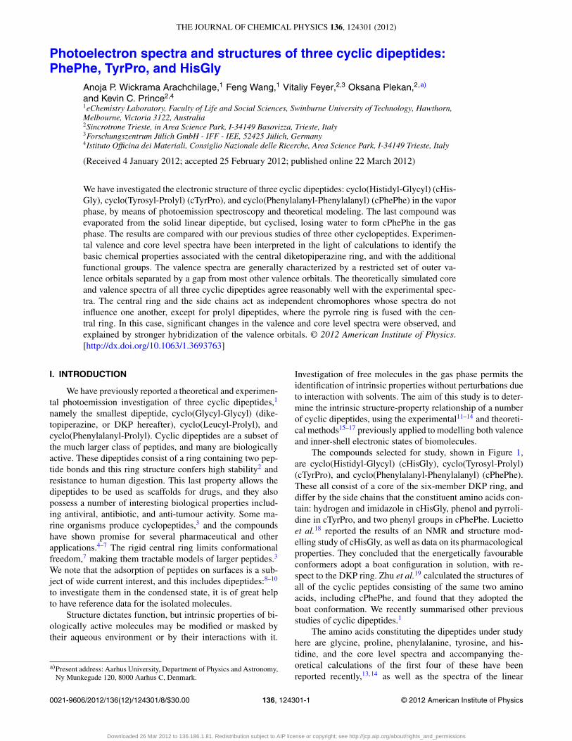
FIG. 1. (a)–(h) Three-dimensional views and schematic structures ofcTyrPro, cHisGly, cPhePhe, and lPhePhe. Double click the three-dimensionalfigures on the left to view the interactive 3D structures.
peptide Glycyl-Glycine.20 Here we investigate cyclic dimersof these amino acids. cHisGly can also be considered as DKPmodified by the addition of an imidazole ring. In cTyrPro, thepentagonal pyrrolidine ring is fused to the DKP ring to pro-duce a rather rigid double ring structure, a motif also foundin two of our previously studied compounds, cyclo(Prolyl-Phenylalanyl) and cyclo(Leucyl-Prolyl). In proline, the fourlowest energy conformers are constructed from two structuralelements: the rotation of the carboxylic acid group and thepuckering of the pyrrolidine ring.21 The carboxylic group isnot present in the dipeptide, so we expect that the main con-formational element will be the puckering of the proline ringto an “up” or “down” geometry with respect to the twist inthe DKP ring, giving rise to two possible conformers. In addi-tion, the phenol moiety of cTyrPro contributes to the confor-mational landscape. The third compound, cPhePhe, containstwo aromatic side groups; like DKP, it is formed from twoidentical amino acids.
II. EXPERIMENTAL AND COMPUTATIONAL METHODS
The measurements were performed at the Gas PhasePhotoemission beamline, Elettra, Trieste,22 using apparatusand calibration methods described previously.11–13 The sam-ples were supplied by Bachem (www.bachem.com) and usedwithout further purification. The compounds used were thosenominated except for cPhePhe, for which the sample insertedin the furnace and heated was Phenylalanyl-Phenylalanine alinear, non-cyclic dipeptide. They were evaporated at temper-atures of 473 (cHisGly), 448 (cTyrPro), and 443 (lPhePhe)
K, respectively, and checked for signs of thermal decompo-sition (spectral changes as a function of time, discolorationafter heating, etc.). No evidence was found for decompositionof cHisGly and cTyrPro, but for the third compound, exces-sive degassing of water was observed. This was interpreted asa sign of structural alteration, and it will be shown below thatthis was indeed the case, as cyclisation with water eliminationwas occurring. No other sign of thermal change was observedfor this compound, such as discoloration or spectral changesas a function of time, once the water was eliminated. Detailsof the energy resolution and calibration procedure have beengiven in Refs. 23 and 24, in brief, the resolution was estimatedto be 0.32, 0.46, and 0.78 eV at hν 382 (C 1s), 495 (N 1s), and628 eV (O 1s), respectively.
The computational methods have been discussedpreviously,1 and will not be repeated here but are summa-rized briefly. All the geometries were optimized using theB3LYP/cc-pVTZ model, followed by harmonic vibrationalfrequency calculations, which is incorporated in GAUSSIAN
03 computational package.25 The interactive 3D-pdf struc-tures for the molecules, shown in Figure 1 are produced asdescribed by Selvam et al.26 Adobe Acrobat 8.1 or higher issuitable for viewing. Single point calculation was performed,based on the LB94/et-pVQZ model,27, 28 which is incorpo-rated in the Amsterdam Density Functional (ADF) computa-tional chemistry package29 to produce core vertical ionizationenergies. The outer valence vertical ionization energies of thedipeptides were calculated using the outer valence Green’sfunction OVGF/6-31G* model,30–34 while the complete va-lence vertical ionization energies were calculated using theHF/6-311G** model.
III. RESULTS
Figure 1 displays the optimized structures of the threedipeptides in three-dimensional space using a recently de-veloped interactive 3D-pdf technique.26 Double clickingFigures 1(a), 1(c), 1(e), or 1(g) online or the pdf file will al-low viewing of the structures embedded in the pdf file. Ascan be seen from their 3D structures, all the cyclic dipep-tides share a common six-member DKP ring, which prefers aboat conformation. The dipeptides, cTyrPro, cHisGly, and lin-ear PhePhe, exhibit C1 point group symmetry, while cPhePheadopts a boat conformation, with C2 point group symmetry.
Table I of the supplementary material35 presents a sum-mary of some characteristic geometric parameters of thedipeptides. Although all the cyclic dipeptides contain a six-member DKP ring, their attached functional groups are notthe same. For example, cHisGly is composed of a DKP ring,connected to a five-member imidazole ring via the C(8)-C(7)-C(6) carbon bridge. The DKP ring of cTyrPro is fused with apyrrole ring sharing the N(2)-C(3) bond, while the other endof the C(8)-C(7)-C(6) carbon bridge is a para-phenol moi-ety. As stated, cPhePhe exhibits C2 point group symmetry:the symmetric DKP ring links two phenyl moieties throughthe C-C-C bridge. Regardless of the type of functional moi-ety linked to the DKP ring, the bridge dihedral angle, C(1)-C(6)-C(7)-C(8), does not change significantly for cTyrPro and
Downloaded 26 Mar 2012 to 136.186.1.81. Redistribution subject to AIP license or copyright; see http://jcp.aip.org/about/rights_and_permissions
124301-3 Wickrama Arachchilage et al. J. Chem. Phys. 136, 124301 (2012)
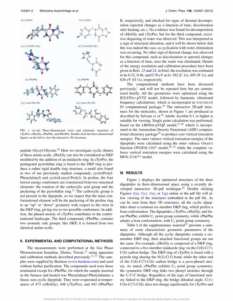
FIG. 2. O 1s spectra of cPhePhe, cHisGly, and cTyrPro. Theoretical curves(cPhePhe, linear PhePhe) and histograms (cHisGly, cTyrPro) are also shown.The FWHM of the simulated spectra of cPhePhe and linear PhePhe is 0.87 eV.The shifts of the theoretical spectra are: cPhePhe, +2.95 eV; linear PhePhe,+2.2 eV; cHisGly, +2.97 eV; cTyrPro, +3.04 eV.
cPhePhe but is different in cHisGly, where the imidazole ringis bonded to the DKP ring.
Briefly, the same functional groups as those in the previ-ously studied dipeptides have very nearly the same structuralparameters. For example, the perimeters of the central DKPring and the benzene ring do not vary by more than 0.03 and0.01 Å, respectively.
In Figures 2 and 4, we show the experimental and theo-retical O, N, and C 1s photoemission spectra of the three com-pounds, and key data are summarized in Tables I–III; Table 2of the supplementary material35 gives a complete listing. The-oretical spectra have been offset by the energies indicated inthe captions and tables, to give the best overall agreement withexperiment. In the calculated O 1s spectra of linear and cyclicPhePhe, the linear form contains two peaks with an intensityratio of 2:1, with the larger peak due to the carbonyl and pep-tide oxygen atoms, and the weaker peak due to the hydroxyoxygen; the spectrum is similar to that of the linear peptideGlyGly.20 Our theoretical spectrum of cPhePhe agrees withthe experimental data (within an offset of 3.00 ± 0.05 eVfor all compounds), which shows a single peak at binding en-
ergy 537.0 eV, whereas the lPhePhe spectrum is quite differ-ent from the experimental data. We therefore conclude that onheating the linear form cyclised with the loss of water, so thatthe cyclic form is produced in the vapor, and we refer to thissample as cPhePhe from here on.
The O 1s spectrum of cHisGly also shows a peak at537.0 eV, and again this is a single peak as expected from thesimilarity of the chemical environments of the two oxygenatoms. The theory predicts that the splitting is only 0.27 eV,clearly too small to be resolved experimentally.
The spectrum of cTyrPro contains two O 1s peaks, ofwhich the stronger at 536.8 eV is assigned to the two peptideoxygen atoms, while the weaker feature at 539.2 eV is dueto the oxygen atom in the phenol side chain. The theoreticalsplitting of the two peptide oxygen peaks is 0.1 eV, again toosmall to be resolved. The experimental intensity ratio is 1.8,slightly below the stoichiometric value of 2. We attribute thissmall difference to the transfer of oscillator strength from themain peak to satellites, which are stronger for peptide oxygencompared to the hydroxyl oxygen.
All three compounds show peaks due to the peptideoxygen atoms, with binding energies within 0.2 eV of oneanother, and these values are very similar to those of the cy-clopeptides c(Leucyl-Prolyl) and c(Phenylalanyl-Prolyl) stud-ied previously.1 We conclude that these O 1s binding energiesdo not vary much within this class of compounds, and are sub-stantially independent of the amino acid side chain, providedit is larger than a hydrogen atom. DKP, where the side chainsare hydrogen atoms, showed higher O 1s binding energy(537.45 eV), attributed to reduced screening in the final state.
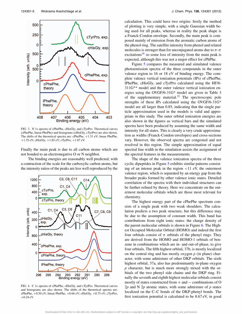
The experimental and calculated N 1s core level spectraare shown in Figure 3 and energies are listed in Table II. Thevertical bars are the calculated N 1s core levels for cHisGlyand cTyrPro whereas simulated core N 1s spectra are givenfor cPhePhe and lPhePhe. For cPhePhe, theory predicts a sin-gle spectral peak, and this is observed. The alternative linearstructure contains a peptide and an amino nitrogen atom, andthe calculated spectrum has two peaks, which were not ob-served experimentally. This is further evidence that the sam-ple is cyclic in the gas phase.
cHisGly contains four nitrogen atoms, two of which arein the central DKP ring, and are chemically very similar. Theother two are amino and imino nitrogen atoms in the imi-dazole side chain. Thus we expect three peaks with a stoi-chiometric ratio of 1:2:1, and experimentally three peaks areobserved, with fitted intensities of 0.85:2.24:0.90. This is rea-sonable agreement, considering that the peaks are not fully
TABLE I. Experimental and theoretical O 1s vertical ionization energies of cPhePhe, cHisGly, and cTyrProand theoretical values for linear PhePhe (eV). Theoretical values have been calculated with the LB94/et-pVQZmodel. O 1s values are shifted by +2.95 eV (cPhePhe); +2.2 eV (linear PhePhe); +2.97 eV (cHisGly); +3.04eV (cTyrPro).
cPhePhe lPhePhe cHisGly cTyrPro
Expt. Theory Theory Expt. Theory Expt. Theory
537.0 O(1) 537.00 O(1) 537.02 537.0 O(1) 536.98 536.8 O(1) 537.00O(4) 537.0 O(4) 536.82 O(4) 537.01 O(4) 537.10
O(4)-H 538.06 539.2 O(11) 538.94
Downloaded 26 Mar 2012 to 136.186.1.81. Redistribution subject to AIP license or copyright; see http://jcp.aip.org/about/rights_and_permissions
124301-4 Wickrama Arachchilage et al. J. Chem. Phys. 136, 124301 (2012)
TABLE II. Experimental and theoretical N 1s vertical ionization energies of cPhePhe, cHisGly, and cTyrProand theoretical values for linear PhePhe (eV).). Theoretical values have been calculated with the LB94/et-pVQZmodel. N 1s values are shifted by +1.31 eV (cPhePhe); +1.35 eV (lPhePhe); +1.85 (cHisGly); +1.07 (cTyrPro).
cPhePhe cPhePhe lPhePhe cHisGly cHisGly cTyrPro cTyrPro(Expt.) (Theory) Theory (Expt.) Theory (Expt.) (Theory)
405.65 N(2) 405.65 N(2) 406.38 404.45 N(12) 403.74 405.45 N(2) 406.44N(5) 405.65 N(5) 403.08 N(5) 405.29
405.80 N(2) 405.44N(5) 405.52
406.75 N(10) 405.85
resolved. The calculation underestimates the splitting be-tween the peptide and amino nitrogen core levels, N(2) andN(5) versus N(10).
cTyrPro also displays a single peak due to the two pep-tide nitrogen atoms at 405.45 eV, and the calculated split-ting is 0.15 eV. The center of the peak is 0.2 eV lower inbinding energy than the peak of cPhePhe, and about 0.3 eVlower than the corresponding peak in cHisGly. Thus the or-der of the peptide N 1s binding energy is cTyrPro < cPhePhe< cHisGly.
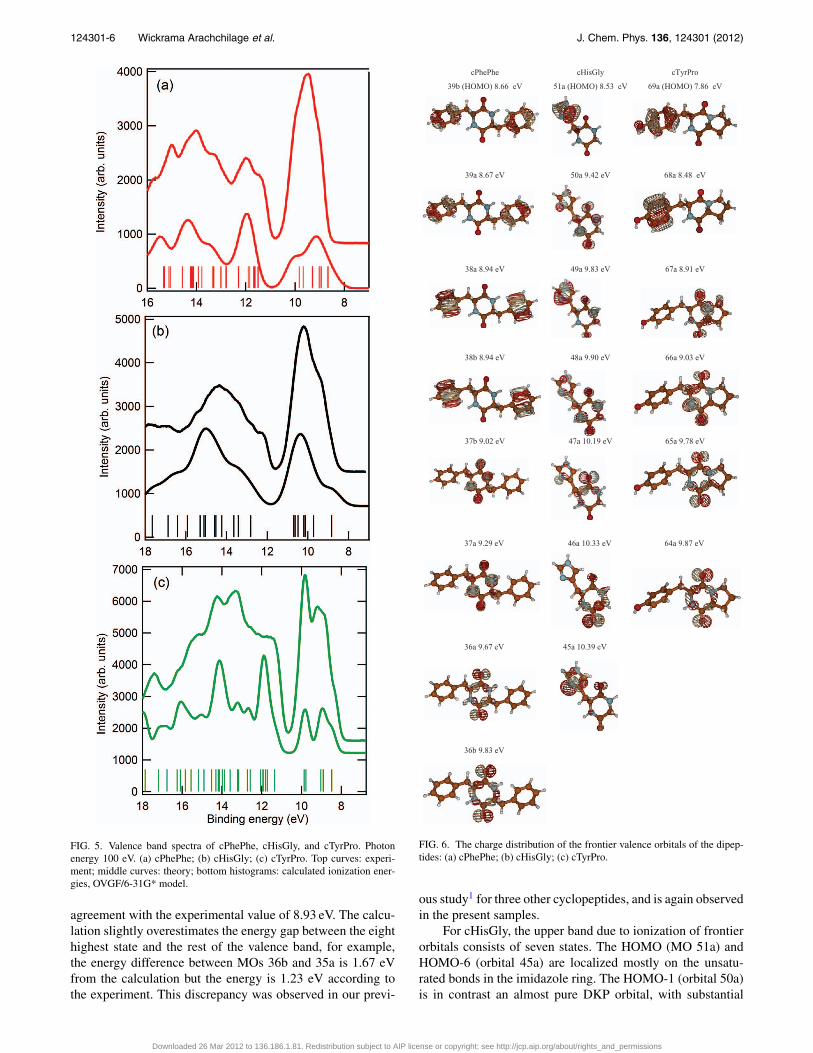
Figure 4 shows the theoretical and experimental C 1sspectra, and the energies are listed in Table III. In the spec-trum of cPhePhe there are three peaks: the molecule contains18 carbon atoms, with nine chemically distinct types of atomsfor the conformation with C2 symmetry. However, the chemi-cal environments of the carbon atoms in the two phenyl ringsare very similar, and the core levels of C(7) and C(14) are un-likely to be much shifted from the energies of the phenyl car-bon atoms, so we expect a peak due to 14 carbon atoms. C(1)and C(4) are chemically equivalent, and so are C(3) and C(6).These considerations predict three peaks with a stoichiomet-ric ratio of 2:2:14, as predicted by theory. The experimentalratio, obtained by fitting the peaks and integrating the areas,is 1.9:2.2:13.9, in good agreement with the calculation andexpectation.
The theoretical spectra for both linear and cyclic PhePheare shown, and although the spectrum for the linear formagrees better with the experimental data, we have alreadyestablished above that the cyclic form is present in the gasphase. The calculations generally tend to predict a more com-
pressed energy scale than that observed in the experiment, andthis is the case for cPhePhe.
In cHisGly, there are eight carbon atoms, giving rise tothree peaks in the experimental spectrum. The two carbonylcarbon atoms of the central ring, C(1) and C(4), appear as asingle peak at 293.75 eV; the theoretical splitting is 70 meV.The peak at 292.00 eV is assigned to the carbon atoms in thecentral ring, C(3) and C(6), with a contribution from C(11),located between the two nitrogen atoms of the imidazole ring.The peak at lowest binding energy is composed of contribu-tions from C(7), C(8), and C(9). The latter two are bonded tonitrogen in the imidazole ring, while C(7) is the only carbonatom bonded solely to carbon and hydrogen, with no bonds tonitrogen or oxygen.
The cTyrPro molecule contains 14 carbon atoms andthree peaks are observed experimentally. The peak at293.34 eV is assigned to the carbonyl atoms of the centralring and it has an intensity ratio of 2.1:11.9 with respect to theother peaks, close to the expected value of 2:12. The bindingenergy is lower than that of cHisGly and cPhePhe and closerto the values measured for cyclo(Leucyl-Prolyl), 293.52 eV,and cyclo(Phenylalanyl-Prolyl), 293.40 eV.1 cTyrPro has incommon with these compounds a fused pyrrolidine ring sowe conclude that this structural feature tends to decrease theC 1s binding energy by a small amount. The second peak con-sists of C 1s emission from C(3), C(6), and C(11), which arelocated in the central ring, or bonded to oxygen in the phe-nol ring, C(11). A peak due to C(14), the carbon atom in thepyrrolidine ring which is bonded to nitrogen, is not resolved,but theory predicts it to lie between the two main peaks.
TABLE III. Experimental and theoretical C 1s vertical ionization energies of cPhePhe, cHisGly, and cTyrPro and theoretical values for linear PhePhe (eV).Theoretical values have been calculated with the LB94/et-pVQZ model. C 1s values are shifted by +0.50 eV (cPhePhe); +0.66 eV (lPhePhe); +0.75 eV(cHisGly,); +0.24 eV (cTyrPro).
ehPehPc ylGsiHc orPryTc
Atom Theory Expt.Relative intensity Atom Theory Expt.
Relative intensity Atom Theory Expt.
Relative intensity
Other 8 atoms
289.70-290.18
290.26 7.37 C(7), C(8), C(9)
290.24-290.39
290.96 2.84 Other 14 atoms
289.70-290.11
290.34 13.94
C(14) 290.92 - C(6) 291.46
}292.00 }3.2 C(3), C(6) 291.60 291.96 2.17
C(3) 291.46
}291.65 4.51 11.2 37.392 46.292 )4(C ,)1(C 04.192 )3(C
30.192 )11(C 15.192 )6(C 36.292 )1(C 44.192 )11(C }293.75 }1.96
C(1) 292.54 }293.34 2.11
65.292 )4(C 93.292 )4(C
Downloaded 26 Mar 2012 to 136.186.1.81. Redistribution subject to AIP license or copyright; see http://jcp.aip.org/about/rights_and_permissions
124301-5 Wickrama Arachchilage et al. J. Chem. Phys. 136, 124301 (2012)
FIG. 3. N 1s spectra of cPhePhe, cHisGly, and cTyrPro. Theoretical curves(cPhePhe, linear PhePhe) and histograms (cHisGly, cTyrPro) are also shown.The shifts of the theoretical spectra are: cPhePhe, +1.31 eV; linear PhePhe,+1.35 eV; cHisGly, +1.85 eV; cTyrPro, +1.07 eV.
Finally the main peak is due to all carbon atoms which arenot bonded to an electronegative O or N neighbor.
The binding energies are reasonably well predicted, witha contraction of the scale for the carboxylic carbon atoms, butthe intensity ratios of the peaks are less well reproduced by the
FIG. 4. C 1s spectra of cPhePhe, cHisGly, and cTyrPro. Theoretical curvesand histograms are also shown. The shifts of the theoretical spectra are:cPhePhe, +0.50 eV; linear PhePhe, +0.66 eV; cHisGly, +0.75 eV; cTyrPro,+0.24 eV.
calculation. This could have two origins: firstly the methodof plotting is very simple, with a single Gaussian width be-ing used for all peaks, whereas in reality the peak shape isa Franck-Condon envelope. Secondly, the main peak is com-posed mainly of emission from the aromatic carbon atoms ofthe phenol ring. The satellite intensity from phenol and relatedmolecules is stronger than for unconjugated atoms due to π -πexcitations36 so some loss of intensity from the main peak isexpected, although this was not a major effect for cPhPhe.
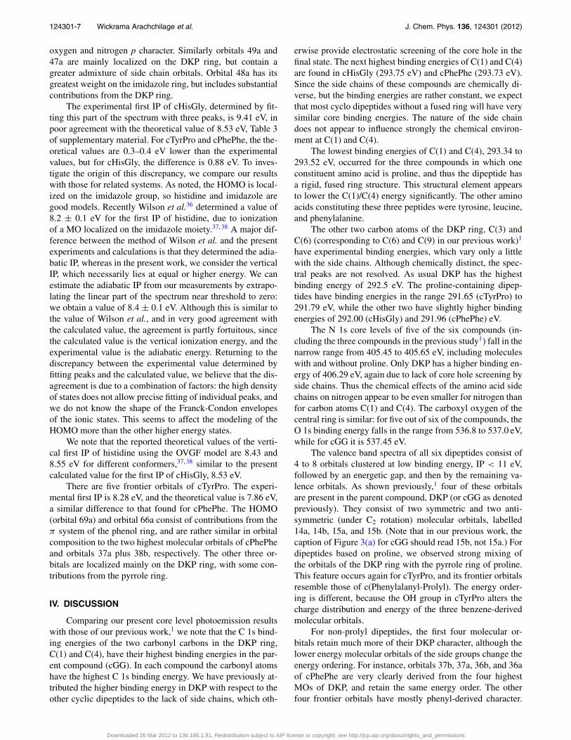
Figure 5 compares the measured and simulated valencephotoemission spectra of the three compounds in the outervalence region to 16 or 18 eV of binding energy. The com-plete valence vertical ionization potentials (IPs) of cPhePhe,lPhePhe, cHisGly, and cTyrPro calculated using the HF/6-311G** model and the outer valence vertical ionization en-ergies using the OVGF/6-31G* model are given in Table 3of the supplementary material.35 The spectroscopic polestrengths of these IPs calculated using the OVGF/6-31G*model are all larger than 0.85, indicating that the single par-ticle approximation used in the models is valid and appro-priate in this study. The outer orbital ionization energies arealso shown in the figures as vertical bars and the simulatedspectra have been produced by assuming the same width andintensity for all states. This is clearly a very crude approxima-tion as widths (Franck-Condon envelopes) and cross-sectionsvary. However, the observed spectra are congested and notresolved in this region. The simple approximation of equalspectral line width in the simulation assists the assignment ofthe spectral features in the measurements.
The shape of the valence ionization spectra of the threecyclic dipeptides in Figure 5 exhibits similar patterns consist-ing of an intense peak in the region <11 eV, the outermostvalence region, which is separated by an energy gap from thebroader peaks formed by other valence ionic states. Detailedcorrelation of the spectra with their individual structures canbe further refined by theory. Here we concentrate on the out-ermost molecular orbitals which are those most relevant forchemistry.
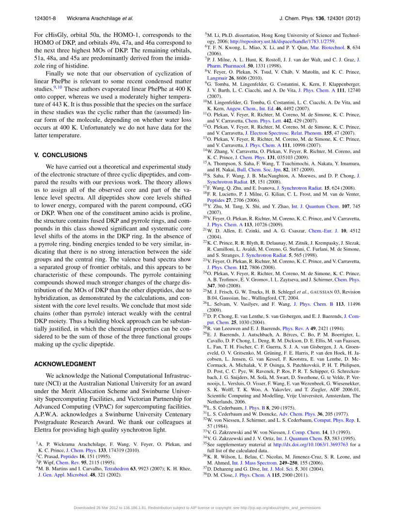
The highest energy part of the cPhePhe spectrum con-sists of a single peak with two weak shoulders. The calcu-lation predicts a two peak structure, but this difference maybe due to the assumption of constant width. This band hascontributions from eight ionic states: the charge density ofthe parent molecular orbitals is shown in Figure 6. The High-est Occupied Molecular Orbital (HOMO) and indeed the firstfour orbitals consist of π orbitals of the phenyl rings. Theyare derived from the HOMO and HOMO-1 orbitals of ben-zene in combinations which are in- and out-of-phase, to givefour orbitals. The fifth highest orbital, 37b, is mostly localizedon the central ring and has mostly oxygen p (in plane) char-acter, with some admixture of other DKP orbitals. The sixthhighest orbital, 37a, also has predominantly in-plane oxygenp character, but is much more strongly mixed with the or-bitals of the two phenyl side chains and the DKP ring. Fi-nally the seventh and eighth highest molecular orbitals consistmostly of states constructed from + and − combinations of O2p and N 2p atomic states, with some admixture of p stateslocalized on the C–C bonds of the DKP-phenyl bonds. Thefirst ionization potential is calculated to be 8.67 eV, in good
Downloaded 26 Mar 2012 to 136.186.1.81. Redistribution subject to AIP license or copyright; see http://jcp.aip.org/about/rights_and_permissions
124301-6 Wickrama Arachchilage et al. J. Chem. Phys. 136, 124301 (2012)
FIG. 5. Valence band spectra of cPhePhe, cHisGly, and cTyrPro. Photonenergy 100 eV. (a) cPhePhe; (b) cHisGly; (c) cTyrPro. Top curves: experi-ment; middle curves: theory; bottom histograms: calculated ionization ener-gies, OVGF/6-31G* model.
agreement with the experimental value of 8.93 eV. The calcu-lation slightly overestimates the energy gap between the eighthighest state and the rest of the valence band, for example,the energy difference between MOs 36b and 35a is 1.67 eVfrom the calculation but the energy is 1.23 eV according tothe experiment. This discrepancy was observed in our previ-
cPhePhe cHisGly cTyrPro
39b (HOMO) 8.66 eV 51a (HOMO) 8.53 eV 69a (HOMO) 7.86 eV
39a 8.67 eV 50a 9.42 eV 68a 8.48 eV
38a 8.94 eV 49a 9.83 eV 67a 8.91 eV
38b 8.94 eV 48a 9.90 eV 66a 9.03 eV
37b 9.02 eV 47a 10.19 eV 65a 9.78 eV
37a 9.29 eV 46a 10.33 eV 64a 9.87 eV
36a 9.67 eV 45a 10.39 eV
36b 9.83 eV
FIG. 6. The charge distribution of the frontier valence orbitals of the dipep-tides: (a) cPhePhe; (b) cHisGly; (c) cTyrPro.
ous study1 for three other cyclopeptides, and is again observedin the present samples.
For cHisGly, the upper band due to ionization of frontierorbitals consists of seven states. The HOMO (MO 51a) andHOMO-6 (orbital 45a) are localized mostly on the unsatu-rated bonds in the imidazole ring. The HOMO-1 (orbital 50a)is in contrast an almost pure DKP orbital, with substantial
Downloaded 26 Mar 2012 to 136.186.1.81. Redistribution subject to AIP license or copyright; see http://jcp.aip.org/about/rights_and_permissions
124301-7 Wickrama Arachchilage et al. J. Chem. Phys. 136, 124301 (2012)
oxygen and nitrogen p character. Similarly orbitals 49a and47a are mainly localized on the DKP ring, but contain agreater admixture of side chain orbitals. Orbital 48a has itsgreatest weight on the imidazole ring, but includes substantialcontributions from the DKP ring.
The experimental first IP of cHisGly, determined by fit-ting this part of the spectrum with three peaks, is 9.41 eV, inpoor agreement with the theoretical value of 8.53 eV, Table 3of supplementary material. For cTyrPro and cPhePhe, the the-oretical values are 0.3–0.4 eV lower than the experimentalvalues, but for cHisGly, the difference is 0.88 eV. To inves-tigate the origin of this discrepancy, we compare our resultswith those for related systems. As noted, the HOMO is local-ized on the imidazole group, so histidine and imidazole aregood models. Recently Wilson et al.36 determined a value of8.2 ± 0.1 eV for the first IP of histidine, due to ionizationof a MO localized on the imidazole moiety.37, 38 A major dif-ference between the method of Wilson et al. and the presentexperiments and calculations is that they determined the adia-batic IP, whereas in the present work, we consider the verticalIP, which necessarily lies at equal or higher energy. We canestimate the adiabatic IP from our measurements by extrapo-lating the linear part of the spectrum near threshold to zero:we obtain a value of 8.4 ± 0.1 eV. Although this is similar tothe value of Wilson et al., and in very good agreement withthe calculated value, the agreement is partly fortuitous, sincethe calculated value is the vertical ionization energy, and theexperimental value is the adiabatic energy. Returning to thediscrepancy between the experimental value determined byfitting peaks and the calculated value, we believe that the dis-agreement is due to a combination of factors: the high densityof states does not allow precise fitting of individual peaks, andwe do not know the shape of the Franck-Condon envelopesof the ionic states. This seems to affect the modeling of theHOMO more than the other higher energy states.
We note that the reported theoretical values of the verti-cal first IP of histidine using the OVGF model are 8.43 and8.55 eV for different conformers,37, 38 similar to the presentcalculated value for the first IP of cHisGly, 8.53 eV.
There are five frontier orbitals of cTyrPro. The experi-mental first IP is 8.28 eV, and the theoretical value is 7.86 eV,a similar difference to that found for cPhePhe. The HOMO(orbital 69a) and orbital 66a consist of contributions from theπ system of the phenol ring, and are rather similar in orbitalcomposition to the two highest molecular orbitals of cPhePheand orbitals 37a plus 38b, respectively. The other three or-bitals are localized mainly on the DKP ring, with some con-tributions from the pyrrole ring.
IV. DISCUSSION
Comparing our present core level photoemission resultswith those of our previous work,1 we note that the C 1s bind-ing energies of the two carbonyl carbons in the DKP ring,C(1) and C(4), have their highest binding energies in the par-ent compound (cGG). In each compound the carbonyl atomshave the highest C 1s binding energy. We have previously at-tributed the higher binding energy in DKP with respect to theother cyclic dipeptides to the lack of side chains, which oth-
erwise provide electrostatic screening of the core hole in thefinal state. The next highest binding energies of C(1) and C(4)are found in cHisGly (293.75 eV) and cPhePhe (293.73 eV).Since the side chains of these compounds are chemically di-verse, but the binding energies are rather constant, we expectthat most cyclo dipeptides without a fused ring will have verysimilar core binding energies. The nature of the side chaindoes not appear to influence strongly the chemical environ-ment at C(1) and C(4).
The lowest binding energies of C(1) and C(4), 293.34 to293.52 eV, occurred for the three compounds in which oneconstituent amino acid is proline, and thus the dipeptide hasa rigid, fused ring structure. This structural element appearsto lower the C(1)/C(4) energy significantly. The other aminoacids constituting these three peptides were tyrosine, leucine,and phenylalanine.
The other two carbon atoms of the DKP ring, C(3) andC(6) (corresponding to C(6) and C(9) in our previous work)1
have experimental binding energies, which vary only a littlewith the side chains. Although chemically distinct, the spec-tral peaks are not resolved. As usual DKP has the highestbinding energy of 292.5 eV. The proline-containing dipep-tides have binding energies in the range 291.65 (cTyrPro) to291.79 eV, while the other two have slightly higher bindingenergies of 292.00 (cHisGly) and 291.96 (cPhePhe) eV.
The N 1s core levels of five of the six compounds (in-cluding the three compounds in the previous study1) fall in thenarrow range from 405.45 to 405.65 eV, including moleculeswith and without proline. Only DKP has a higher binding en-ergy of 406.29 eV, again due to lack of core hole screening byside chains. Thus the chemical effects of the amino acid sidechains on nitrogen appear to be even smaller for nitrogen thanfor carbon atoms C(1) and C(4). The carboxyl oxygen of thecentral ring is similar: for five out of six of the compounds, theO 1s binding energy falls in the range from 536.8 to 537.0 eV,while for cGG it is 537.45 eV.
The valence band spectra of all six dipeptides consist of4 to 8 orbitals clustered at low binding energy, IP < 11 eV,followed by an energetic gap, and then by the remaining va-lence orbitals. As shown previously,1 four of these orbitalsare present in the parent compound, DKP (or cGG as denotedpreviously). They consist of two symmetric and two anti-symmetric (under C2 rotation) molecular orbitals, labelled14a, 14b, 15a, and 15b. (Note that in our previous work, thecaption of Figure 3(a) for cGG should read 15b, not 15a.) Fordipeptides based on proline, we observed strong mixing ofthe orbitals of the DKP ring with the pyrrole ring of proline.This feature occurs again for cTyrPro, and its frontier orbitalsresemble those of c(Phenylalanyl-Prolyl). The energy order-ing is different, because the OH group in cTyrPro alters thecharge distribution and energy of the three benzene-derivedmolecular orbitals.
For non-prolyl dipeptides, the first four molecular or-bitals retain much more of their DKP character, although thelower energy molecular orbitals of the side groups change theenergy ordering. For instance, orbitals 37b, 37a, 36b, and 36aof cPhePhe are very clearly derived from the four highestMOs of DKP, and retain the same energy order. The otherfour frontier orbitals have mostly phenyl-derived character.
Downloaded 26 Mar 2012 to 136.186.1.81. Redistribution subject to AIP license or copyright; see http://jcp.aip.org/about/rights_and_permissions
124301-8 Wickrama Arachchilage et al. J. Chem. Phys. 136, 124301 (2012)
For cHisGly, orbital 50a, the HOMO-1, corresponds to theHOMO of DKP, and orbitals 49a, 47a, and 46a correspond tothe next three highest MOs of DKP. The remaining orbitals,51a, 48a, and 45a are predominantly derived from the imida-zole ring of histidine.
Finally we note that our observation of cyclization oflinear PhePhe is relevant to some recent condensed matterstudies.9, 10 These authors evaporated linear PhePhe at 400 Konto copper, whereas we used a moderately higher tempera-ture of 443 K. It is thus possible that the species on the surfacein these studies was the cyclic rather than the (assumed) lin-ear form of the molecule, depending on whether water lossoccurs at 400 K. Unfortunately we do not have data for thelatter temperature.
V. CONCLUSIONS
We have carried out a theoretical and experimental studyof the electronic structure of three cyclic dipeptides, and com-pared the results with our previous work. The theory allowsus to assign all of the observed core and part of the va-lence level spectra. All dipeptides show core levels shiftedto lower energy, compared with the parent compound, cGGor DKP. When one of the constituent amino acids is proline,the structure contains fused DKP and pyrrole rings, and com-pounds in this class showed significant and systematic corelevel shifts of the atoms in the DKP ring. In the absence ofa pyrrole ring, binding energies tended to be very similar, in-dicating that there is no strong interaction between the sidegroups and the central ring. The valence band spectra showa separated group of frontier orbitals, and this appears to becharacteristic of these compounds. The pyrrole containingcompounds showed much stronger changes of the charge dis-tribution of the MOs of DKP than the other dipeptides, due tohybridization, as demonstrated by the calculations, and con-sistent with the core level results. We conclude that most sidechains (other than pyrrole) interact weakly with the centralDKP moiety. Thus a building block approach can be substan-tially justified, in which the chemical properties can be con-sidered to be the sum of those of the three functional groupsmaking up the cyclic dipeptide.
ACKNOWLEDGMENT
We acknowledge the National Computational Infrastruc-ture (NCI) at the Australian National University for an awardunder the Merit Allocation Scheme and Swinburne Univer-sity Supercomputing Facilities, and Victorian Partnership forAdvanced Computing (VPAC) for supercomputing facilities.A.P.W.A. acknowledges a Swinburne University CentenaryPostgraduate Research Award. We thank our colleagues atElettra for providing high quality synchrotron light.
1A. P. Wickrama Arachchilage, F. Wang, V. Feyer, O. Plekan, andK. C. Prince, J. Chem. Phys. 133, 174319 (2010).
2C. Prasad, Peptides 16, 151 (1995).3P. Wipf, Chem. Rev. 95, 2115 (1995).4M. B. Martins and I. Carvalho, Tetrahedron 63, 9923 (2007); K. H. Rhee,J. Gen. Appl. Microbiol. 48, 321 (2002).
5M. Li, Ph.D. dissertation, Hong Kong University of Science and Technol-ogy, 2006; http://repository.ust.hk/dspace/handle/1783.1/2759.
6T. F. N. Kwong, L. Miao, X. Li, and P. Y. Qian, Mar. Biotechnol. 8, 634(2006).
7P. J. Milne, A. L. Hunt, K. Rostoll, J. J. van der Walt, and C. J. Graz, J.Pharm. Pharmacol. 50, 1331 (1998).
8V. Feyer, O. Plekan, N. Tsud, V. Cháb, V. Matolín, and K. C. Prince,Langmuir 26, 8606 (2010).
9G. Tomba, M. Lingenfelder, G. Costantini, K. Kern, F. Klappenberger,J. V. Barth, L. C. Ciacchi, and A. De Vita, J. Phys. Chem. A 111, 12740(2007).
10M. Lingenfelder, G. Tomba, G. Costantini, L. C. Ciacchi, A. De Vita, andK. Kern, Angew. Chem., Int. Ed. 46, 4492 (2007).
11O. Plekan, V. Feyer, R. Richter, M. Coreno, M. de Simone, K. C. Prince,and V. Carravetta, Chem. Phys. Lett. 442, 429 (2007).
12O. Plekan, V. Feyer, R. Richter, M. Coreno, M. de Simone, K. C. Prince,and V. Carravetta, J. Electron Spectrosc. Relat. Phenom. 155, 47 (2007).
13O. Plekan, V. Feyer, R. Richter, M. Coreno, M. de Simone, K. C. Prince,and V. Carravetta, J. Phys. Chem. A 111, 10998 (2007).
14W. Zhang, V. Carravetta, O. Plekan, V. Feyer, R. Richter, M. Coreno, andK. C. Prince, J. Chem. Phys. 131, 035103 (2009).
15A. Thompson, S. Saha, F. Wang, T. Tsuchimochi, A. Nakata, Y. Imamura,and H. Nakai, Bull. Chem. Soc. Jpn. 82, 187 (2009).
16S. Saha, F. Wang, J. B. MacNaughton, A. Moewes, and D. P. Chong, J.Synchrotron Radiat. 15, 151 (2008).
17F. Wang, Q. Zhu, and E. Ivanova, J. Synchrotron Radiat. 15, 624 (2008).18F. R. Lucietto, P. J. Milne, G. Kilian, C. L. Frost, and M. van de Venter,
Peptides 27, 2706 (2006).19Y. Zhu, M. Tang, X. Shi, and Y. Zhao, Int. J. Quantum Chem. 107, 745
(2007).20V. Feyer, O. Plekan, R. Richter, M. Coreno, K. C. Prince, and V. Carravetta,
J. Phys. Chem. A 113, 10726 (2009).21W. D. Allen, E. Czinki, and A. G. Csaszar, Chem.-Eur. J. 10, 4512
(2004).22K. C. Prince, R. R. Blyth, R. Delaunay, M. Zitnik, J. Krempasky, J. Slezak,
R. Camilloni, L. Avaldi, M. Coreno, G. Stefani, C. Furlani, M. de Simone,and S. Stranges, J. Synchrotron Radiat. 5, 565 (1998).
23V. Feyer, O. Plekan, R. Richter, M. Coreno, K. C. Prince, and V. Carravetta,J. Phys. Chem. 112, 7806 (2008).
24O. Plekan, V. Feyer, R. Richter, M. Coreno, M. de Simone, K. C. Prince,A. B. Trofimov, E. V. Gromov, I. L. Zaytseva, and J. Schirmer, Chem. Phys.347, 360 (2008).
25M. J. Frisch, G. W. Trucks, H. B. Schlegel et al., GAUSSIAN 03, RevisionB.04, Gaussian, Inc., Wallingford, CT, 2004.
26L. Selvam, V. Vasilyev, and F. Wang, J. Phys. Chem. B 113, 11496(2009).
27D. P. Chong, E. van Lenthe, S. van Gisbergen, and E. J. Baerends, J. Com-put. Chem. 25, 1030 (2004).
28R. van Leeuwen and E. J. Baerends, Phys. Rev. A 49, 2421 (1994).29E. J. Baerends, J. Autschbach, A. Bérces, C. Bo, P. M. Boerrigter, L.
Cavallo, D. P. Chong, L. Deng, R. M. Dickson, D. E. Ellis, M. van Faassen,L. Fan, T. H. Fischer, C. F. Guerra, S. J. A. van Gisbergen, J. A. Groen-eveld, O. V. Gritsenko, M. Grüning, F. E. Harris, P. van den Hoek, H. Ja-cobsen, L. Jensen, G. van Kessel, F. Kootstra, E. van Lenthe, D. Mc-Cormack, A. Michalak, V. P. Osinga, S. Patchkovskii, P. H. T. Philipsen,D. Post, C. C. Pye, W. Ravenek, P. Ros, P. R. T. Schipper, G. Schrecken-bach, J. G. Snijders, M. Solà, M. Swart, D. Swerhone, G. te Velde, P. Ver-nooijs, L. Versluis, O. Visser, F. Wang, E. van Wezenbeek, G. Wiesenekker,S. K. Wolff, T. K. Woo, A. Yakovlev, and T. Ziegler, ADF 2006.01,Scientific Computing and Modelling, Vrije Universiteit, Amsterdam, TheNetherlands, 2006.
30L. S. Cederbaum, J. Phys. B 8, 290 (1975).31L. S. Cederbaum and W. Domcke, Adv. Chem. Phys. 36, 205 (1977).32W. von Niessen, J. Schirmer, and L. S. Cederbaum, Comput. Phys. Rep. 1,
57 (1984).33V. G. Zakrzewski and W. von Niessen, J. Comp. Chem. 14, 13 (1993).34V. G. Zakrzewski and J. V. Ortiz, Int. J. Quantum Chem. 53, 583 (1995).35See supplementary material at http://dx.doi.org/10.1063/1.3693763 for a
full list of the calculated data.36K. R. Wilson, L. Belau, C. Nicolas, M. Jimenez-Cruz, S. R. Leone, and
M. Ahmed, Int. J. Mass Spectrom. 249–250, 155 (2006).37D. Dehareng and G. Dive, Int. J. Mol. Sci. 5, 301 (2004).38D. M. Close, J. Phys. Chem. A 115, 2900 (2011).
Downloaded 26 Mar 2012 to 136.186.1.81. Redistribution subject to AIP license or copyright; see http://jcp.aip.org/about/rights_and_permissions