Embed Size (px)
Citation preview
THE .JOURNAI. OF BIOLOGICAL CHEMISTRY Vol. 251, No 4, Issue of February 25, pp. 1207-1216,1976
Prmted in U.S.A.
31P NMR of Phosphate and Phosphonate Complexes of Metalloalkaline Phosphatases”
(Received for publication, June 23, 1975)
JAN F. CHLEBOWSKI, IAN M. ARMITAGE, PHILIP P. TWA, AND JOSEPH E. COLEMAN
From the Department of Molecular Biophysics and Biochemistry and the Section of Physical Sciences, Yale University School of Medicine, New Haven, Connecticut 06510
31P NMR spectra of phosphate and phosphonate complexes of Escherichia coli alkaline phosphatase
have been obtained by Fourier transform NMR methods. One equivalent of P,, bound to Zn(I1) alkaline
phosphatase, pH 8, gives rise to a single ‘lP resonance 2 ppm downfield from that for P,, and assignable to
the noncovalent complex, E.P. Inorganic phosphate in excess of 1 eq per enzyme dimer gives rise to a
resonance at the position expected for free P,. At pH 5.1, a second resonance appears 8.5 ppm downfield
from that for free P,, and is assignable to the covalent complex, E-P. The large downfield shift suggests
that the enzyme phosphoryl group is highly strained with an O-P-O bond angle of under 100”.
At pH 6.5, Cd(I1) alkaline phosphatase forms 1 mol of a stable phosphoryl group per mol of enzyme,
with a single ‘lP resonance 8 ppm downfield from the resonance for P,. Formation of the apophosphoryl
enzyme by removal of the metal ion from the Cd(I1) phosphoryl enzyme shifts the 31P resonance upfield
by -2 ppm. While the metal ion induces additional strain in E-P, the protein environment of the active
site serine induces much of the unusual chemical shift for E-P. The apophosphoryl enzyme is stable
between pH 2 and 9. Complete pH titrations of the 31P resonance show that the enzyme phosphoserine
cannot be protonated until the enzyme dissociates and unfolds below pH 3. The phosphorus nucleus in
the apophosphoryl enzyme is coupled to the p protons of the serine with a coupling constant of 13 Hz, has a nuclear Overhauser enhancement (s+l) of 1.22, and a T, of 1.5 s from which an effective rotational
correlation time, 7,, of 5 x 10e9 s can be calculated. Thus, the phosphoryl group of the apoenzyme has
considerable rotational mobility relative to the protein. In contrast, the 31P nucleus of the Cd(I1)
phosphoryl enzyme shows a much broader 31P-(1H) resonance and has less rotational freedom.
Addition of 1 eq of P, to Co(I1) or Mn(I1) alkaline phosphatase results in complete disappearance of the
31P resonance, suggesting that the phosphate is bound within the first or second coordination spheres of
the metal ion. The 31P resonance of a 2nd eq of P, added to the Co(U) or Mn(I1) enzymes appears in the
position expected for free Pi, and there is no paramagnetic broadening of this resonance. Thus the Co(I1)
and Mn(I1) enzymes show absolute negative cooperativity at pH 8.0, and rapid exchange between the
phosphate at the active site and free P, does not occur. In contrast, there is rapid exchange between
p-aminobenzylphosphonate bound at the active site of the Co(I1) or Mn(I1) enzymes and free inhibitor.
From the T,, value determined for the 31P nucleus of phosphonate bound to the Mn(II) enzyme, a Mn(II)-3’P distance of 8 A can be calculated, suggesting outer sphere coordination for the inhibitor. Large
hyperfine contributions to T,, prohibit direct metal-31P distance calculations from line broadening data
for both the Co(U) and Mn(I1) enzymes. The linewidth for P, bound to the Co(I1) enzyme is significantly
larger than that for bound phosphonate, suggesting that phosphate may be directly coordinated to the
metal ion. Coordination of the phosphate group by the metal ion, and induction by the protein of unusual
strain in the bond angles of the phosphoserine intermediate, appear to play roles in the catalytic
mechanism of alkaline phosphatase.
Enzyme ‘phosphate complexes are the major intermedi- and a covalent complex, E-P, appear to lie on the reaction
ates in the hydrolysis of phosphate monoesters by the Zn(I1) pathway (l-3). E-P has been shown to form by the phosphoryl-
metalloenzyme alkaline phosphatase, as demonstrated by the ation of Ser 99l of the enzyme (4). The relative stability of E.P
ejection of the alcohol in a rapid pre-steady state phosphoryla-
tion of the enzyme (l-3). Both a noncovalent complex, E.P, ‘The phosphorylated serine is residue 99 from the NH,-terminal
threonine residue in the preliminary numbering of the sequence as it is *This work was supported by Grants AM 09070-11 from the presently available from the work of R. A. Bradshaw, P. A. Neumann,
National Institutes of Health and GB 43481 from the National Science F. Cancedda. K. Schrifla. J. D. Hecht. and M. J. Schlesineer (oersonal Foundation. communication from R. A. Bradshaw).
1207
by guest on February 3, 2020http://w
ww
.jbc.org/D
ownloaded from
1208 a’P NMR of Phosphate Complexes of Alkaline Phosphatase
versus E-P varies greatly with pH; E-P is the more stable
species at acid pH, E-P the more stable species at alkaline pH.
As discussed in detail in the preceding paper, both the binding
of phosphate and the phosphorylation and dephosphorylation
of Ser 99 require a metal ion at the active site (5, 6). A powerful
spectroscopic method for determining the unique chemical
features of these reactive enzyme-phosphate intermediates is
s1P NMR. The s1P NMR spectra of both phosphate and
phosphonate complexes of Escherichia coli alkaline phospha-
tase are reported in this paper.
MATERIALS AND METHODS
Enzymes and Chemicals-Isolation of native alkaline phosphatase, preparation of apo- and metalloalkaline phosphatases, and determina- tion of protein concentrations were carried out as described in the previous paper (5) with the following exception. For the NMR studies, the Co(R) and Mn(II) enzymes were prepared by the addition of slightly less than 2 eq of Me(R) per apoenzyme dimer. Titration of apoalkaline phosphatase with Me(R) ions as followed by several spectroscopic techniques shows that the first two metal ions added were tightly bound at the sites occupied by the catalytically active Zn(I1) ions of the native enzyme (6-11). When using paramagnetic metal ions to reconstitute apoalkaline phosphatase, addition of more than 2 eq of Me(R) per enzyme dimer gives rise to an ESR signal significantly different from that generated by the first 2 eq of metal ion (8,9). Concentrations of Me(R) above 2 eq per enzyme dimer also lead to broadening of the NMR lines of nuclei carried on active site ligands which is similar to that produced by free metal ions in solution (10, 11). The same phenomenon was observed in the present work with both phosphate and phosphonate as ligands. Addition of more than 2 eq of Co(R) to the enzyme resulted in broadening of the a1P resonances for both compounds similar to that observed with hydrated Co(R) (see under “Results”). All NMR samples and equipment were prepared metal-free following procedures described in the preceding paper (5, 11). Such precautions are particularly important because of artifacts generated in the NMR spectra due to paramagnetic contaminants. 0-Phosphoserine was obtained from Sigma Chemical Co., St. Louis, MO. All other chemicals were reagent grade. p-Aminobenzylphospho- nate was prepared by acid hydrolysis of diethyl p-aminobenzylphos- phonate (Aldrich Chemical Co., Milwaukee, Wise.), and the final product was recrystallized from water.
NMR Spectrometelc3’P NMR spectra were recorded on a FT- Bruker HFX-90 spectrometer operating at 36.4 MHz. Deuterium oxide (D,O), present in the sample or in a 3-mm coaxial capillary insert, was used as a field-frequency lock. All spectra were obtained under conditions of proton noise decoupling unless otherwise indicated using the Fourier transform method. A spectra1 width of 5000 Hz was used with an acquisition time of 0.2 s, This sweep width was chosen to maximize the S/N improvement from a 5000-Hz bandwidth crystal filter. For all spectra shown, an interpolation expansion routine was employed providing a resolution of 1.22 Hz/point (13). Measurements were made at 25 * 2” on 0.8. to 1.0.ml samples contained in IO-mm sample tubes fitted with Vortex plugs to confine the solution within the transmitter coils. Solutions of E.P and E-P complexes ranged from 0.5 x 10es M to 1.2 x 10d8 M in concentration. To obtain satisfactory spectra, up to 250,000 transients were collected on individual samples. 3LP chemical shifts were determined relative to external 85% H,PO,, and were found to be identical for both the internal and external *H lock, eliminating corrections for possible changes in magnetic suscepti- bility. 3’P-iH coupling constants (Urn) were determined by computer simulation of the observed spectra for the most simple case of an A,X spin system using values for the spin-spin relaxation times (T,) derived from the decoupled linewidths. Spin-lattice relaxation times (T,) were obtained by progressive saturation (14, 15) and inversion-recovery (16) methods. Dynamic nuclear Overhauser enhancements (NOE) were measured by a previously described gating technique (17).
RESULTS
s1P NMR of Phosphate Complexes of Zn(IZ) Alkaline Phos- phatase-Because of the micromolar binding constant for P,, alkaline phosphatase can easily become contaminated with
phosphate. This phosphate can be removed by extensive
dialysis. Phosphate is also removed when the metal ion is
removed, since phosphate binding is metal ion-dependent, as
documented in detail by Applebury et al. (6). Thus, all
metallophosphatases made from the apoenzyme are initially
phosphate-free. The s*P NMR of a millimolar solution of the
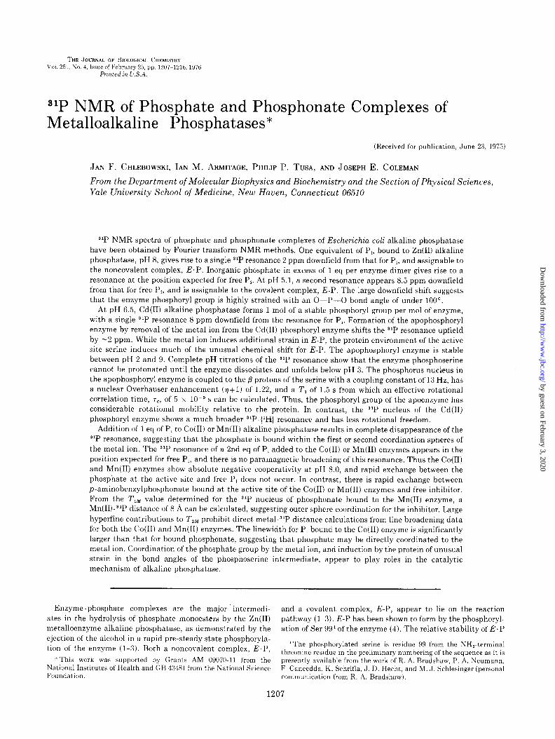
phosphate-free Zn(I1) enzyme is shown in Fig. 1A under the
same conditions as used to record the spectra of the phosphate
complexes. Addition of 1 eq of PJmol of enzyme dimer, pH 8,
results in the appearance of a single resonanck 2.1 ppm
downfield from the resonance position for free inorganic phosphate (Fig. 1B). Addition of a 2nd eq of phosphate per
enzyme dimer, pH 8, gives rise to a second resonance at the
position expected for free inorganic phosphate (Fig. 1C). The
resonance from the initial enzyme-bound phosphate is unaf-
+ IrqP‘
+ I eg P,. pH 5.1
CoOI) AP, pH 8 0 l I f%q P‘
1 COOI) AP. pH 8.0
+ 2rq Pi
III& -10 -5 0 5
8 (ppm)
FIG. 1. JL P NMR spectra obtained on interaction of phosphate with Zn(I1) and Co(R) alkaline phosphatases (At’). Conditions: 0.01 M Tris/l.O M NaC1. A, 1.19 x lOma M Zn(II) alkaline phosphatase, pH 8.0; B, 1.10 x 10v9 M Zn(II) alkaline phosphatase/l.22 x lOMa M K,HPO,, pH 8.0; C, 1.08 x lOma M Zn(II) alkaline phosphatase/2.40 x 10es M
K,HPO,, pH 8.0; D, 6.20 x lo-’ M Zn(II) alkaline phosphatase/6.20 x lo-’ M K,HPO,, pH 5.1, (- - -) add’t’ i ional s’P resonance observed with 1.10 x lo-$ M K,HPO, present; E, 1.10 x lOis M Co(R) alkaline phosphatase/l.04 x lo-* M K,HPO,, pH 8.0; F, 1.09 x lo-$ M Co(I1) alkaline phosphatase/2.38 x lo-* M K,HPO,, pH 8.0.
by guest on February 3, 2020http://w
ww
.jbc.org/D
ownloaded from
31P NMR of Phosphate Complexes of Alkaline Phosphatase 1209
fected by the 2nd equivalent of P,. I f 1 eq of phosphate is added to the Zn(I1) enzyme at pH 5.1, two resonances appear (Fig. 1D). One of these is -2 ppm upfield from the position of the resonance for enzyme-bound phosphate observed at pH 8 and -2 ppm downfield from the resonance position of orthophos- phate at pH 5.1, which suggests that the noncovalently bound phosphate has become protonated. The second resonance is over 8 ppm downfield from the resonance position for free inorganic phosphate at pH 5.1 (Fig. 1D). The latter resonance must represent that of the covalently bound phosphate com- plex, E-P, since previous evidence shows that E-P becomes the predominant species at low pH (5, 6).
Effect of Co(ZZ) and Mn(ZZ) on s1P Resonance of Enzyme- bound Phosphate-Addition of 1 eq of phosphate to Co(I1) alkaline phosphatase (pH 8.0) containing 2 g at Co(II)/mol of enzyme dimer results in complete disappearance of the 31P resonance (Fig. 1E). Under the present system of data collec- tion, and at millimolar concentrations, the linewidth would have to be at least 300 Hz to be undetectable. The addition of a 2nd eq of P, to the Co(R) enzyme results in the appearance of a highly resolved resonance at the position expected for free inorganic phosphate at this pH (Fig. 1F). This resonance is not detectably broadened, showing that the 2nd eq of P, is not in rapid exchange with the 1st eq, and that there is negligible free Co(I1) present in solution. Addition of any more than 2 eq of Co(I1) to the enzyme broadens this line in a manner similar to that observed on the addition of Co(I1) to a solution of inorganic phosphate. Exactly the same phenomena are ob- served when Mn(I1) is used instead of Co(I1).
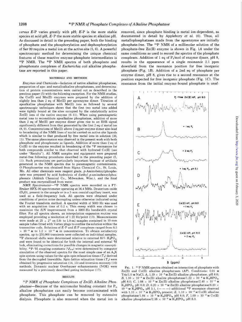
31P NMR of Phosphate Complexes of Cd(ZZ) and Apoalkaline Phosphatase-Since Cd(R) alkaline phosphatase forms a sta- ble and well characterized phosphoryl enzyme near neutral pH (5), the 31P NMR characteristics of the phosphoryl enzyme were further explored by preparation of the phosphorylated Cd(I1) enzyme. Addition of 2 eq of P, to Cd(I1) alkaline phosphatase at pH 6.5 results in the appearance of two SIP resonances; one at the position of the resonance for inorganic phosphate, and the other of similar amplitude 8 ppm downfield from the resonance position for inorganic phosphate (Fig. W). Dialysis of the Cd(I1) enzyme at pH 6.5 against metal-free buffer results in removal of the resonance at the position of inorganic phosphate and retention of the downfield resonance (Fig. 2B). As demonstrated by S2P-labeling (5), this resonance must represent the phosphorus nucleus of the Cd(I1) phospho- ryl enzyme.
The Cd(I1) ion can be completely removed from the dialyzed Cd(R) phosphoryl enzyme by dialysis against 5 x 1O-s M
l,lO-phenanthroline to form the apophosphoryl enzyme (5). The s1P resonance of the apophosphoryl enzyme (Fig. 2C) shifts -2.3 ppm (88 Hz) upfield from the resonance position of the Cd(R) phosphoryl enzyme (Fig. 2B), but it is still -5 ppm downfield from the resonance for free inorganic phosphate at this pH.
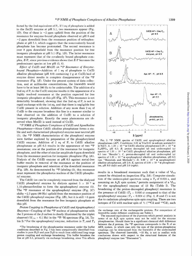
Proton Coupling to Phosphorus of Cd(ZZ) and Apophosphoryl Enzymes-Coupling of the S1P nucleus of phosphoserine with
the 2 protons of the /3 carbon is clearly illustrated by the triplet
observed (VP” = 6.1 Hz) in the slP-lH spectrum (Fig. 3A, Ta- ble I).2 For the apophosphoryl enzyme, reduction in T, and T,
*The broadening of the phosphoserine resonance under the buffer conditions described in Fig. 3 has been unequivocally identified from studies in pure H,O and pure D,O to result from additional long range proton coupling and exchange broadening. Tris buffer broadens the line at pH 6.5, primarily via exchange broadening, since Tris affects
Cd(II) AP + 2sq P‘, dmlyzed, pH 6 5
FIG. 2. $lP NMR spectra of Cd(H) and apophosphoryl alkaline phosphatases (AP). Conditions: 0.01 M Tris/O.Ol M sodium acetate/O.1 M NaCl. A, 1.20 x 10ms M Cd(R) alkaline phosphatase/2.28 x 10ms M K,HPO,, pH 6.5; B, proton decoupled (d) and undecoupled (u) spectra of 1.20 x lOma M Cd(R) phosphoryl alkaline phosphatase, pH 6.5 (see “Results”); C, proton-decoupled (d) and undecoupled (u) spectra of 1.20 x lOma M apophosphoryl alkaline phosphatase, pH 6.5 (see “Materials and Methods”); D, 9.88 x lo-’ M apophosphoryl alkaline phosphatase, pH 2.0; E, spectra of 3.00 x lo-’ M phosphoser- ine, pH 8.0 (left) and pH 2.0 (right).
results in a broadened resonance such that a value of V,, cannot be obtained on inspection (Fig. 3A). Computer simula- tion of the undecoupled spectrum using a T, of 0.024 s, and assuming an A,X spin system,2 permits assignment of a Vr, for the apophosphoryl enzyme of 13 Hz (Table I). The broadening of the proton-decoupled phosphoryl resonance in the presence of Cd(I1) (T, = 0.016 s) compared to that of the apophosphoryl enzyme (T, = 0.024 s) (Fig. 2, B and C) is not due to cadmium-phosphorus spin-spin coupling. There are two isotopes of Cd with nuclear spin of i/z (l%d and lllCd), each
the exchange rate of the exchangeable protons. For comparison of linewidths under different conditions see Table I.
The assumed equivalence of the @ protons which permit analysis in terms of an A,X spin system may not be valid for the enzyme phosphoserine. Should there be a significant chemical shift difference between the B protons, the s1P spectrum corresponds to the X of an ARX system, in which case only the sum of the proton-phosphorus couplings can be determined from the linewidth of the undecoupled spectrum. The observed linewidth, -33 Hz, is not in conflict with conclusions drawn with respect to the structural features of the phosphoryl enzyme (see “Discussion”).
by guest on February 3, 2020http://w
ww
.jbc.org/D
ownloaded from
1210 31P NMR of Phosphate Complexes of Alkaline Phosphatase
FIG. 3. Effect of proton-decoupling on the s’P NMR spectra of phosphoserine and apophosphoryl alkaline phosphatase (ApoAP-I’). Conditions: 0.01 M T&/O.01 M sodium acetate/O.1 M NaCl, pH 6.5/1.1 x 10es M apophosphoryl enzyme/3.0 x 10m2 M phosphoserine. A, 31P-1H spectra; B, 3LP-(‘H)spectra; C, gated 31P- (‘HJspectra. Amplitudes of spectra given in A are increased by a factor of -2.5 for purposes of display.
present at a natural abundance of 1‘2%. Using pure 118Cd(II) to
reconstitute the phosphoryl enzyme, we have determined that
the S1P-113Cd coupling constant at the active site of alkaline
phosphatase is not large enough to significantly broaden the
line when the natural abundance of Cd isotopes is present.
The 31P-(1H)3 spectra of phosphoserine and the apophospho-
ryl enzyme yield T, vaiues of 0.53 and 0.024 s, respectively,
from the measured linewidth (see Table I for comparison of
buffer conditions). Comparison of the intensities of the gated
31P-(1H) spectra (Fig. 3C) with the corresponding SIP-(lH)
spectra (Fig. 3B) allow direct determination of the NOE (v+l)
for phosphoserine and the apophosphoryl enzyme of 1.83 and
1.22, respectively. Determination of T, by both inversion
recovery and progressive saturation gives values of 19.0 s for
phosphoserine and 1.5 s for the apophosphoryl enzyme. From
the NOE (assuming isotropic motion), an effective rotational
correlation time, 7, = 7,, of 5 x 10m9 s can be calculated for the
phosphoryl group on the apoenzyme using published methods
(19) (see footnotes to Table I).
Zonization State of Enzyme Phosphoserine as a Function of pH-Because the apophosphoryl enzyme is stable between pH
2 and 9 (5), a complete pH titration of the apophosphoryl
enzyme can be carried out, and the chemical shifts of the alp
nucleus of the enzyme phosphoserine can be compared with the
9 S’P-lH, undecoupled phosphoryl group; s’P-(‘Hl, proton decou- pled phosphoryl group.
ApoAP-P Ser-P
undecoupled 1
& decouple1
6 I I I I I , I I I I -7 -6 -5 -4 -3
6 (ppm)
corresponding changes induced in the model compound, phos-
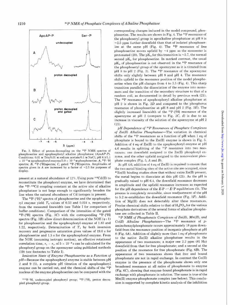
phoserine. The results are shown in Fig. 4. The slP resonance of
the phosphoseryl group in apoalkaline phosphatase at pH 8 is
-2.5 ppm further downfield than that of isolated phosphoser-
ine at the same pH (Fig. 4). The 31P resonance of free
phosphoserine moves upfield by -4 ppm as the monoester is
protonated (20). The pK, for this transition is -5.7, the normal
second pK, for phosphoserine. In marked contrast, the usual
pK, of phosphoserine is not observed in the 31P resonance of
the phosphoseryl group of the apoenzyme as it is titrated from
pH 8 to pH 2 (Fig. 3). The 31P resonance of the apoenzyme
shifts only slightly between pH 8 and pH 4. The resonance
shifts upfield to the resonance position of the model phospho-
serine when the pH changes from 4 to 3.5 (Fig. 4). This sharp
transition parallels the dissociation of the enzyme into mono-
mers and the transition of the secondary structure to that of a
random coil, as documented in detail by previous work (21).
The 31P resonance of apophosphoryl alkaline phosphatase at
pH 2 is shown in Fig. 20 and compared to the phosphorus
resonance of phosphoserine at pH 8 and pH 2 (Fig. 2E). The
slightly increased linewidth of the 31P-(1H) resonance of the
apoenzyme at pH 2 (compare to Fig. 2C, cl) is due to an
increase in viscosity of the solution of the apoenzyme at pH 2
(21).
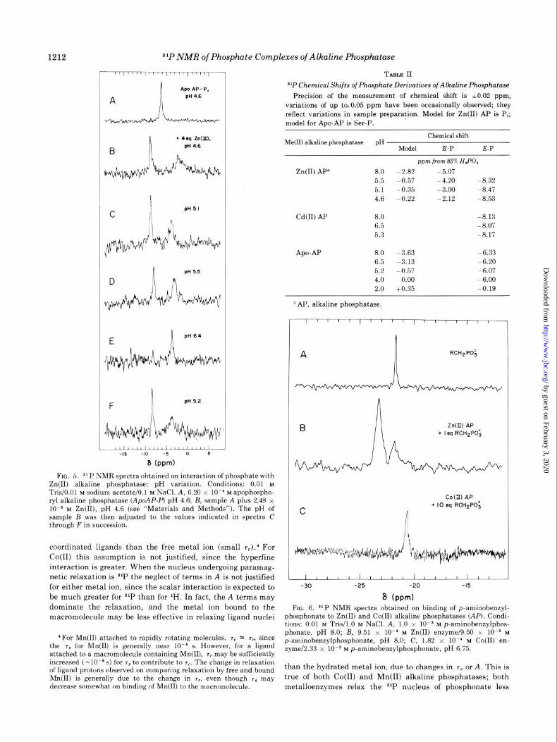
pH Dependence of 31P Resonances of Phosphate Complexes of Zn(I4 Alkaline Phosphatase-The variation in chemical
shifts of the 31P resonances as a function of pH when 1 eq of
phosphate is bound to the Zn(I1) enzyme is shown in Fig. 5.
Addition of 4 eq of Zn(I1) to the apophosphoryl enzyme at pH
4.6 results in splitting of the 31P resonance into two reso-
nances: one downfield assigned to the Zn(I1) phosphoryi en-
zyme, and the other upfield assigned to the noncovalent phos-
phate complex (Fig. 5, A and B).
At pH 4.6, addition of 4 eq of Zn(I1) is required to ensure that
the two metal binding sites of the active site are occupied, since
65Zn(II) binding studies show that without extra Zn(I1) present,
the metal begins to dissociate at this pH (21). As the pH is
gradually raised to pH 6.4, the downfield resonance decreases
in amplitude and the upfield resonance increases as expected for the pH dependence of the E-P + E.P equilibrium (5). The
process is completely reversible, since readjustment of the pH
to 5.2 re-establishes the downfield resonance (Fig. 5F). Addi-
tion of Mg(I1) does not detectably alter these resonances.
Precise chemical shifts relative to that of H,PO, for the various phosphate derivatives of the several forms of alkaline phospha-
tase are collected in Table II.
$‘P NMR of Phosphonate Complexes of Zn(II), Mn(Z4, and Co(ZI) Alkaline Phosphatases-The 31P resonance of p- aminobenzylphosphonate occurs approximately 18 ppm down-
field from the resonance position of inorganic phosphate at pH
8 (Fig. 6A). Addition of slightly more than 1 eq of phosphonate
to the native Zn(I1) alkaline phosphatase results in the
appearance of two resonances; a major one 2.2 ppm (81 Hz)
downfield from that for free phosphonate; and a second at the
position of the resonance for free phosphonate (Fig. 6B). The
appearance of two resonances shows that free and bound
phosphonate are not in rapid exchange. In contrast the Co(B)
enzyme in the presence of the phosphonate shows only one
broadened resonance at all ratios of phosphonate to enzyme
(Fig. 6C), showing that enzyme-bound phosphonate is in rapid
exchange with phosphonate in solution. The same is true of the
Mn(I1) enzyme phosphonate complex (see below). This conclu-
sion is supported by complete kinetic analysis of the inhibition
by guest on February 3, 2020http://w
ww
.jbc.org/D
ownloaded from
$‘P NMR of Phosphate Complexes of Alkaline Phosphutase 1211
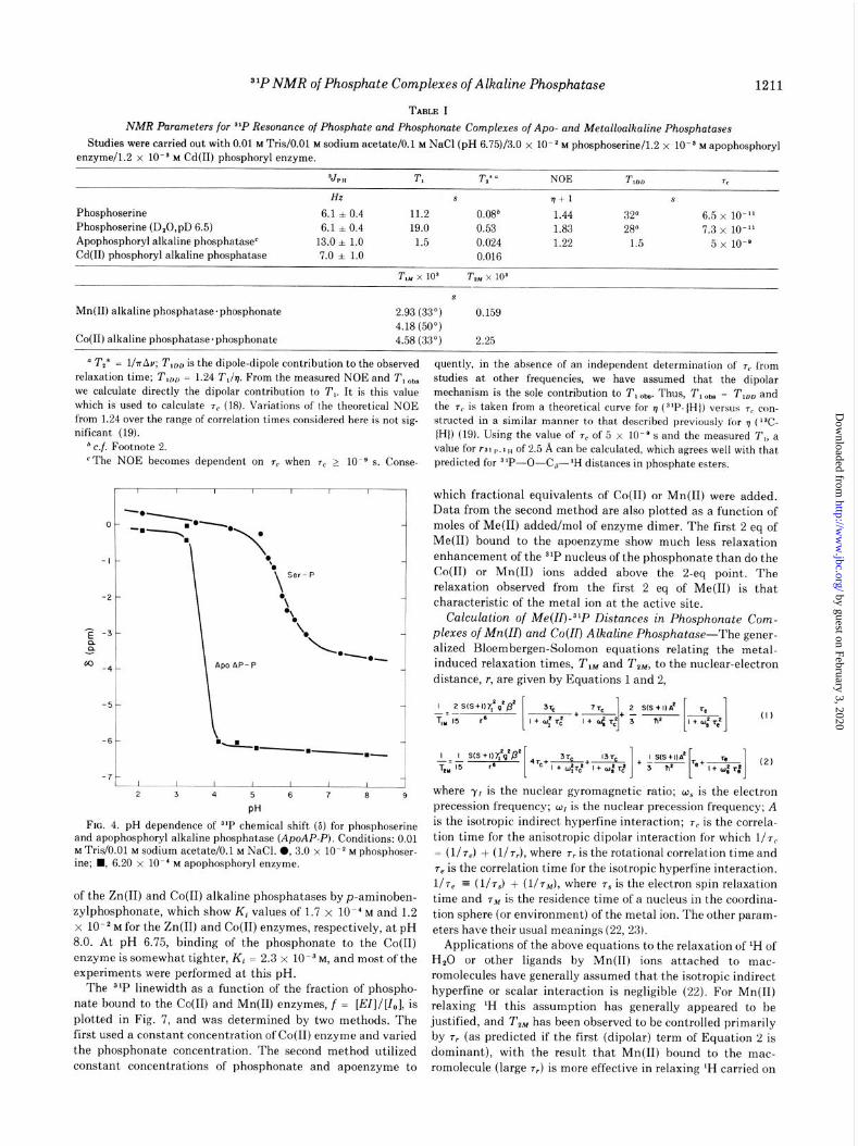
TABLE I
NMR Parameters for 3’P Resonance of Phosphate and Phosphonute Complexes of Apo- and Metalloalkaline Phosphatases
Studies were carried out with 0.01 M Tris/O.Ol M sodium acetate/O.1 M NaCl (pH 6.75)/X0 x 10mz M phosphoserine/l.2 x 10e3 M apophosphoryl enzyme/l.2 x 10e3 M Cd(B) phosphoryl enzyme.
sJP” T, T,* o NOE T I** Tc
HZ s rl+1 s
Phosphoserine 6.1 + 0.4 11.2 0.08" 1.44 32" 6.5 x lo-” Phosphoserine (D,O,pD 6.5) 6.1 i 0.4 19.0 0.53 1.83 28" 7.3 x 10-I’ Apophosphoryl alkaline phosphatasee 13.0 * 1.0 1.5 0.024 1.22 1.5 5 x 10-e Cd(B) phosphoryl alkaline phosphatase 7.0 l 1.0 0.016
T,, x lo3 T m x 10s
s
Mn(I1) alkaline phosphatase.phosphonate 2.93(33O) 0.159 4.18 (50’)
Co(I1) alkaline phosphatase.phosphonate 4.58(33') 2.25
D T,* = l/lrAv; T,,, is the dipole-dipole contribution to the observed relaxation time; T,,, = 1.24 T,/q. From the measured NOE and T, obS we calculate directly the dipolar contribution to T,. It is this value which is used to calculate T, (18). Variations of the theoretical NOE from 1.24 over the range of correlation times considered here is not sig- nificant (19).
b c.f. Footnote 2. ‘The NOE becomes dependent on ,c when T, > lo-’ s. Conse-
_I .5 4 5 6 7 6 9
PH
FIG. 4. pH dependence of slP chemical shift (S) for phosphoserine and apophosphoryl alkaline phosphatase (ApoAP-P). Conditions: 0.01 M Tris/O.Ol M sodium acetate/O.1 M NaCl. 0, 3.0 x lo-* M phosphoser- ine; n , 6.20 x lo-’ M apophosphoryl enzyme.
of the Zn(I1) and Co(I1) alkaline phosphatases by p-aminoben- zylphosphonate, which show K, values of 1.7 x lo-’ M and 1.2 x 10m2 M for the Zn(II) and Co(B) enzymes, respectively, at pH 8.0. At pH 6.75, binding of the phosphonate to the Co(I1) enzyme is somewhat tighter, K, = 2.3 x 10m3M, and most of the experiments were performed at this pH.
The 3*P linewidth as a function of the fraction of phospho- nate bound to the Co(B) and Mn(I1) enzymes, f = [EZ]l[Z,], is plotted in Fig. 7, and was determined by two methods. The first used a constant concentration of Co(I1) enzyme and varied the phosphonate concentration. The second method utilized constant concentrations of phosphonate and apoenzyme to
quently, in the absence of an independent determination of T, from studies at other frequencies, we have assumed that the dipolar mechanism is the sole contribution to T, Obs. Thus, T, Obs = T,,, and the 7c is taken from a theoretical curve for q (“‘P-(H)) versus T, con- structed in a similar manner to that described previously for TJ ( 13Cv lHl) (19). Using the value of rC of 5 x 10M9 s and the measured T,, a value for r31 p.~H of 2.5 A can be calculated, which agrees well with that predicted for 31P-O-C,-‘H distances in phosphate esters.
which fractional equivalents of Co(I1) or Mn(I1) were added. Data from the second method are also plotted as a function of moles of Me(B) added/m01 of enzyme dimer. The first 2 eq of Me(B) bound to the apoenzyme show much less relaxation enhancement of the 31P nucleus of the phosphonate than do the Co(I1) or Mn(I1) ions added above the 2-eq point. The relaxation observed from the first 2 eq of Me(B) is that characteristic of the metal ion at the active site.
Calculation of Me(ZZ)-a’P Distances in Phosphonate Com- plexes of Mn(ZZ) and Co(ZZ) Alkaline Phosphatase-The gener- alized Bloembergen-Solomon equations relating the metal- induced relaxation times, T,, and TZM, to the nuclear-electron distance, r, are given by Equations 1 and 2,
where y, is the nuclear gyromagnetic ratio; wS is the electron precession frequency; wI is the nuclear precession frequency; A is the isotropic indirect hyperfine interaction; T, is the correla- tion time for the anisotropic dipolar interaction for which l/re = (l/rJ + (l/r?), where rr is the rotational correlation time and re is the correlation time for the isotropic hyperfine interaction. l/r1 = ( ~/TJ + (l/7,,,), where rS is the electron spin relaxation time and T,,, is the residence time of a nucleus in the coordina- tion sphere (or environment) of the metal ion. The other param- eters have their usual meanings (22, 23).
Applications of the above equations to the relaxation of ‘H of H,O or other ligands by Mn(I1) ions attached to mac- romolecules have generally assumed that the isotropic indirect hyperfine or scalar interaction is negligible (22). For Mn(II) relaxing ‘H this assumption has generally appeared to be justified, and T,, has been observed to be controlled primarily by TV (as predicted if the first (dipolar) term of Equation 2 is dominant), with the result that Mn(I1) bound to the mac- romolecule (large 7,) is more effective in relaxing ‘H carried on
by guest on February 3, 2020http://w
ww
.jbc.org/D
ownloaded from
1212 S1P NMR of Phosphate Complexes of Alkaline Phosphutase
Apo AP- P. PH 4.6
b + 4eq znm.
B pH 4.6
1 pti55
F
k ILLLLLLLI
-15 -10 -5 0 6
8 (ppm)
FE. 5. 31P NMR spectra obtained on interaction of phosphate with Zn(I1) alkaline phosphatase: pH variation. Conditions: 0.01 M Tris/O.Ol M sodium acetate/O.1 M NaCl. A, 6.20 x lo-’ M apophospho- ryl alkaline phosphatase (ApoAP-P) pH 4.6; B, sample A plus 2.48 x 10~~ M Zn(II), pH 4.6 (see “Materials and Methods”). The pH of sample B was then adjusted to the values indicated in spectra C through Fin succession.
coordinated ligands than the free metal ion (small 7?).’ For Co(I1) this assumption is not justified, since the hyperfine interaction is greater. When the nucleus undergoing paramag- netic relaxation is 81P the neglect of terms in A is not justified for either metal ion, since the scalar interaction is expected to be much greater for $‘P than for ‘H. In fact, the A terms may dominate the relaxation, and the metal ion bound to the macromolecule may be less effective in relaxing ligand nuclei
‘For Mn(II) attached to rapidly rotating molecules, T, e 7,. since the ra for Mn(II) is generally near 1Om8 s. However, for a ligand attached to a macromolecule containing Mn(II), T, may be sufficiently increased (-1O-8 s) for rb to contribute to TV. The change in relaxation of ligand protons observed on comparing relaxation by free and bound Mn(I1) is generally due to the change in r,, even though r6 may decrease somewhat on binding of Mn(I1) to the macromolecule.
TABLE II
a’P Chemical Shifts of Phosphate Derivatives of Alkaline Phosphatase
Precision of the measurement of chemical shift is 10.02 ppm, variations of up to.0.05 ppm have been occasionally observed; they reflect variations in sample preparation. Model for Zn(I1) AP is P,; model for Apo-AP is Ser-P.
Chemical shift Me(R) alkaline phosphatase pH
Model E.P E-P
ppm from 85% HJ’O,
Zn(I1) AP” 8.0 -2.82 -5.07
5.5 -0.57 -4.20 -8.32 5.1 -0.35 -3.00 -8.41
4.6 -0.22 -2.12 -8.53
Cd(R) AP 8.0 -8.13
6.5 -8.07 5.3 -8.17
Apo-AP 8.0 -3.63 -6.33
6.5 -3.13 -6.20 5.2 -0.57 -6.07
4.0 0.00 -6.00
2.0 +0.35 -0.19
O1 AP, alkaline phosphatase.
Zn(II) BP + I eq RCH*PO;
C
CoGI) AP + IO eq RCH2PO;
II h I IL1 I I IIIId II II -30 -25 -20 -15
8 (ppm)
FIG. 6. 31P NMR spectra obtained on binding of p-aminobenzyl- phosphonate to Zn(I1) and Co(R) alkaline phosphatases (AP). Condi- tions: 0.01 M Tris/l.O M NaCl. A, 1.0 x 10es M p-aminobenzylphos- phonate, pH 8.0; B, 9.51 x lo-’ M Zn(II) enzyme/9.50 x lOma M p-aminobenzylphosphonate, pH 8.0; C, 1.82 x lo-’ M Co(R) en- zyme/2.33 x 10e3 M p-aminobenzylphosphonate, pH 6.75.
than the hydrated metal ion, due to changes in 7, or A. This is true of both Co(I1) and Mn(I1) alkaline phosphatases; both metalloenzymes relax the s1P nucleus of phosphonate less
by guest on February 3, 2020http://w
ww
.jbc.org/D
ownloaded from
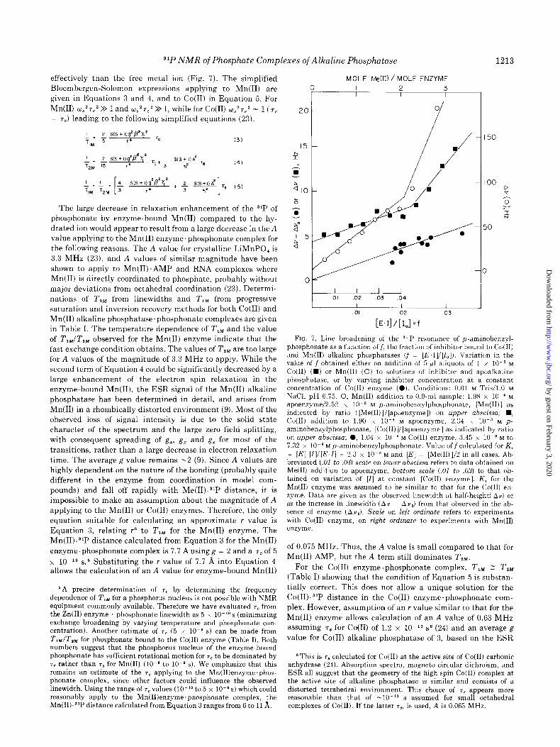
31P NMR of Phosphate Complexes of Alkaline Phosphatase
effectively than the free metal ion (Fig. 7). The simplified
Bloembergen-Solomon expressions applying to Mn(I1) are
given in Equations 3 and 4, and to Co(I1) in Equation 5. For
Mn(I1) w,,~T,~ >> 1 and w~‘T~~ >> 1, while for Co(I1) 0~~7~~ - 1 (7,
= 7,) leading to the following simplified equations (23).
The large decrease in relaxation enhancement of the 31P of
phosphonate by enzyme-bound Mn(I1) compared to the hy-
drated ion would appear to result from a large decrease in the A
value applying to the Mn(I1) enzyme.phosphonate complex for
the following reasons. The A value for crystalline LiMnPO, is
3.3 MHz (23), and A values of similar magnitude have been
shown to apply to Mn(II).AMP and RNA complexes where
Mn(I1) is directly coordinated to phosphate, probably without
major deviations from octahedral coordination (23). Determi-
nations of T,, from linewidths and T,, from progressive
saturation and inversion recovery methods for both Co(I1) and
Mn(I1) alkaline phosphatase.phosphonate complexes are given
in Table I. The temperature dependence of T,, and the value
of T,,/T,, observed for the Mn(I1) enzyme indicate that the
fast exchange condition obtains. The values of T,, are too large
for A values of the magnitude of 3.3 MHz to apply. While the
second term of Equation 4 could be significantly decreased by a
large enhancement of the electron spin relaxation in the
enzyme-bound Mn(II), the ESR signal of the Mn(I1) alkaline
phosphatase has been determined in detail, and arises from
Mn(I1) in a rhombically distorted environment (9). Most of the observed loss of signal intensity is due to the solid state
character of the spectrum and the large zero field splitting,
with consequent spreading of g,, g, and g, for most of the
transitions, rather than a large decrease in electron relaxation
time. The average g value remains -2 (9). Since A values are
highly dependent on the nature of the bonding (probably quite
different in the enzyme from coordination in model com-
pounds) and fall off rapidly with Me(II)-31P distance, it is
impossible to make an assumption about the magnitude of A
applying to the Mn(I1) or Co(I1) enzymes. Therefore, the only
equation suitable for calculating an approximate r value is
Equation 3, relating r6 to T,, for the Mn(I1) enzyme. The
Mn(II)-“P distance calculated from Equation 3 for the Mn(I1)
enzyme’phosphonate complex is 7.7 A using g = 2 and a 7, of 5
x lo-l0 s5 Substituting the r value of 7.7 b, into Equation 4 allows the calculation of an A value for enzyme-bound Mn(I1)
0
‘A precise determination of 7. by determining the frequency dependence of T,, for a phosphorus nucleus is not possible with NMR equipment commonly available. Therefore we have evaluated T, from the Zn(I1) enzyme phosphonate linewidth as 5 x lo-‘O s (minimizing exchange broadening by varying temperature and phosphonate con- centration). Another estimate of 7e (5 x lo-@ s) can be made from TIM/T,, for phosphonate bound to the Co(I1) enzyme (Table I). Both numbers suggest that the phosphorus nucleus of the enzyme-bound phosphonate has sufficient rotational motion for T, to be dominated by 7r rather than T, for Mn(I1) (lOma to lo-* s). We emphasize that this remains an estimate of the rc applying to the Mn(II)enzyme.phos- phonate complex, since other factors could influence the observed linewidth. Using the range of TV values (lo-” to 5 x 10m8s) which could reasonably apply to the Mn(II)enzyme.phosphonate complex, the Mn(II)-3’P distance calculated from Equation 3 ranges from 6 to 11 A.
MOLE Me(D) / MOLE ENZYME
1
-I(
1213
I 50
I 00 e
3 V I N
50
3
I I L I 01 .02 03 .04
1 I 1 01 02 03
[E.I]/[I.]=f
FIG. 7. Line broadening of the ‘l P resonance of p-aminobenzyl- phosphonate as a function off, the fraction of inhibitor bound to Co(I1) and Mn(II) alkaline phosphatases (f = [E,IV[I,]). Variation in the value of f obtained either on addition of 5.~1 aliquots of 1 x 10.’ M Co(I1) (m) or Mn(I1) (0) to solutions of inhibitor and apoalkaline phosphatase, or by varying inhibitor concentration at a constant concentration of Co(I1) enzyme (0). Conditions: 0.01 M Tris/l.O M NaCl, pH 6.75. 0, Mn(II) addition to 0.9.ml sample: 1.98 x lo-’ M apoenzyme/2.52 x 10m3 M p-aminobenzylphosphonate, [Mn(II)] as indicated by ratio ([Me(II)]/[ p a oenzyme]) on upper abscissa; n , Co@) addition to 1.90 x lo-’ M apoenzyme, 2.34 x 10m3 M p- aminobenzylphosphonate, [Co(II)V[apoenzyme] as indicated by ratio on upper abscissa; 0, 1.04 x lo-’ M Co@) enzyme, 3.45 x 10m3 M to 7.32 x 10. ’ M p-aminobenzylphosphonate. Value off calculated for K, = [El [I]l[E.I] = 2.3 x 10 3 M and [E] = [Me(II)]/2 in all cases. Ab- breviated (.OI to .04) scale on lower abscissa refers to data obtained on Me(I1) addition to apoenzyme, bottom scale (.01 to .03) to that ob- tained on variation of [I] at constant [Co(II) enzyme]. K1 for the Mn(I1) enzyme was assumed to be similar to that for the Co(I1) en- zyme. Data are given as the observed linewidth at half-height( Au) or as the increase in linewidth (Av ~ Av,) from that observed in the ab- sence of enzyme (Av,). Scale on left ordinate refers to experiments with Co(I1) enzyme, on right ordinate to experiments with Mn(I1) enzyme.
of 0.075 MHz. Thus, the A value is small compared to that for
Mn(I1) AMP, but the A term still dominates T,,.
For the Co(I1) enzyme .phosphonate complex, T,, 1 T2,,, (Table I) showing that the condition of Equation 5 is substan-
tially correct. This does not allow a unique solution for the
Co(II)-“P distance in the Co(I1) enzyme.phosphonate com-
plex. However, assumption of an r value similar to that for the
Mn(I1) enzyme allows calculation of an A value of 0.63 MHz
assuming 7g for Co(I1) of 1.2 x lo-” sB (24) and an average g
value for Co(I1) alkaline phosphatase of 3, based on the ESR
‘This is 7B calculated for Co(I1) at the active site of Co(I1) carbonic anhydrase (24). Absorption spectra, magneto circular dichroism, and ESR all suggest that the geometry of the high spin Co(I1) complex at the active site of alkaline phosphatase is similar and consists of a distorted tetrahedral environment. This choice of 7g appears more reasonable than that of -1Om’3 s assumed for small octahedral complexes of Co(I1). If the latter TV, is used, A is 0.063 MHz.
by guest on February 3, 2020http://w
ww
.jbc.org/D
ownloaded from
1214 ‘lP NMR of Phosphate Complexes of Alkaline Phosphatase
signal at helium temperature of the enzyme containing two,
Co(I1) ions per dimer. As for Mn(I1) enzyme, the hyperfine
term dominates TZM.
DISCUSSION
31P NMR is a particularly suitable method for detecting the
number and stoichiometry of the species of phosphate com-
plexes present in alkaline phosphatase because all the chemi-
cally distinct species of bound phosphate are detected simulta-
neously in an enzyme sample unperturbed by the method of
detection. At pH 8, phosphate-free alkaline phosphatase can
bind only one noncovalently linked inorganic phosphate dian-
ion in an unusual chemical environment (Fig. 1, A and B).
Thus, phosphate above 1 eq per enzyme dimer is either not
bound to the enzyme or is bound at a site of different chemical
environment so that it does not alter the chemical shift from
that expected for inorganic phosphate at the same pH. This
finding suggests that negative allosteric interactions exist
between initially identical phosphate binding sites, one on
each of the two identical subunits of the enzyme. This has
previously been suggested by extensive spectral data (6, 8, 10).
The electron density map of the dimer at 3 A resolution shows
that at this resolution apparent 2.fold symmetry exists be-
tween the monomers of the dimer of alkaline phosphatase.?
When one phosphate ion is bound to the dimer, a second
phosphate binding site of similar chemical environment is not
available even at millimolar concentrations of phosphate (Fig.
1). This conclusion is strikingly confirmed by the use of 31P
NMR to follow phosphate binding to the Mn(I1) or Co(II)
enzymes (Fig. 1). Only the 1st eq of phosphate binds to the
dimer in a position for its resonance to be broadened by the
paramagnetic effect of Co(I1) or Mn(I1) (Fig. 1). The second
phosphate is unaffected by the paramagnetic ion, and thus
clearly cannot be near the second Me(I1) ion, implying absolute
negative cooperativity between the active sites at neutral pH
even in the presence of millimolar phosphate. We have
determined the stoichiometry of phosphate binding by these
NMR methods on several samples of each of the metallophos-
phatases discussed in this paper, and in each case only one chemically distinct phosphate binding site per dimer is pres-
ent. The observed negative cooperativity may be a reflection of
an underlying molecular mechanism aiding the dissociation of
the product complex, E’P, since binding of ROP to the second
site may reduce the affinity of P, for the alternate site.
The phosphoryl enzyme formed by both the Zn(I1) and
Cd(I1) enzymes is a most unusual phosphate monoester. The
31P resonance of this species is shifted 5 to 6 ppm downfield
from the 31P resonances observed for model phosphate mono-
esters (Figs. I, 2, and 5 and Table II). Assignment of this
resonance to the enzyme phosphoserine is confirmed by the
interchange between this species and the noncovalent en-
zyme.phosphate complex as a function of pH, which follows
the known pH stability of E-P (Fig. 5) (4, 6).
Formation of the phosphorylated enzyme giving rise to the
downfield resonance in stoichiometric quantities by Cd(R)
further confirms this species as E-P (Fig. 2). While the metal
ion contributes to the downfield shift of the phosphorus
resonance of E-P, the 31P resonance of the phosphoryl group on
the apoenzyme remains 3 to 6 ppm (depending on pH) down-
field from that observed for free phosphoserine (Figs. 2 and
‘W. Carlson, M. Chou, J. Coleman, and H. Wyckoff, work in progress.
4). Thus, the protein structure itself contributes significantly
to the unusual chemical characteristics of this phosphoseryl
residue. The structural basis for the failure to protonate the
phosphoryl residue on the apoenzyme as long as the enzyme
retains its native conformation (F,ig. 4), is not obvious in terms
of known protein structure. Speculative suggestions might
include hydrogen bonding to an adjacent hydroxyl group, in a
structure similar to that occurring between the carboxyl and
phenolic hydroxyl of salicylic acid, which lowers the pK, of the
carboxyl (25). A distribution of positive charges around the
active site seryl residue might also prevent protonation.
It has recently been proposed that an empirical correlation
can be made between the chemical shift of the 31P resonance
and the magnitude of the smallest O-P-O bond angle in
phosphate monoesters, acyclic diesters, and five- and six-mem-
bered cyclic diesters (20). The 31P resonance shifts progres- sively downfield as this angle decreases from 106”. Chemical
shifts observed on association of metal ions and hydrogen
bonding donors with the phosphate group are accompanied by
a shift in the phosphoryl pK,, and fall within the range
predicted by this correlation for changes in the ionization state
of the phosphoryl group (20). While a divalent metal ion is in
close proximity to phosphate bound to alkaline phosphatase
(see below), comparison of the chemical shifts observed for the
Cd(II), Zn(II), and apophosphoryl enzymes (Figs. 1 and 2) suggests that other factors must influence the chemical nature
of the phosphoryl group. The magnitude of the observed
chemical shifts suggests that the surrounding protein structure
induces a significant compression of the O-P-O bond angle.
Thus, the Zn(I1) phosphoryl enzyme, with a 31P chemical shift
approaching that observed for five-membered cyclic diesters
having an O-P-O bond angle less than loo”, appears to be a
highly strained phosphate ester. The Cd(I1) phosphoryl en-
zyme is slightly less strained, and the strain is substantially
relieved in the apophosphoryl enzyme (Fig. 2). The apparent
distortion of the phosphoryl group in the Zn(I1) enzyme may
provide the driving force for its rapid turnover (see below).
Reduction of strain in the Cd(I1) phosphoryl enzyme relative to
the Zn(I1) phosphoryl enzyme might be related to the slow
turnover of the Cd(I1) enzyme.
The 31P chemical shift of E.P is -2 ppm downfield from that
observed for free P, (Fig. 1). This cannot be attributed simply
to a shift, in the pK, of E. P, since the chemical shift difference
is relatively constant from pH 4.6 to pH 8.0 (Table II). Rather,
it would appear that the forces operative in inducing strain in
E-P are also functional, albeit to a limited degree, in perturb-
ing the structure of E.P in a similar manner.
Enzyme-Phosphate Interactions as Determined by 3iP NMR-The magnitude of 7, for the apophosphoryl enzyme
(Table I) suggests that the mobility of the enzyme-bound
phosphoryl group is not governed by the rotational correlation
time of the macromolecule (7, for a molecule of average radius
33 A, calculated from the Stokes-Einstein equation, is 3 x
10.’ s). Thus, the data in Table I reflect the dynamic
properties of a relatively mobile phosphoryl group at the active
center, and can be interpreted with reference to the model
compound phosphoserine.
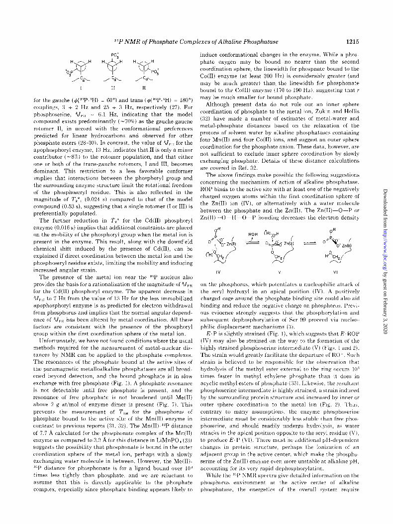
The molecular fragment ‘H-G-O--31P has been demon-
strated to show an angular dependence for “‘P-‘H spin-spin
coupling similar to that given by the Karplus equation for
vicinal ‘H-‘H coupling (26-28). Thus, direct assessment of the
fractional population of the minimum energy rotomers (I, II, and III) can be made on the basis of the reported values of 3Jr,
by guest on February 3, 2020http://w
ww
.jbc.org/D
ownloaded from
31P NMR of Phosphate Complexes of Alkaline Phosphatase 1215
‘03PY
/ 5-
I
for the gauche (G(~‘P-‘H) = 60”) and tram ( $(31P-‘H) = 180”)
couplings, 3 + 2 Hz and 25 i 3 Hz, respectively (27). For
phosphoserine, 3JpH = 6.1 Hz, indicating that the model
compound exists predominantly (-70%) as the gauche-gauche
rotomer II, in accord with the conformational preferences
predicted for linear hydrocarbons and observed for other
phosphate esters (28-30). In contrast, the value of ?J,, for the
apophosphoryl enzyme, 13 Hz, indicates that II is only a minor
contributor (-8%) to the rotomer population, and that either
one or both of the trans.gauche rotomers, I and III, becomes
dominant. This restriction to a less favorable conformer
implies that interactions between the phosphoryl group and
the surrounding enzyme structure limit the rotational freedom
of the phosphoseryl residue. This is also reflected in the
magnitude of T,‘, (0.024 s) compared to that of the model
compound (0.53 s), suggesting that a single rotomer (I or III) is
preferentially populated. The further reduction in T,* for the Cd(I1) phosphoryl
enzyme (0.016 s) implies that additional constraints are placed
on the mobility of the phosphoryl group when the metal ion is
present in the enzyme. This result, along with the downfield
chemical shift induced by the presence of Cd(II), can be
explained if direct coordination between the metal ion and the
phosphoseryl residue exists, limiting the mobility and inducing
increased angular strain.
The presence of the metal ion near the 31P nucleus also
provides the basis for a rationalization of the magnitude of 3J,,
for the Cd(I1) phosphoryl enzyme. The apparent decrease in
‘J,,, to 7 Hz from the value of 13 Hz for the less immobilized
apophosphoryl enzyme is as predicted for electron withdrawal
from phosphorus and implies that the normal angular depend-
ence of 3J,X,, has been altered by metal coordination. All these
factors are consistent with the presence of the phosphoryl
group within the first coordination sphere of the metal ion.
Unfortunately, we have not found conditions where the usual
methods required for the measurement of metal-nuclear dis-
tances by NMR can be applied to the phosphate complexes.
The resonances of the phosphate bound at the active sites of
the paramagnetic metalloalkaline phosphatases are all broad-
ened beyond detection, and the bound phosphate is in slow
exchange with free phosphate (Fig. 1). A phosphate resonance
is not detectable until free phosphate is present, and the
resonance of free phosphate is not broadened until Me(R)
above 2 g at/mol of enzyme dimer is present (Fig. 7). This
prevents the measurement of T,, for the phosphorus of phosphate bound to the active site of the Mn(I1) enzyme in
contrast to previous reports (31, 32). The Mn(II)-31P distance
of 7.7 A ca!culated for the phosphonate complex of the Mn(I1)
enzyme as compared to 3.3 A for this distance in LiMnPO, (23)
suggests the possibility that, phosphonate is bound in the outer
coordination sphere of the metal ion, perhaps with a slowly exchanging water molecule in between. However, the Me(II)-
31P distance for phosphonate is for a ligand bound over lo3 times less tightly than phosphate, and we are reluctant to
assume that this is directly applicable to the phosphate
complex, especially since phosphate binding appears likely to
induce conformational changes in the enzyme. While a phos-
phate oxygen may be bound no nearer than the second
coordination sphere, the linewidth for phosphate bound to the
Co(I1) enzyme (at least 300 Hz) is considerably greater (and
may be much greater) than, the linewidth for phosphonate
bound to the Co(R) enzyme (170 to 190 Hz), suggesting that r
may be much smaller for bound phosphate.
Although present data do not rule out an inner sphere
coordination of phosphate to the metal ion, Zukin and Hollis
(32) have made a number of estimates of metal-water and
metal-phosphate distances based on the relaxation of the
protons of solvent water by alkaline phosphatases containing four Mn(I1) and four Co(I1) ions, and suggest an outer sphere
coordination for the phosphate anion. These data, however, are
not sufficient to exclude inner sphere coordination by slowly
exchanging phosphate. Details of these distance calculations
are covered in Ref. 32.
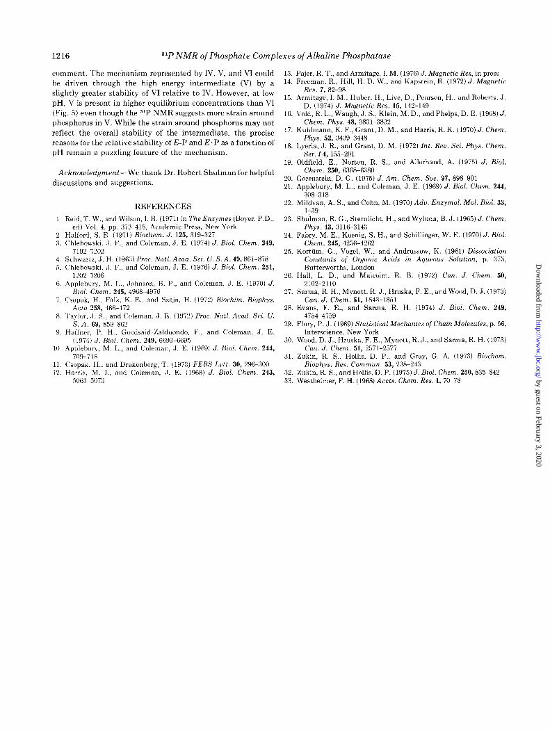
The above findings make possible the following suggestions
concerning the mechanism of action of alkaline phosphatase.
ROP binds to the active site with at least one of the negatively
charged oxygen atoms within the first coordination sphere of
the Zn(I1) ion (IV), or alternatively ivith a water molecule
between the phosphate and the Zn(I1). The Zn(Ii)-O-P or
Zn(II)-O-H-O-P bonding decreases the electron density
IV v VI
on the phosphorus, which potentiates a nucleophilic attack of
the seryl hydroxyl in an apical position (IV). A positively
charged cage around the phosphate binding site could also aid
binding and reduce the negative charge on phosphorus. Previ-
ous evidence strongly suggests that the phosphorylation and
subsequent dephosphorylation of Ser 99 proceed via nucleo-
philic displacement mechanisms (3).
E.P is slightly strained (Fig. l), which suggests that E.ROP
(IV) may also be strained on the way to the formation of the
highly strained phosphoserine intermediate (V) (Figs. 1 and 2).
The strain would greatly facilitate the departure of RO Such
strain is believed to be responsible for the observation that
hydrolysis of the methyl ester external to the ring occurs 10’
times faster in methyl ethylene phosphate than it does in
acyclic methyl esters of phosphate (33). Likewise, the resultant phosphoserine intermediate is highly strained, a strain induced
by the surrounding protein structure and increased by inner or
outer sphere coordination to the metal ion (Fig. 2). Thus,
contrary to many assumptions, the enzyme phosphoserine
intermediate must be considerably less stable than free phos-
phoserine, and should readily undergo hydrolysis, as water
attacks in the apical position opposite to the seryl residue (V),
to produce E.P (VI). There must be additional pH-dependent
changes in protein structure, perhaps the ionization of an
adjacent group in the active center, which make the phospho-
serine of the Zn(I1) enzyme even more unstable at alkaline pH,
accounting for its very rapid dephosphorylation.
While the 31P NMR spectra give detailed information on the
phosphorus environment at the active center of alkaline
phosphatase, the energetics of the overall system require
by guest on February 3, 2020http://w
ww
.jbc.org/D
ownloaded from
1216 ‘lP NMR of Phosphate Complexes of Alkaline Phosphatase
comment. The mechanism represented by IV, V, and VI could
be driven through the high energy intermediate (V) by a
slightly greater stability of VI relative to IV. However, at low
pH, V is present in higher equilibrium concentrations than VI
(Fig. 5) even though the 31P NMR suggests more strain around
phosphorus in V. While the strain around phosphorus may not
reflect the overall stability of the intermediate, the precise
reasons for the relative stability of E-P and E.P as a function of
pH remain a puzzling feature of the mechanism.
Acknowledgment-We thank Dr. Robert Shulman for helpful
discussions and suggestions.
REFERENCES
1. Reid, T. W., and Wilson, I. B. (1971) in The Enzymes (Boyer, P.D., ed) Vol. 4, pp. 373-415, Academic Press, New York
2. Halford, S. E. (1971) Biochem. J. 125, 319-327 3. Chlebowski, J. F., and Coleman, J. E. (1974) J. Biol. Chem. 249,
7192-7202 4. Schwartz, J. H. (1963) Proc. N&l. Acad. Sci. U. S. A. 49,861-878 5. Chlebowski, J. F., and Coleman, J. E. (1976) J. Biol. Chem. 251,
1202-1206 6. Applebury, M. L., Johnson, B. P., and Coleman, J. E. (1970) J.
Bid. Chem. 245, 4968-4976 ‘i. Csopak, H., Falk, K.-E., and Szajn, H. (1972) Biochim. Biophys.
Acta 258, 466-472 8. Taylor, J. S., and Coleman, J. E. (1972) Proc. N&l. Acad. Sci. U.
S. A. 69, 859-862 9. Haffner. P. H., Goodsaid-Zalduondo. F., and Coleman, J. E.
(1974) J. Biol. Chem. 249, 6693-6695 10. ADplebury. M. L.. and Coleman, J. E. (1969) J. Biol. Chem. 244,
‘jog-718 II. Csopak, H., and Drakenberg, T. (1973) FEBS Lett. 30, 296-300 12. Harris, M. I., and Coleman, J. E. (1968) J. Biol. Chem. 243,
5063-5073
13. 14.
15.
16.
17.
18.
19.
20. 21.
22.
23.
24.
25.
26.
27.
28.
29.
30.
31.
32. 33.
Pajer, R. T., and Armitage, I. M. (1976) J. Magnetic Res, in press Freeman, R., Hill, H. D. W., and Kapstein, R. (1972) J. Magnetic
Res. 7, 82-98 Armitage, I. M., Huber, H., Live, D., Pearson, H., and Roberts, J.
D. (1974) J. Magnetic Res. 15, 142-149 Vold, R. L., Waugh, J. S., Klein, M. D., and Phelps, D. E. (1968) J.
Chem. Phys. 48, 3831-3832 Kuhlmann, k. F., Grant, D. M., and Harris, R. K. (1970) J. Chem.
Phys. 52, 3439-3448 Lyerla, J. R., and Grant, D. M. (1972) ht. Reu. Sci. Phys. Chem.
Ser. 14,155&201 Oldfield, E., Norton, R. S., and Allerhand, A. (1975) J. Biol.
Chem. 250, 6368-6380 Gorenstein, D. G. (1975) J. Am. Chem. Sot. 97, 898-901 Applebury, M. L., and Coleman, J. E. (1969) J. Biol. Chem. 244,
308-318 Mildvan, A. S., and Cohn, M. (1970) Adu. Enzynol. Mol. Biol. 33,
l-39 Shulman, R. G., Sternlicht, H., and Wyluda, B. J. (1965) J. Chem.
Phys. 43, 3116-3143 Fabry, M. E., Koenig, S. H., and Schillinger, W. E. (1970) J. Biol.
Chem. 245, 4256-4262 Kortiim, G., Vogel, W., and Andrussow, K. (1961) Dissociation
Constants of Organic Acids in Aqueous Solution, p. 373, Butterworths, London
Hall, L. D., and Malcolm, R. B. (1972) Can. J. Chem. 50, 2102~2110
Sarma, R. H., Mynott, R. J., Hruska, F. E., and Wood, D. J. (1973) Can. J. Chem. 51, 1843-1851
Evans, F. E., and Sarma, R. H. (1974) J. Biol. Chem. 249, 4754-4759
Flory, P. J. (1969) Statistical Mechanics of Chain Molecules, p. 56, Interscience, New York
Wood, D. J., Hruska, F. E., Mynott, R. J., and Sarma, R. H. (1973) Can. J. Chem. 51, 2571-2577
Zukin, R. S., Hollis, D. P., and Gray, G. A. (1973) Biochem. Riophys. Res. Commun. 53, 238-243
Zukin, R. S., and Hollis, D. P. (1975) J. Biol. Chem. 250,835-842 Westheimer, F. H. (1968) Accts. Chem. Res. 1,70-78 by guest on February 3, 2020
http://ww
w.jbc.org/
Dow
nloaded from
J F Chlebowski, I M Armitage, P P Tusa and J E Colemanphosphatases.
31P NMR of phosphate and phosphonate complexes of metalloalkaline
1976, 251:1207-1216.J. Biol. Chem.
http://www.jbc.org/content/251/4/1207Access the most updated version of this article at
Alerts:
When a correction for this article is posted•
When this article is cited•
to choose from all of JBC's e-mail alertsClick here
http://www.jbc.org/content/251/4/1207.full.html#ref-list-1
This article cites 0 references, 0 of which can be accessed free at
by guest on February 3, 2020http://w
ww
.jbc.org/D
ownloaded from