Embed Size (px)
Citation preview
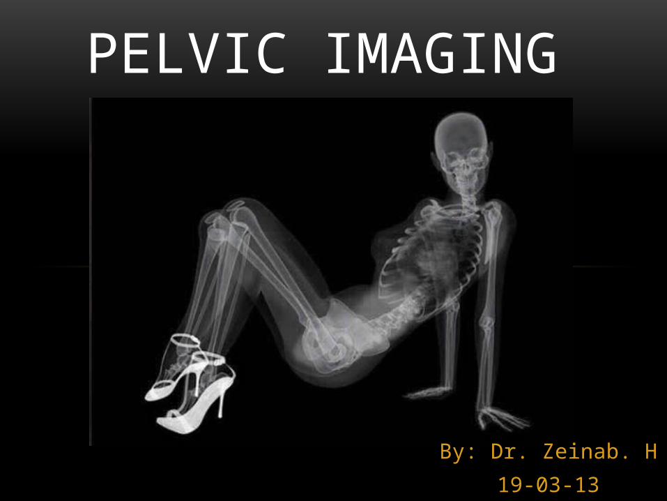
PELVIC IMAGING
By: Dr. Zeinab. H
19-03-13
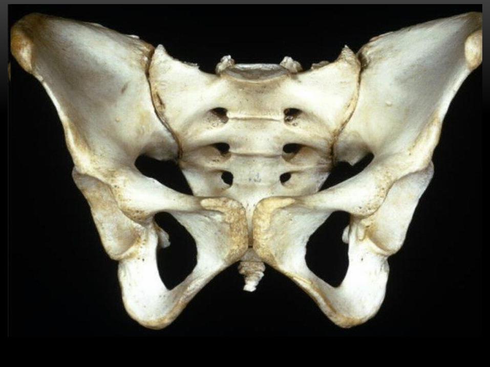
the pelvic skeleton is formed
Posteriorly: by the sacrum and the coccyx laterally and anteriorly: by a pair of hip bones, the lower extremity.
In an adult human being, the pelvic skeleton is thus composed of three large bones, and the coccyx (3–5 bones)
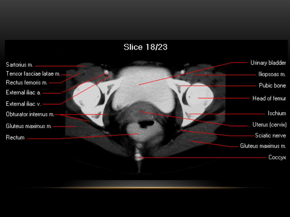
The gap enclosed by the pelvic skeleton, called the pelvic cavity, is the section of the body underneath the abdomen and mainly consists of the reproductive organs (sex organs), urinary bladder and the rectum.
IMAGING STUDIES
• Plain X-ray of pelvis
• Contrast X-ray
i. Retrograde Cysto-urethrography
ii. Hysterosalpingography
• CT scan
• MRI
• Ultrasound
PLAIN X-RAY
• AP pelvis image can display a variety of puzzling features which can be usefully separated into
• normal anatomical features
• normal anatomical variants
• artifacts
• foreign bodies
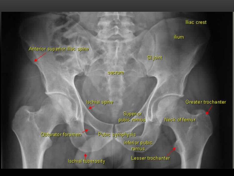
AP VIEW OF PELVIS SHOWING IMPORTANT ANATOMICAL LINES
• The five bones of pelvis are the ilium, ischium, pubis, sacrum, and coccyx.
• Most trauma to the pelvis and hips can be evaluated with an AP view.
• CT of the pelvis is the technique of choice for evaluating complex fracture patterns, degree of displacement and soft tissue injury.
• The femurs should be internally rotated when obtaining an AP pelvis film so that the femoral necks can be appropriately assessed for fractures.
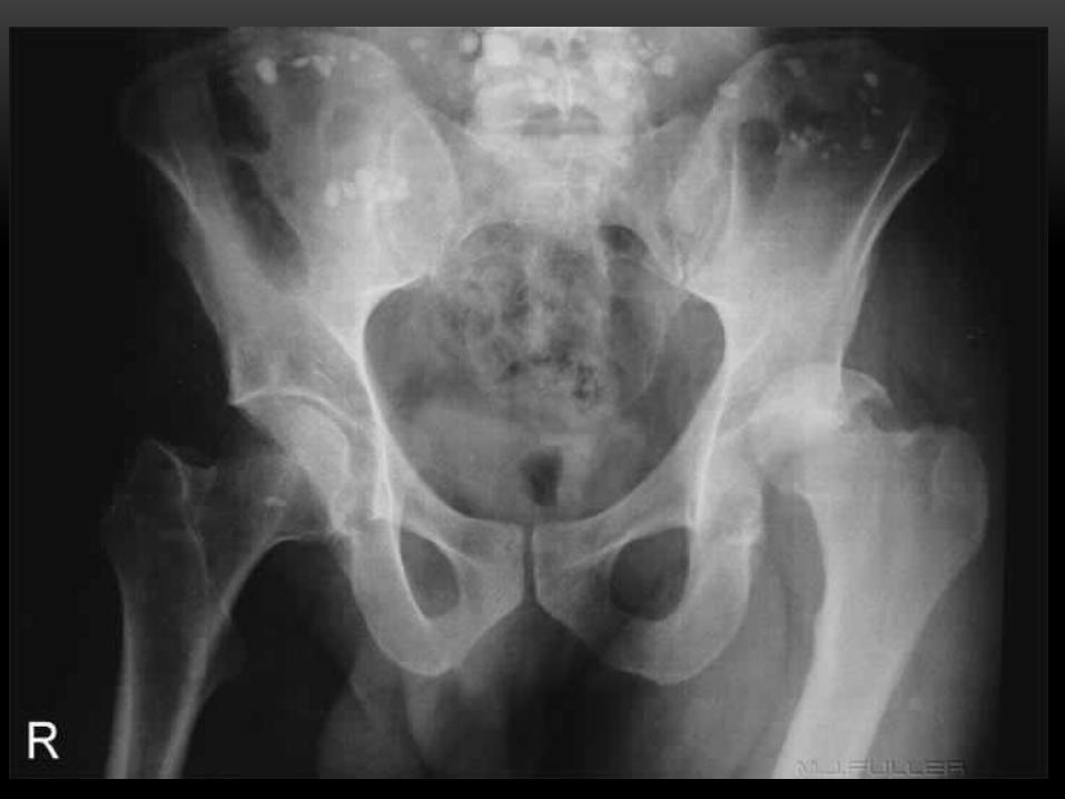
CASE.1
28 years old male was involved in motor vehicle accident, he is complaining of sever pain on the left hip and he is not able to move his left leg. On examination his left leg was adducted and internally rotated.
• Which imaging modality is appropriate for this case?
• What you will see ?
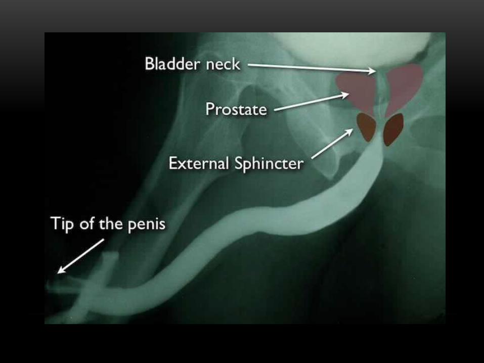
CONTRAST X-RAYi. Retrograde urethrography
• A retrograde urethrogram is a routine radiologic procedure (most typically in males) used to image the integrity of the urethra. Hence a retrograde urethrogram is essential for diagnosis of urethral injury, or urethral stricture.
• The procedure involves the insertion of a Foley catheter into the distal urethra and minimally inflating it. This is followed by instillation of 30mL of water soluble contrast and a plain radiograph is obtained; leakage of the contrast suggests urethral injury (usually secondary to pelvic trauma) and is an indication for surgical intervention.
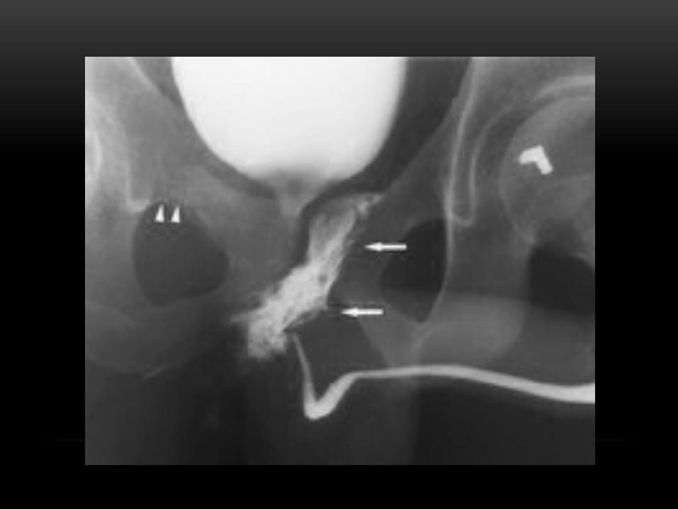
CASE.2
31 years old male presented with history of fall from height. He is complaining of bleeding per urethra, no urine is passed since the accident and associated with pain.
• What is the imaging study of choice for this case?
• Mention the abnormal finding if any.
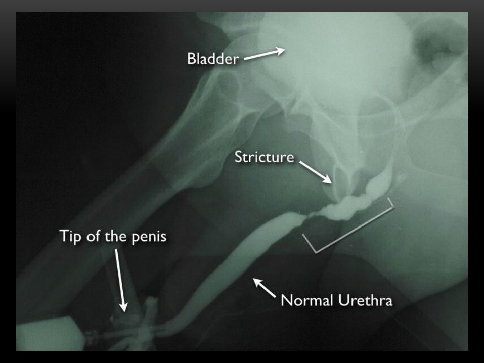
CASE.3
76 years old male complaining of inability to pass urine.
he mention in his PMHx , he was catheterized few times to relieve his urinary retention.
• Imaging of choice?
• What is the abnormality you will find?
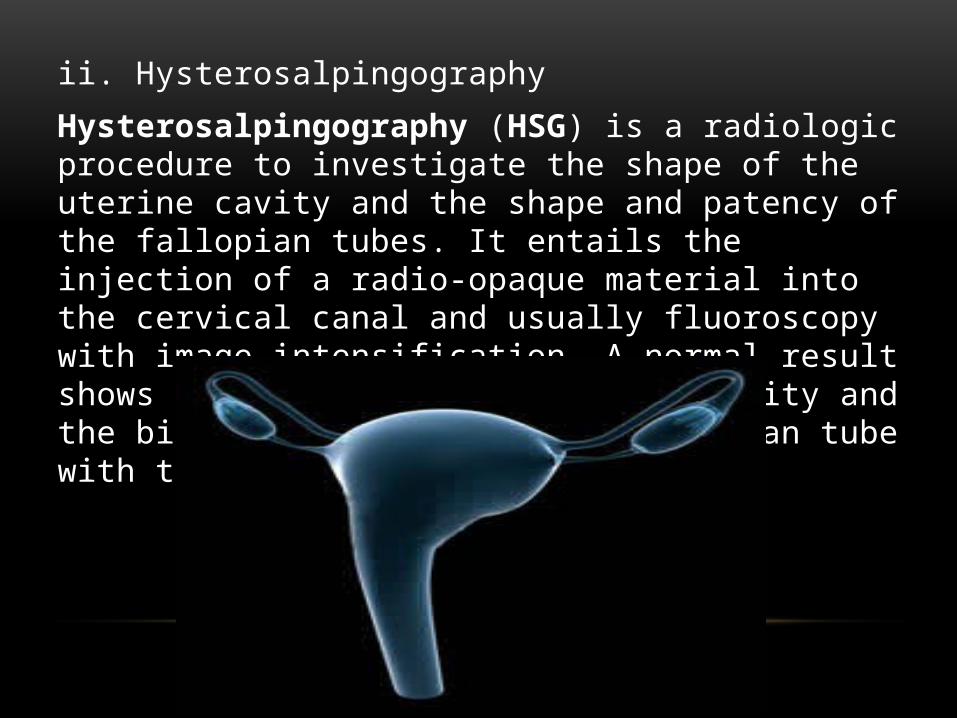
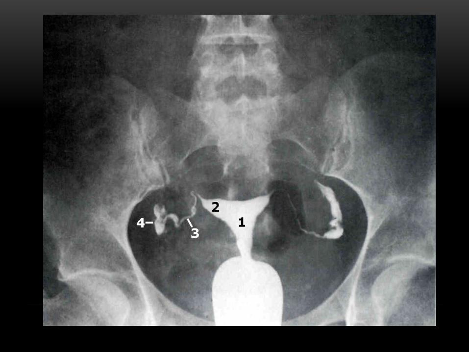
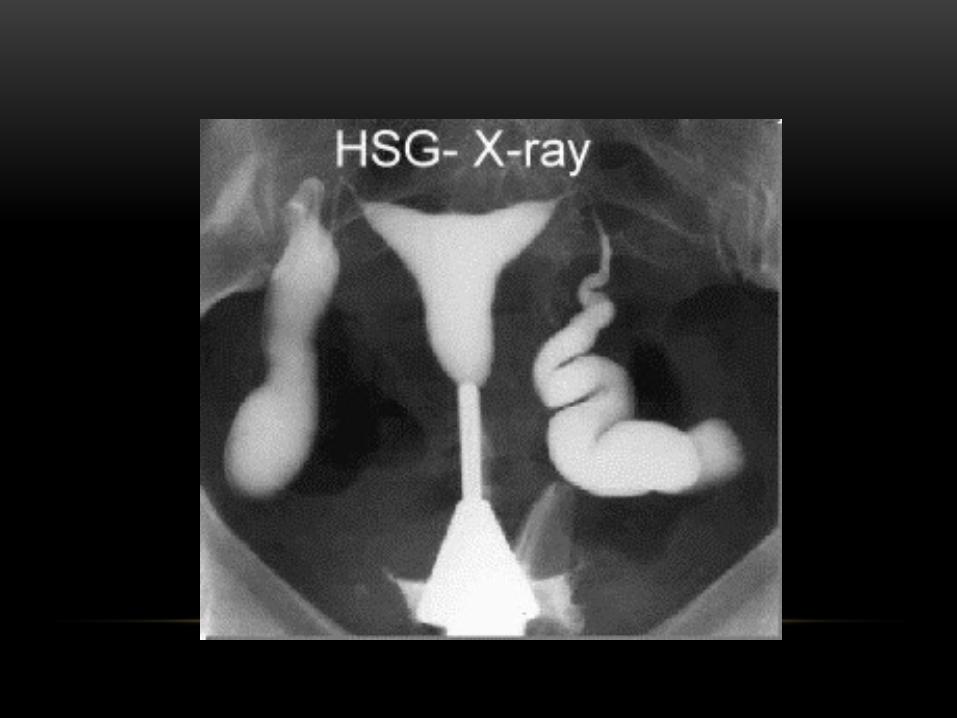
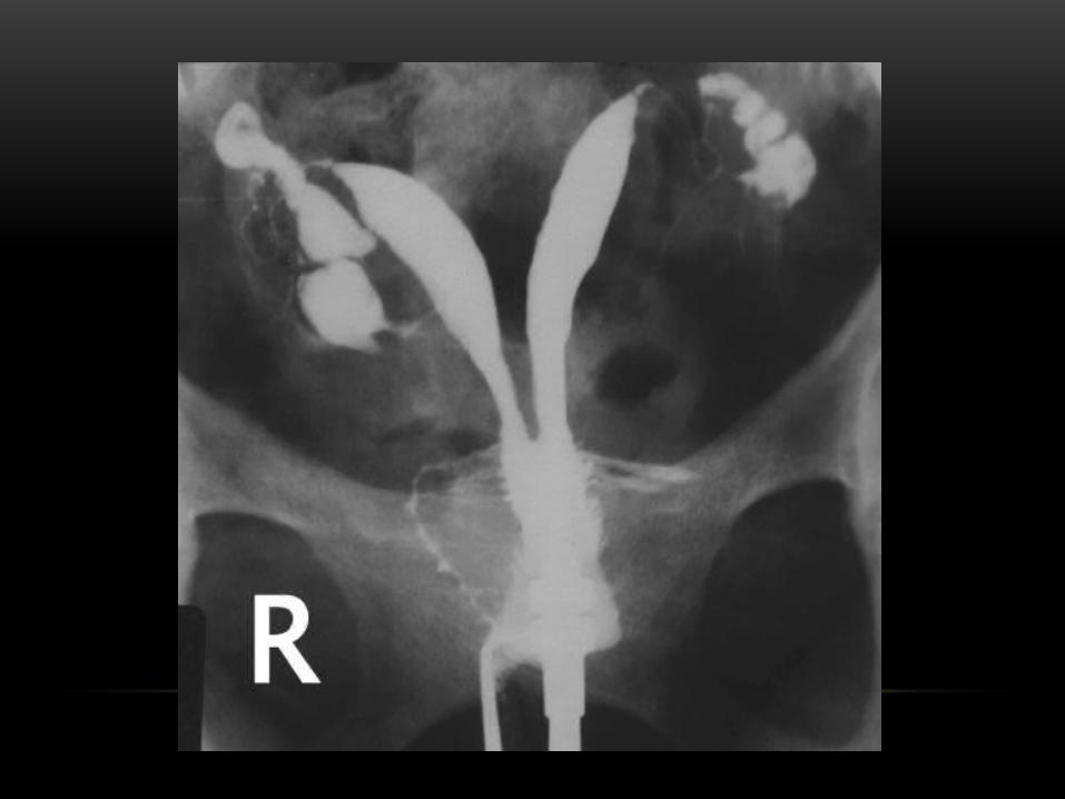
ii. Hysterosalpingography
Hysterosalpingography (HSG) is a radiologic procedure to investigate the shape of the uterine cavity and the shape and patency of the fallopian tubes. It entails the injection of a radio-opaque material into the cervical canal and usually fluoroscopy with image intensification. A normal result shows the filling of the uterine cavity and the bilateral filling of the fallopian tube with the injection material.
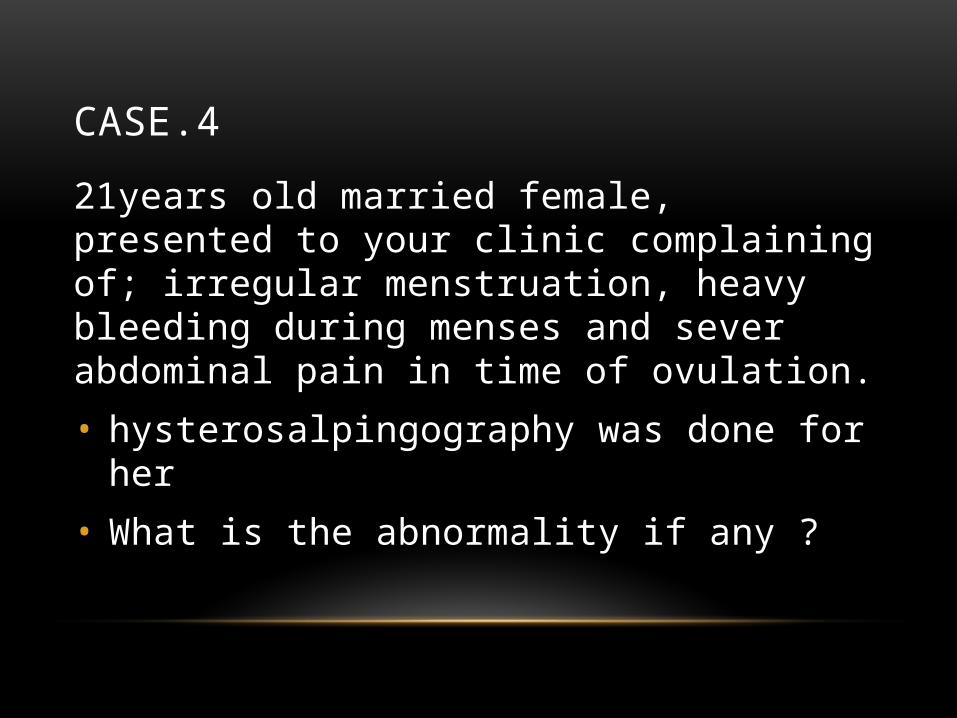
CASE.4
21years old married female, presented to your clinic complaining of; irregular menstruation, heavy bleeding during menses and sever abdominal pain in time of ovulation.
• hysterosalpingography was done for her
• What is the abnormality if any ?
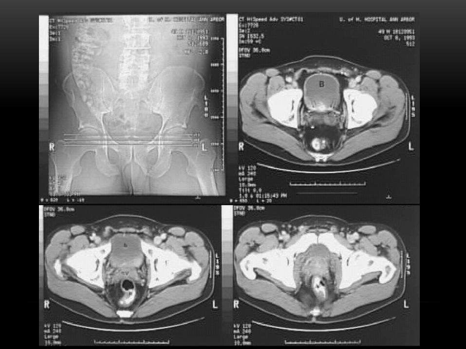
COMPUTED TOMOGRAPHY OF PELVIS
CT has limited application in the evaluation of the pelvis. For the female pelvis in particular, ultrasound and MRI are the imaging modalities of choice. Nevertheless, it may be part of abdominal scanning (e.g., for tumors), and has uses in assessing fractures.
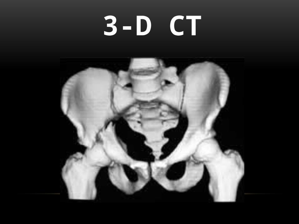
3-D CT
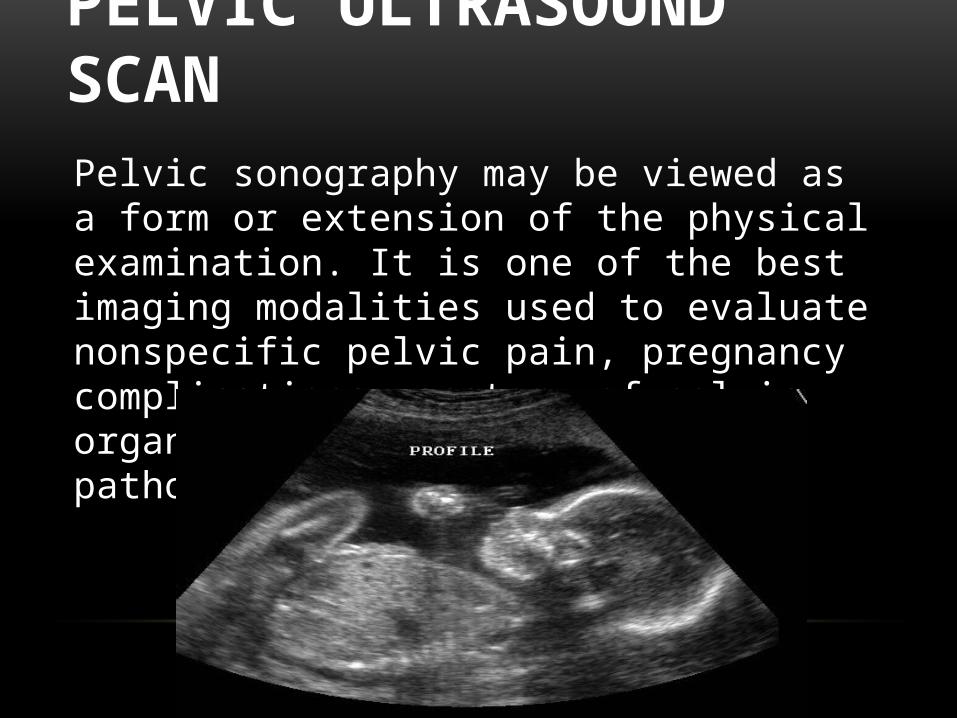
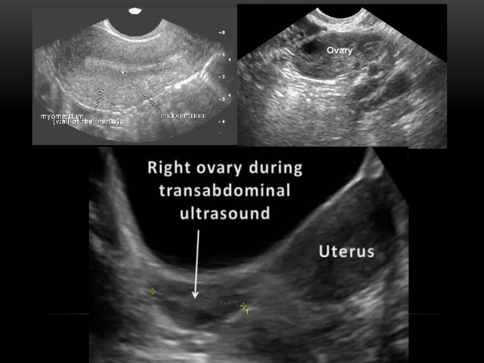
PELVIC ULTRASOUND SCANPelvic sonography may be viewed as a form or extension of the physical examination. It is one of the best imaging modalities used to evaluate nonspecific pelvic pain, pregnancy complications, anatomy of pelvic organs, and various ovarian pathologies
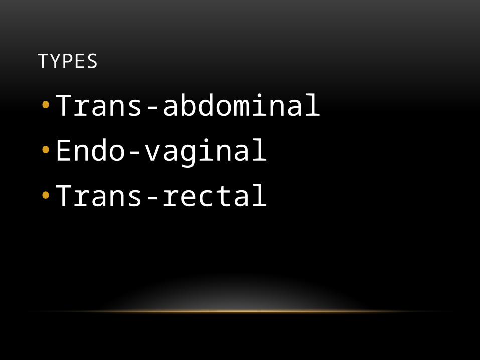
TYPES
• Trans-abdominal• Endo-vaginal• Trans-rectal
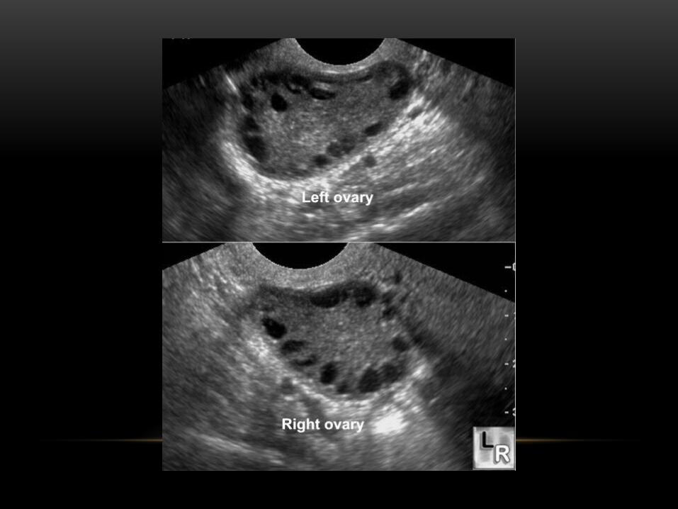
CASE.5
23 years old female complaining of irregular menstruation and excessive facial hair and acne in the past 3 months,
She also noticed weight gain and fatiguness.
• Imaging of modality ?
• Describe your finding.
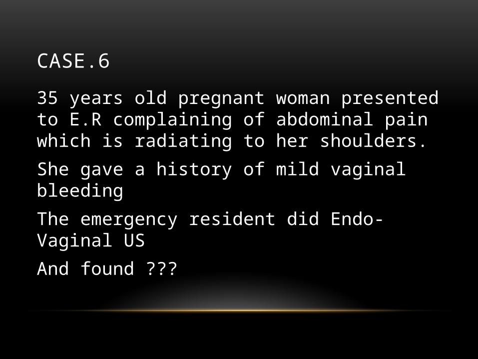
CASE.6
35 years old pregnant woman presented to E.R complaining of abdominal pain which is radiating to her shoulders.
She gave a history of mild vaginal bleeding
The emergency resident did Endo-Vaginal US
And found ???
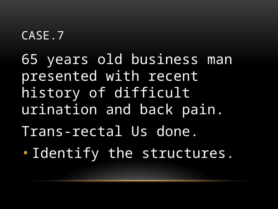
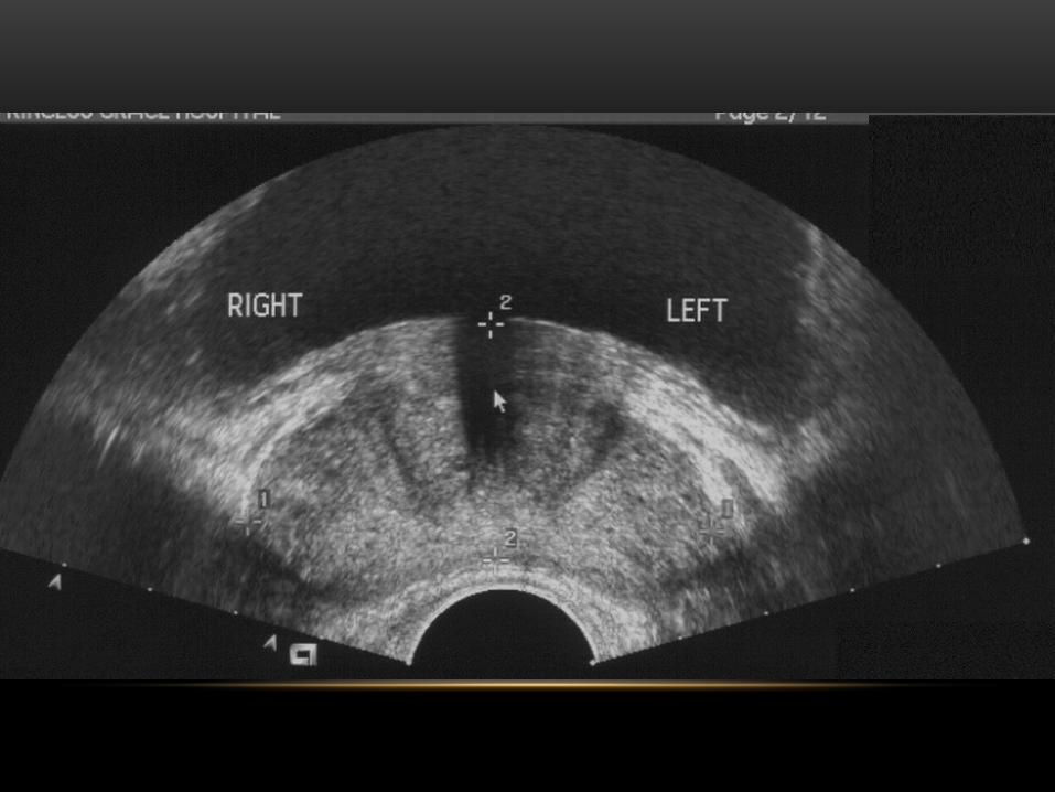
CASE.7
65 years old business man presented with recent history of difficult urination and back pain.
Trans-rectal Us done. • Identify the structures.
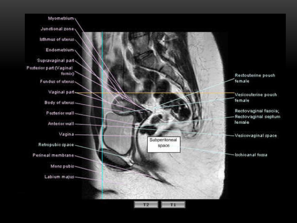
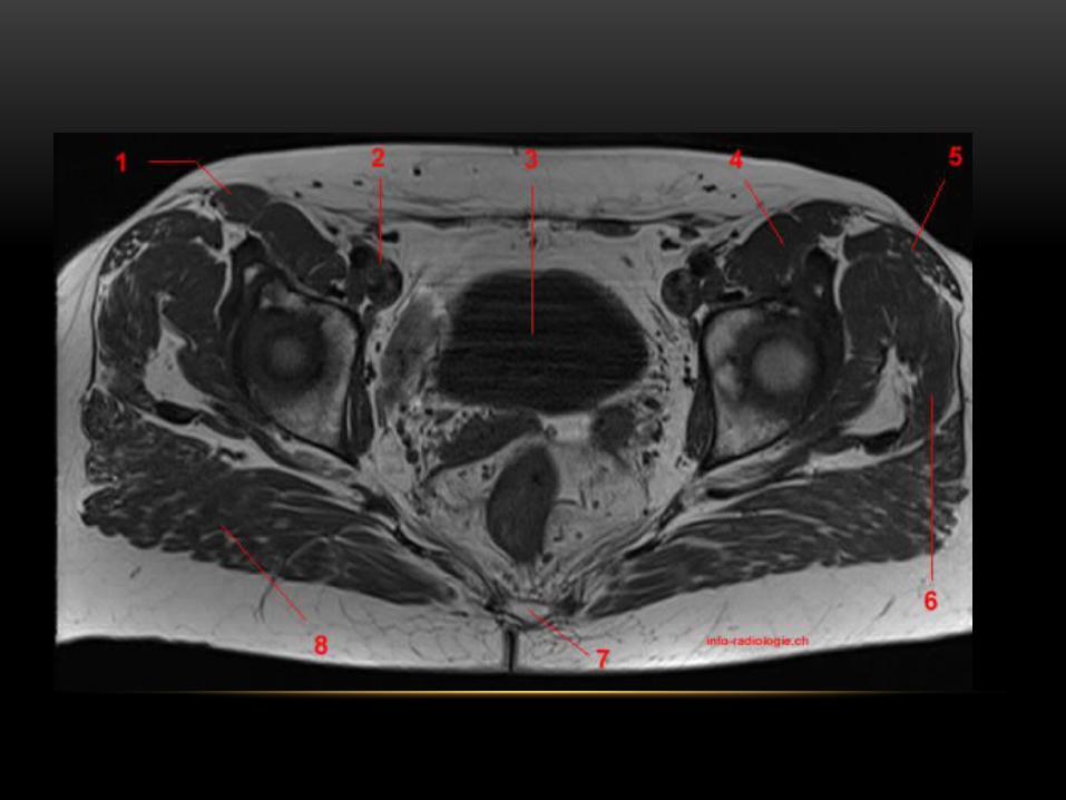
MAGNETIC RESONANCE IMAGING • A pelvis MRI (magnetic resonance imaging) scan is a imaging
test that uses powerful magnets and radio waves to create pictures of the area between the hip bones. This part of the body is called the pelvic area.
• The pelvic area contains the reproductive organs.
• In women, it includes the womb (uterus), cervix, ovaries, and fallopian tubes.
• In men, it includes the prostate gland and testicles.
DONE

Thanks