Embed Size (px)
Citation preview
22nd Annual Northeast Regional Nurse Practitioner Conference – May 6-8, 2015
Pediatric Cardiology for the Primary Care ProviderMary Anne Milbert, RN, MSN, CPNP-PC
D I S C L O S U R E S
• There has been no commercial support or sponsorship for this program.
• The planners and presenters have declared that no conflicts of interest exist.
• The program co-sponsors do not endorse any products in conjunction with any educational activity.

A C C R E D I TAT I O N
Boston College Connell School of Nursing Continuing Education Program is accredited as a provider of continuing nursing education by the American Nurses Association Massachusetts, an accredited approver by the American Nurses Credentialing Center’s Commission on Accreditation.
22nd Annual Northeast Regional Nurse Practitioner Conference – May 6-8, 2015
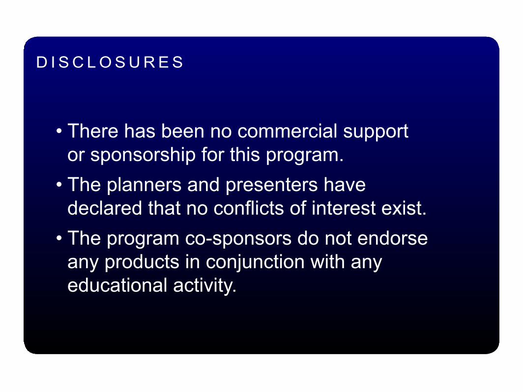
S E S S I O N O B J E C T I V E S
• Name four most common congenital heart defects.
• Identify when to refer for chest pain, syncope and murmur.
Mary Anne Milbert RN, MSN, CPNP-PC Pediatric Nurse Practitioner
Cardiovascular Program Boston Children’s Hospital
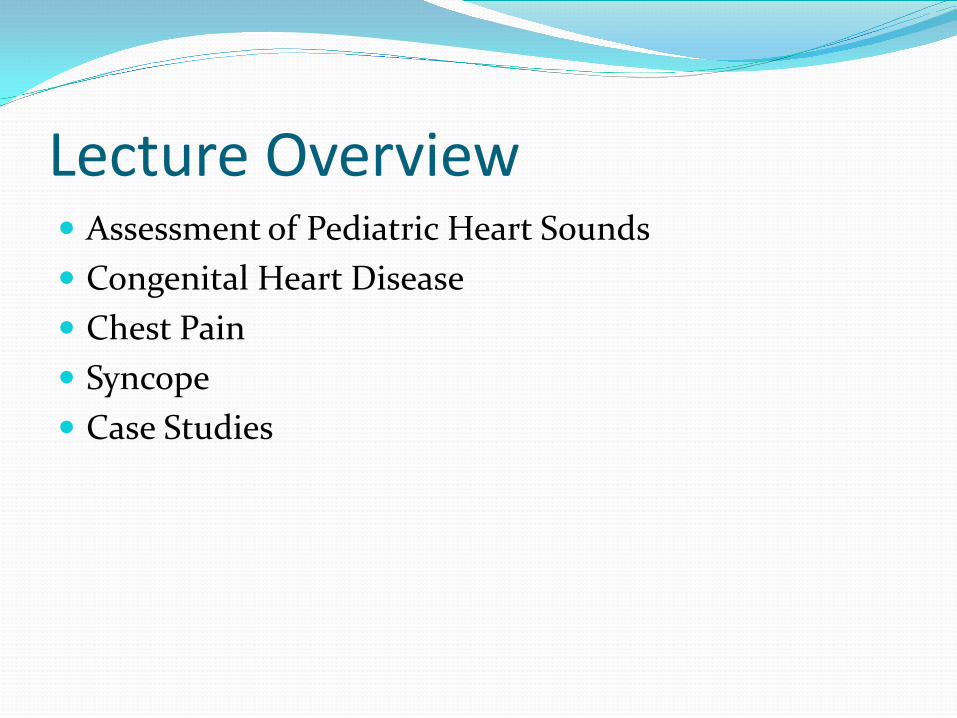
Lecture Overview Assessment of Pediatric Heart Sounds Congenital Heart Disease Chest Pain Syncope Case Studies
Assessment of Pediatric Heart Sounds
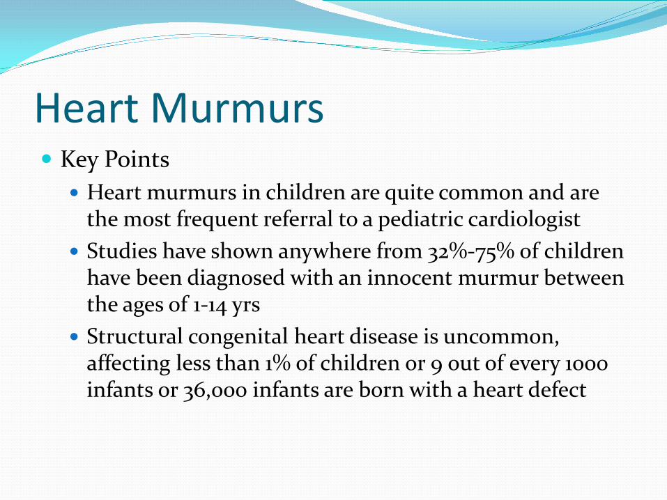
Heart Murmurs Key Points
Heart murmurs in children are quite common and are the most frequent referral to a pediatric cardiologist
Studies have shown anywhere from 32%-75% of children have been diagnosed with an innocent murmur between the ages of 1-14 yrs
Structural congenital heart disease is uncommon, affecting less than 1% of children or 9 out of every 1000 infants or 36,000 infants are born with a heart defect
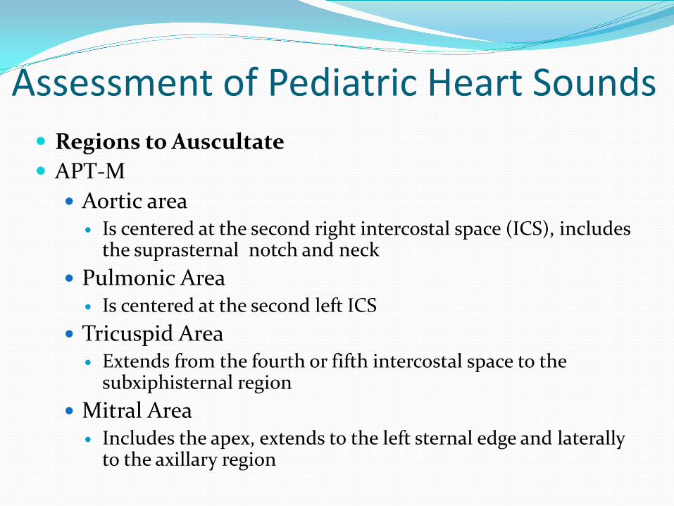
Assessment of Pediatric Heart Sounds Regions to Auscultate APT-M
Aortic area Is centered at the second right intercostal space (ICS), includes
the suprasternal notch and neck Pulmonic Area
Is centered at the second left ICS Tricuspid Area
Extends from the fourth or fifth intercostal space to the subxiphisternal region
Mitral Area Includes the apex, extends to the left sternal edge and laterally
to the axillary region
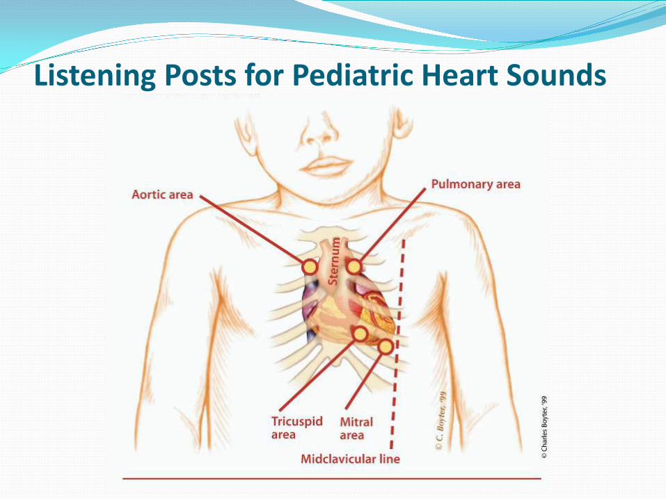
Listening Posts for Pediatric Heart Sounds
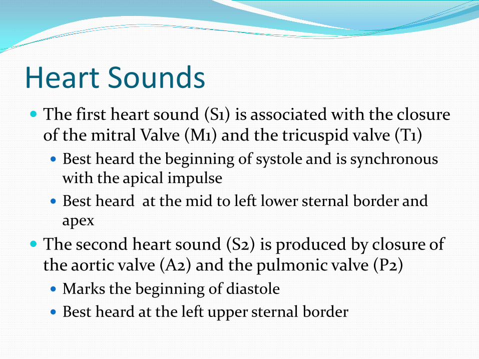
Heart Sounds The first heart sound (S1) is associated with the closure
of the mitral Valve (M1) and the tricuspid valve (T1) Best heard the beginning of systole and is synchronous
with the apical impulse Best heard at the mid to left lower sternal border and
apex The second heart sound (S2) is produced by closure of
the aortic valve (A2) and the pulmonic valve (P2) Marks the beginning of diastole Best heard at the left upper sternal border
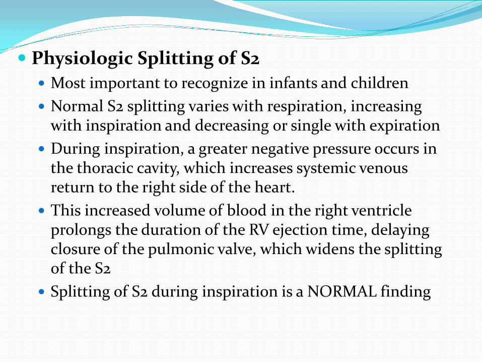
Physiologic Splitting of S2 Most important to recognize in infants and children Normal S2 splitting varies with respiration, increasing
with inspiration and decreasing or single with expiration During inspiration, a greater negative pressure occurs in
the thoracic cavity, which increases systemic venous return to the right side of the heart.
This increased volume of blood in the right ventricle prolongs the duration of the RV ejection time, delaying closure of the pulmonic valve, which widens the splitting of the S2
Splitting of S2 during inspiration is a NORMAL finding
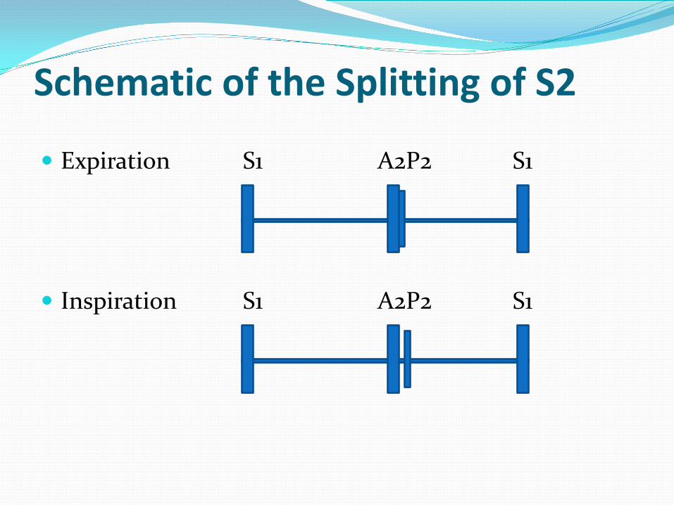
Schematic of the Splitting of S2
Expiration S1 A2P2 S1
Inspiration S1 A2P2 S1
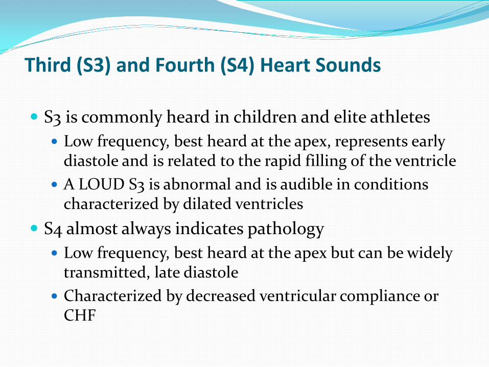
S3 is commonly heard in children and elite athletes Low frequency, best heard at the apex, represents early
diastole and is related to the rapid filling of the ventricle A LOUD S3 is abnormal and is audible in conditions
characterized by dilated ventricles S4 almost always indicates pathology
Low frequency, best heard at the apex but can be widely transmitted, late diastole
Characterized by decreased ventricular compliance or CHF
Third (S3) and Fourth (S4) Heart Sounds
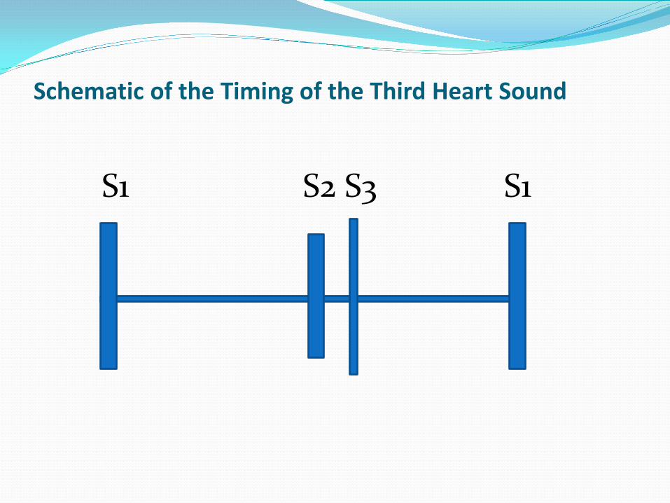
Schematic of the Timing of the Third Heart Sound
S1 S2 S3 S1
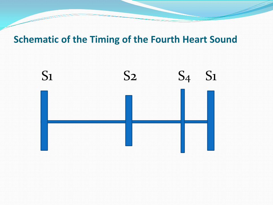
Schematic of the Timing of the Fourth Heart Sound
S1 S2 S4 S1
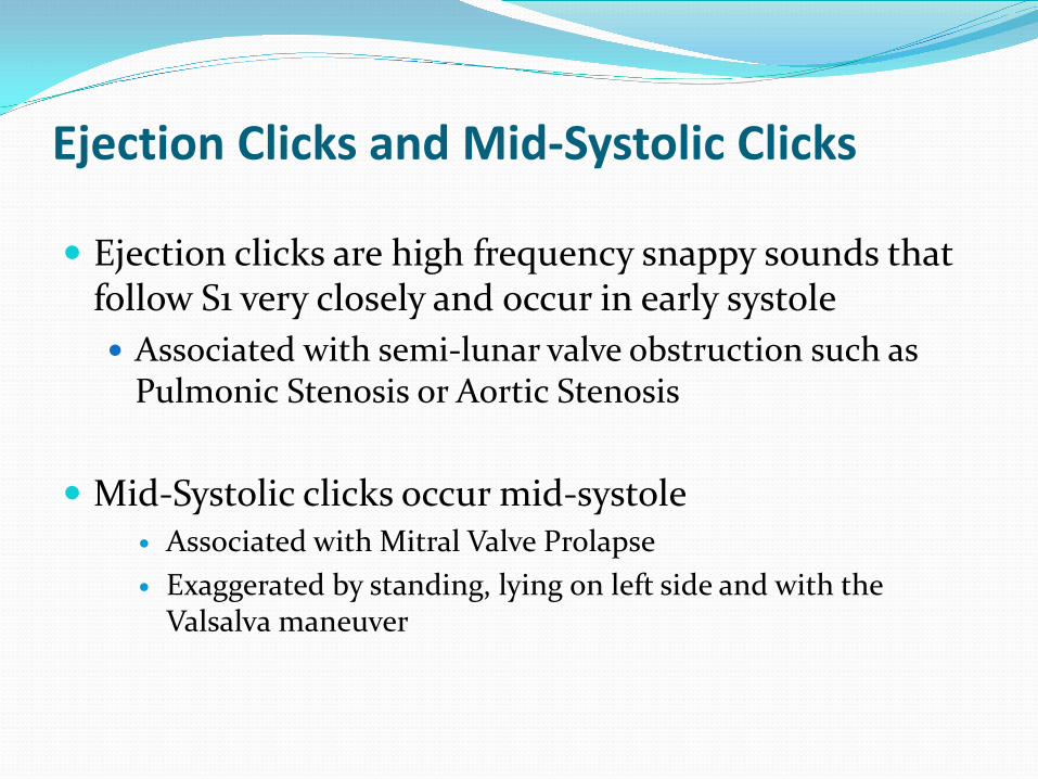
Ejection Clicks and Mid-Systolic Clicks
Ejection clicks are high frequency snappy sounds that follow S1 very closely and occur in early systole Associated with semi-lunar valve obstruction such as
Pulmonic Stenosis or Aortic Stenosis
Mid-Systolic clicks occur mid-systole Associated with Mitral Valve Prolapse Exaggerated by standing, lying on left side and with the
Valsalva maneuver
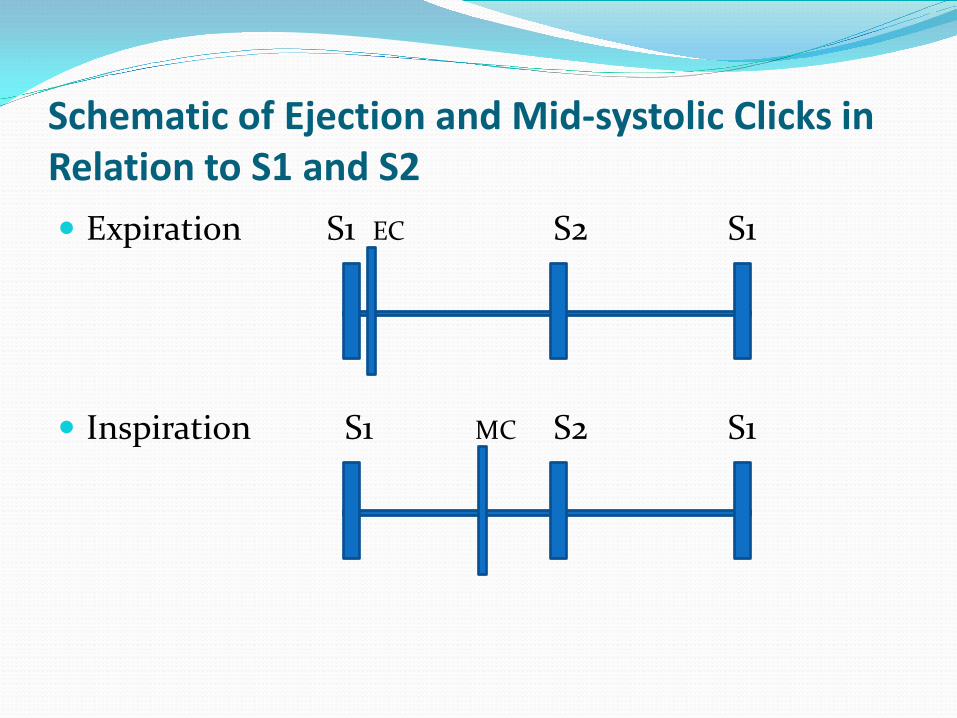
Schematic of Ejection and Mid-systolic Clicks in Relation to S1 and S2 Expiration S1 EC S2 S1
Inspiration S1 MC S2 S1
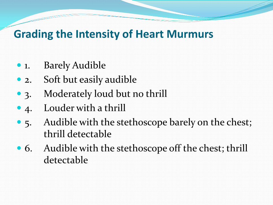
Grading the Intensity of Heart Murmurs
1. Barely Audible 2. Soft but easily audible 3. Moderately loud but no thrill 4. Louder with a thrill 5. Audible with the stethoscope barely on the chest;
thrill detectable 6. Audible with the stethoscope off the chest; thrill
detectable
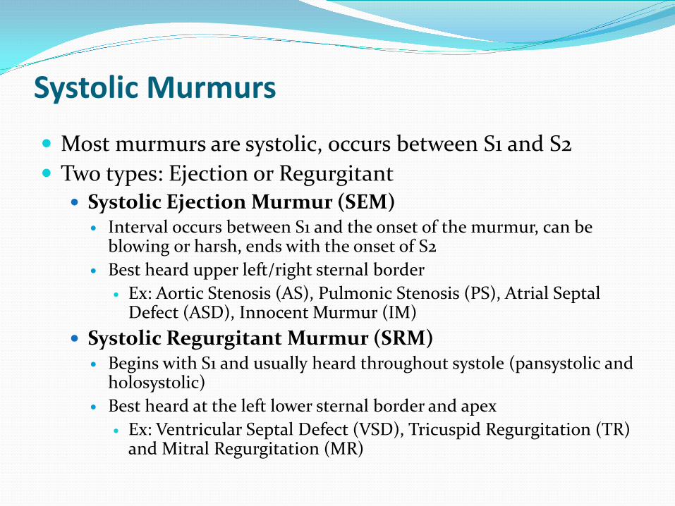
Systolic Murmurs Most murmurs are systolic, occurs between S1 and S2 Two types: Ejection or Regurgitant
Systolic Ejection Murmur (SEM) Interval occurs between S1 and the onset of the murmur, can be
blowing or harsh, ends with the onset of S2 Best heard upper left/right sternal border
Ex: Aortic Stenosis (AS), Pulmonic Stenosis (PS), Atrial Septal Defect (ASD), Innocent Murmur (IM)
Systolic Regurgitant Murmur (SRM) Begins with S1 and usually heard throughout systole (pansystolic and
holosystolic) Best heard at the left lower sternal border and apex
Ex: Ventricular Septal Defect (VSD), Tricuspid Regurgitation (TR) and Mitral Regurgitation (MR)
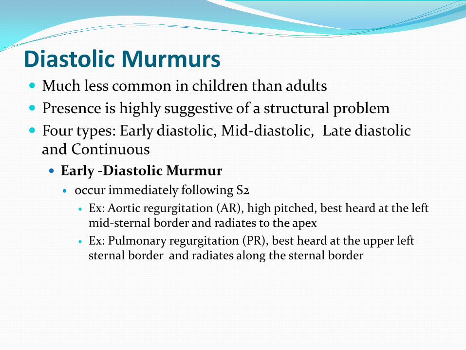
Diastolic Murmurs Much less common in children than adults Presence is highly suggestive of a structural problem Four types: Early diastolic, Mid-diastolic, Late diastolic
and Continuous Early -Diastolic Murmur
occur immediately following S2 Ex: Aortic regurgitation (AR), high pitched, best heard at the left
mid-sternal border and radiates to the apex Ex: Pulmonary regurgitation (PR), best heard at the upper left
sternal border and radiates along the sternal border
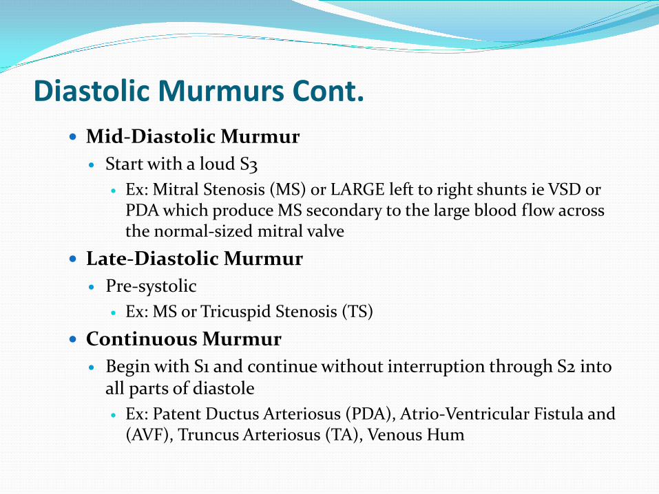
Diastolic Murmurs Cont. Mid-Diastolic Murmur
Start with a loud S3 Ex: Mitral Stenosis (MS) or LARGE left to right shunts ie VSD or
PDA which produce MS secondary to the large blood flow across the normal-sized mitral valve
Late-Diastolic Murmur Pre-systolic
Ex: MS or Tricuspid Stenosis (TS)
Continuous Murmur Begin with S1 and continue without interruption through S2 into
all parts of diastole Ex: Patent Ductus Arteriosus (PDA), Atrio-Ventricular Fistula and
(AVF), Truncus Arteriosus (TA), Venous Hum
Congenital Heart Disease
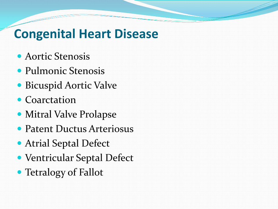
Congenital Heart Disease Aortic Stenosis Pulmonic Stenosis Bicuspid Aortic Valve Coarctation Mitral Valve Prolapse Patent Ductus Arteriosus Atrial Septal Defect Ventricular Septal Defect Tetralogy of Fallot
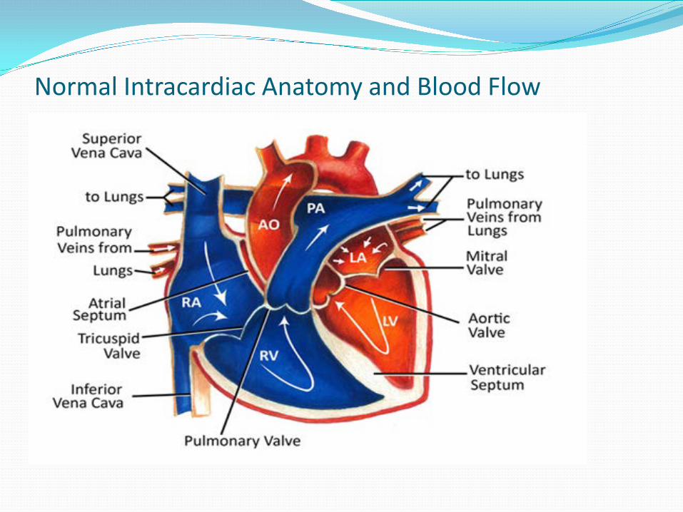
Normal Intracardiac Anatomy and Blood Flow
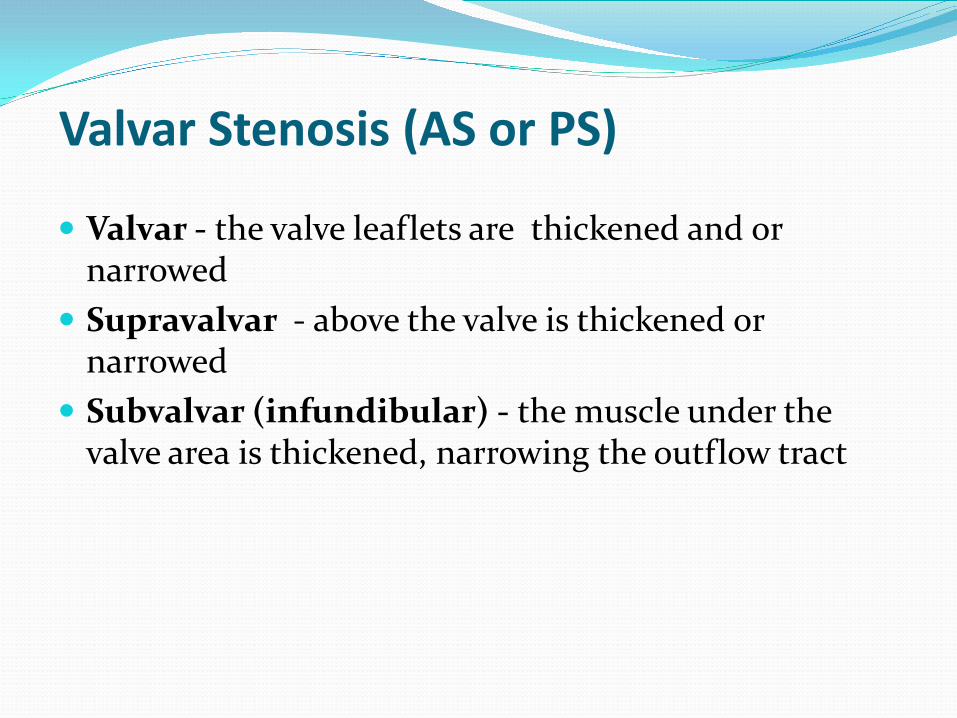
Valvar Stenosis (AS or PS)
Valvar - the valve leaflets are thickened and or narrowed
Supravalvar - above the valve is thickened or narrowed
Subvalvar (infundibular) - the muscle under the valve area is thickened, narrowing the outflow tract
Aortic Stenosis/Bicuspid Aortic Valve Overall incidence is 0.2-0.5 per 1000 children. Most cases of
aortic valve stenosis in young people are due to a bicuspid aortic valve, the aortic valve has only 2 leaflets instead of the normal 3
Familial incidence with left ventricular outflow obstruction (LVOO) 3-5%, recommend screening for first degree relatives
The murmur of aortic stenosis is typically a mid-systolic ejection murmur, heard best over the “aortic area” or right second intercostal space, with radiation into the right neck
Grading of AS is mild, moderate and severe and is dependent on the degree of narrowing
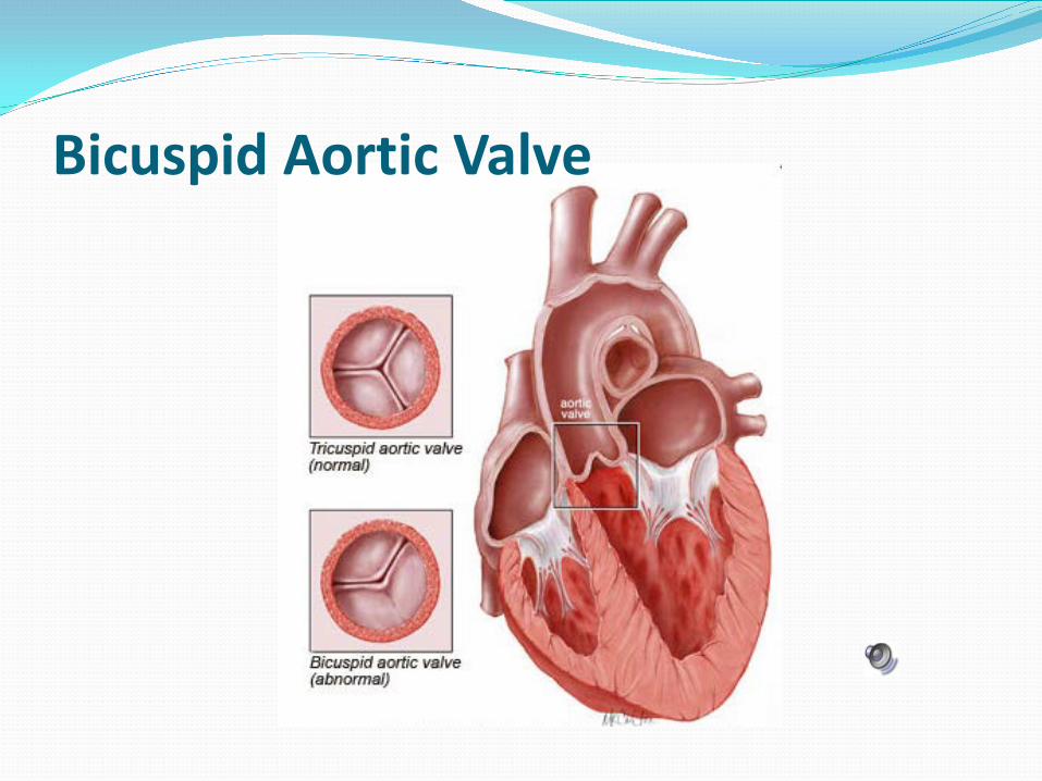
Bicuspid Aortic Valve
Treatment Options for AS Wide spectrum of treatment options Most children with mild aortic valve stenosis require
bi-annual follow-up (with or without aortic regurgitation), the degree of stenosis often gradually worsens over time
Severe or moderate to severe aortic valve stenosis require intervention to prevent long term damage to the heart, initial intervention is a balloon valvuloplasty through a cardiac catheterization
Valvotomy is surgical release of scar tissue within the aortic valve leaflets that is preventing the valve leaflets from opening properly
Pulmonic Stenosis Overall incidence is about 8-10% of all cases of
congenital heart defects, one of the most common defects
Thickened leaflets with commisural fusion, post-stenotic dilatation common
Classic systolic ejection murmur in pulmonic area, with an ejection click present, diastolic murmur is related to the “leaking” PR,
Connective tissue disorders i.e. Marfan’s Syndrome, Ehlers-Danolos have a high incidence of MVP, more than 50%
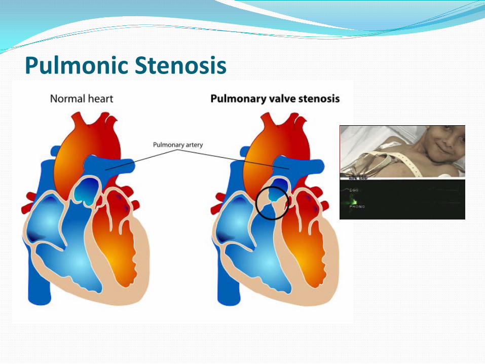
Pulmonic Stenosis
Treatment for PS Mild pulmonary stenosis often does not require treatment Moderate or severe stenosis is treated with repair of the
pulmonic valve, balloon valvuloplasty most common Valvotomy is surgical release of scar tissue within the
pulmonary valve leaflets that is preventing the valve leaflets from opening properly
Patch enlargement: Subvalvar PS, an incision is made into the right ventricle and
a patch is sewn into the cut edges of the right ventricle to enlarge the area below the pulmonary valve where the narrowing was
Supravalvar PS, the narrowing is in the artery just beyond the pulmonary valve, patches are sewn into this artery to enlarge its diameter and relieve the narrowing
Coarctation of the Aorta Overall incidence is 5 in 10,000 births or 5% of all
congenital heart defects Cause is unknown, certain chromosomal abnormalities
such as Turner's syndrome high incidence, more than 50% have a Bicuspid Aortic Valve
The murmur of a Co-Arc is typically a systolic ejection murmur, heard best over the upper sternal border with intrascapular murmur, high blood pressure in upper extremities and decreased femoral pulses
elastic tissue from the ductus arteriosus may encircle the aorta and cause a "lasso effect" on it as the ductus closes, resulting in a coarctation of the aorta
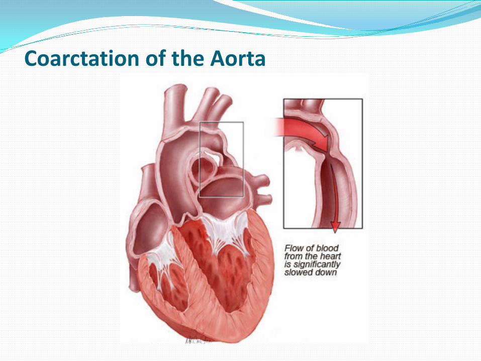
Coarctation of the Aorta
Treatment options for Co-Arc In a critically ill newborn, prostaglandin (PGE-1), is used to
open the ductus arteriosus allowing blood to flow to areas beyond the coarctation
severity and length of the coarctation segment will dictate the most appropriate technique, surgical vs balloon dilation
recurrence of coarctation at the area of the repair is possible, even years following treatment, re-stenosis is highest among newborns, and decreases in older children
the majority of cases can be managed with cardiac catheterization and balloon dilation
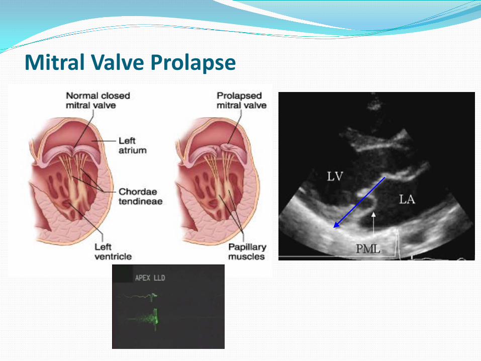
Mitral Valve Prolapse Mitral valve prolapse is the most prevalent cardiac valve
disorder Overall incidence is about 3-5% of pediatric patients likely
have clinically significant mitral valve prolapse, recent article in CA however stated 0.6%
The prolapse occurs from various underlying causes affecting one or more portions of the mitral valve leaflets, chordae tendineae, papillary muscle, and/or valve annulus
Classic mid-systolic click heard at the apex, regurgitant systolic murmur is related to the “leaking” MR,
Connective tissue disorders i.e. Marfan’s Syndrome, Ehlers-Danolos have a high incidence of MVP, more than 50%
Mitral Valve Prolapse
Treatment for MVP/MR Asymptomatic patients require no specific treatment
and they should be reassured of their excellent prognosis
MVP patients without mitral regurgitation should be evaluated every 3 to 5 years
Mild-mild+ should be evaluated every 2 years Moderate to severe mitral regurgitation or high-risk
features should be reviewed with an echocardiogram yearly or more often
When surgery is required, mitral valve repair is usually feasible, repair is characterized by low mortality and long-lasting durability; the 10-year reoperation-free survival rate ranges between 93% and 96%
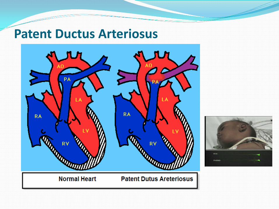
Patent Ductus Arteriosus
Abnormal blood flow between the aorta and pulmonary artery, typically closes in a couple of days after birth
Occurs in about 8 out of every 1,000 premature babies, compared with 2 out of every 1,000 full-term babies, more girls than boys
Infants with genetic disorders, such as Down syndrome, and whose mothers had rubella during pregnancy are at higher risk for PDA
Classic continuous murmur, machinery sounding, in premature infants, a heart murmur may not be heard
Patent Ductus Arteriosus
Treatment for PDA If treatment is needed, medications such
as indomethacin or a special form of ibuprofen are often the first choice, works well with little side effects, the earlier treatment is given, the more likely it is to succeed
Transcatheter device closure; few different types small metal coil or other blocking device
Surgery may be needed if the catheter procedure does not work or it cannot be used, surgery involves making a small cut between the ribs to repair the PDA (Trans-thoracic)
If other heart problems or defects, keeping the ductus arteriosus open may be lifesaving. Medicine may be used to stop it from closing
SCAMP Criteria for PDA Isolated PDA on any ECHO If unexplained poor wgt gain then cath for HD/PDA
closure ECHO reveals: LVEDV z score >2.5 or PDA >2mm (<2
y/o) OR > 3mm (>2 y/o) or RVp > ½ systemic Yes No LVEDV z score > 1.8 Yes No RTC < 2 y/o at 3 yrs of age 2-4 y/o in 18 mons >4 y/o in 3 yrs, then 5 yrs then exit
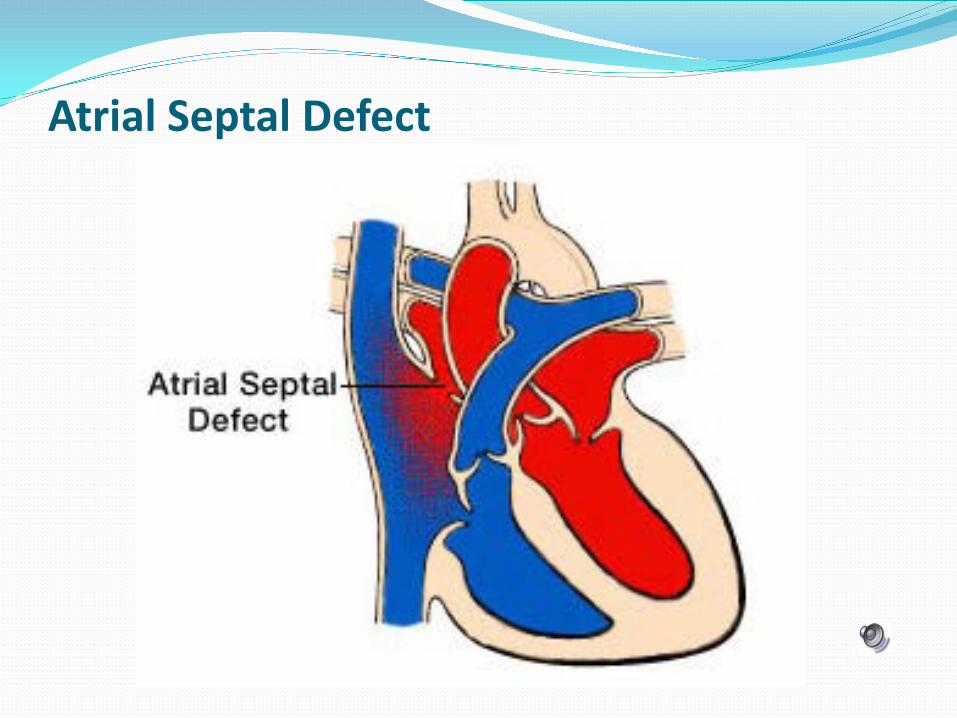
Atrial Septal Defect Overall incidence is 2-3 per 1000 children, second
most common congenital heart defect Cause is unknown, certain genetic disorders have a
high incidence ex. Down syndrome, trisomy 13, and trisomy 18
The murmur of an ASD is typically a systolic ejection murmur, heard best over the upper sternal border with radiation, fixed or widely splitS2
Size of defect determines treatment options small, moderate and large
Atrial Septal Defect
Treatment options for ASD Wide spectrum of treatment options, small no treatment
generally close spontaneously, typically surgery vs cath closure between 2-5 yrs of age
Surgical closure is quite rare, done for large defects or defects with not enough septum to attach device
Device closure if small to moderate, new the last 10-15 years, cath procedure which is minimally invasive Advantages: less discomfort and shorter stay Disadvantages: lack of long term follow-up on the safety and
efficacy of device closure compared with surgery ie: Amplatzer device recently issues with erosion of walls
Ventricular Septal Defects A VSD is the most common type of congenital heart defect
5-7 per 1,000 infants born have a VSD Two types: Muscular and Membranous
Muscular: small-medium sized defects rarely cause problems, close spontaneously up to 70%, never get bigger only get smaller
Membranous: same as above, close spontaneously only 30-40% of the time
Large Defects: cause CHF, generally need medications, Lasix, Aldactone and Digoxin and increased caloric feeds, defects will become smaller over time, will electively close at 4-6 mons if not controlled with meds
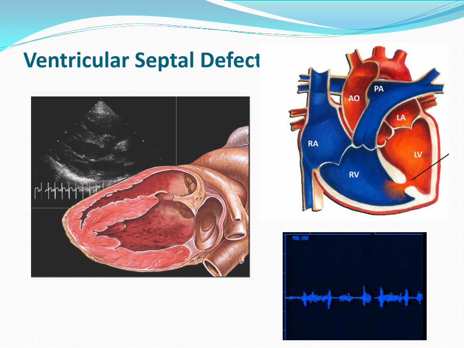
Ventricular Septal Defect
Treatment for VSD Surgical closure of isolated ventricular septal defects is
uncomplicated in 99 percent or more of cases Some ventricular septal defects may be closed using an
FDA approved closure device Since the 2007 SBE recommendations at BCH:
Small hemodynamically insignificant muscular defects are D/C at 3 y/o
Small membranous defects are followed every 2-3 yrs Can develop LVOT obstruction, Sub Ao membrane Increase in RV/LV muscle bundle
Tetralogy of Fallot Clinical picture of this defect is wide, from asymptomatic forms
to extreme forms in which the anatomical and functional communication between right ventricle and pulmonary artery is absent (pulmonary atresia with Tetralogy of Fallot), with severe clinical expression
Pulmonary artery stenosis is constantly present and represents the central element of Tetralogy of Fallot
It may be a pulmonary valve stenosis in the right ventricular outflow tract or a hypoplasia of the pulmonary artery trunk
May be associated with chromosomal abnormalities, such as 22q11 deletion syndrome otherwise known as DiGeorge Syndrome
Cyanotic TOF
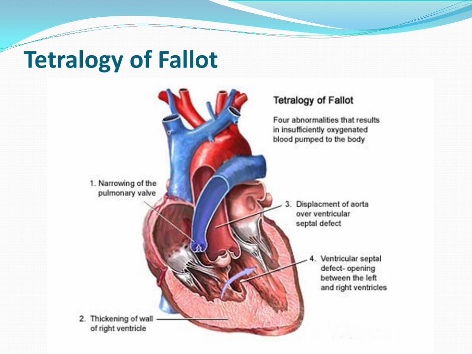
Tetralogy of Fallot
Treatment for TOF Clinical picture of this defect is wide, acyanotic infants to
cyanotic Depending on O2 levels determines timing of surgery, if
critically low surgical repair in the neonatal period Infants with normal oxygen levels or only mild cyanosis are
usually able to go home in the first week of life and elective surgery at 6 mons of life
Closure of the ventricular septal defect with a synthetic Dacron patch so that the blood can flow normally from the left ventricle to the aorta
The narrowing of the pulmonary valve and right ventricular outflow tract is then augmented (enlarged) by a combination of cutting away (resecting) obstructive muscle tissue in the right ventricle and by enlarging the outflow pathway with a patch
SBE Guidelines New guidelines in 2007 the American Heart
Association simplified its recommendations Dental procedure prophylaxis:
Artificial valve or a valve repaired with artificial material History of endocarditis Heart transplant with abnormal heart valve function Congenital heart defects including:
Cyanotic heart disease, not fully repaired, has a conduit or shunt
S/P repair with artificial material or a device for 6 mons Residual defects, leaks or abnormal flow at/or adjacent to a
prosthetic patch or device
Chest Pain
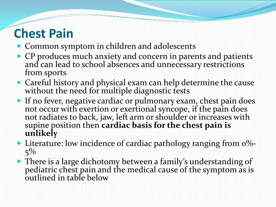
Chest Pain Common symptom in children and adolescents CP produces much anxiety and concern in parents and patients
and can lead to school absences and unnecessary restrictions from sports
Careful history and physical exam can help determine the cause without the need for multiple diagnostic tests
If no fever, negative cardiac or pulmonary exam, chest pain does not occur with exertion or exertional syncope, if the pain does not radiates to back, jaw, left arm or shoulder or increases with supine position then cardiac basis for the chest pain is unlikely
Literature: low incidence of cardiac pathology ranging from 0%-5%
There is a large dichotomy between a family’s understanding of pediatric chest pain and the medical cause of the symptom as is outlined in table below
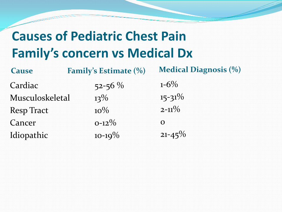
Causes of Pediatric Chest Pain Family’s concern vs Medical Dx Cause Family’s Estimate (%) Medical Diagnosis (%)
Cardiac 52-56 % Musculoskeletal 13% Resp Tract 10% Cancer 0-12% Idiopathic 10-19%
1-6% 15-31% 2-11% 0 21-45%
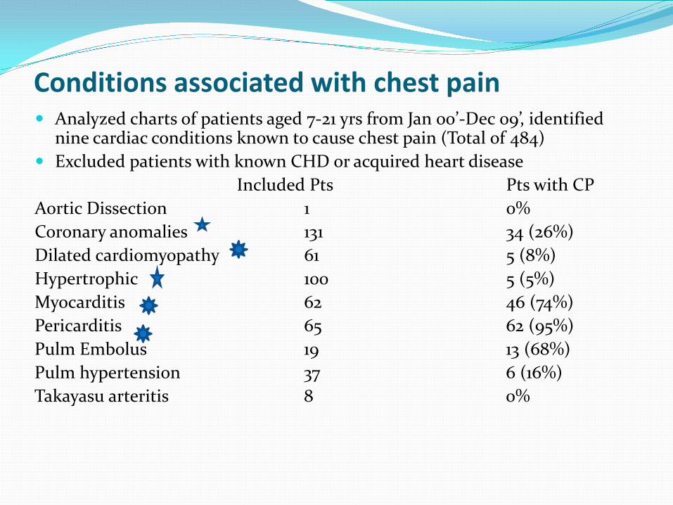
Conditions associated with chest pain Analyzed charts of patients aged 7-21 yrs from Jan 00’-Dec 09’, identified
nine cardiac conditions known to cause chest pain (Total of 484) Excluded patients with known CHD or acquired heart disease Included Pts Pts with CP Aortic Dissection 1 0% Coronary anomalies 131 34 (26%) Dilated cardiomyopathy 61 5 (8%) Hypertrophic 100 5 (5%) Myocarditis 62 46 (74%) Pericarditis 65 62 (95%) Pulm Embolus 19 13 (68%) Pulm hypertension 37 6 (16%) Takayasu arteritis 8 0%
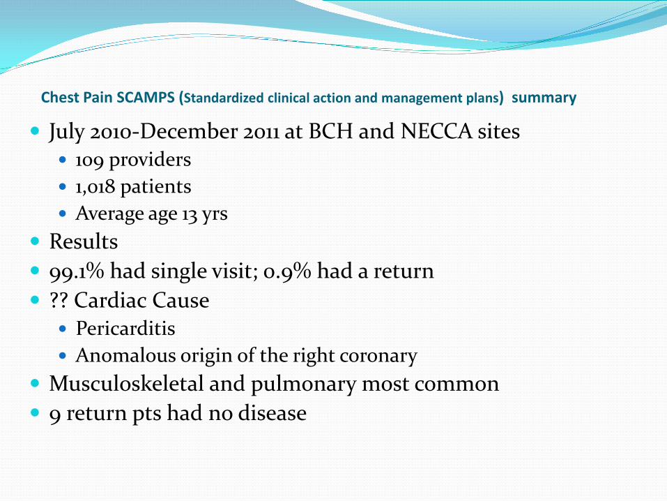
Chest Pain SCAMPS (Standardized clinical action and management plans) summary
July 2010-December 2011 at BCH and NECCA sites 109 providers 1,018 patients Average age 13 yrs
Results 99.1% had single visit; 0.9% had a return ?? Cardiac Cause
Pericarditis Anomalous origin of the right coronary
Musculoskeletal and pulmonary most common 9 return pts had no disease
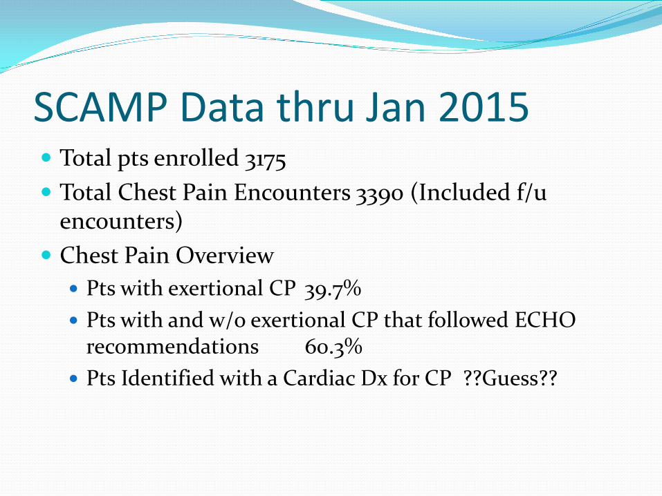
SCAMP Data thru Jan 2015 Total pts enrolled 3175 Total Chest Pain Encounters 3390 (Included f/u
encounters) Chest Pain Overview
Pts with exertional CP 39.7% Pts with and w/o exertional CP that followed ECHO
recommendations 60.3% Pts Identified with a Cardiac Dx for CP ??Guess??
Syncope
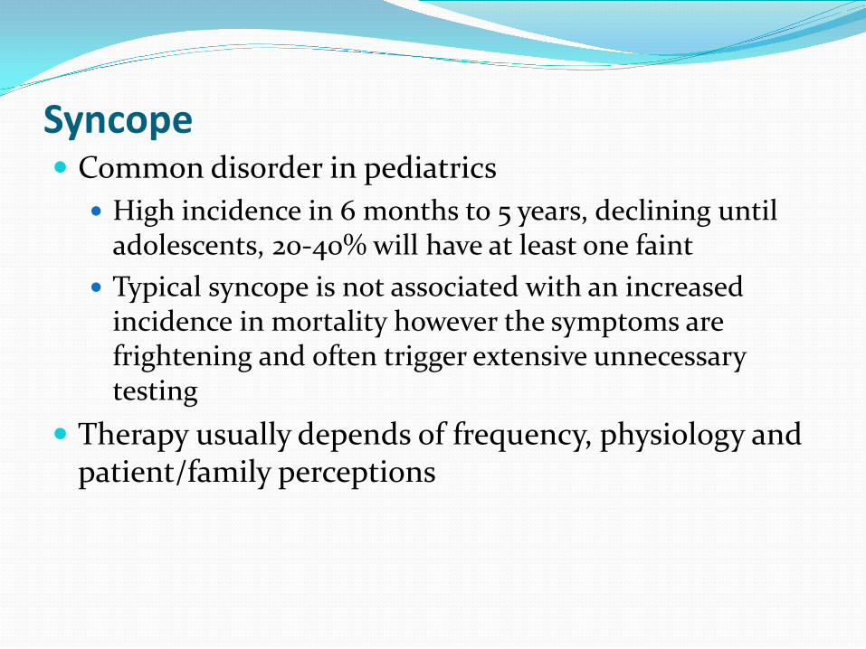
Syncope Common disorder in pediatrics
High incidence in 6 months to 5 years, declining until adolescents, 20-40% will have at least one faint
Typical syncope is not associated with an increased incidence in mortality however the symptoms are frightening and often trigger extensive unnecessary testing
Therapy usually depends of frequency, physiology and patient/family perceptions
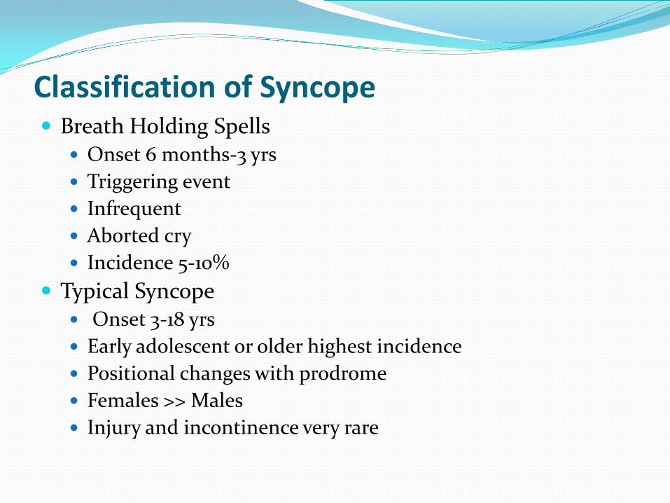
Classification of Syncope Breath Holding Spells
Onset 6 months-3 yrs Triggering event Infrequent Aborted cry Incidence 5-10%
Typical Syncope Onset 3-18 yrs Early adolescent or older highest incidence Positional changes with prodrome Females >> Males Injury and incontinence very rare
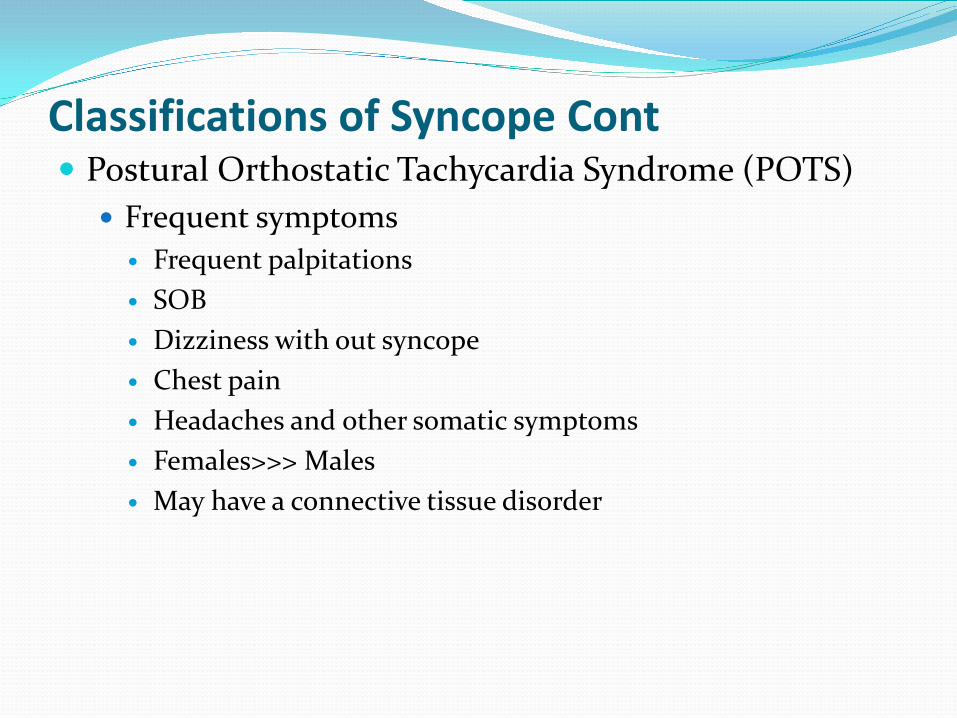
Classifications of Syncope Cont Postural Orthostatic Tachycardia Syndrome (POTS)
Frequent symptoms Frequent palpitations SOB Dizziness with out syncope Chest pain Headaches and other somatic symptoms Females>>> Males May have a connective tissue disorder
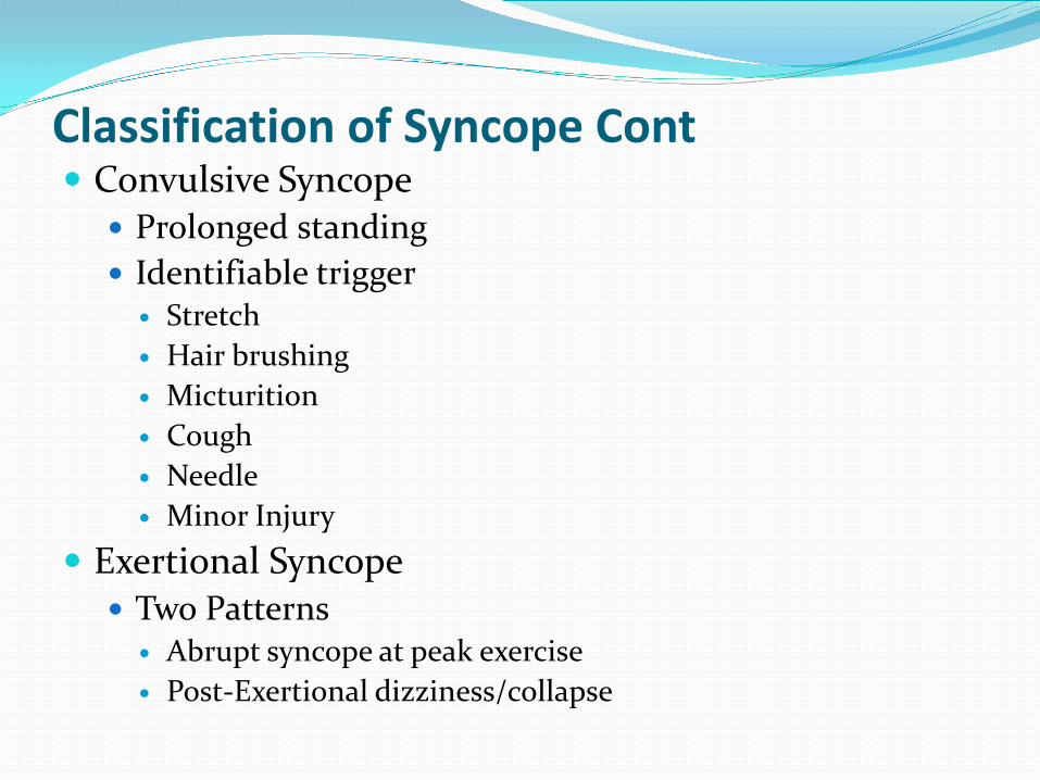
Classification of Syncope Cont Convulsive Syncope
Prolonged standing Identifiable trigger
Stretch Hair brushing Micturition Cough Needle Minor Injury
Exertional Syncope Two Patterns
Abrupt syncope at peak exercise Post-Exertional dizziness/collapse
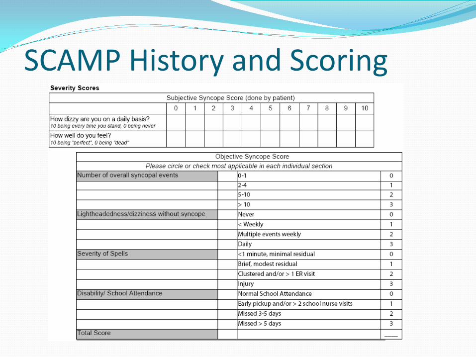
SCAMP History and Scoring
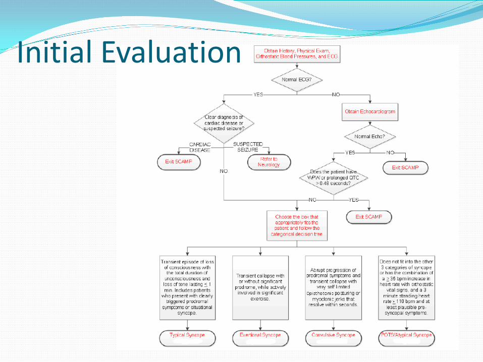
Initial Evaluation
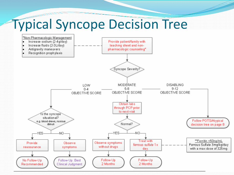
Typical Syncope Decision Tree
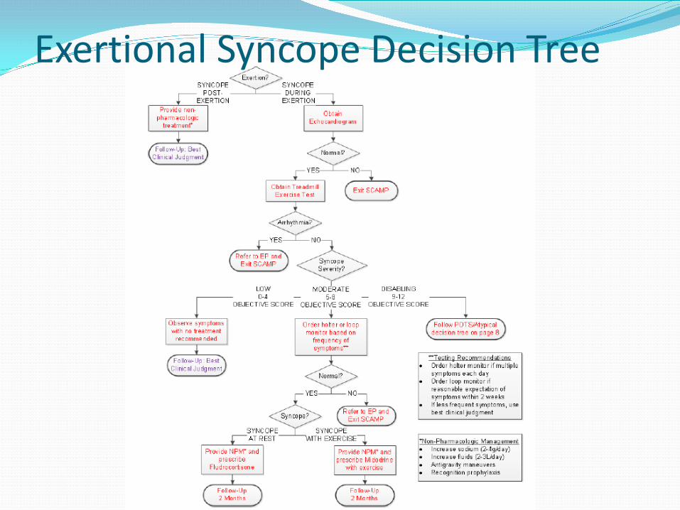
Exertional Syncope Decision Tree
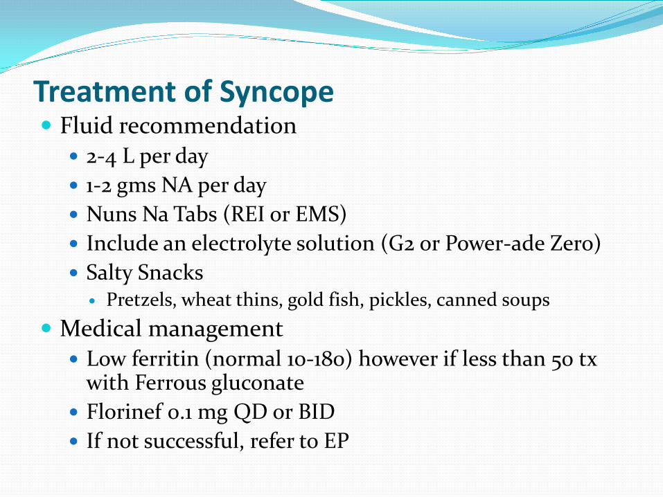
Treatment of Syncope Fluid recommendation
2-4 L per day 1-2 gms NA per day Nuns Na Tabs (REI or EMS) Include an electrolyte solution (G2 or Power-ade Zero) Salty Snacks
Pretzels, wheat thins, gold fish, pickles, canned soups Medical management
Low ferritin (normal 10-180) however if less than 50 tx with Ferrous gluconate
Florinef 0.1 mg QD or BID If not successful, refer to EP

Case Studies
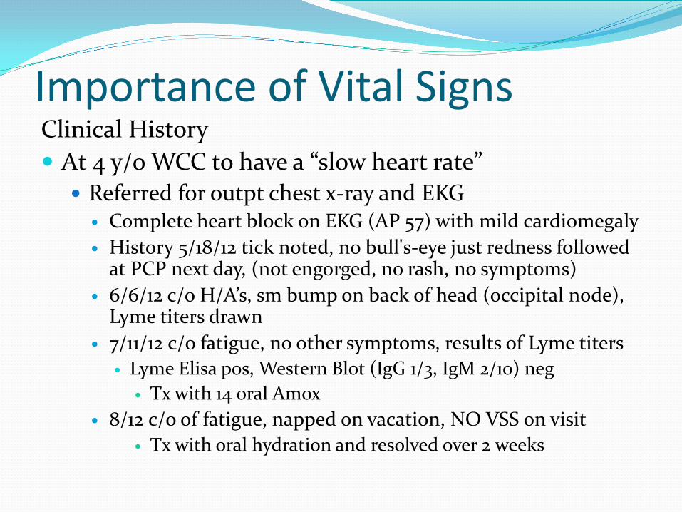
Importance of Vital Signs Clinical History At 4 y/o WCC to have a “slow heart rate”
Referred for outpt chest x-ray and EKG Complete heart block on EKG (AP 57) with mild cardiomegaly History 5/18/12 tick noted, no bull's-eye just redness followed
at PCP next day, (not engorged, no rash, no symptoms) 6/6/12 c/o H/A’s, sm bump on back of head (occipital node),
Lyme titers drawn 7/11/12 c/o fatigue, no other symptoms, results of Lyme titers
Lyme Elisa pos, Western Blot (IgG 1/3, IgM 2/10) neg Tx with 14 oral Amox
8/12 c/o of fatigue, napped on vacation, NO VSS on visit Tx with oral hydration and resolved over 2 weeks
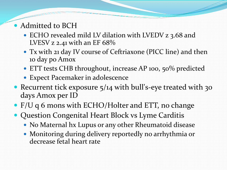
Admitted to BCH ECHO revealed mild LV dilation with LVEDV z 3.68 and
LVESV z 2.41 with an EF 68% Tx with 21 day IV course of Ceftriaxone (PICC line) and then
10 day po Amox ETT tests CHB throughout, increase AP 100, 50% predicted Expect Pacemaker in adolescence
Recurrent tick exposure 5/14 with bull's-eye treated with 30 days Amox per ID
F/U q 6 mons with ECHO/Holter and ETT, no change Question Congenital Heart Block vs Lyme Carditis
No Maternal hx Lupus or any other Rheumatoid disease Monitoring during delivery reportedly no arrhythmia or
decrease fetal heart rate
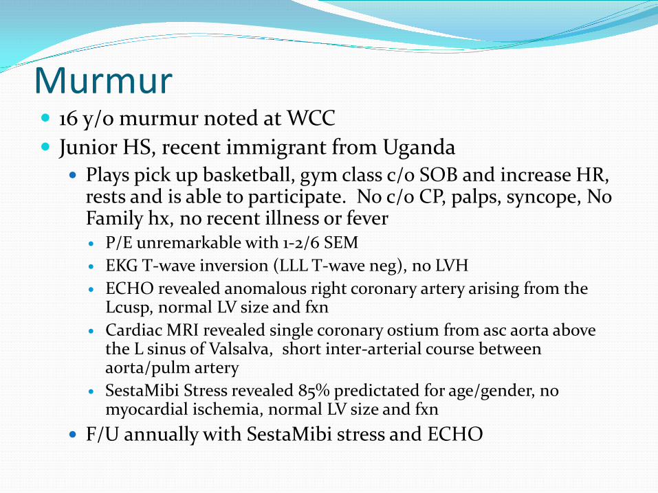
Murmur 16 y/o murmur noted at WCC Junior HS, recent immigrant from Uganda
Plays pick up basketball, gym class c/o SOB and increase HR, rests and is able to participate. No c/o CP, palps, syncope, No Family hx, no recent illness or fever P/E unremarkable with 1-2/6 SEM EKG T-wave inversion (LLL T-wave neg), no LVH ECHO revealed anomalous right coronary artery arising from the
Lcusp, normal LV size and fxn Cardiac MRI revealed single coronary ostium from asc aorta above
the L sinus of Valsalva, short inter-arterial course between aorta/pulm artery
SestaMibi Stress revealed 85% predictated for age/gender, no myocardial ischemia, normal LV size and fxn
F/U annually with SestaMibi stress and ECHO
Chest Pain 17 y/o with c/o CP at PE, No other concerns, Tx with 600mg
Advil TID for 2 wks, no improvement Normal EKG/Chest x-ray
Junior in HS daily c/o CP since December, episodes lasts few hours to the entire day
Missed 100 days of school (no feeling well c/o migraines, backaches and overall pain), no organized or rec sports, Gym class describes a squeezing CP no other symptoms, sits out of gym and does not return
PE: Wgt 93%, Hgt 23% and BMI 96%, No Murmur, EKG nrml EHCO revealed a RACA arising from the L SoV, looks to have
a longer inter-arterial course, nrml LV size and fxn Scheduled Cardiac MRI and SestaMibi Stress in April
Syncope 12 y/o with 4-5 episodes of syncope
c/o of tunnel vision, ringing in ears, all with postural changes, no c/o cp or palps, no loss b/b, no injury, awake and alert within a min
Family history…..Dad with an LVAD, Dx with Cardiomyopathy in 2008 when ICD placed and on LVAD in 2012. No genetic testing done ECHO normal F/U based on Dad’s genetic testing, however if no testing or Dad
negative then f/u 2 years 15 y/o with 3-4 episodes of syncope
Most recent episodes occurred a few wks ago with blood draw, another during health class, c/o tunnel vision and ringing in ears, no c/o cp or palps, no loss b/b, no injury, awake and alert within a min , first one in 9/14 while playing hockey, felt knees get weak, tunnel vision and went down face first. ECHO normal inter-cardiac anatomy, nrml LV size and fxn ETT scheduled
Easiest Heart Patient
Thank You!


![Pediatric Cardiology Dysfunction for Students--2011[1]](https://img.pdfslide.us/doc/110x75/577d21651a28ab4e1e9523b1/pediatric-cardiology-dysfunction-for-students-20111.jpg)