Embed Size (px)
Citation preview
Regional Implementation of a PediatricCardiology Syncope Algorithm Using
Standardized Clinical Assessment andManagement Plans (SCAMPS) Methodology
The Harvard community has made thisarticle openly available. Please share howthis access benefits you. Your story matters
Citation Paris, Yvonne, Olga H. Toro#Salazar, Naomi S. Gauthier, KathleenM. Rotondo, Lucy Arnold, Rose Hamershock, David E. Saudek, DavidR. Fulton, Ashley Renaud, and Mark E. Alexander. 2016. “RegionalImplementation of a Pediatric Cardiology Syncope AlgorithmUsing Standardized Clinical Assessment and ManagementPlans (SCAMPS) Methodology.” Journal of the American HeartAssociation: Cardiovascular and Cerebrovascular Disease 5 (2):e002931. doi:10.1161/JAHA.115.002931. http://dx.doi.org/10.1161/JAHA.115.002931.
Published Version doi:10.1161/JAHA.115.002931
Citable link http://nrs.harvard.edu/urn-3:HUL.InstRepos:26860043
Terms of Use This article was downloaded from Harvard University’s DASHrepository, and is made available under the terms and conditionsapplicable to Other Posted Material, as set forth at http://nrs.harvard.edu/urn-3:HUL.InstRepos:dash.current.terms-of-use#LAA
Regional Implementation of a Pediatric Cardiology Syncope AlgorithmUsing Standardized Clinical Assessment and Management Plans(SCAMPS) MethodologyYvonne Paris, MD; Olga H. Toro-Salazar, MD; Naomi S. Gauthier, MD; Kathleen M. Rotondo, MD; Lucy Arnold, MD;Rose Hamershock, MA; David E. Saudek, MD; David R. Fulton, MD; Ashley Renaud, RN; Mark E. Alexander, MD;on behalf of the New England Congenital Cardiology Association (NECCA)*
Background-—Pediatric syncope is common. Cardiac causes are rarely found. We describe and assess a pragmatic approach tothese patients first seen by a pediatric cardiologist in the New England region, using Standardized Clinical Assessment andManagement Plans (SCAMPs).
Methods and Results-—Ambulatory patients aged 7 to 21 years initially seen for syncope at participating New EnglandCongenital Cardiology Association practices over a 2.5-year period were evaluated using a SCAMP. Findings were iterativelyanalyzed and the care pathway was revised. The vast majority (85%) of the 1254 patients had typical syncope. A minority hadexercise-related or more problematic symptoms. Guideline-defined testing identified one patient with cardiac syncope. SyncopeSeverity Scores correlated well between physician and patient perceived symptoms. Orthostatic vital signs were of limited use.Largely incidental findings were seen in 10% of ECGs and 11% of echocardiograms. The 10% returning for follow-up, by design,reported more significant symptoms, but did not have newly recognized cardiac disease. Iterative analysis helped refine theapproach.
Conclusions-—SCAMP methodology confirmed that the vast majority of children referred to the outpatient pediatric cardiologysetting had typical low-severity neurally mediated syncope that could be effectively evaluated in a single visit using minimalresources. A simple scoring system can help triage patients into treatment categories. Prespecified criteria permitted the effectivediagnosis of the single patient with a clear cardiac etiology. Patients with higher syncope scores still have a very low risk of cardiacdisease, but may warrant attention. ( J Am Heart Assoc. 2016;5:e002931 doi: 10.1161/JAHA.115.002931)
Key Words: adolescence • ambulatory care • pediatrics • syncope (fainting)
N eurally mediated syncope—an abrupt, reflex-mediatedtransient loss of consciousness—is very common with
a cumulative incidence of 35% by age 18 and nearly 50% byage 21.1 Newly recognized cardiac disease is rare. While notall pediatric patients with syncope come to medical attention,the mismatch of an exceptionally common cardinal symptom
and a rare but potentially life-threatening cause motivatesfrequent referral and cardiac evaluation.
The evaluation of these patients can be extensive,expending considerable time and resources.2–4 Previousreports on syncope have been mostly observational.4–6 Toaddress this, a group of pediatric cardiologists from around
From the Division of Pediatric Cardiology, Baystate Medical Center, Springfield, MA (Y.P.); Department of Pediatrics, Tufts Medical School, Boston, MA (Y.P.); PediatricCardiology, Connecticut Children’s Medical Center, Hartford, CT (O.H.T.-S.); Department of Pediatrics, University of Connecticut School of Medicine, Farmington, CT(O.H.T.-S.); Pediatric Cardiology, Children’s Hospital at Dartmouth-Hitchcock, Dover, NH, (N.S.G.); Department of Pediatrics, Geisel School of Medicine at Dartmouth,Hanover, NH (N.S.G.); Pediatric Cardiology, Hasbro Children’s Hospital, Providence, RI (K.M.R.); Department of Pediatrics, Warren Alpert Medical School at BrownUniversity, Providence, RI (K.M.R.); Pediatric Cardiology, Harvard Vanguard Medical Associates, Boston, MA (L.A.); Institute for Relevant Clinical Data Analytics, Inc,Boston, MA (R.H., A.R.); Pediatric Cardiology, Children’s Hospital of Wisconsin, Milwaukee, WI (D.E.S.); Department of Pediatrics, Medical College of Wisconsin,Milwaukee, WI (D.E.S.); Department of Cardiology (D.R.F., M.E.A.) and Arrhythmia Service (M.E.A.), Boston Children’s Hospital, Boston, MA; Department of Pediatrics,Harvard Medical School, Boston, MA (D.R.F., M.E.A.); on behalf of the New England Congenital Cardiology Association (NECCA).
*Accompanying Appendices S1, S2 and list of additional participants in the New England Congenital Cardiology Association (NECCA) are available athttp://jaha.ahajournals.org/content/5/2/e002931/suppl/DC1
Correspondence to: Mark E. Alexander, MD, Department of Cardiology, Boston Children’s Hospital, Boston, MA 02115. E-mail: [email protected]
Received November 12, 2015; accepted December 21, 2015.
ª 2016 The Authors. Published on behalf of the American Heart Association, Inc., by Wiley Blackwell. This is an open access article under the terms of the CreativeCommons Attribution-NonCommercial License, which permits use, distribution and reproduction in any medium, provided the original work is properly cited and isnot used for commercial purposes.
DOI: 10.1161/JAHA.115.002931 Journal of the American Heart Association 1
ORIGINAL RESEARCH
New England collaborated to design and implement a qualityimprovement tool utilizing Standardized Clinical Assessmentand Management Plans (SCAMPs) to guide the care ofpatients presenting to a pediatric cardiology outpatient clinicwith a complaint of syncope. SCAMPs methodology usesprospective acquisition of defined data that are analyzed on arecurring basis. A sound initial approach is combined withspecific, testable, targeted data statements about eitherdiagnostic or therapeutic approaches. Changes to the carepathways are based on this analysis.7,8
New England Congenital Cardiology Association (NECCA),an organization of �16 academic and community-basedpractices representing all 6 New England states, previouslysuccessfully implemented a SCAMP on chest pain resulting ina decrease in practice variation and resource utilization.7,9,10
The syncope SCAMP objective was to develop a standard-ized approach to evaluation of syncope in the outpatientsetting. The pathway which emphasized the critical aspects ofhistory, focused family history, physical exam, orthostatic vitalsigns, and an ECG was asserted to have a high negativepredictive value. This would allow identification of the smallernumber of patients with higher probability of significantcardiac disease or inherited arrhythmia syndromes and limitthe more complex testing. Once the cause of syncope wasidentified, specific approaches to management were outlinedwith an emphasis on education and nonpharmacologicmanagement for patients with typical syncope.
MethodsWe developed a SCAMP clinical care pathway for syncopeusing history, physical examination, and ECG.11,12 Thisevaluation targeted pediatric, adolescent, and young adultpatients 7 to 21 years of age who present to pediatriccardiology outpatient practices for a chief complaint ofsyncope. The SCAMP was designed to explore multiplequestions simultaneously by targeted data statements. Theseformed the basis for the clinical care pathway and SCAMPData Form (SDF) used to track the SCAMP. The strategies forimplementation were shared and the guideline was revisedbased on feedback. Flexibility in implementation of theSCAMP was necessary due to the variation of resourcesavailable at different sites. Specific pathways were outlinedfor typical, exertional, convulsive, atypical/refractory syn-cope, and postural tachycardia syndrome (POTS)(Appendix S1).
This care pathway was specifically designed to classify thetype of syncope and to identify cardiac causes of syncopeusing the resources in the outpatient cardiology clinicalsetting. The intent was to complete the evaluation in a singlevisit for patients with typical, low-severity syncope.
The institutional review boards at Boston Children’sHospital and the participating NECCA sites evaluated theproject and waived review designating the SCAMP method-ology as a quality improvement initiative. Data use agree-ments limited reporting age/sex from NECCA sites.
All patient data were de-identified. Activity at the NECCAsites was facilitated, tracked, and coordinated throughmonthly conference calls that ensured uniformity in use ofthe SCAMP. Following a pilot period, minor revisions weremade and the SCAMP was deployed throughout NECCA for15 months followed by data analysis and revision of theprocess with continued tracking for 18 months. Practicesentered the SCAMP at different times, so that not all practiceswere involved the entire 33 months.
There were 2 phases of analysis. Phase 1 used the initialclinical pathways with interim analyses that motivatedmodification to the revised Phase 2 pathway. Phase 1analysis included patients enrolled between March 2012and July 2013, with Phase 2 analysis representing patientsentered between August 2013 and December 2014. Interimanalyses were also performed. Each analysis was presentedbroadly within NECCA for feedback followed by modificationof the algorithm by consensus.
Patient SelectionAmbulatory patients between 7 and 21 years of age present-ing to an outpatient pediatric cardiology practice for a first-time evaluation of the principal complaint of syncope wereenrolled. A complete history and physical exam wereperformed and an ECG was reviewed.
Patients were excluded for cardiac arrest needing sustainedcardiopulmonary resuscitation, previously diagnosed seizure,or known cardiac disease. Those with WPW (Wolff ParkinsonWhite) or QT interval >480 ms (Long QT Syndrome) on initialECG were also excluded. The SDF did not track exclusions.
Data Collection
Clinical evaluation and classifications
Based on initial history and exam, each patient was classified ashaving typical, exertional, convulsive, atypical/refractory syn-cope, or POTS. Clinical workflow was completely at theclinician’s discretion, such that echocardiogramor other testingcould be scheduled prior to the history and physical.
Demographics and clinical characteristics were collectedincluding signs, symptoms and consequences of the synco-pal event as well as pertinent past medical and familyhistory. Historical data were also noted with regard to pastevaluations for syncope and previous specialist or emer-gency room evaluation. Pertinent physical exam findings,
DOI: 10.1161/JAHA.115.002931 Journal of the American Heart Association 2
Prospective Pediatric Syncope SCAMP Paris et alORIG
INALRESEARCH
ECG interpretation, and orthostatic vital signs were recordedeither manually or using automated equipment. Orthostaticblood pressure measurements were discontinued duringPhase 2 based on analysis of Phase 1 data. The patientjudged their symptom severity using 2 subjective, 10-pointsyncope scales for daily dizziness and overall well-being/wellness. The provider assessed a 0 to 12 point SyncopeSeverity Score (SSS) based on frequency, severity, anddisability associated with the symptoms (Appendix S2).
The initial ECG was evaluated and any abnormalities werenoted. Pertinent abnormalities were tabulated, but specificcriteria for each ECG diagnosis were neither mandated norindependently reviewed. Echocardiograms were recom-mended for predefined, generally accepted consensus-basedcriteria focusing on exertional symptoms or concerningfeatures of the presentation (abrupt onset, injury), abnormalECG or examination, and potentially concerning family history.Echocardiograms done for family/clinician anxiety weretracked. Patients defined as having typical low-severitysyncope were provided with teaching materials and reassur-ance and not scheduled for follow-up. A complete blood countand ferritin level were recommended for those defined withmoderate to severe typical syncope.13,14 Provider preferenceregarding medical treatment or conservative managementwas recorded, and patient follow-up in 2 months to reviewresponse to treatment was recommended.
If patients reported a syncopal event with or immediatelyfollowing exercise, they were classified as having exertionalsyncope. Echocardiograms were recommended for thosepatients who were noted to have syncope during exercise. Ifno abnormalities were noted on the echocardiogram, thentreadmill exercise testing was recommended. Patients exitedthe SCAMP care pathway if either a structural abnormality ora clinically important arrhythmia were identified. Otherwise,management mirrored typical syncope.
Syncope was classified as convulsive if the patient wasnoted to stiffen or have brief tonic–clonic movements duringthe event. Repeated episodes, sustained tonic–clonic events,or neurological findings dictated neurology referral. Repeatedepisodes directed an exercise test and an echocardiogram aswell as ambulatory arrhythmia testing with a loop recorder ifthe frequency of convulsive syncope was greater than twice amonth.
POTS was initially defined as a change in heart rate (HR) of>30 beats per minute during the orthostatic testing processdelineated in the SCAMP care pathway with plausiblesymptoms.15 Atypical syncope was defined as SSS >8 withcombinations of disabling symptoms, syncope while supine,delayed recovery, or sustained “jerking/stiffening” combinedwith frequent events. More expansive laboratory screeningand monitoring was recommended with SSS >4 with partic-ular focus on those with SSS ≥8.
Treatment with iron was suggested if ferritin levels were<50 ng/mL. Patients with endocrinopathies were referredand exited the SCAMP. Provider preference regarding medicaltreatment or conservative management was recorded.Patients were recommended for follow-up in 2 months toreview response to treatment if the patient had moderate ormore severe syncope.
Test interpretation
ECG and ancillary testing interpretations were tabulatedbased on results recorded on the SDFs. Testing was generallyobtained at the time of the initial visit or soon after. Testresults performed prior to the pediatric cardiology visit werealso noted. There was no attempt to validate results.Ambiguity on critical data was resolved by querying thesubmitting clinical site.
SCAMP data collection and analysis
Data were collected on an SDF completed by the provider atthe initial visit (Appendix S2). Patients who had scheduledfollow-up or who returned after discharge from the clinic wereevaluated using a second SDF documenting interval historyself-reports/assigned SSS. Data were collated by the Institutefor Relevant Clinical Data Analytics for NECCA and operationsstaff at Boston Children’s Hospital.
Results of testing were tracked to assess adherence to therecommendations of the algorithm and to analyze outcomes.
Statistical AnalysisSummary statistics were tabulated. Differences in patient andfamily history by site and syncope type were examined withv2 and Fisher exact tests. Chi-square and Fisher exact testswere also used to analyze the difference in ECG results oversyncope types. Bubble plots and correlation analysis wereperformed on the relationship between physician-assignedand patient-assessment scores. Logistic regression analyzeddifferential medication use. Lastly, the receiver operatingcharacteristic c-statistic was calculated to determine whetherthe differences in orthostatic supine values and 3-minutestanding orthostatic values were useful in determiningsyncope classifications. With the exception of specific anal-yses of follow-up, all analyses were performed on the initialepisode of care to maintain independence.
Results
Patient PopulationBetween March of 2012 and December of 2014 a total of1292 patients (1437 total visits) were enrolled with 91%
DOI: 10.1161/JAHA.115.002931 Journal of the American Heart Association 3
Prospective Pediatric Syncope SCAMP Paris et alORIG
INALRESEARCH
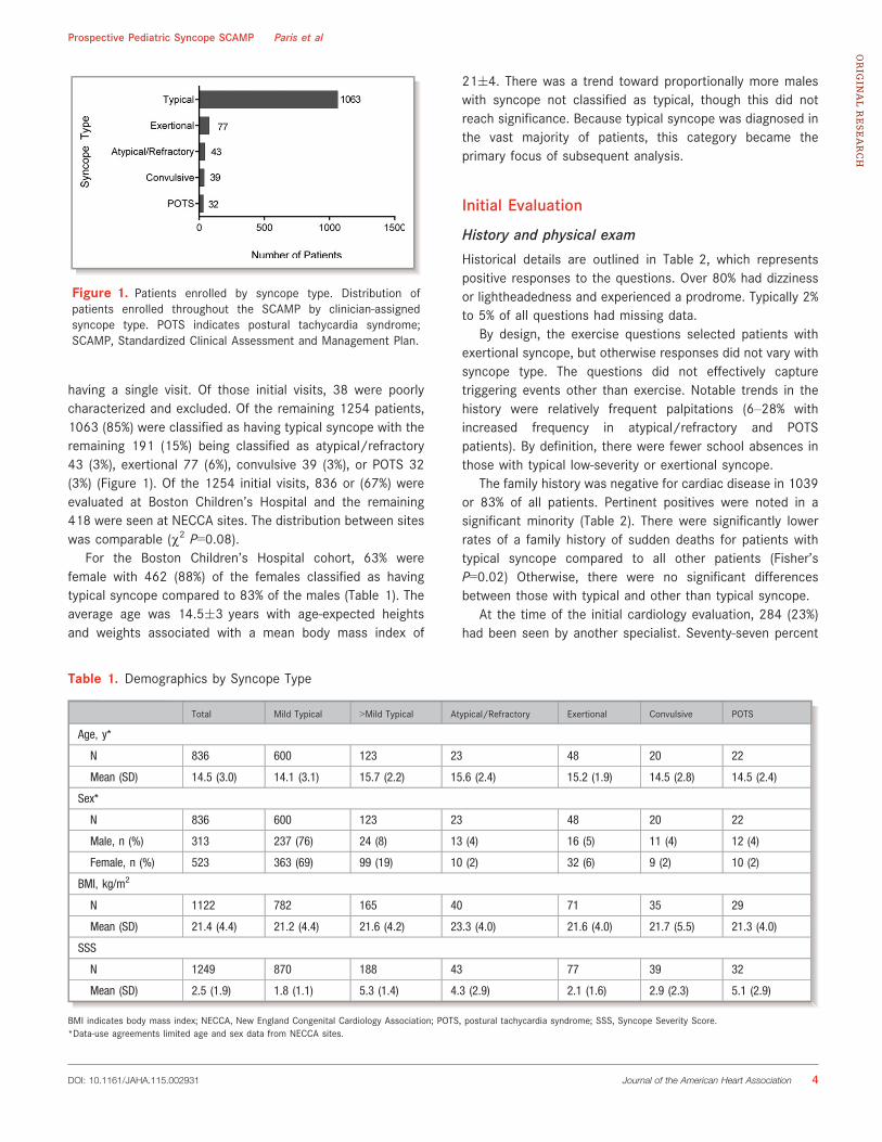
having a single visit. Of those initial visits, 38 were poorlycharacterized and excluded. Of the remaining 1254 patients,1063 (85%) were classified as having typical syncope with theremaining 191 (15%) being classified as atypical/refractory43 (3%), exertional 77 (6%), convulsive 39 (3%), or POTS 32(3%) (Figure 1). Of the 1254 initial visits, 836 or (67%) wereevaluated at Boston Children’s Hospital and the remaining418 were seen at NECCA sites. The distribution between siteswas comparable (v2 P=0.08).
For the Boston Children’s Hospital cohort, 63% werefemale with 462 (88%) of the females classified as havingtypical syncope compared to 83% of the males (Table 1). Theaverage age was 14.5�3 years with age-expected heightsand weights associated with a mean body mass index of
21�4. There was a trend toward proportionally more maleswith syncope not classified as typical, though this did notreach significance. Because typical syncope was diagnosed inthe vast majority of patients, this category became theprimary focus of subsequent analysis.
Initial Evaluation
History and physical exam
Historical details are outlined in Table 2, which representspositive responses to the questions. Over 80% had dizzinessor lightheadedness and experienced a prodrome. Typically 2%to 5% of all questions had missing data.
By design, the exercise questions selected patients withexertional syncope, but otherwise responses did not vary withsyncope type. The questions did not effectively capturetriggering events other than exercise. Notable trends in thehistory were relatively frequent palpitations (6–28% withincreased frequency in atypical/refractory and POTSpatients). By definition, there were fewer school absences inthose with typical low-severity or exertional syncope.
The family history was negative for cardiac disease in 1039or 83% of all patients. Pertinent positives were noted in asignificant minority (Table 2). There were significantly lowerrates of a family history of sudden deaths for patients withtypical syncope compared to all other patients (Fisher’sP=0.02) Otherwise, there were no significant differencesbetween those with typical and other than typical syncope.
At the time of the initial cardiology evaluation, 284 (23%)had been seen by another specialist. Seventy-seven percent
Figure 1. Patients enrolled by syncope type. Distribution ofpatients enrolled throughout the SCAMP by clinician-assignedsyncope type. POTS indicates postural tachycardia syndrome;SCAMP, Standardized Clinical Assessment and Management Plan.
Table 1. Demographics by Syncope Type
Total Mild Typical >Mild Typical Atypical/Refractory Exertional Convulsive POTS
Age, y*
N 836 600 123 23 48 20 22
Mean (SD) 14.5 (3.0) 14.1 (3.1) 15.7 (2.2) 15.6 (2.4) 15.2 (1.9) 14.5 (2.8) 14.5 (2.4)
Sex*
N 836 600 123 23 48 20 22
Male, n (%) 313 237 (76) 24 (8) 13 (4) 16 (5) 11 (4) 12 (4)
Female, n (%) 523 363 (69) 99 (19) 10 (2) 32 (6) 9 (2) 10 (2)
BMI, kg/m2
N 1122 782 165 40 71 35 29
Mean (SD) 21.4 (4.4) 21.2 (4.4) 21.6 (4.2) 23.3 (4.0) 21.6 (4.0) 21.7 (5.5) 21.3 (4.0)
SSS
N 1249 870 188 43 77 39 32
Mean (SD) 2.5 (1.9) 1.8 (1.1) 5.3 (1.4) 4.3 (2.9) 2.1 (1.6) 2.9 (2.3) 5.1 (2.9)
BMI indicates body mass index; NECCA, New England Congenital Cardiology Association; POTS, postural tachycardia syndrome; SSS, Syncope Severity Score.*Data-use agreements limited age and sex data from NECCA sites.
DOI: 10.1161/JAHA.115.002931 Journal of the American Heart Association 4
Prospective Pediatric Syncope SCAMP Paris et alORIG
INALRESEARCH
of these patients were classified as having typical syncope.Patients with atypical or convulsive syncope and POTS weresignificantly more likely to have seen other specialists (40% to60%). Exertional syncope patients were least likely to havebeen referred to another specialist at 12% (overall P<0.01).Overall, prior emergency room visits had occurred in 40% ofpatients. Those with typical syncope were less likely to havehad prior emergency care (P<0.01).
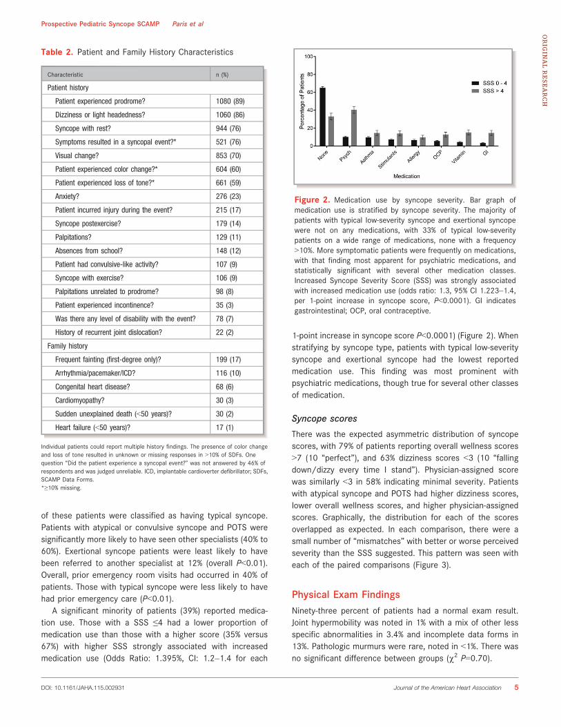
A significant minority of patients (39%) reported medica-tion use. Those with a SSS ≤4 had a lower proportion ofmedication use than those with a higher score (35% versus67%) with higher SSS strongly associated with increasedmedication use (Odds Ratio: 1.395%, CI: 1.2–1.4 for each
1-point increase in syncope score P<0.0001) (Figure 2). Whenstratifying by syncope type, patients with typical low-severitysyncope and exertional syncope had the lowest reportedmedication use. This finding was most prominent withpsychiatric medications, though true for several other classesof medication.
Syncope scores
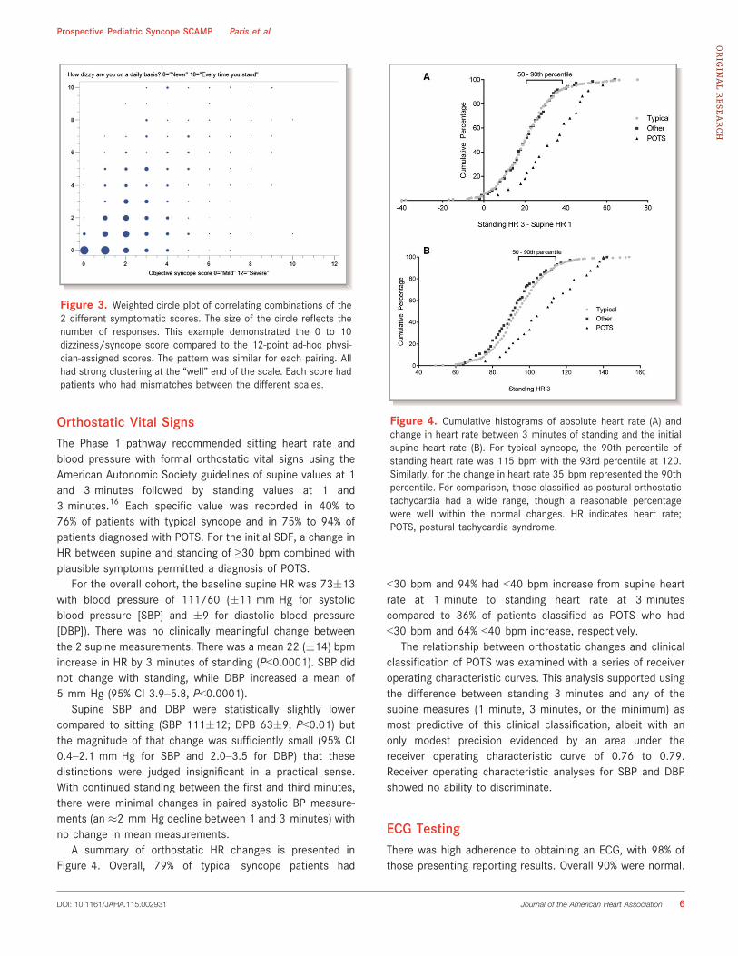
There was the expected asymmetric distribution of syncopescores, with 79% of patients reporting overall wellness scores>7 (10 “perfect”), and 63% dizziness scores <3 (10 “fallingdown/dizzy every time I stand”). Physician-assigned scorewas similarly <3 in 58% indicating minimal severity. Patientswith atypical syncope and POTS had higher dizziness scores,lower overall wellness scores, and higher physician-assignedscores. Graphically, the distribution for each of the scoresoverlapped as expected. In each comparison, there were asmall number of “mismatches” with better or worse perceivedseverity than the SSS suggested. This pattern was seen witheach of the paired comparisons (Figure 3).
Physical Exam FindingsNinety-three percent of patients had a normal exam result.Joint hypermobility was noted in 1% with a mix of other lessspecific abnormalities in 3.4% and incomplete data forms in13%. Pathologic murmurs were rare, noted in <1%. There wasno significant difference between groups (v2 P=0.70).
Table 2. Patient and Family History Characteristics
Characteristic n (%)
Patient history
Patient experienced prodrome? 1080 (89)
Dizziness or light headedness? 1060 (86)
Syncope with rest? 944 (76)
Symptoms resulted in a syncopal event?* 521 (76)
Visual change? 853 (70)
Patient experienced color change?* 604 (60)
Patient experienced loss of tone?* 661 (59)
Anxiety? 276 (23)
Patient incurred injury during the event? 215 (17)
Syncope postexercise? 179 (14)
Palpitations? 129 (11)
Absences from school? 148 (12)
Patient had convulsive-like activity? 107 (9)
Syncope with exercise? 106 (9)
Palpitations unrelated to prodrome? 98 (8)
Patient experienced incontinence? 35 (3)
Was there any level of disability with the event? 78 (7)
History of recurrent joint dislocation? 22 (2)
Family history
Frequent fainting (first-degree only)? 199 (17)
Arrhythmia/pacemaker/ICD? 116 (10)
Congenital heart disease? 68 (6)
Cardiomyopathy? 30 (3)
Sudden unexplained death (<50 years)? 30 (2)
Heart failure (<50 years)? 17 (1)
Individual patients could report multiple history findings. The presence of color changeand loss of tone resulted in unknown or missing responses in >10% of SDFs. Onequestion “Did the patient experience a syncopal event?” was not answered by 46% ofrespondents and was judged unreliable. ICD, implantable cardioverter defibrillator; SDFs,SCAMP Data Forms.*≥10% missing.
Figure 2. Medication use by syncope severity. Bar graph ofmedication use is stratified by syncope severity. The majority ofpatients with typical low-severity syncope and exertional syncopewere not on any medications, with 33% of typical low-severitypatients on a wide range of medications, none with a frequency>10%. More symptomatic patients were frequently on medications,with that finding most apparent for psychiatric medications, andstatistically significant with several other medication classes.Increased Syncope Severity Score (SSS) was strongly associatedwith increased medication use (odds ratio: 1.3, 95% CI 1.223–1.4,per 1-point increase in syncope score, P<0.0001). GI indicatesgastrointestinal; OCP, oral contraceptive.
DOI: 10.1161/JAHA.115.002931 Journal of the American Heart Association 5
Prospective Pediatric Syncope SCAMP Paris et alORIG
INALRESEARCH
Orthostatic Vital SignsThe Phase 1 pathway recommended sitting heart rate andblood pressure with formal orthostatic vital signs using theAmerican Autonomic Society guidelines of supine values at 1and 3 minutes followed by standing values at 1 and3 minutes.16 Each specific value was recorded in 40% to76% of patients with typical syncope and in 75% to 94% ofpatients diagnosed with POTS. For the initial SDF, a change inHR between supine and standing of ≥30 bpm combined withplausible symptoms permitted a diagnosis of POTS.
For the overall cohort, the baseline supine HR was 73�13with blood pressure of 111/60 (�11 mm Hg for systolicblood pressure [SBP] and �9 for diastolic blood pressure[DBP]). There was no clinically meaningful change betweenthe 2 supine measurements. There was a mean 22 (�14) bpmincrease in HR by 3 minutes of standing (P<0.0001). SBP didnot change with standing, while DBP increased a mean of5 mm Hg (95% CI 3.9–5.8, P<0.0001).
Supine SBP and DBP were statistically slightly lowercompared to sitting (SBP 111�12; DPB 63�9, P<0.01) butthe magnitude of that change was sufficiently small (95% CI0.4–2.1 mm Hg for SBP and 2.0–3.5 for DBP) that thesedistinctions were judged insignificant in a practical sense.With continued standing between the first and third minutes,there were minimal changes in paired systolic BP measure-ments (an �2 mm Hg decline between 1 and 3 minutes) withno change in mean measurements.
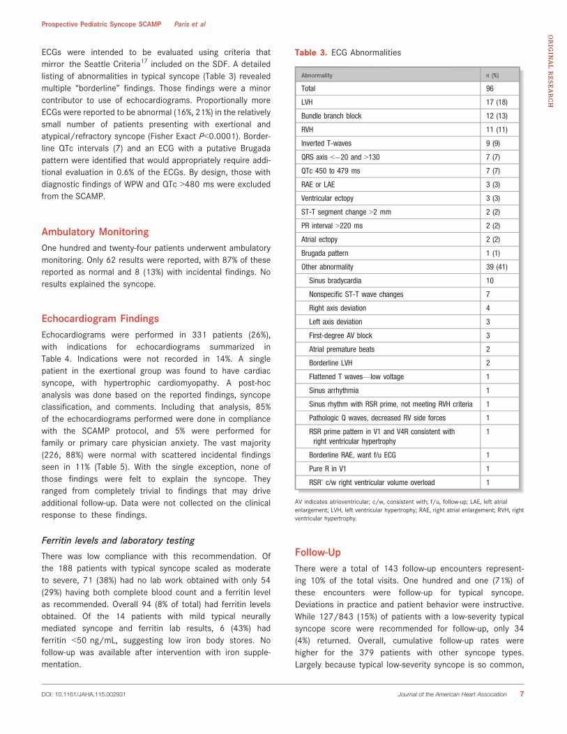
A summary of orthostatic HR changes is presented inFigure 4. Overall, 79% of typical syncope patients had
<30 bpm and 94% had <40 bpm increase from supine heartrate at 1 minute to standing heart rate at 3 minutescompared to 36% of patients classified as POTS who had<30 bpm and 64% <40 bpm increase, respectively.
The relationship between orthostatic changes and clinicalclassification of POTS was examined with a series of receiveroperating characteristic curves. This analysis supported usingthe difference between standing 3 minutes and any of thesupine measures (1 minute, 3 minutes, or the minimum) asmost predictive of this clinical classification, albeit with anonly modest precision evidenced by an area under thereceiver operating characteristic curve of 0.76 to 0.79.Receiver operating characteristic analyses for SBP and DBPshowed no ability to discriminate.
ECG TestingThere was high adherence to obtaining an ECG, with 98% ofthose presenting reporting results. Overall 90% were normal.
Figure 3. Weighted circle plot of correlating combinations of the2 different symptomatic scores. The size of the circle reflects thenumber of responses. This example demonstrated the 0 to 10dizziness/syncope score compared to the 12-point ad-hoc physi-cian-assigned scores. The pattern was similar for each pairing. Allhad strong clustering at the “well” end of the scale. Each score hadpatients who had mismatches between the different scales.
Figure 4. Cumulative histograms of absolute heart rate (A) andchange in heart rate between 3 minutes of standing and the initialsupine heart rate (B). For typical syncope, the 90th percentile ofstanding heart rate was 115 bpm with the 93rd percentile at 120.Similarly, for the change in heart rate 35 bpm represented the 90thpercentile. For comparison, those classified as postural orthostatictachycardia had a wide range, though a reasonable percentagewere well within the normal changes. HR indicates heart rate;POTS, postural tachycardia syndrome.
DOI: 10.1161/JAHA.115.002931 Journal of the American Heart Association 6
Prospective Pediatric Syncope SCAMP Paris et alORIG
INALRESEARCH
ECGs were intended to be evaluated using criteria thatmirror the Seattle Criteria17 included on the SDF. A detailedlisting of abnormalities in typical syncope (Table 3) revealedmultiple “borderline” findings. Those findings were a minorcontributor to use of echocardiograms. Proportionally moreECGs were reported to be abnormal (16%, 21%) in the relativelysmall number of patients presenting with exertional andatypical/refractory syncope (Fisher Exact P<0.0001). Border-line QTc intervals (7) and an ECG with a putative Brugadapattern were identified that would appropriately require addi-tional evaluation in 0.6% of the ECGs. By design, those withdiagnostic findings of WPW and QTc >480 ms were excludedfrom the SCAMP.
Ambulatory MonitoringOne hundred and twenty-four patients underwent ambulatorymonitoring. Only 62 results were reported, with 87% of thesereported as normal and 8 (13%) with incidental findings. Noresults explained the syncope.
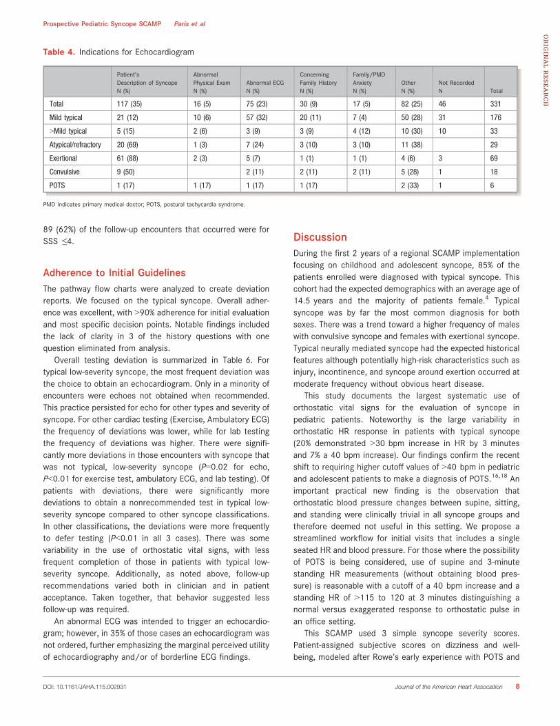
Echocardiogram FindingsEchocardiograms were performed in 331 patients (26%),with indications for echocardiograms summarized inTable 4. Indications were not recorded in 14%. A singlepatient in the exertional group was found to have cardiacsyncope, with hypertrophic cardiomyopathy. A post-hocanalysis was done based on the reported findings, syncopeclassification, and comments. Including that analysis, 85%of the echocardiograms performed were done in compliancewith the SCAMP protocol, and 5% were performed forfamily or primary care physician anxiety. The vast majority(226, 88%) were normal with scattered incidental findingsseen in 11% (Table 5). With the single exception, none ofthose findings were felt to explain the syncope. Theyranged from completely trivial to findings that may driveadditional follow-up. Data were not collected on the clinicalresponse to these findings.
Ferritin levels and laboratory testing
There was low compliance with this recommendation. Ofthe 188 patients with typical syncope scaled as moderateto severe, 71 (38%) had no lab work obtained with only 54(29%) having both complete blood count and a ferritin levelas recommended. Overall 94 (8% of total) had ferritin levelsobtained. Of the 14 patients with mild typical neurallymediated syncope and ferritin lab results, 6 (43%) hadferritin <50 ng/mL, suggesting low iron body stores. Nofollow-up was available after intervention with iron supple-mentation.
Follow-UpThere were a total of 143 follow-up encounters represent-ing 10% of the total visits. One hundred and one (71%) ofthese encounters were follow-up for typical syncope.Deviations in practice and patient behavior were instructive.While 127/843 (15%) of patients with a low-severity typicalsyncope score were recommended for follow-up, only 34(4%) returned. Overall, cumulative follow-up rates werehigher for the 379 patients with other syncope types.Largely because typical low-severity syncope is so common,
Table 3. ECG Abnormalities
Abnormality n (%)
Total 96
LVH 17 (18)
Bundle branch block 12 (13)
RVH 11 (11)
Inverted T-waves 9 (9)
QRS axis <�20 and >130 7 (7)
QTc 450 to 479 ms 7 (7)
RAE or LAE 3 (3)
Ventricular ectopy 3 (3)
ST-T segment change >2 mm 2 (2)
PR interval >220 ms 2 (2)
Atrial ectopy 2 (2)
Brugada pattern 1 (1)
Other abnormality 39 (41)
Sinus bradycardia 10
Nonspecific ST-T wave changes 7
Right axis deviation 4
Left axis deviation 3
First-degree AV block 3
Atrial premature beats 2
Borderline LVH 2
Flattened T waves—low voltage 1
Sinus arrhythmia 1
Sinus rhythm with RSR prime, not meeting RVH criteria 1
Pathologic Q waves, decreased RV side forces 1
RSR prime pattern in V1 and V4R consistent withright ventricular hypertrophy
1
Borderline RAE, want f/u ECG 1
Pure R in V1 1
RSR’ c/w right ventricular volume overload 1
AV indicates atrioventricular; c/w, consistent with; f/u, follow-up; LAE, left atrialenlargement; LVH, left ventricular hypertrophy; RAE, right atrial enlargement; RVH, rightventricular hypertrophy.
DOI: 10.1161/JAHA.115.002931 Journal of the American Heart Association 7
Prospective Pediatric Syncope SCAMP Paris et alORIG
INALRESEARCH
89 (62%) of the follow-up encounters that occurred were forSSS ≤4.
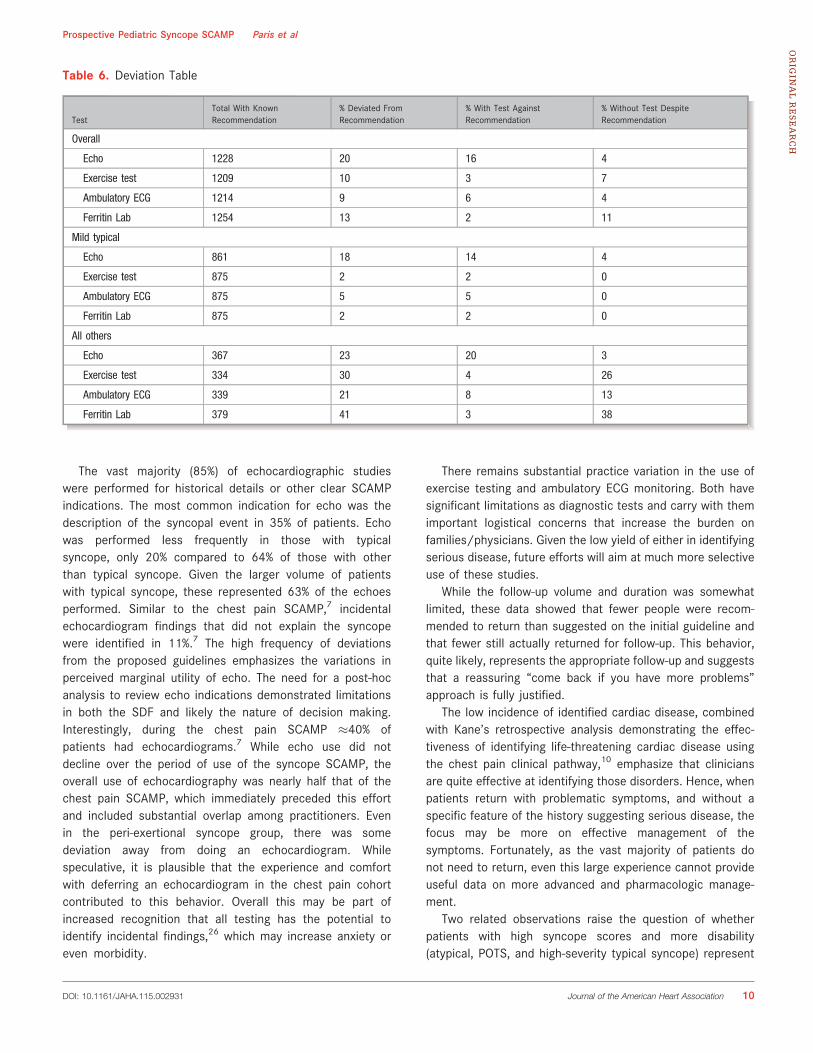
Adherence to Initial GuidelinesThe pathway flow charts were analyzed to create deviationreports. We focused on the typical syncope. Overall adher-ence was excellent, with >90% adherence for initial evaluationand most specific decision points. Notable findings includedthe lack of clarity in 3 of the history questions with onequestion eliminated from analysis.
Overall testing deviation is summarized in Table 6. Fortypical low-severity syncope, the most frequent deviation wasthe choice to obtain an echocardiogram. Only in a minority ofencounters were echoes not obtained when recommended.This practice persisted for echo for other types and severity ofsyncope. For other cardiac testing (Exercise, Ambulatory ECG)the frequency of deviations was lower, while for lab testingthe frequency of deviations was higher. There were signifi-cantly more deviations in those encounters with syncope thatwas not typical, low-severity syncope (P=0.02 for echo,P<0.01 for exercise test, ambulatory ECG, and lab testing). Ofpatients with deviations, there were significantly moredeviations to obtain a nonrecommended test in typical low-severity syncope compared to other syncope classifications.In other classifications, the deviations were more frequentlyto defer testing (P<0.01 in all 3 cases). There was somevariability in the use of orthostatic vital signs, with lessfrequent completion of those in patients with typical low-severity syncope. Additionally, as noted above, follow-uprecommendations varied both in clinician and in patientacceptance. Taken together, that behavior suggested lessfollow-up was required.
An abnormal ECG was intended to trigger an echocardio-gram; however, in 35% of those cases an echocardiogram wasnot ordered, further emphasizing the marginal perceived utilityof echocardiography and/or of borderline ECG findings.
DiscussionDuring the first 2 years of a regional SCAMP implementationfocusing on childhood and adolescent syncope, 85% of thepatients enrolled were diagnosed with typical syncope. Thiscohort had the expected demographics with an average age of14.5 years and the majority of patients female.4 Typicalsyncope was by far the most common diagnosis for bothsexes. There was a trend toward a higher frequency of maleswith convulsive syncope and females with exertional syncope.Typical neurally mediated syncope had the expected historicalfeatures although potentially high-risk characteristics such asinjury, incontinence, and syncope around exertion occurred atmoderate frequency without obvious heart disease.
This study documents the largest systematic use oforthostatic vital signs for the evaluation of syncope inpediatric patients. Noteworthy is the large variability inorthostatic HR response in patients with typical syncope(20% demonstrated >30 bpm increase in HR by 3 minutesand 7% a 40 bpm increase). Our findings confirm the recentshift to requiring higher cutoff values of >40 bpm in pediatricand adolescent patients to make a diagnosis of POTS.16,18 Animportant practical new finding is the observation thatorthostatic blood pressure changes between supine, sitting,and standing were clinically trivial in all syncope groups andtherefore deemed not useful in this setting. We propose astreamlined workflow for initial visits that includes a singleseated HR and blood pressure. For those where the possibilityof POTS is being considered, use of supine and 3-minutestanding HR measurements (without obtaining blood pres-sure) is reasonable with a cutoff of a 40 bpm increase and astanding HR of >115 to 120 at 3 minutes distinguishing anormal versus exaggerated response to orthostatic pulse inan office setting.
This SCAMP used 3 simple syncope severity scores.Patient-assigned subjective scores on dizziness and well-being, modeled after Rowe’s early experience with POTS and
Table 4. Indications for Echocardiogram
Patient’sDescription of SyncopeN (%)
AbnormalPhysical ExamN (%)
Abnormal ECGN (%)
ConcerningFamily HistoryN (%)
Family/PMDAnxietyN (%)
OtherN (%)
Not RecordedN Total
Total 117 (35) 16 (5) 75 (23) 30 (9) 17 (5) 82 (25) 46 331
Mild typical 21 (12) 10 (6) 57 (32) 20 (11) 7 (4) 50 (28) 31 176
>Mild typical 5 (15) 2 (6) 3 (9) 3 (9) 4 (12) 10 (30) 10 33
Atypical/refractory 20 (69) 1 (3) 7 (24) 3 (10) 3 (10) 11 (38) 29
Exertional 61 (88) 2 (3) 5 (7) 1 (1) 1 (1) 4 (6) 3 69
Convulsive 9 (50) 2 (11) 2 (11) 2 (11) 5 (28) 1 18
POTS 1 (17) 1 (17) 1 (17) 1 (17) 2 (33) 1 6
PMD indicates primary medical doctor; POTS, postural tachycardia syndrome.
DOI: 10.1161/JAHA.115.002931 Journal of the American Heart Association 8
Prospective Pediatric Syncope SCAMP Paris et alORIG
INALRESEARCH
Chronic Fatigue,19 generally correlated well with an ad-hocphysician-assigned 12-point scale. The clinician behavior andpatient follow-up choices supported a physician-assignedscore of ≤4 representing low-severity syncope. Practically,this designation included up to 2 to 3 episodes of syncopewith minimal school loss, no injury, and no late residualsymptoms. This physician-assigned SSS correlated with the
self-assigned scores for well-being and dizziness. These datasupport using simple scores to help assess the level ofdisability (whether perceived or absolute) for their syncopesymptoms. The SSS summarizes the actual and perceiveddisability related to the syncope syndrome and likely will helppredict which patients may be interested in therapy or follow-up. Importantly, because of the exceptionally low frequency ofidentified cardiac disease, these scores do not affect prob-ability of critical cardiac diagnoses. The 12-point SSS requiressome modification to effectively reflect follow-up visits, as thenumber of faints was not limited to a defined time period.
There are ample data to support that typical neurallymediated syncope in the school-age child and adolescentresults in excessive testing,20–22 with a systematic trendtoward more testing in the last 40 years.4 Adult groups havedeveloped reasonable predictive rules; however, they alsohave disease incidence and severity that are orders ofmagnitude higher than seen in pediatrics.23 Given the lowprior probability of significant disease, once the ECG is nearlynormal, serious cardiac disorders will always be rare inpediatrics.24,25 Using SCAMP methodology, we can thereforedecrease practice variation and focus toward both an efficientand a safe approach.
The ECG was asserted to have a high negative predictivevalue for serious heart disease24 with early exclusion ofpatients with WPW and Long QT Syndrome (defined as QTc≥480 ms). Using predefined criteria, �10% of the ECGsdemonstrated a wide range of “borderline” abnormalitiesnot likely contributing to syncope. An abnormal ECG was aminor contributor to the decision to perform an echocar-diogram. The one new cardiac diagnosis had both high-risksymptoms and a clearly abnormal ECG with T-waveinversion and left ventricular hypertrophy. Interestingly,16% of ECGs in patients with exertional symptoms wereread as abnormal. These may be systematic changes inathletes’ ECGs, but could also suggest that cardiologistsmay interpret the ECG differently when evaluating a patientwith a potentially higher probability of significant disease.Neither bias would change practice, as history drives thedecision to obtain an echocardiogram or other testing inthose with exertional syncope.
The exclusion of WPW and LQTS, 2 of the most obviousECG diagnoses, represents the reality that in clinical evalu-ation of a patient, any of those findings will drive decisionmaking away from typical syncope. A small number (7borderline QTc and 1 potential Brugada pattern or 0.6%) of theECGs suggested additional investigations for ion channeldisorders. The overall prevalence of those disorders (�1/700for WPW and �1/2000 for LQTS) informs choices aboutwhether an ECG is “mandatory” following an event thatotherwise meets clinical criteria for typical, low-severity,neurocardiogenic syncope.
Table 5. Incidental Abnormalities Found by Echocardiogram
SyncopeType Incidental Abnormality
Mildtypical
Borderline MVP, no MR
Tricuspid valve prolapse
Mild central aortic regurgitation, mild ascending aorticdilation
Mild aortic dilation
Premature ventricular contractions
Borderline apical LV noncompaction
Coronary arises from ST junctional, normal courses (??)
Trivial AI, normal aortic valve
Secundum atrial septal defect
Mild aortic regurgitation and likely partial fusion of his aorticvalve commissure
Small fenestration in the atrial septum with left-to-rightflow
Moderate TR and tiny patent foramen ovale
Mildly dilated LV, low normal function
Bicuspid aortic valve (mild aortic insufficiency, no aorticstenosis). No root dilation.
Mild central aortic regurgitation and mild descending aorticdilation
Mild LV dilation
Normal structure, but some ectopy
Mild tricuspid regurgitation. Low right ventricular pressure.
Hypertrabeculation pattern
>Mildtypical
Mild pulmonary branch stenosis
Exertional High takeoff of right coronary
Top normal to mild root dilation
Trivial aortic regurgitation
Possible small patent foramen ovale
Aortic insufficiency
Partial fusion of L & R coronary cusp
Mildly dilated left ventricle, normal mass:volume ratio
Small ASD
Small left coronary system of concern, but not definitive
AI indicates aortic insufficiency; ASD, atrial septal defect; LV, left ventricular; MR, mitralinsufficiency; MVP, mitral valve prolapse.
DOI: 10.1161/JAHA.115.002931 Journal of the American Heart Association 9
Prospective Pediatric Syncope SCAMP Paris et alORIG
INALRESEARCH
The vast majority (85%) of echocardiographic studieswere performed for historical details or other clear SCAMPindications. The most common indication for echo was thedescription of the syncopal event in 35% of patients. Echowas performed less frequently in those with typicalsyncope, only 20% compared to 64% of those with otherthan typical syncope. Given the larger volume of patientswith typical syncope, these represented 63% of the echoesperformed. Similar to the chest pain SCAMP,7 incidentalechocardiogram findings that did not explain the syncopewere identified in 11%.7 The high frequency of deviationsfrom the proposed guidelines emphasizes the variations inperceived marginal utility of echo. The need for a post-hocanalysis to review echo indications demonstrated limitationsin both the SDF and likely the nature of decision making.Interestingly, during the chest pain SCAMP �40% ofpatients had echocardiograms.7 While echo use did notdecline over the period of use of the syncope SCAMP, theoverall use of echocardiography was nearly half that of thechest pain SCAMP, which immediately preceded this effortand included substantial overlap among practitioners. Evenin the peri-exertional syncope group, there was somedeviation away from doing an echocardiogram. Whilespeculative, it is plausible that the experience and comfortwith deferring an echocardiogram in the chest pain cohortcontributed to this behavior. Overall this may be part ofincreased recognition that all testing has the potential toidentify incidental findings,26 which may increase anxiety oreven morbidity.
There remains substantial practice variation in the use ofexercise testing and ambulatory ECG monitoring. Both havesignificant limitations as diagnostic tests and carry with themimportant logistical concerns that increase the burden onfamilies/physicians. Given the low yield of either in identifyingserious disease, future efforts will aim at much more selectiveuse of these studies.
While the follow-up volume and duration was somewhatlimited, these data showed that fewer people were recom-mended to return than suggested on the initial guideline andthat fewer still actually returned for follow-up. This behavior,quite likely, represents the appropriate follow-up and suggeststhat a reassuring “come back if you have more problems”approach is fully justified.
The low incidence of identified cardiac disease, combinedwith Kane’s retrospective analysis demonstrating the effec-tiveness of identifying life-threatening cardiac disease usingthe chest pain clinical pathway,10 emphasize that cliniciansare quite effective at identifying those disorders. Hence, whenpatients return with problematic symptoms, and without aspecific feature of the history suggesting serious disease, thefocus may be more on effective management of thesymptoms. Fortunately, as the vast majority of patients donot need to return, even this large experience cannot provideuseful data on more advanced and pharmacologic manage-ment.
Two related observations raise the question of whetherpatients with high syncope scores and more disability(atypical, POTS, and high-severity typical syncope) represent
Table 6. Deviation Table
TestTotal With KnownRecommendation
% Deviated FromRecommendation
% With Test AgainstRecommendation
% Without Test DespiteRecommendation
Overall
Echo 1228 20 16 4
Exercise test 1209 10 3 7
Ambulatory ECG 1214 9 6 4
Ferritin Lab 1254 13 2 11
Mild typical
Echo 861 18 14 4
Exercise test 875 2 2 0
Ambulatory ECG 875 5 5 0
Ferritin Lab 875 2 2 0
All others
Echo 367 23 20 3
Exercise test 334 30 4 26
Ambulatory ECG 339 21 8 13
Ferritin Lab 379 41 3 38
DOI: 10.1161/JAHA.115.002931 Journal of the American Heart Association 10
Prospective Pediatric Syncope SCAMP Paris et alORIG
INALRESEARCH
a comprehensive pediatric “problem” more than a subspe-cialty cardiology problem. These subgroups had high use ofprior specialty referrals and a high use of medications. Thereare at least 3 potential causes for that association, whichcannot be answered here. These patients may have a medicalor psychiatric diagnosis that makes them more prone tosyncope; their therapy may predispose them to syncope(certainly a common cause of syncope in the elderly), andlastly, they may be excessively medicalized with syncope partof a pattern of somatization. All of those potential causessuggest careful evaluation of problematic patients in theirmedical home prior to or coincident with referral tocardiology.
The Boston Children’s Hospital emergency department, inparallel with these efforts, demonstrated that a systematicapproach to syncope patients can decrease resource utiliza-tion and limit low-yield testing.27 A multicenter experience inChina included a relatively large number with cardiacsyncope.28 With a tiered protocol that included frequenthead-up tilt testing, typical syncope was identified in 73% ofpatients. They assert that this approach decreased resourceutilization, even if they were more interventional than currentlocal practice. The guideline used here specifically acceptsthat typical low-to-moderate-severity syncope is almostcertainly neurocardiogenic, and does not require confirmatorytesting. That choice was made both because head-up tilttesting is increasingly discredited as a routine test for typicalsyncope29 and to limit the medical burden associated withphysiology that overlaps with normal.30 This workflow permitssome uncertainty regarding the precise noncardiac diagnosis,with the trade-off of limiting patient, family, and medicalresource use. Implicit in that overarching plan is that morecomplicated pictures (whether POTS, somatoform, or difficult-to-identify arrhythmias) will have recurrent symptoms and canreturn for care.
LimitationsSerious cardiac disorders both are likely identified immedi-ately and may present less frequently in the office setting. Bydesign, WPW and Long QT Syndrome, the 2 diagnoses mostapparent on ECG, were excluded from the SCAMP. Clinicianschose to enroll patients in this process, so that if theyimmediately classified them as having heart disease thepatients are not included in this experience. This initial reportfocuses on the majority with typical syncope and cannotprovide significant data on the more problematic patients witheither other forms of syncope or those who had follow-uprecommended or chose to return for follow-up.
Explicit in an approach like this is that there will always berare diseases that either are not seen on ECG or exam(anomalous coronary, catecholaminergic polymorphic VT as 2
classic examples). Both are rare, both are classically associ-ated with at least peri-exertional if not midexertional symp-toms,31,32 and hence, in this clinical pathway, should bedetected with proscribed testing. All diagnostic testing carrieswith it the risk of incidental findings, false positive/falsenegative results, and ineffective interpretation (whether atechnical or provider issue). While these data incompletelytabulate the burden of “positive” tests, they do not addressthose issues. Test results are not adjudicated or confirmed.Rather, the SCAMPS approach focuses on the behavior of theclinician in reacting to the data.
During pathway development, we invested significantenergy in suggesting more advanced therapy options andsomewhat creative approaches to advanced testing. Thepaucity of patients with more persistent symptoms limitedfurther analysis. The limited data on ferritin levels support theidea that considering deficient iron stores may be a contrib-utor to more symptomatic patients.13,14 The poor adherenceto those recommendations similarly suggested (at least to thecardiologists) that iron studies are best done in a primary caresetting. The enthusiasm for developing these more advancedoptions speaks to the challenge of treating patients withhigher SSS despite representing a small group.
Experienced clinicians rapidly identify precise details of thehistory that shift their thinking toward more serious diag-noses.5,32,33 Given the low incidence of serious disease, thisexperience has a limited role in refining that algorithm. Thefrequency of palpitations, injury, and incontinence seen onlyemphasizes that those findings have a limited positivepredictive value.
ConclusionsUsing SCAMP methodology, we have demonstrated thattypical neurally mediated syncope represents the vast major-ity of children referred to pediatric cardiologists for syncope.Cardiac etiology is rare, and in this study one patient withhypertrophic cardiomyopathy was identified using the sug-gested evaluation plan. Syncope can be evaluated in theambulatory setting pragmatically and effectively using mini-mal resources and in the vast majority in a single initial visit.
This experience confirms and solidifies many of thetraditional historical details that define typical neurallymediated syncope. Subjective and physician-assigned Syn-cope Severity Scores appear to provide a rapid way to stratifypatients likely to require additional care. The relatively highfrequency of potentially concerning family history, mildabnormalities on ECG, and incidental findings on echocardio-graphy and ambulatory monitoring serve as a cautionary noteregarding the need to place both the decision to obtain a test,and its interpretation in context.
DOI: 10.1161/JAHA.115.002931 Journal of the American Heart Association 11
Prospective Pediatric Syncope SCAMP Paris et alORIG
INALRESEARCH
Finally, our findings demonstrate that orthostatic vitalsigns add little if anything to the initial evaluation. Thoseshould be reserved for more symptomatic patients in whom astreamlined workflow with a single baseline blood pressurefollowed by use of supine and 3-minute standing HRmeasurements is appropriate. Using that technique, a stand-ing HR <120 bpm and a change from supine to standing of<40 bpm is within the 93rd percentile, and <115 bpm and HRchange of <35 bpm is within the 90th percentile. Thoseranges provide data-driven and easy-to-remember thresholdsfor those in whom orthostatic intolerance is the more likelycontributor to daily symptoms.
Evolving and explicit clinical pathways such as SCAMPScan be implemented regionally across a wide range of clinicalpractice settings, and this approach can overcome a numberof barriers often limiting traditional clinical guideline imple-mentation and prospective randomized controlled trials. Theyrepresent a way to relatively rapidly gain and disseminateexperience among providers and centers.
AcknowledgmentsThe authors acknowledge the many individual clinicians within NECCAwho contributed patients to this process and the Institute for RelevantClinical Data Analytics staff who effectively tabulated the data.
Sources of FundingThis work was supported by the Boston Children’s HeartFoundation, the Institute for Relevant Clinical Data Analytics,Tommy Kaplan Fund and the New England CongenitalCardiology Research Foundation (NECCRF).
DisclosuresNone.
References1. Ganzeboom KS, Colman N, Reitsma JB, Shen WK, Wieling W. Prevalence
and triggers of syncope in medical students. Am J Cardiol. 2003;91:1006–1008.
2. Friedman KG, Alexander ME. Chest pain and syncope in children: a practicalapproach to the diagnosis of cardiac disease. J Pediatr. 2013;163:896–901.e891-893.
3. Ritter S, Tani LY, Etheridge SP, Williams RV, Craig JE, Minich LL. What is theyield of screening echocardiography in pediatric syncope? Pediatrics.2000;105:E58.
4. Driscoll DJ, Jacobsen SJ, Porter CJ, Wollan PC. Syncope in children andadolescents. J Am Coll Cardiol. 1997;29:1039–1045.
5. Hurst D, Hirsh DA, Oster ME, Ehrlich A, Campbell R, Mahle WT, Mallory M,Phelps H. Syncope in the pediatric emergency department—can we predictcardiac disease based on history alone? J Emerg Med. 2015;49:1–7.
6. Tretter JT, Kavey RE. Distinguishing cardiac syncope from vasovagal syncope ina referral population. J Pediatr. 2013;163:1618–1623.e1611.
7. Angoff GH, Kane DA, Giddins N, Paris YM, Moran AM, Tantengco V, RotondoKM, Arnold L, Toro-Salazar OH, Gauthier NS, Kanevsky E, Renaud A, Geggel RL,
Brown DW, Fulton DR. Regional implementation of a pediatric cardiology chestpain guideline using SCAMPs methodology. Pediatrics. 2013;132:e1010–e1017.
8. James BC, Savitz LA. How intermountain trimmed health care costs throughrobust quality improvement efforts. Health Aff. 2011;30:1185–1191.
9. Verghese GR, Friedman KG, Rathod RH, Meiri A, Saleeb SF, Graham DA,Geggel RL, Fulton DR. Resource utilization reduction for evaluation of chestpain in pediatrics using a novel standardized clinical assessment andmanagement plan (SCAMP). J Am Heart Assoc. 2012;1(2):jah3-e000349. doi:10.1161/JAHA.111.000349.
10. Kane DA, Fulton DR, Saleeb S, Zhou J, Lock JE, Geggel RL. Needles in hay:chest pain as the presenting symptom in children with serious underlyingcardiac pathology. Congenit Heart Dis. 2010;5:366–373.
11. Farias M, Jenkins K, Lock J, Rathod R, Newburger J, Bates DW, Safran DG,Friedman K, Greenberg J. Standardized clinical assessment and managementplans (SCAMPs) provide a better alternative to clinical practice guidelines.Health Aff (Millwood). 2013;32:911–920.
12. Farias M, Friedman KG, Powell AJ, de Ferranti SD, Marshall AC, Brown DW,Kulik TJ. Dynamic evolution of practice guidelines: analysis of deviations fromassessment and management plans. Pediatrics. 2012;130:93–98.
13. Grondin MA, Ruivard M, Perreve A, Rumeaux-Burel H, Perthus I, Roblin J,Thiollieres F, Gerbaud L. Prevalence of iron deficiency and health-relatedquality of life among female students. J Am Coll Nutr. 2008;27:337–341.
14. Jarjour IT, Jarjour LK. Low iron storage in children and adolescents withneurally mediated syncope. J Pediatr. 2008;153:40–44.
15. Low PA, Opfer-Gehrking TL, Textor SC, Benarroch EE, Shen WK, Schondorf R,Suarez GA, Rummans TA. Postural tachycardia syndrome (POTS). [review].Neurology. 1995;45(suppl 5):S19–S25.
16. Freeman R, Wieling W, Axelrod FB, Benditt DG, Benarroch E, Biaggioni I,Cheshire WP, Chelimsky T, Cortelli P, Gibbons CH, Goldstein DS, Hainsworth R,Hilz MJ, Jacob G, Kaufmann H, Jordan J, Lipsitz LA, Levine BD, Low PA, MathiasC, Raj SR, Robertson D, Sandroni P, Schatz I, Schondorff R, Stewart JM, vanDijk JG. Consensus statement on the definition of orthostatic hypotension,neurally mediated syncope and the postural tachycardia syndrome. Clin AutonRes. 2011;21:69–72.
17. Drezner JA, Ackerman MJ, Anderson J, Ashley E, Asplund CA, Baggish AL,Borjesson M, Cannon BC, Corrado D, DiFiori JP, Fischbach P, Froelicher V,Harmon KG, Heidbuchel H, Marek J, Owens DS, Paul S, Pelliccia A, Prutkin JM,Salerno JC, Schmied CM, Sharma S, Stein R, Vetter VL, Wilson MG.Electrocardiographic interpretation in athletes: the ‘Seattle criteria’. Br JSports Med. 2013;47:122–124.
18. Sheldon RS, Grubb BP II, Olshansky B, Shen WK, Calkins H, Brignole M, Raj SR,Krahn AD, Morillo CA, Stewart JM, Sutton R, Sandroni P, Friday KJ, Hachul DT,Cohen MI, Lau DH, Mayuga KA, Moak JP, Sandhu RK, Kanjwal K. 2015 HeartRhythm Society expert consensus statement on the diagnosis and treatmentof postural tachycardia syndrome, inappropriate sinus tachycardia, andvasovagal syncope. Heart Rhythm. 2015;12:e41–e63.
19. Rowe PC, Calkins H, DeBusk K, McKenzie R, Anand R, Sharma G, CuccheriniBA, Soto N, Hohman P, Snader S, Lucas KE, Wolff M, Straus SE.Fludrocortisone acetate to treat neurally mediated hypotension in chronicfatigue syndrome: a randomized controlled trial. JAMA. 2001;285:52–59.
20. Johnson PC, Ammar H, Zohdy W, Fouda R, Govindu R. Yield of diagnostic testsand its impact on cost in adult patients with syncope presenting to acommunity hospital. South Med J. 2014;107:707–714.
21. Steinberg LA, Knilans TK. Syncope in children: diagnostic tests have a highcost and low yield. J Pediatr. 2005;146:355–358.
22. Anderson JB, Czosek RJ, Cnota J, Meganathan K, Knilans TK, Heaton PC.Pediatric syncope: national hospital ambulatory medical care survey results.J Emerg Med. 2012;43:575–583.
23. Brignole M, Alboni P, Benditt DG, Bergfeldt L, Blanc JJ, Bloch Thomsen PE, Gertvan DJ, Fitzpatrick A, Hohnloser S, Janousek J, Kapoor W, Anne KR, KulakowskiP, Masotti G, Moya A, Raviele A, Sutton R, Theodorakis G, Ungar A, Wieling W.Task Force on Syncope, European Society of Cardiology. Guidelines onmanagement (diagnosis and treatment) of syncope––update 2004. Europace.2004;6:467–537.
24. Rodday AM, Triedman JK, Alexander ME, Cohen JT, Ip S, Newburger JW,Parsons SK, Trikalinos TA, Wong JB, Leslie LK. Electrocardiogram screening fordisorders that cause sudden cardiac death in asymptomatic children: a meta-analysis. Pediatrics. 2012;129:e999–e1010.
25. Colman N, Bakker A, Linzer M, Reitsma JB, Wieling W, Wilde AA. Value ofhistory-taking in syncope patients: in whom to suspect long QT syndrome?Europace. 2009;11:937–943.
26. Ronai C, Baker AL, Friedman KG, Fulton DR, Newburger JW, Lang P. Prevalenceof undiagnosed structural heart disease in children undergoing echocardio-graphy for Kawasaki disease. Clin Pediatr (Phila). 2015; doi:10.1177/0009922815594588.
DOI: 10.1161/JAHA.115.002931 Journal of the American Heart Association 12
Prospective Pediatric Syncope SCAMP Paris et alORIG
INALRESEARCH
27. Guse SE, Neuman MI, O’Brien M, Alexander ME, Berry M, Monuteaux MC, FineAM. Implementing a guideline to improve management of syncope in theemergency department. Pediatrics. 2014;134:e1413–e1421.
28. Zhang Q, Du J, Wang C, Du Z, Wang L, Tang C. The diagnostic protocol inchildren and adolescents with syncope: a multi-centre prospective study. ActaPaediatr. 2009;98:879–884.
29. Batra AS, Balaji S. Usefulness of tilt testing in children with syncope: a surveyof pediatric electrophysiologists. Indian Pacing Electrophysiol J. 2008;8:242–246.
30. Anderson JB, Czosek RJ, Knilans TK, Marino BS. The effect of paediatricsyncope on health-related quality of life. Cardiol Young. 2012;22:583–588.
31. Miyake CY, Motonaga KS, Fischer-Colbrie ME, Chen L, Hanisch DG, Balise RR,Kim JJ, Dubin AM. Risk of cardiac disease and observations on lack of potentialpredictors by clinical history among children presenting for cardiac evaluation
of mid-exertional syncope. Cardiol Young. 2015; doi:10.1017/S1047951115001481 [Epub ahead of print].
32. Johnson ER, Etheridge SP, Minich LL, Bardsley T, Heywood M, Menon SC.Practice variation and resource use in the evaluation of pediatric vasovagalsyncope: are pediatric cardiologists over-testing? Pediatr Cardiol.2014;35:753–758.
33. Strickberger SA, Benson DW, Biaggioni I, Callans DJ, Cohen MI, Ellenbogen KA,Epstein AE, Friedman P, Goldberger J, Heidenreich PA, Klein GJ, Knight BP,Morillo CA, Myerburg RJ, Sila CA. AHA/ACCF Scientific Statement on theevaluation of syncope: from the American Heart Association Councils onClinical Cardiology, Cardiovascular Nursing, Cardiovascular Disease in theYoung, and Stroke, and the Quality of Care and Outcomes ResearchInterdisciplinary Working Group; and the American College of CardiologyFoundation: in collaboration with the Heart Rhythm Society: endorsed by theAmerican Autonomic Society. Circulation. 2006;113:316–327.
DOI: 10.1161/JAHA.115.002931 Journal of the American Heart Association 13
Prospective Pediatric Syncope SCAMP Paris et alORIG
INALRESEARCH