Embed Size (px)
Citation preview
PATHOLOGY OF THE BREAST
Dr. Melinda Hajdu
1st Dept of Pathology and ExperimentalCancer Research
April 23, 2020
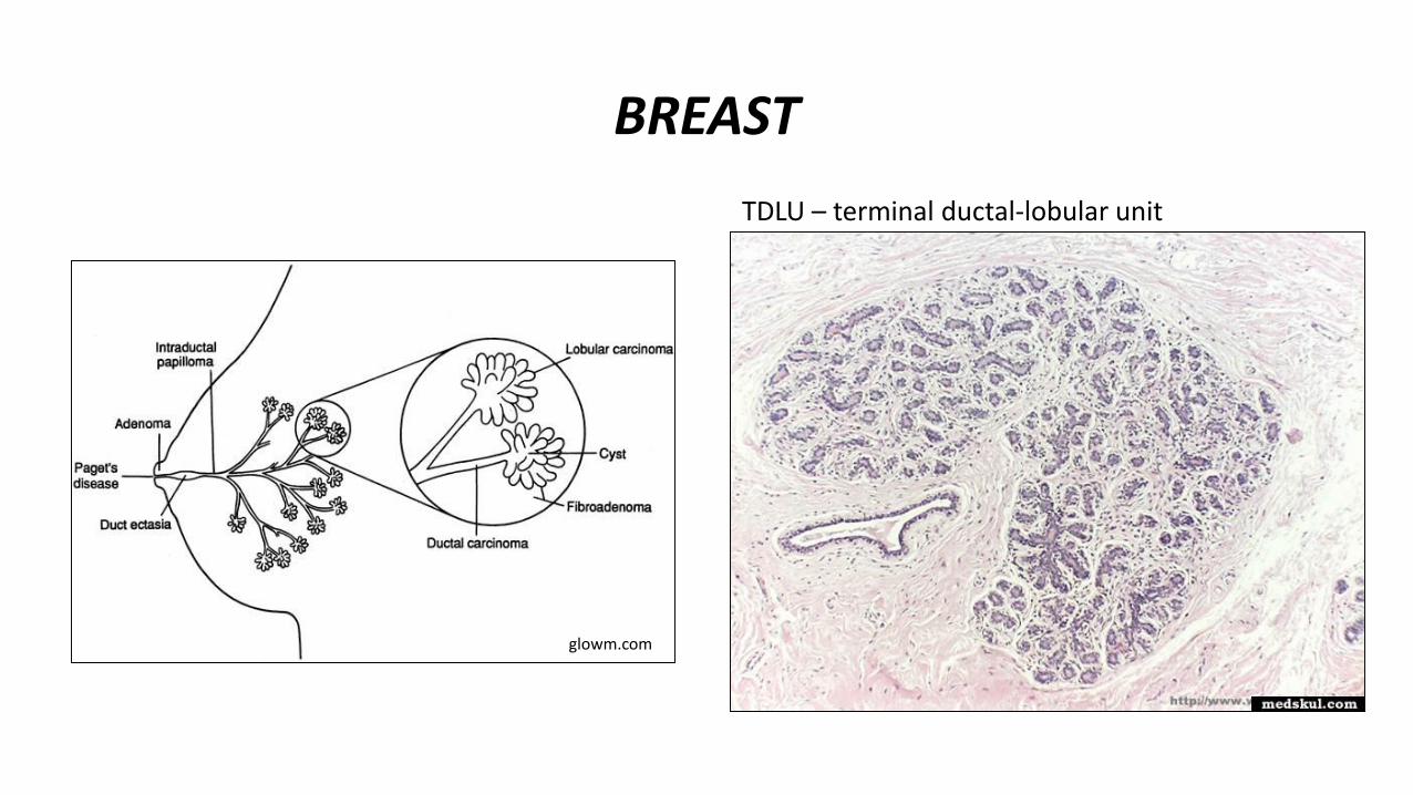
BREAST
glowm.com
TDLU – terminal ductal-lobular unit
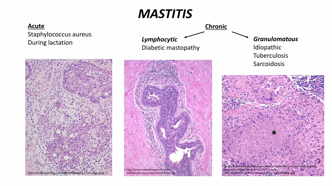
MASTITISAcuteStaphylococcus aureusDuring lactation
Chronic
LymphocyticDiabetic mastopathy
GranulomatousIdiopathicTuberculosisSarcoidosis
https://farm9.staticflickr.com/8341/8199838977_111f2c7916_b.jpghttp://www.webpathology.com/slides-13/slides/Breast_LymphocyticMastitis2.jpg
https://pubs.rsna.org/na101/home/literatum/publisher/rsna/journals/content/radiographics/2005/radiographics.2005.25.issue-2/rg.252045077/20141024/images/medium/g05mr09g8b.jpeg
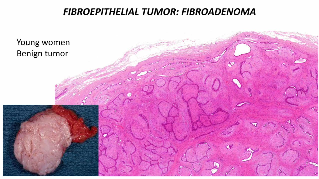
FIBROEPITHELIAL TUMOR: FIBROADENOMA
Young womenBenign tumor
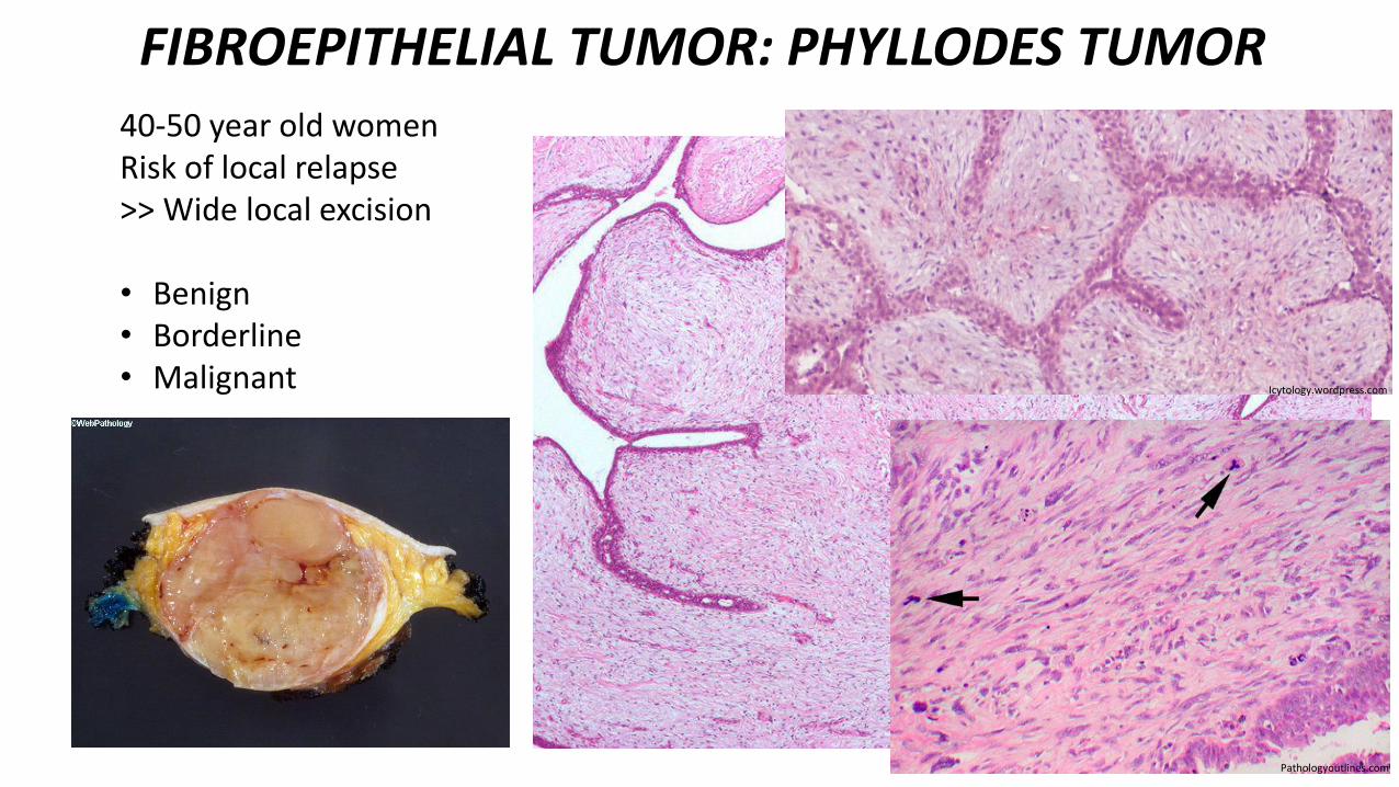
FIBROEPITHELIAL TUMOR: PHYLLODES TUMOR40-50 year old womenRisk of local relapse>> Wide local excision
• Benign• Borderline• Malignant
En.wikipedia.org
Icytology.wordpress.com
Pathologyoutlines.com
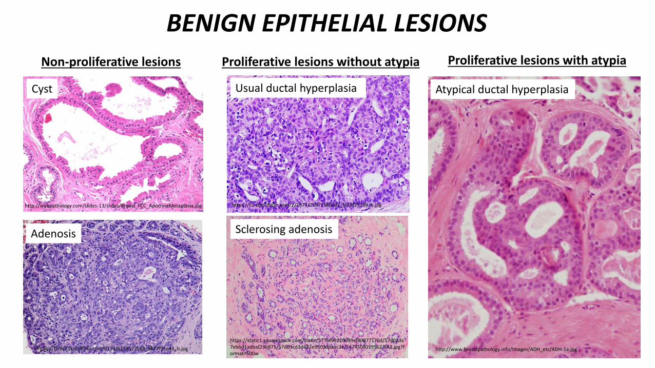
BENIGN EPITHELIAL LESIONS
Non-proliferative lesions
http://webpathology.com/slides-13/slides/Breast_FCC_ApocrineMetaplasia.jpg
https://farm7.staticflickr.com/6153/6158572518_8b77f95e43_b.jpg
Cyst
Adenosis
Proliferative lesions without atypia Proliferative lesions with atypia
https://c1.staticflickr.com/2/1674/25877580402_fa7df25109_b.jpg
Sclerosing adenosis
https://static1.squarespace.com/static/577bf99220099ef80877176d/57d02da7ebbd1adbaf23b871/57db3cd3d482e9509ddaac3e/1474508019562/SA3.jpg?format=500w
Atypical ductal hyperplasia
http://www.breastpathology.info/Images/ADH_etc/ADH-1a.jpg
Usual ductal hyperplasia
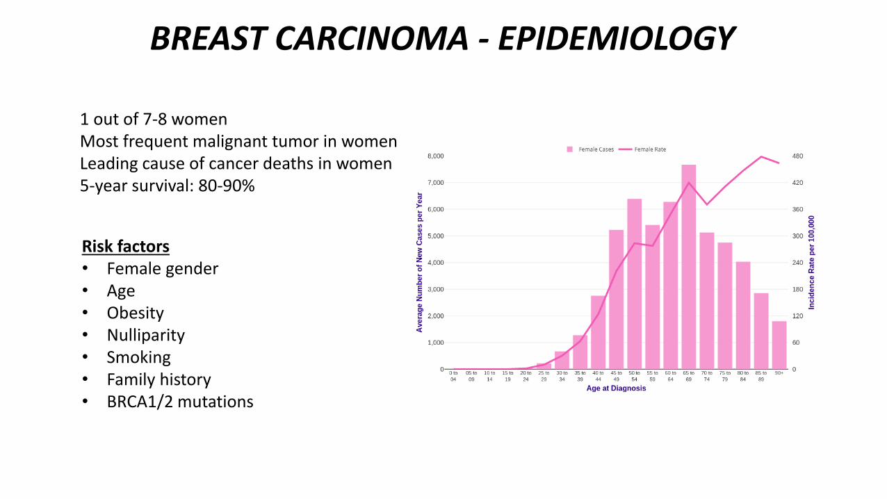
BREAST CARCINOMA - EPIDEMIOLOGY
1 out of 7-8 womenMost frequent malignant tumor in womenLeading cause of cancer deaths in women5-year survival: 80-90%
Risk factors• Female gender• Age• Obesity• Nulliparity• Smoking• Family history• BRCA1/2 mutations
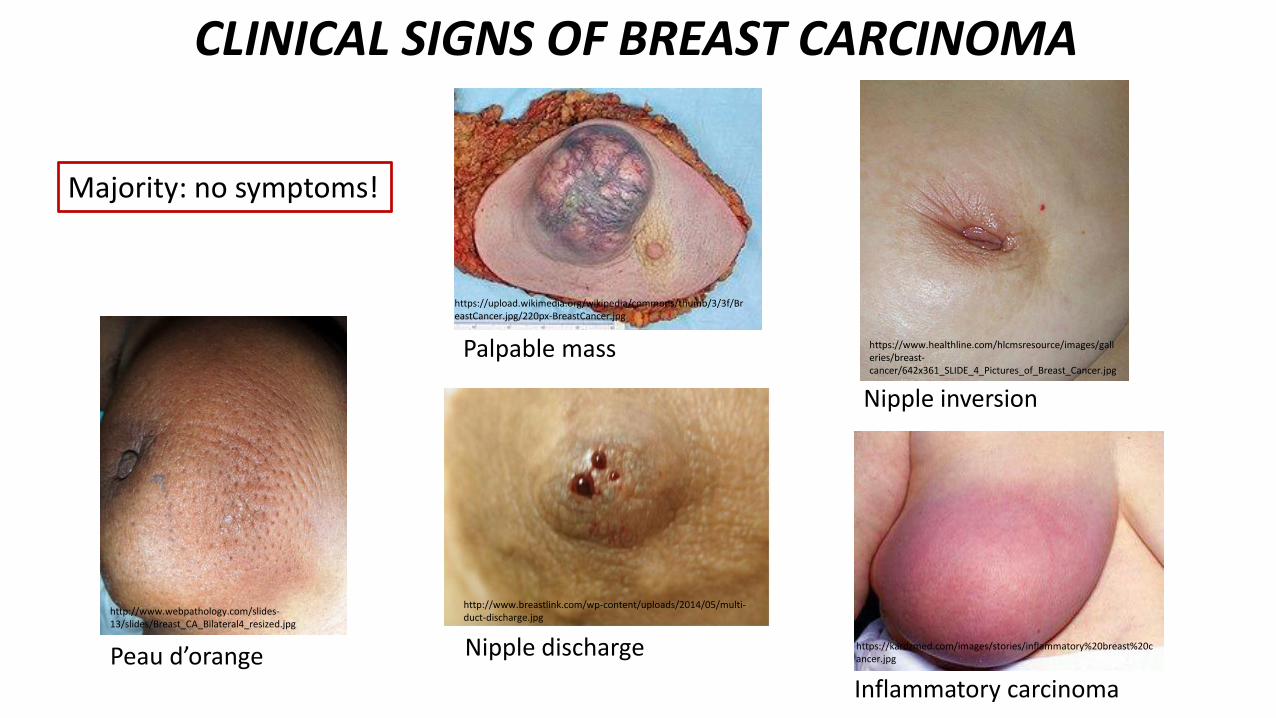
CLINICAL SIGNS OF BREAST CARCINOMA
http://www.webpathology.com/slides-13/slides/Breast_CA_Bilateral4_resized.jpg
https://www.healthline.com/hlcmsresource/images/galleries/breast-cancer/642x361_SLIDE_4_Pictures_of_Breast_Cancer.jpg
Peau d’orangeInflammatory carcinoma
Nipple discharge
http://www.breastlink.com/wp-content/uploads/2014/05/multi-duct-discharge.jpg
https://upload.wikimedia.org/wikipedia/commons/thumb/3/3f/BreastCancer.jpg/220px-BreastCancer.jpg
Palpable mass
Nipple inversion
https://kardzmed.com/images/stories/inflammatory%20breast%20cancer.jpg
Majority: no symptoms!
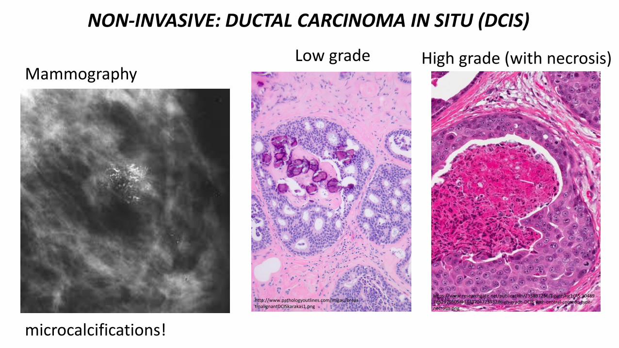
NON-INVASIVE: DUCTAL CARCINOMA IN SITU (DCIS)
http://www.pathologyoutlines.com/imgau/breastmalignantDCISkarakas1.png
https://www.researchgate.net/publication/235897256/figure/fig1/AS:304897862979605@1449704723332/High-grade-DCIS-with-central-comedo-type-necrosis.png
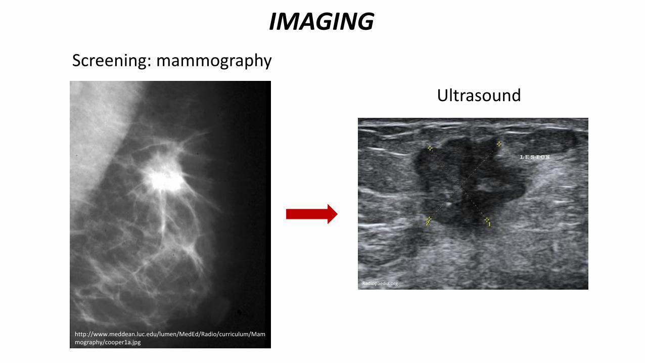
Mammography
microcalcifications!
Low grade High grade (with necrosis)
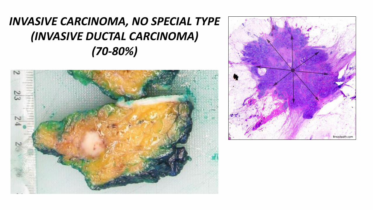
INVASIVE CARCINOMA, NO SPECIAL TYPE(INVASIVE DUCTAL CARCINOMA)
(70-80%)
Breastpath.com
http://pathology.jhu.edu/breast/grade.php http://tgmouse.compmed.ucdavis.edu/jensen-mamm2000/brca-1/brca-1.html
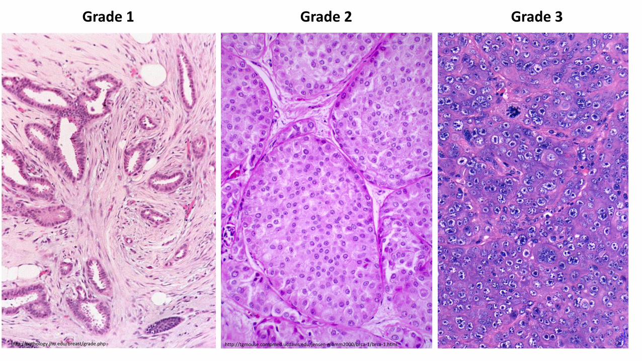
Grade 1 Grade 2 Grade 3
http://www.pathologyoutlines.com/caseofweek/case413image8.jpg
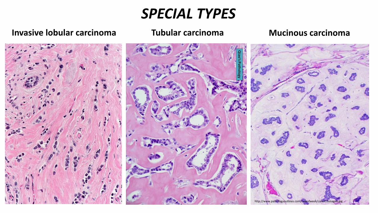
Tubular carcinoma Mucinous carcinoma
SPECIAL TYPESInvasive lobular carcinoma
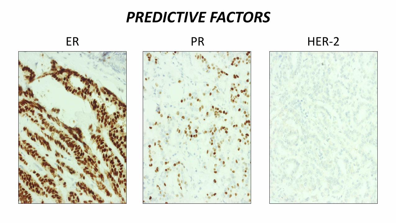
ER HER-2PR
PREDICTIVE FACTORS
https://www.researchgate.net/figure/HER2-protein-expression-detected-by-IHC-400-A-Negative-score-0-B-Negative-score_fig6_280031002
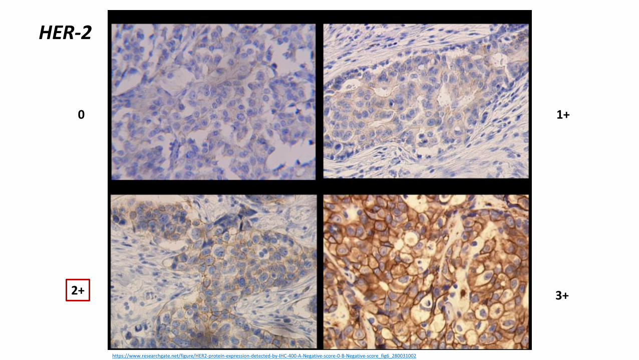
HER-2
0 1+
3+2+
wjso.com
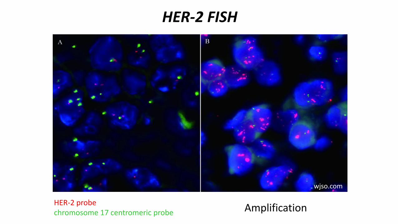
HER-2 FISH
AmplificationHER-2 probechromosome 17 centromeric probe
https://breastcancersurgery.com/wp-content/uploads/2016/12/img-lymph-node.jpg
http://cdn.healthandsymptoms.com/wp-content/uploads/2016/09/The-Basics-of-Stage-4-Breast-Cancer-in-the-Lungs-featured-720x381.jpg
https://www.aerzteblatt.de/image.asp?id=57107&w=550
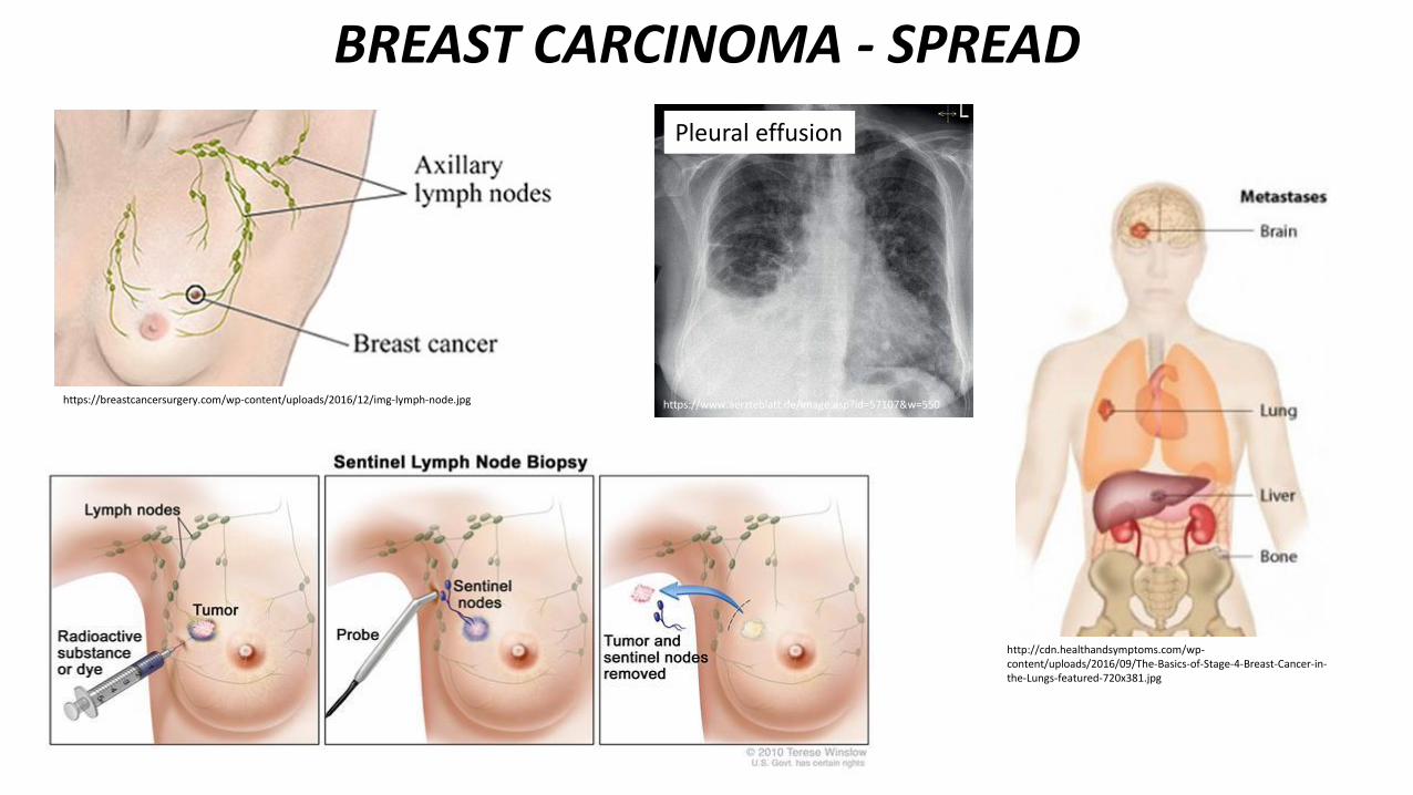
BREAST CARCINOMA - SPREAD
Pleural effusion
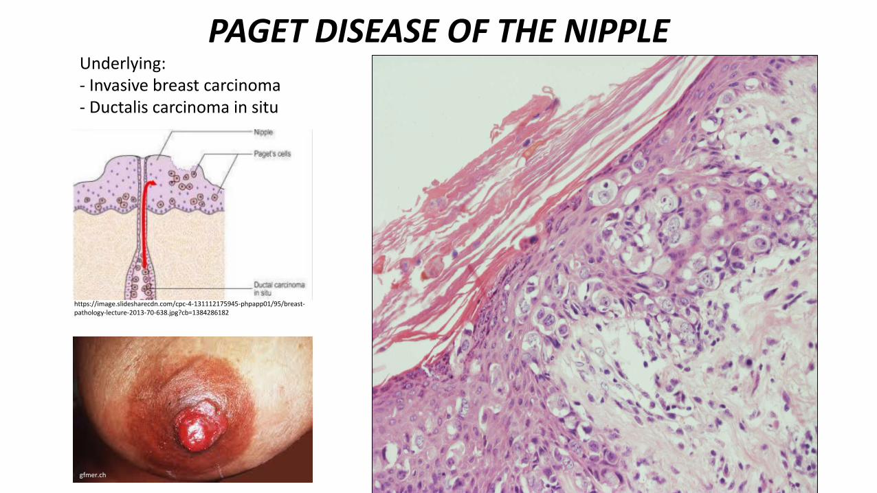
PAGET DISEASE OF THE NIPPLE
gfmer.ch
Underlying:- Invasive breast carcinoma- Ductalis carcinoma in situ
https://image.slidesharecdn.com/cpc-4-131112175945-phpapp01/95/breast-pathology-lecture-2013-70-638.jpg?cb=1384286182
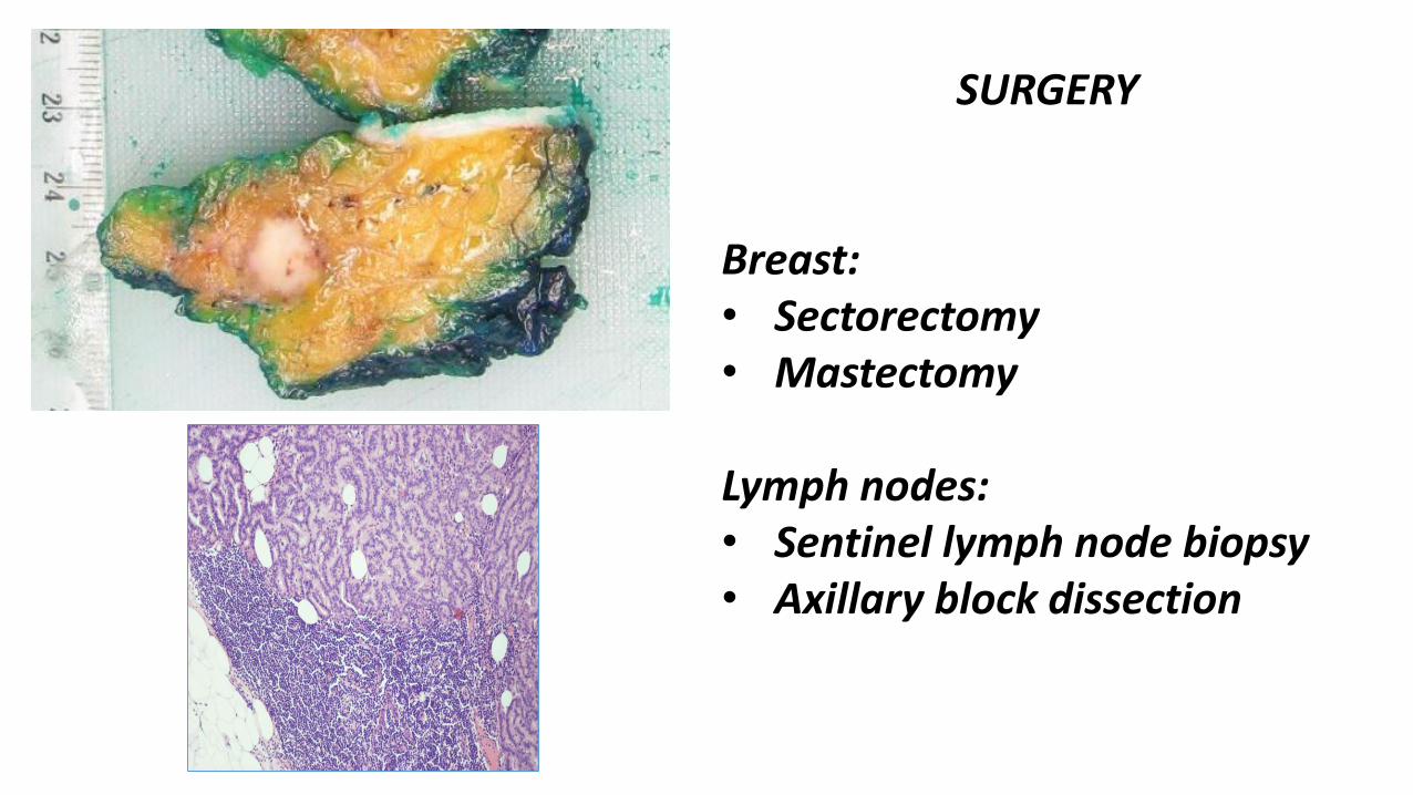
SURGERY
Breast:• Sectorectomy• Mastectomy
Lymph nodes: • Sentinel lymph node biopsy• Axillary block dissection
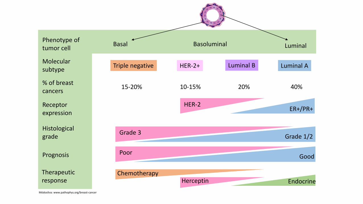
Triple negative HER-2+ Luminal B Luminal A
15-20% 10-15% 20% 40%
HER-2ER+/PR+
Grade 3Grade 1/2
PoorGood
EndocrineHerceptinChemotherapy
Basal LuminalBasoluminal
Molecularsubtype
% of breastcancers
Receptor expression
Histologicalgrade
Prognosis
Therapeuticresponse
Phenotype of tumor cell
Módosítva: www.pathophys.org/breast-cancer
http://www.meddean.luc.edu/lumen/MedEd/Radio/curriculum/Mammography/cooper1a.jpg
Screening: mammography
Ultrasound
IMAGING
Radiopaedia.org
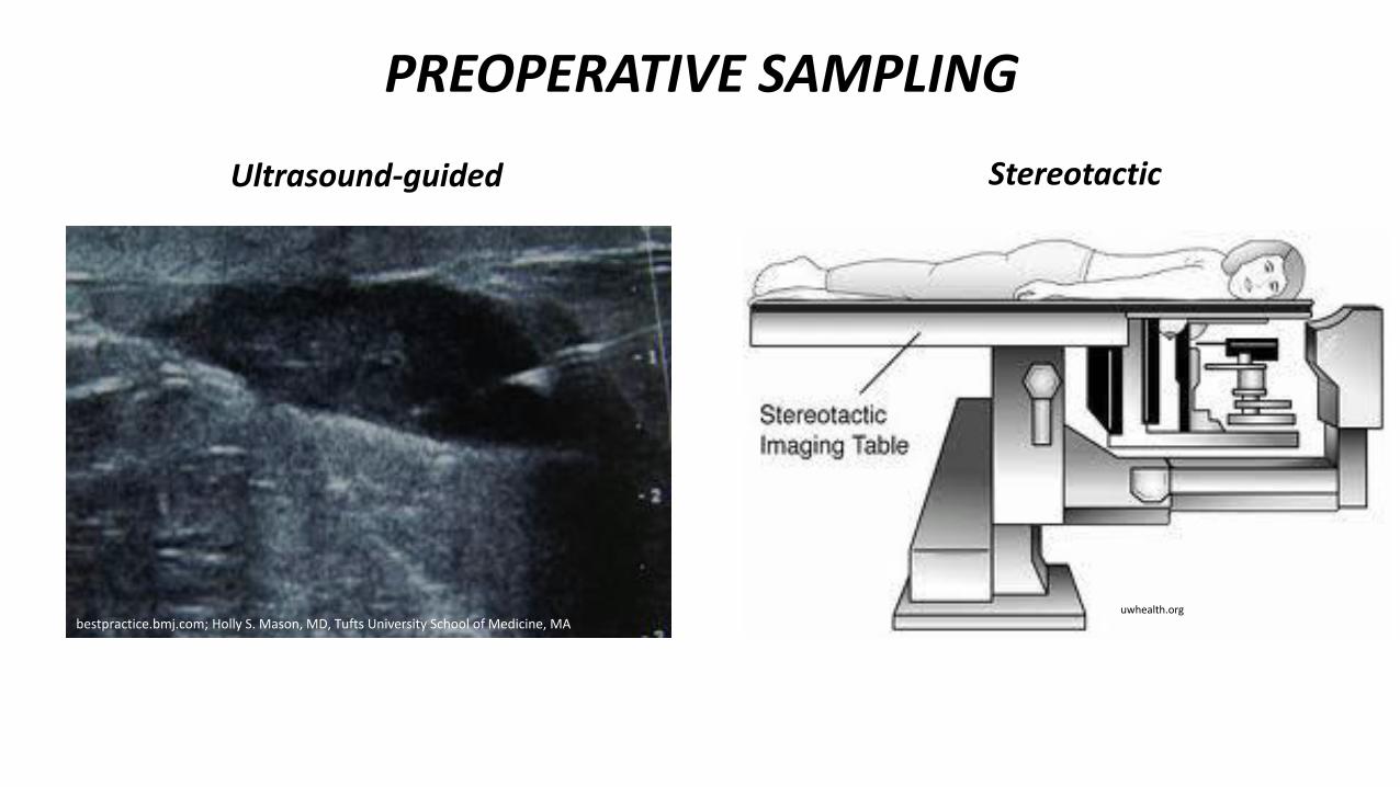
bestpractice.bmj.com; Holly S. Mason, MD, Tufts University School of Medicine, MAuwhealth.org
PREOPERATIVE SAMPLING
Ultrasound-guided Stereotactic
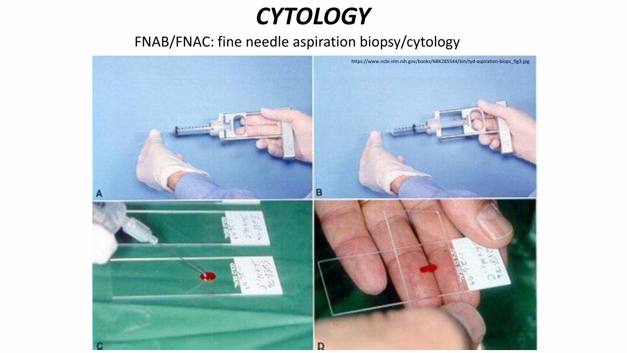
CYTOLOGYFNAB/FNAC: fine needle aspiration biopsy/cytology
https://www.ncbi.nlm.nih.gov/books/NBK285544/bin/tyd-aspiration-biops_fig3.jpg
https://posterng.netkey.at/esr/viewing/index.php?module=viewimage&task=&mediafile_id=412254&201201302221.jpg
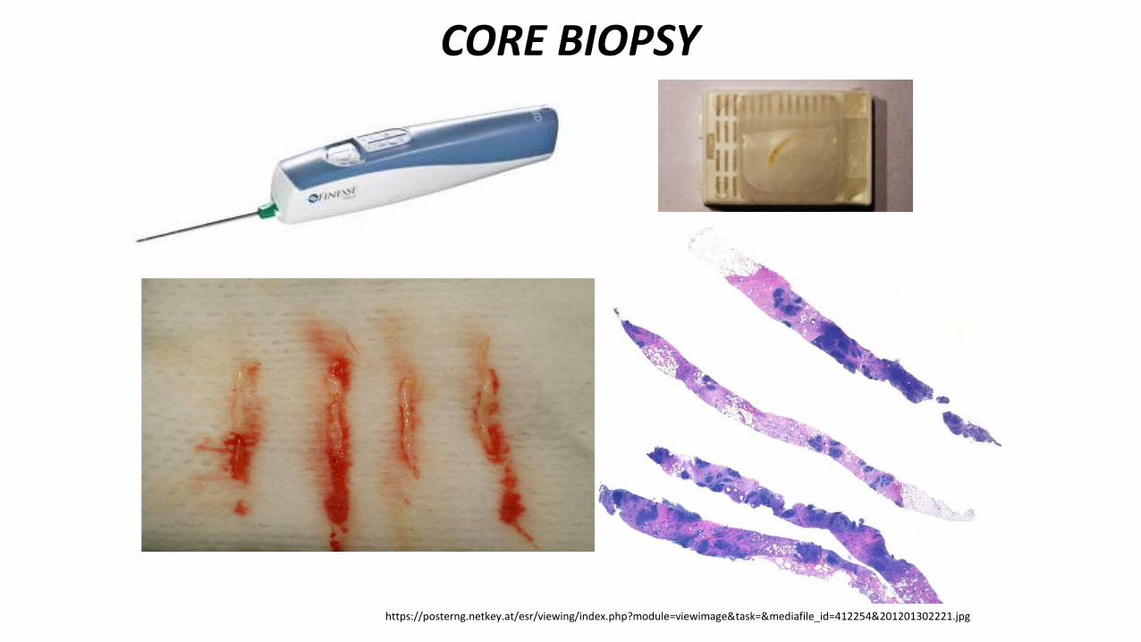
CORE BIOPSY
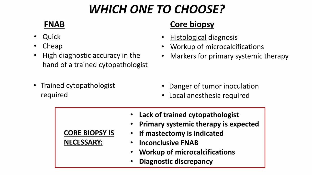
WHICH ONE TO CHOOSE?
• Histological diagnosis• Workup of microcalcifications• Markers for primary systemic therapy
FNAB Core biopsy
• Danger of tumor inoculation• Local anesthesia required
• Quick • Cheap• High diagnostic accuracy in the
hand of a trained cytopathologist
• Trained cytopathologistrequired
• Lack of trained cytopathologist• Primary systemic therapy is expected• If mastectomy is indicated• Inconclusive FNAB• Workup of microcalcifications• Diagnostic discrepancy
CORE BIOPSY IS NECESSARY:
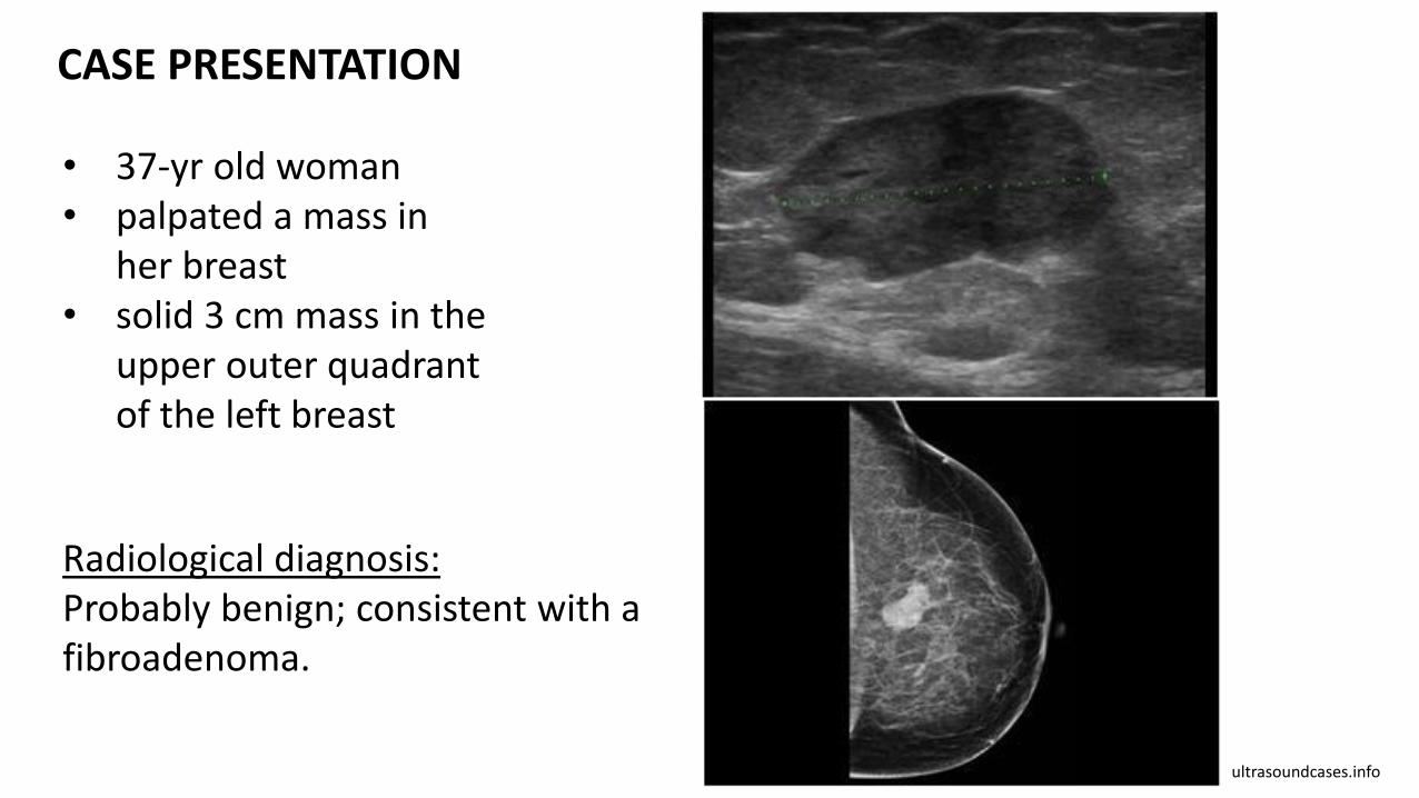
• 37-yr old woman• palpated a mass in
her breast• solid 3 cm mass in the
upper outer quadrantof the left breast
ultrasoundcases.info
Radiological diagnosis:Probably benign; consistent with a fibroadenoma.
CASE PRESENTATION
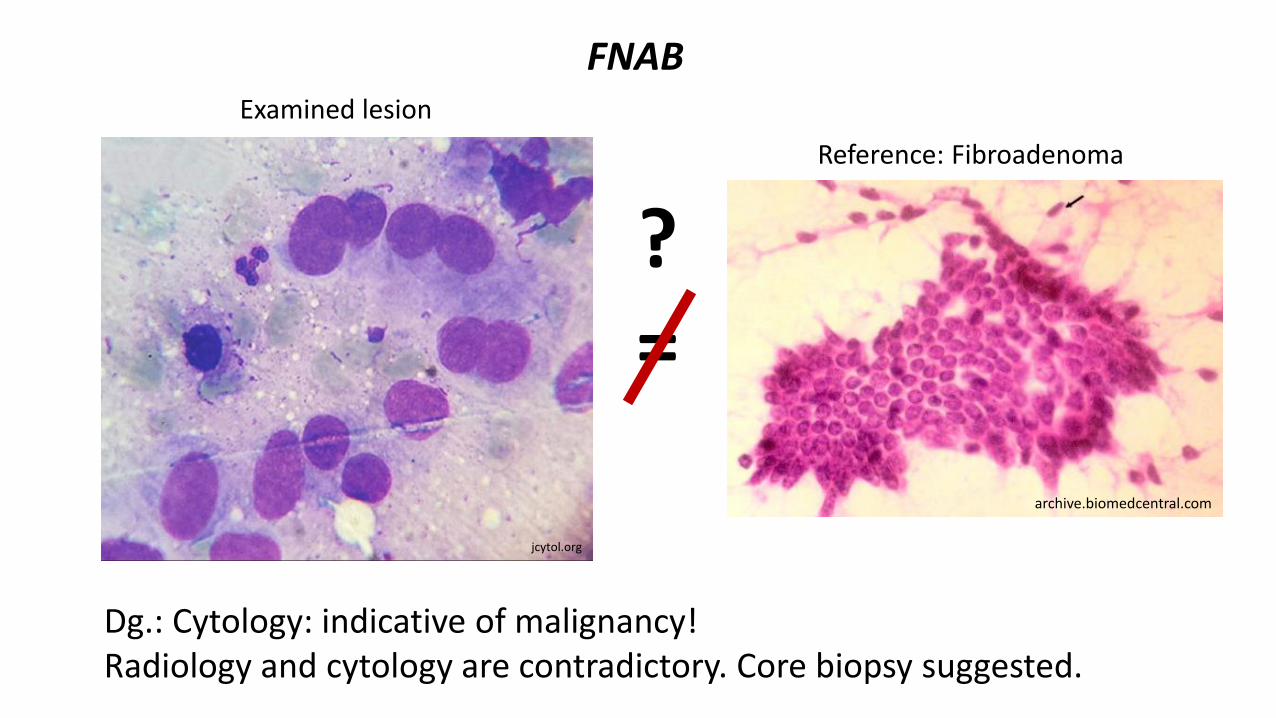
Dg.: Cytology: indicative of malignancy!Radiology and cytology are contradictory. Core biopsy suggested.
FNAB
jcytol.org
archive.biomedcentral.com
Reference: Fibroadenoma
Examined lesion
?=
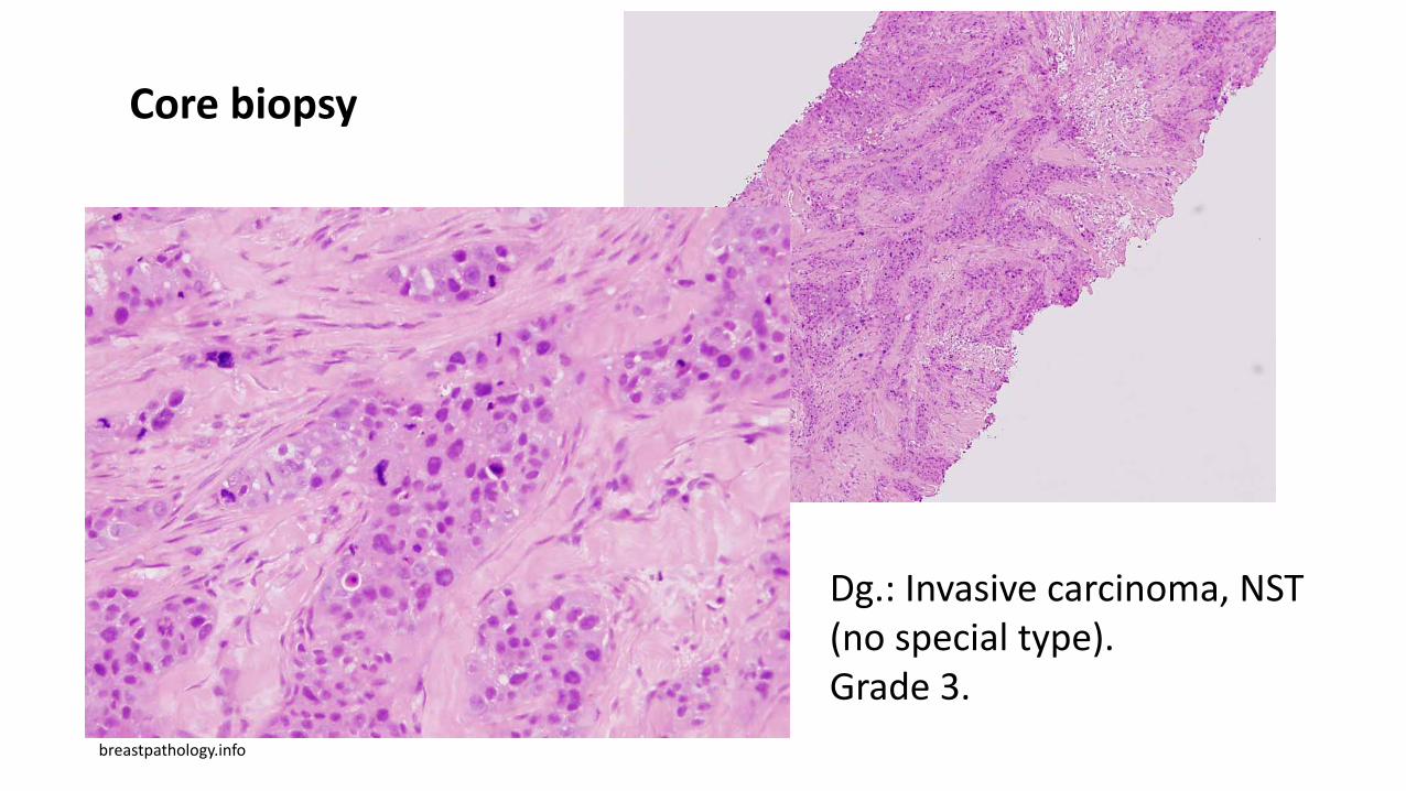
Core biopsy
breastpathology.info
Dg.: Invasive carcinoma, NST (no special type). Grade 3.
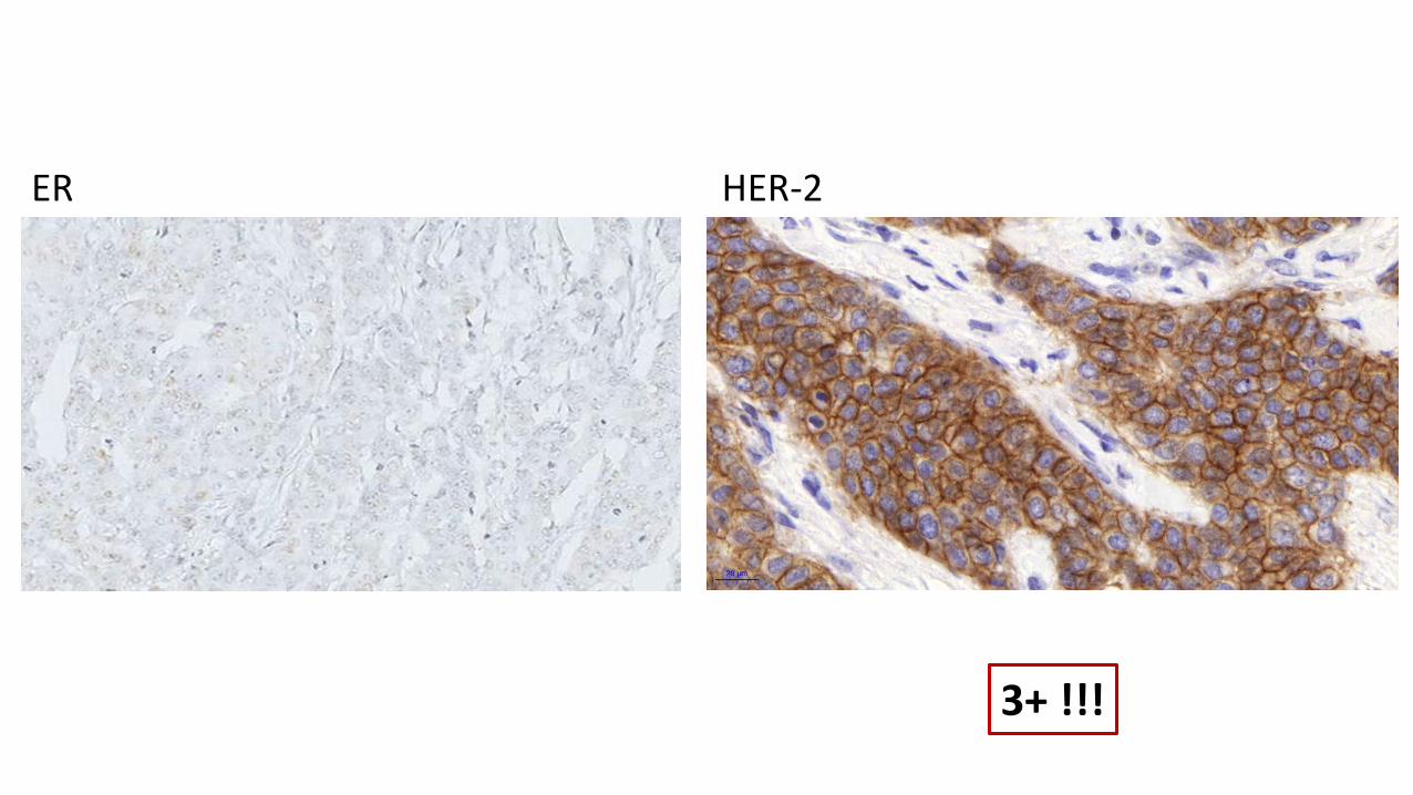
ER HER-2
3+ !!!