Embed Size (px)
Citation preview

ORIGINAL RESEARCH
12 WOUNDS® www.woundsresearch.com
Abstract: Introduction. Wounds are a major problem at the end of life, affecting nearly one-third of hospice patients. The main patient com-plaints associated with these wounds are pain, infection, and odor, often making wound care difficult due to patient discomfort and the complicated nature of the wounds. Materials and Methods. A method of wound treatment invented in 2001 for palliative wound care was designed to meet the goals of pain relief, odor control, and infection prevention. This method, which involved application of viscous lido-caine and topical polymyxin/bacitracin to gauze, was used on hun-dreds of hospice patients and, later, on non-hospice patients as well. A retrospective review of results was undertaken. Results. This paper presents 2 descriptive retrospective observational reviews. One is a retrospective review of 323 wounds in hospice patients treated over a period of 30 months using the palliative wound treatment method. In this cohort, more than 40% of pressure ulcers healed to closure or were healing by the end of the study; 30% of ischemic ulcers were healed to closure or were healing by the end of the study,; and more than 40% of neuropathic ulcers were healing by the end of the study. In total, nearly 40% of all wounds were in the process of heal-ing or healed to closure. The second study is a retrospective review of nursing home data using the palliative wound treatment method. In a cohort of 72 patients with 156 mixed wound types followed for 1 year, rates of healing to closure approached 95% after 12 weeks of treatment. Conclusion. While meeting palliative goals of reducing pain, preventing infection, and controlling odor, use of this method also provided unexpected healing. Further investigation on use of this method is warranted.
Key words: palliative wound care, pressure ulcers, diabetic ulcers, ischemic ulcer
WOUNDS 2015;27(1):12-19
From AT Squared, LLC, Blue Ash, OH
Address correspondence to:Aletha W. Tippett, MDAT Squared10274 Alliance RoadBlue Ash, OH [email protected]
Disclosure: The author discloses she is owner and president of AT Squared, but this presents no financial or other conflict of interest in relation to this manuscript.
This material was previously presented in a lecture, “Novel Palliative Wound Dressing Promotes Healing,” at the Symposium on Advanced Wound Care & Wound Healing Society 2007; April 28-May 1, 2007; Tampa, FL.
Palliative Wound Treatment Promotes Healing
Aletha W. Tippett, MD
Published data report 35% of hospice patients have wounds. Of those wounds, 50% are pressure ulcers and 20% are ischemic.1,2 The remaining 30% are a mix of various wound types, including
surgical, stasis, skin tears, and tumors.1,2 With nearly 2 million patients currently in hospice care,3 this correlates to approximately 700,000 wounds needing care.
Most chronic wounds are pressure ulcers, a tragic problem for millions of Americans. Five million to seven million Americans develop a chronic
DO NOT D
UPLICATE

Tippett
Vol. 27, No. 1 January 2015 13
wound each year, with the incidence increasing by 10% per year,4 of which 2.5 million of these chronic wounds are pressure ulcers, remarkably close to the 50% noted in hospice.1,5 Pressure ulcers are a signifi-cant problem for people who are frail and elderly,6 but also for people who are paralyzed from spinal cord or brain injury,7 or from neurodegenerative diseases such as multiple sclerosis or Huntington’s Disease.8 Pres-sure ulcers typically occur over a bony prominence that has unrelieved pressure from normal gravitational body force,9 such as the sacrum and heels from lying in bed and ischial tuberosities from sitting. In the United States alone, the pressure ulcer burden is staggering, costing in excess of $9.1 billion for treatment annu-ally.5 These pressure ulcers are full-thickness, often very deep, painful, and prone to infection. Most com-monly, these wounds are located on the sacrum or in the pelvic region.1 These wound locations can make treatment very difficult. Contamination with urine and feces is a frequent problem, and fastening a dressing to the wound may be quite difficult due to size require-ments, body shape, moisture, fragile/thin skin that tears easily, or the presence of body hair.
A dressing to effectively treat wounds in the sacral and pelvic region is highly sought after by wound care providers.10,11 There are currently thousands11,12 of pressure ulcer products on the market, yet, while wounds and wound care are fraught with pain, few dressings provide much-needed pain relief. Patients have described pain as the worst part of living with a chronic wound.12 Pain is a complex biopsychosocial phenomenon that requires multiple pharmacological and nonpharmacological management approaches.12
All open wounds are contaminated with bacte-ria, sometimes leading to wound infection which is a scourge to patients and caregivers, with some esti-mates as high as 30% of wounds affected.13 A wound dressing with antimicrobial activity against common wound pathogens would be advantageous.
For hospice patients at the end of life, goals of care are pain relief, infection prevention, comfort, and dig-nity. Healing of wounds or wound closure are not con-sidered primary goals for terminal patients, whose average life expectancy is approximately 67 days.3 In 2001, a hospice physician developed a method to meet the palliative objectives of pain reduction, odor con-trol, and infection prevention for wounds in hospice patients.1 Since 2001 this method has been in use on hundreds, if not thousands, of patients in hospice and
later for patients not in hospice by the physician who did the original study and invented the treatment.
Development of wound treatment. The method was developed in 2001 at the bedside, primarily to provide pain relief in dying patients with painful pressure ul-cers. At that time there were no guidelines for palliative wound care, and wound healing was seen as an unreal-istic goal of treatment in this patient population.14 This technique was not developed from clinical trials or lab-oratory review, but from a clinical and pharmacologi-cal knowledge base of using lidocaine to achieve the level of pain relief desired for palliative wound care. The method involved application of viscous lidocaine and a topical antibiotic to gauze.
Prior to use, an extensive literature search was con-ducted to investigate the use of lidocaine in wounds. Animal studies showed mixed results for adequate healing in wounds treated with lidocaine.15-19 Human studies using lidocaine for burns showed it was safe for topical use on a wound, as long as it is below toxicity level.20,21 Nothing in the literature was found to either support or contradict use of lidocaine in the wound bed for pain relief, yet it is known that topical lidocaine can decrease pain.22 Great care was taken to ensure the amounts of lidocaine used were below toxicity level. Brofeldt et al20 studied application of 5% lidocaine to burns up to 28% of total body surface area (BSA). Se-rum levels never reached toxicity level in his study.20
The palliative wound treatment has less than 2% lido-caine in application, so is safe for very large wound ar-
Figure 1. Total healing results by wound type. Study A results in the treatment of 231 wounds in hospice patients. De-picted are the 3 most prevalent wound types treated: pres-sure, arterial (ischemic), and neuropathic (diabetic). Used with permission.1
DO NOT D
UPLICATE

Tippett
14 WOUNDS® www.woundsresearch.com
eas, greater than 28% BSA. A viscous lidocaine (contain-ing 2% lidocaine) was selected because of its known safety in common use with humans, low percentage of lidocaine, and because it contains carboxymethylcel-lulose, which makes a hydrogel that adheres well in an open wound. Hydrogels have an observed benefit in treating various types of wounds, believed to be due to the promotion of the body’s intrinsic autolysis pro-cess.23,24 When viscous lidocaine was used, the mixture contained 1% lidocaine, an amount that has shown safe reduction of pain when applied on a wound.22
Because of the theoretical risk of increased infec-tion due to an altered inflammatory response,25 anti-biotics were added with the lidocaine. A simple mix-ture of polymyxin and bacitracin was created by hand, squeezing approximately 3-4 g viscous lidocaine on gauze, then sprinkling approximately 0.1 g of antibi-otic powder over the lidocaine. The mixture was based on reports suggesting this may improve wound heal-ing.26 In vitro efficacy testing later performed on this mixture showed inhibition of 4 of the major wound pathogens: Escherichia coli, Staphylococcus aureus, Candida albicans, and Pseudomonas aeruginosa.27 The addition of antibiotics provided good inhibitory effects against major wound pathogens, but the lido-caine can also contribute to antibacterial effects and bacterial minimization.28,29
Malodor is a known problem with wounds but the general consensus at the time of this invention was that good cleansing and effective treatment of infec-tion helped reduce malodor. Using an agent such as topical metronidazole for odor was first reported30 in 1993 but was not yet standard of care at the time of development of this method. For the population in this treatment cohort, relieving the patient of the burden of foul odor was paramount. With that rationale, oil of wintergreen was added, using several drops for each
wound treated. Oil of wintergreen has a strong and cooling aroma, and it contains 85%-99% methyl salicy-late which is aspirin-like and can contribute to pain relief as promoted in commercial literature.31 The Na-tional Center for Biotechnology Information features scientific information on the main ingredient in oil of wintergreen, methyl salicylate.32 Since 2001, more re-search has been conducted on options for controlling odor with different agents,33,34
An important characteristic is that since the proce-dure was useful for any type of wound, it was not nec-essary to characterize the wound. Depending on the type and location of the wound dressed and the top dressing used, the dressing required changing every 1-7 days.
Materials and MethodsThis is a retrospective observational chart review of
2 different cohorts. Study A included 192 hospice pa-tients with 323 wounds treated over 30 months; Study B included treatment of 156 pressure ulcers and other wounds in 72 nursing home patients over 12 months. Study A is from previously published data and results;1
the current report details specific treatment not cov-ered in the previous report.
Study A. Study A consisted of 192 hospice patients with 323 wounds treated over a 30-month period. Types of wounds included pressure, neuropathic, isch-emic, and other. Wound treatment selection was deter-mined by hospice physician recommendation and pa-tient/caregiver consent. Consent was obtained as part of standard practice for any treatment offered. With patient or caregiver permission, wounds were treated with the mixture of lidocaine and topical antibiotic, moistened as needed with normal saline, and applied on a gauze to fit the wound. Wound dressings were ei-ther changed daily, less frequently if the wound was dry, or more frequently if there was heavy drainage. Not all patients or caregivers gave permission for use of this treatment; some were afraid of using lidocaine in the wound, some did not perceive it as aggressive treatment, and some thought it seemed too unusual to work. Therefore there were 92 (28%) wounds treated with another modality based on their physician’s or-ders, including various types of dressings such as wet-to-dry, hydrocolloids, alginates, or vacuum-assisted clo-sure (VAC).1 The new method, which will be referred to as the novel dressing, was used to treat 231 (72%) wounds. Ninety-two wounds were treated with other
Keypoints
• For hospice patients at the end of life, goals of care are pain relief, infection prevention, comfort, and dignity.
• A method involving the application of viscous lido-caine and a topical antibiotic to gauze was devel-oped in 2001, primarily to provide pain relief in dying patients.
• Relieving the patient of the burden of foul odor was also paramount; oil of wintergreen was added, using several drops for each wound treated.
DO NOT D
UPLICATE

Tippett
Vol. 27, No. 1 January 2015 15
methods. Wound healing was determined by the tradi-tional method of measuring length and width, evaluat-ing the amount of drainage, and assessing the wound tissue for granulation, slough, or necrosis. A wound was considered to be healing if it showed contracture with granulation and decreased size. Wounds were assessed weekly by a nurse, and every 1-3 weeks by the hospice physician. Since the palliative goals of care were to re-lieve pain, control odor, and prevent infection, these parameters were evaluated with each assessment. Since the majority of patients were nonverbal no ana-log scales were used; evaluations depended on nursing assessments. Pain assessment was made by observing patient facial expression, body movement, and noting if the patient moaned or cried during treatment.
This method provided a primary wound dressing, but a secondary dressing was usually needed. In keep-ing with the desire for simplicity and maintaining com-fort, the top dressing was provided by applying a thick layer of zinc oxide ointment around the wound with a sheet of plastic wrap placed on top and pressed into the zinc oxide ointment.
Study B. As stated, use of this treatment method expanded from hospice to non-hospice in the wound center of a long term care facility (Clovernook Health Care Pavilion, Cincinnati, OH) which used it exclu-sively. Over a 1-year period in this nursing home, 156 wounds—including venous, diabetic, surgical, isch-emic, and stage 2-4 pressure ulcers—in 72 patients were treated with the lidocaine and antibiotic mixture on gauze, which was changed daily. Some of these pa-tients actually were in hospice, but not identified in Study B. Wound healing was judged by the traditional method of measuring length and width, evaluating the amount of drainage, and assessing the wound tissue for granulation, slough, or necrosis. A wound was consid-
ered to be healing if it showed contracture with gran-ulation and decreased size. Pain and odor were also observed, but not reported in Study B. Wounds were assessed weekly by the wound nurse, who held wound care certification (WCC) from the National Alliance of Wound Care and Ostomy, and every 1-2 weeks by the director of wound care for the nursing home. Wounds were followed to closure. In this nursing home, all pa-tients with wounds also had static air pressure support with mattress overlays and chair cushions (EHOB, Inc, Indianapolis, IN). This data was from nursing home wound records, and was not published.
ResultsStudy A. For Study A, the median age of patients
treated was 82. Most patients lived less than 30 days, with a median of 31 days. Healing to closure was not an endpoint in treatment of this cohort as most of the patients would not live long enough for their wound to achieve closure. According to nursing reports, odor and pain were reduced. Pain relief was immediate in most cases, and persisted with use of the novel dressing. In the cases treated, no infections occurred with use of the dressing. Odor was also well controlled. Besides re-ducing pain, preventing infection, and controlling odor, healing results were surprising. More than 40% of pres-sure ulcers healed to closure or were healing; 30% of ischemic ulcers were healed to closure or were healing; and more than 40% of neuropathic ulcers were healing (Figure 1). There were no allergic reactions reported. Allergic reaction to lidocaine has been found to be low, < 0.7%.22 The top dressing used was very successful in
Keypoints
• This is a retrospective observational chart review of 2 different cohorts who were treated with the palliative wound care method.
• Study A included 192 hospice patients with 323 wounds treated over 30 months; the lidocaine and antibiotic mixture on gauze dressing was used to treat 231 (72%) wounds.
• Study B included treatment of 156 pressure ulcers and other wounds in 72 nursing home patients over 12 months; all wounds were treated with the novel dressing.
Figure 2. Comparison of healing rates of novel dressing vs other treatments.
DO NOT D
UPLICATE
Tippett
16 WOUNDS® www.woundsresearch.com
that it kept the wound dressing moist and warm, which are both adjuvants to healing,35 and prevented trauma to periwound skin with dressing changes.
Included in Study A were 24 diabetic foot wounds treated over the 30-month period. Eleven (46%) healed to closure or were healing, no new wound infections presented, and there were no amputations.
A comparison of 231 wounds treated with the novel dressing vs the 92 wounds receiving other treatments is shown in Figure 2. Overall, 40% of wounds treated with the novel dressing healed to closure or were heal-ing vs 10% of wounds treated with other modalities.
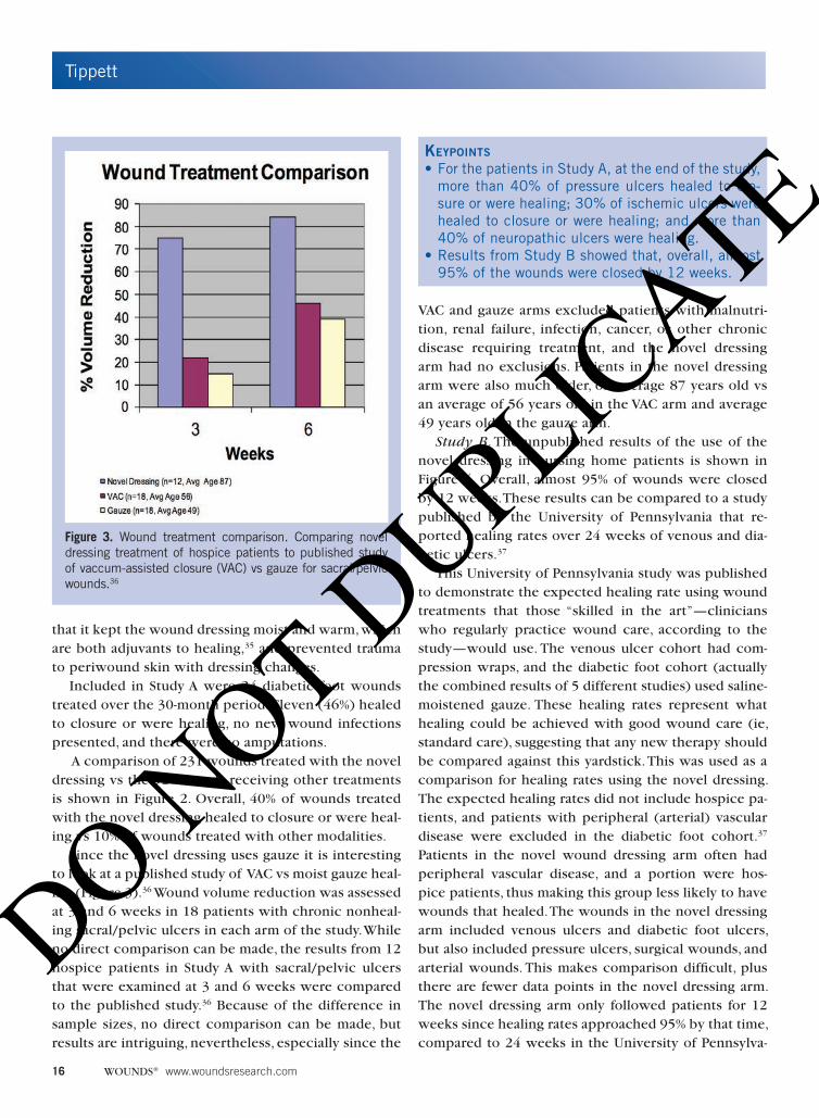
Since the novel dressing uses gauze it is interesting to look at a published study of VAC vs moist gauze heal-ing (Figure 3).36 Wound volume reduction was assessed at 3 and 6 weeks in 18 patients with chronic nonheal-ing sacral/pelvic ulcers in each arm of the study. While no direct comparison can be made, the results from 12 hospice patients in Study A with sacral/pelvic ulcers that were examined at 3 and 6 weeks were compared to the published study.36 Because of the difference in sample sizes, no direct comparison can be made, but results are intriguing, nevertheless, especially since the
VAC and gauze arms excluded patients with malnutri-tion, renal failure, infection, cancer, or other chronic disease requiring treatment, and the novel dressing arm had no exclusions. Patients in the novel dressing arm were also much older, on average 87 years old vs an average of 56 years old in the VAC arm and average 49 years old in the gauze arm.
Study B. The unpublished results of the use of the novel dressing in nursing home patients is shown in Figure 4. Overall, almost 95% of wounds were closed by 12 weeks. These results can be compared to a study published by the University of Pennsylvania that re-ported healing rates over 24 weeks of venous and dia-betic ulcers.37
This University of Pennsylvania study was published to demonstrate the expected healing rate using wound treatments that those “skilled in the art”—clinicians who regularly practice wound care, according to the study—would use. The venous ulcer cohort had com-pression wraps, and the diabetic foot cohort (actually the combined results of 5 different studies) used saline-moistened gauze. These healing rates represent what healing could be achieved with good wound care (ie, standard care), suggesting that any new therapy should be compared against this yardstick. This was used as a comparison for healing rates using the novel dressing. The expected healing rates did not include hospice pa-tients, and patients with peripheral (arterial) vascular disease were excluded in the diabetic foot cohort.37 Patients in the novel wound dressing arm often had peripheral vascular disease, and a portion were hos-pice patients, thus making this group less likely to have wounds that healed. The wounds in the novel dressing arm included venous ulcers and diabetic foot ulcers, but also included pressure ulcers, surgical wounds, and arterial wounds. This makes comparison difficult, plus there are fewer data points in the novel dressing arm. The novel dressing arm only followed patients for 12 weeks since healing rates approached 95% by that time, compared to 24 weeks in the University of Pennsylva-
Keypoints
• For the patients in Study A, at the end of the study, more than 40% of pressure ulcers healed to clo-sure or were healing; 30% of ischemic ulcers were healed to closure or were healing; and more than 40% of neuropathic ulcers were healing.
• Results from Study B showed that, overall, almost 95% of the wounds were closed by 12 weeks.
Figure 3. Wound treatment comparison. Comparing novel dressing treatment of hospice patients to published study of vaccum-assisted closure (VAC) vs gauze for sacral/pelvic wounds.36
DO NOT D
UPLICATE

Tippett
Vol. 27, No. 1 January 2015 17
nia study. While a statistical evalua-tion is not possible, the results are interesting. At 4 weeks, the novel dressing cohort had achieved heal-ing rates equal to the best expect-ed at 24 weeks in the University of Pennsylvania study.
DiscussionThe wound treatment under
discussion is a novel dressing us-ing common, ordinary materials in an uncommon way. The ingre-dients used to make this dress-ing were gauze, viscous lidocaine, polymyxin/bacitracin antibiotic, and oil of wintergreen. Gauze was the substrate, often discounted, but yet very versatile and useful. Gauze can be shaped to any size or form, can be used to moisten or dry a wound, and can easily be combined with other products. Viscous lidocaine not only provided lidocaine for pain relief, but the viscous part is a hydrogel which brings ad-vantages such as maintaining moisture and promoting autolytic debridement. It also controls the pH, making a slightly acidic wound dressing.38 The chosen antibi-otic has broad antibacterial activity and Kaye26 noted it hastened wound healing. Also, in vitro studies with the novel dressing show inhibition of some common wound pathogens, including E. coli, S. aureus, P. aeru-ginosa, and C. albicans.27 The oil of wintergreen is a natural oil added to the antibiotic ointment in small amounts for odor control. Both of the reviews, Study A and Study B, reported that when using the novel dress-ing there were no new infections, odor was controlled, and pain was relieved or reduced.
While the original goals of treating pain and pre-venting infection were met, a surprising outcome was the improved healing of wounds. Initially, there was no goal of healing in terminally ill patients; however, wounds improved and even healed in this very fragile group of patients. All the wounds had prior treatment, some with surgery, and most with multiple types of dressings, such as wet-to-dry gauze, hydrocolloids, al-ginates, silver dressings, or VAC. In all cases, this was the first treatment that had worked to help heal the wound. And in all cases, healing was not expected due to that patients conditions, some with terminal wounds.39 The results seen with this treatment meth-od were surprising to observers, with more than 40%
of the wounds healed or healing at the end of the study period. Because of the success with the hospice patients, this method was used with non-hospice pa-tients as well. The current study reports the results of healing in the hundreds of wounds treated with this method compared to reported results in Study A and Study B.
LimitationsThese studies are all observational, not experimen-
tal. Study A is actually quasi-experimental because the assignment of treatment was not random, but elected by the patient or caregiver. All individuals were simply observed, whether or not they had the intervention, and outcomes subsequently assessed. Data collected for both cohorts was the primary data collected by the investigator.40
Even though this was observational research, issues of bias, precision, validity, confounding, and chance must still be considered. In cohort design such as in
Keypoints
• The novel dressing uses ordinary materials—gauze, viscous lidocaine, polymyxin/bacitracin antibiotic, and oil of wintergreen—in an uncommon way.
• Study A and Study B reported that when using the novel dressing there were no new infections, odor was controlled, and pain was relieved or reduced.
• All the wounds had prior treatment, some with sur-gery, most with multiple types of dressings, such as wet-to-dry gauze, hydrocolloids, alginates, sil-ver dressings, or vacuum-assisted closure. In all cases, this was the first treatment that had worked to help heal the wound.
Figure 4. Comparison of healing rates of novel dressing vs other treatments in 72 patients and 156 wounds.
DO NOT D
UPLICATE
Tippett
18 WOUNDS® www.woundsresearch.com
these studies, selection bias by the investigator must be considered, but in this case the investigator did not se-lect participants, they selected themselves by consent-ing or not consenting to the treatment with the novel dressing or, in the case of cohort B, participation was mandatory per the nursing home. All patients in this cohort had given consent to treatment by the wound center on admission to the nursing home.
Confounding factors can be controlled in an obser-vational study by restricting such factors, or by adjust-ing the analysis. In these cases, possible confounding conditions were noted (ie, comorbidities) but are not really confounders because they are intermediate steps in wound development.
Statistical analysis was not done to rule out chance as a cause of results, but common sense and clinical experience would judge the results as meaningful, though not statistically significant.
These studies have strong internal validity and pre-cision with lack of random error or systematic error. External validity also applies because the sample size is large and representative; however, the external validity will be confirmed when similar results are replicated in different populations, places, and time periods.41 The primary endpoint of Study A was management of wound symptoms in hospice patients. In Study B, the primary endpoint was wound healing. In both respects, the novel dressing was adequate.
ConclusionIn summary, a wound treatment combination de-
veloped to care for pressure ulcers and other severe wounds, using an unexpected mixture of common in-gredients, resulted not only in achieving the intended goals of relieving pain, controlling odor, and preventing infection, but also had the unanticipated consequence of improved wound healing. Additionally, while the treatment was developed primarily for pressure ulcers, it also proved effective for other deep wounds, such as arterial, neuropathic, and venous ulcers.
This ability to impact a variety of wounds is an ad-vantage of the novel dressing that dramatically simpli-fies wound care. Most other dressings are developed for a particular type of wound, and the provider is required to assess and diagnose the wound in order to select the appropriate dressing. This novel dressing eliminates that need, thus making effective care much easier to provide. The results seen with this treatment method suggest that further evaluation is warranted.
References1. Tippett AW. Wounds at the end of life. WOUNDS.
2005;17(4):91-98.
2. Reifsnyder J, Hoplamazian L. Incidence and prevalence
of pressure ulcers in hospice. [poster abstract taken
from J Palliat Med. 2005;8(1):209-244]. J Palliat Med.
2005;8(1):244
3. National Hospice and Palliative Care Association. NHPCP
Facts and Figures: Hospice Care in America. 2012 Edi-
tion. www.nhpco.org/sites/default/files/public/Statis-
tics_Research/2012_Facts_Figures.pdf. Published 2012.
4. Cuddigan J, Berlowitz D, Ayello E. Pressure ulcers in
America: prevalence, incidence, and implications for
the future an executive summary of the National Pres-
sure Ulcer Advisory Panel Monograph. Adv Skin Wound
Care: J Prev Healing. 2001;14(4):208-215.
5. Berlowitz D, VanDeusen Lukas C, Parker V, et al. Prevent-
ing pressure ulcers in hospitals: a toolkit for improv-
ing quality of care. Rockville, MD: Agency for Health-
care Research and Quality; 2011. AHRQ Publication
No. 11-0053-EF. www.ahrq.gov/professionals/systems/
long-term-care/resources/pressure-ulcers/pressureul-
certoolkit/index.html. Updated April 2011.
6. Allman RM. Pressure ulcer prevalence, incidence, risk
factors, and impact. Clin Geriatr Med. 1997;13(3):421-
436.
7. Garber S, Rintala D, Hart K, Fuhrer M. Pressure ulcer risk
in spinal cord injury: Predictors of ulcer status over 3
years. Arch Phys Med Rehabil. 2000;81(4): 465-471.
8. Cramp A, Warke K, Lowe-Strong A. The incidence of
pressure ulcers in people with multiple sclerosis and
persons responsible for their management. Int J MS
Care. 2004:6(2):52-54.
9. Agency for Health Care Policy and Research. Pressure
Ulcers in Adults: Prediction and Prevention. Clinical
Practice Guideline Number 3. AHCPR Publication No.
92-0047. Rockville, MD: US Department of Health and
Human Services, Public Health Service; 1992.
10. Agency for Health Care Policy and Research. Treatment
of Pressure Ulcers. Clinical Practice Guideline Num-
ber 15. AHCPR Publication No. 95-0652. Rockville, MD:
US Department of Health and Human Services, Public
Health Service; 1994.
11. Reddy M, Gill SS, Kalker SR, Wu W, Anderson PJ, Rochon
PA. Treatment of pressure ulcers: a systematic review.
JAMA. 2008;300(22):2647-2662.
12. Woo KY, Abbott LK, Librach L. Evidence-based ap-
proach to manage persistent wound-related pain. Curr
Opin Support Palliat Care. 2013;7(1):86-94.
DO NOT D
UPLICATE

Tippett
Vol. 27, No. 1 January 2015 19
13. Lindholm C. Pressure Ulcers and Infection-Under-
standing Clinical Features. Ostomy Wound Manage.
2003;49(5): 4-7.
14. Rinne C. Laying the foundation: the multidisciplinary
approach to program development. Paper presented at:
Southwest Missouri State University, Four-Day Wound
Management Workshop; September 2001; Warrensburg,
MO.
15. Powell DM, Rodeheaver G, Foresman P, et al. Dam-
age to tissue defenses by EMLA cream. J Emerg Med.
1991;9(4):205-209.
16. Davies B, Guyuron B, Husami T. The role of lidocaine,
epinephrine, and flap elevation in wound healing after
chemical peel. Ann. Plast Surg. 1991; 26(3):273-278.
17. Drucker M, Cardenas E, Arizti P, Valenzuela A, Gamboa
A. Experimental studies on the effect of lidocaine on
wound healing. World J Surg. 1998; 22(4):394-398.
18. Vasseur P B, Paul HA, Dybdal N, Crumley L. Effects of
local anesthetics on healing of abdominal wounds in
rabbits. Am J Vet Res. 1984;45(11):2385-2388.
19. Kanta J, Kopacova L, Patockova M, Bartos F. Effect of
carbanilate local anesthetics on granulation tissue for-
mation. Pol J Pharmacol Pharm. 1984;36(6):659-663.
20. Brofeldt BT, Cornwell P, Doherty D, Batra K, Gunther
RA. Topical lidocaine in the treatment of partial-thick-
ness burns. J Burn Care Rehab. 1989;10(1):63-68.
21. Alekseev AA, Krutikov MG, Bobrovnokov AE, Vasilyeva
TS, Grishina IA, Paitsyn, AA. Activetex wound dress-
ings in burn treatment. Ann Burns Fire Disasters.
2002;15(1). www.medbc.com/annals/review/vol_15/
num_1/text/vol15n1p22.asp.
22. Christensen T, Thorum T, Kubiak E. Lidocaine analgesia
for removal of wound vacuum-assisted closure dress-
ings: a randomized double-blinded placebo-controlled
trial. J Orthop Trauma. 27(2):107-112.
23. Rutterman M, Maier-Hasselmann A, Nink-Grebe B, Burck-
hardt M. Local treatment of chronic wounds in patients
with peripheral artery disease, chronic venous insuffi-
ciency, and diabetes. Dtsch Arztebl Int.2013;110(3):25-
31.
24. Brolmann FE, Ubbink DT, Nelson EA, Munte K, van
der Horst CM, Vermeulen H. Evidence-based decisions
for local and systemic wound care. Br J Surg. 2012;
99(9):1172-1183.
25. Hollmann MW, Durieux ME. Local anesthetics and the
inflammatory response: a new therapeutic indication?
Anesthesiology. 2000;93(3):858-875.
26. Kaye ET. Topical antibacterial agents. Infect Dis Clin
North Am. 2000;14(2):321-339.
27. North American Science Associates, Inc. Laboratory
testing of inhibition of novel dressing against a panel of
microorganisms, using Kirby-Bauer Standard Antimicro-
bial Susceptibility Test. April 2006. Unpublished data.
28. Purcell A, Marshall A, King J, Buckley T. Eutectic mixture
of local anaesthetics (EMLA) 5% cream as a primary
dressing on a painful lower leg ulcer. J Wound Care.
2012;21(7):309-314.
29. Berg J, Mossner BK, Skov MN, Lauridsen J, Gottrup F,
Kolmos HJ. Antibacterial properties of EMLA and lido-
caine in wound tissue biopsies for culturing. Wound
Rep Reg. 2006;14:581-585.
30. Poteete V. Case study: eliminating odors from wounds.
Decubitus. 1993;6(4):43-46.
31. Wintergreen essential oil same as birch? www.experi-
ence-essential-oils.com/wintergreen-essential-oil.html.
32. PubChem Open Chemistry Database. Compound sum-
mary for CID 4133: methyl salicylate. Bethesda, MD: US
National Library of Medicine, National Center for Bio-
technology Information. pubchem.ncbi.nlm.nih.gov/
summary/summary.cgi?cid=4133.
33. Patel B, Cox-Hayley D. Managing wound odor: fast
facts and concepts. www.eperc.mcw.edu/EPERC/Fast-
FactsIndex/ff_218.htm. Published August 2009.
34. Kalinski C, Schnepf M, Laboy D, et al. Effectiveness
of a topical formulation containing metronidazole
for wound odor and exudate control. WOUNDS.
2005;17(4);84-90
35. Bolton, LL. Radiant heat therapy and chronic wound
healing [evidence corner]. WOUNDS. 2005:17(10): A8.
36. Australian Safety and Efficacy Register of New Interven-
tional Procedures-Surgical. Vacuum-Assisted Closure for
the Mangement of Wounds: An Accelerated Systematic
Review. Appendix A: Key Efficay and Safety Findings-
Case Series Studies. ASERNIP-S Report No. 37. The Royal
Australian College of Surgeons; December 2003.
37. Kantor J, Margolis D. Expected healing rates for chronic
wounds. WOUNDS. 2000;12(6):155-158.
38. Milne, S, Connolly, P; The influence of different dress-
ings on the pH of the wound environment. J Wound
Care. 2014;23(2): 53-57.
39. Schim SM, Cullen B. Wound care at end of life. Nurs
Clin North Am. 2004;40(2):281-294.
40. Carlson MDA, Morrison RS. Study design, precision,
and validity in observational studies. J Palliative Med.
2009;12(1):77-82.
41. Jepsen P, Johnsen SP, Gillman MW, Sorensen HT. Inter-
pretation of observational studies. Heart. 2003; 90: 956-
960.
DO NOT D
UPLICATE


![Review Open Access - Microsoft · participate in wound healing, post-inflammatory tissue repair and remodeling[2]. While M1 activity suppresses cell proliferation and promotes tissue](https://img.pdfslide.us/doc/110x75/5ed202838e2a2445f64dd26b/review-open-access-microsoft-participate-in-wound-healing-post-inflammatory-tissue.jpg)