Embed Size (px)
Citation preview
Local Wound Care for Malignant andPalliative Wounds
Kevin Y. Woo, PhD, MSc, RN, ACNP, GNC(C), FAPWCA & Wound Care Consultant & West Park Health Care Centre &Toronto, Ontario, Canada & Research Associate & Toronto Regional Wound Clinics & Toronto & Associate Director, WoundPrevention and Care, Masters of Science in Community Health & Dalla Lana School of Public Health & University of Toronto &Assistant Professor & Lawrence S. Bloomberg Faculty of Nursing & University of TorontoR. Gary Sibbald, BSc, MD, MEd, FRCPC (Med Derm), MACP, FAAD, MAPWCA & Professor of Public HealthScience and Medicine & University of Toronto & Toronto, Ontario, Canada & Director & International InterprofessionalWound Care Course & Masters of Science in Community Health & Dalla Lana School of Public Health & University ofToronto & President & World Union of Wound Healing Societies & Clinical Associate Editor & Advances in Skin & Wound Care &Ambler, Pennsylvania
DrWoo has disclosed that he is/was a recipient of grant/research funding fromMolnlycke, 3M, KCI, BSNMedical, Systagenix, Medline, and Ogenix; he was a recipient of grant/research funding
from the Canadian Association of Wound Care and the Registered Nurses Association of Ontario; he is/was a consultant/advisor to ConvaTec and Hollister; and is a consultant/advisor to
Healthpoint and the Canadian Association of Wound Care; and is/was a member of the speaker’s bureau for Coloplast and Molnlycke. Dr Sibbald has disclosed that he is a recipient of grant/
research funding from 3M, Smith & Nephew, and ConvaTec; is a consultant/advisor to Coloplast, Covidien, andMolnlycke; and is a member of the speaker’s bureau for KCI, Johnson & Johnson
(Systagenix), the Government of Ontario, and the Registered Nurses Association of Ontario. The authors have disclosed they will discuss unlabeled/investigational uses for Biatain Ibu.
All staff and faculty, including spouses/partners (if any), in a position to control the content of this CME activity have disclosed that they have no financial relationshipswith, or financial interests in,
any commercial companies pertaining to this educational activity.
This continuing educational activity will expire for physicians on September 30, 2011.
PURPOSE:
To enhance the clinician’s competence in providing local wound care for malignant and palliative wounds.
TARGET AUDIENCE:
This continuing education activity is intended for physicians and nurses with an interest in skin and
wound care.
OBJECTIVES:
After participating in this educational activity, the participant should be better able to:
1. Apply patient prognosis to realistic outcomes, patient education, and pain management strategies.
2. Demonstrate ability to assess wounds and select appropriate dressings.
3. Analyze patient scenarios for use of various non-dressing wound treatment modalities.
ADV SKIN WOUND CARE 2010;23:417-28; quiz 429-30.
C M ECATEGORY 1
1 Credit
ANCC2.6 Contact Hours
SEPTEMBER 2010
ADVANCES IN SKIN & WOUND CARE & SEPTEMBER 2010417WWW.WOUNDCAREJOURNAL.COM
Copyright @ 20 Lippincott Williams & Wilkins. Unauthorized reproduction of this article is prohibited.10
INTRODUCTIONWounds being treated as part of palliative care, including
malignant wounds, are a subgroup of chronic cutaneous
wounds that are often complex and recalcitrant to healing and
may not follow a predictable trajectory of repair despite
standard interventions and treatment of the underlying
malignancy.1 The exact mechanisms that contribute to poor
wound healing remain elusive but likely involve an interplay
of systemic and local factors. To establish realistic objectives,
wounds are classified as healable,maintenance, and nonhealable,
based on prognostic estimation of the likelihood to achieve
healing.2 Table 1 illustrates wound prognosis and realistic
outcomes.
After reading this article, clinicians will be better able to
evaluate the key challenges and select the appropriate strate-
gies to provide comprehensive care for patients with ma-
lignant wounds.
PALLIATIVE WOUNDSPatients at the end of their lives are vulnerable to skin
breakdown that may not always be prevented, as a result of
the deterioration of the body and multiple systems failure
that are intrinsic to the dying process. Underlying physio-
logical changes lower tissue perfusion that compromise
cutaneous oxygen tension, delivery of vital nutrients, and
removal of metabolic wastes.7 In fact, observable signs of
skin changes and related ulceration have been documented
in more than 50% of individuals within 2 to 6 weeks prior
to death.8
Wounds and associated skin changes that develop in palliative
patients are generally considered as nonhealable in light of poor
health condition and the demands of treatment that may
outweigh the potential benefits. These patients often suffer from
conditions that are incurable but life-limiting including malig-
nancy, severe malnutrition, advanced diseases associated with
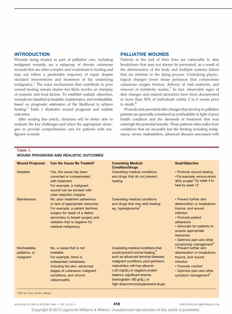
Table 1.
WOUND PROGNOSIS AND REALISTIC OUTCOMES
Wound Prognosis Can the Cause Be Treated? Coexisting MedicalCondition/Drugs
Goal/Objective
Healable Yes, the cause has been
corrected or compensated
with treatment
Coexisting medical conditions
and drugs that do not prevent
healing
& Promote wound healing
For example, a malignant
wound can be excised with
clear resection margins
& For example, venousulcers:
30% smaller3 by week 4 to
heal by week 12
Maintenance No, poor treatment adherence
or lack of appropriate resources
Coexisting medical conditions
and drugs that may stall healing;
eg, hyperglycemia4
& Prevent further skin
deterioration or breakdown,
trauma, and wound
infection
For example, a patient declines
surgery for repair of a defect
secondary to breast surgery and
radiation that is negative for
residual malignancy
& Promote patient
adherence
& Advocate for patients to
acquire appropriate
resources
& Optimize pain and other
symptom(s) management5
Nonhealable,
palliative, or
malignant
No, a cause that is not
treatable
For example, there is
widespread metastasis,
including the skin, advanced
stages of cutaneous malignant
conditions, and chronic
osteomyelitis
Coexisting medical conditions that
would prevent normal healing,6
such as advanced terminal diseases,
malignant conditions, poor perfusion,
malnutrition with low albumin
(<20 mg/dL) or negative protein
balance, significant anemia
(hemoglobin <80 g/dL), or
high-dose immunosuppressive drugs
& Prevent further skin
deterioration or breakdown,
trauma, and wound
infection
& Promote comfort
& Optimize pain and other
symptom management5
*2009 KY Woo and RG Sibbald.
ADVANCES IN SKIN & WOUND CARE & VOL. 23 NO. 9 418 WWW.WOUNDCAREJOURNAL.COM
Copyright @ 20 Lippincott Williams & Wilkins. Unauthorized reproduction of this article is prohibited.10
major organ failure (renal, hepatic, pulmonary, or cardiac), and,
in some cases, profound dementia.9,10 Management of these
cutaneous palliative wounds is challenging to patients and
their healthcare providers. Although wound healing may not
be realistic, it is imperative to maintain patients’ dignity and
quality of life by addressing psychosocial concerns (fear of
dying), empowering patients’ independence, promoting the
highest achievable quality of life and activities of daily living,
and optimizing pain management.11,12
MALIGNANT WOUNDA malignant wound can result from
& tumor necrosis,
& fungating tumor cells,
& ulcerating cancerous wound, or
&malignant cutaneous wound.
Infiltration of malignant cells in these wounds is secondary to
local invasion of a primary cutaneous lesion ormetastatic spread.
The following scenarios should raise the index of suspicion
of malignancy:
&Wounds that are a manifestation of primary skin cancer and
certain types of malignancies: for example, basal cell carcinoma,
squamous cell carcinoma, melanoma, Kaposi sarcoma, cuta-
neous lymphomas, and cutaneous infiltrates associated with
leukemia.13
& Be cautious of wounds in patients with a history of cancer to
rule out cutaneous metastasis. Malignant wounds have been
estimated to affect 5% to 19% of patients with metastatic
disease.14–16 In another study, Lookingbill et al17 reported that
5% of cancer patients develop malignant wounds. The chest
and breasts and the head and neck, followed by the abdomen,
are the most common sites where metastatic malignant
wounds develop.18–20
&Wounds that do not heal over a long time may exhibit chronic
inflammation that can undergo malignant transformation. A
Marjolin ulcer or a squamous cell carcinoma may develop in an
area of chronic inflammation.21,22 These changes have been
documented from a chronic osteomyelitis sinus, persistent
trauma, and burn scar.23,24
& Chronic wounds in patients with chronic immunosup-
pression and drugs that can predispose patients to skin ul-
ceration (eg, azathioprine, methotrexate, and cyclosporine
therapy) and other immunodeficiency disorders, including
HIV infection.14,24
&Wounds secondary to treatment of malignancies, such as late
radiation therapy change breakdown or the development of a
secondary malignancy.25
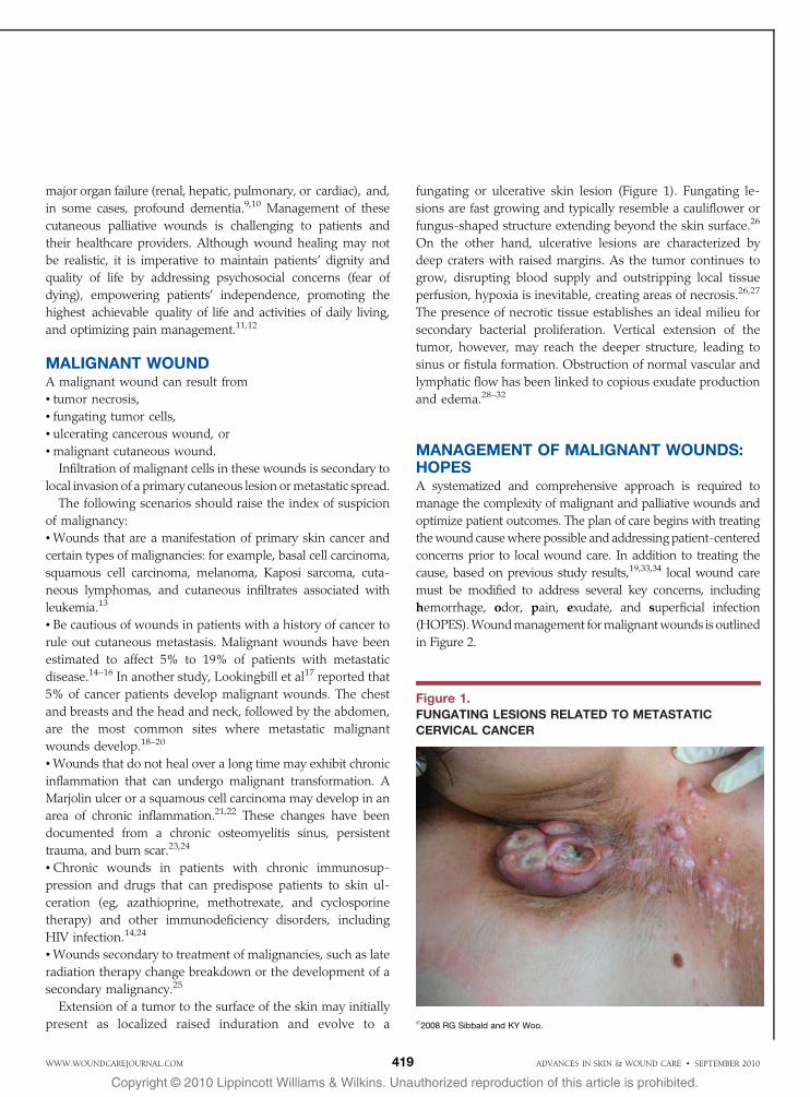
Extension of a tumor to the surface of the skin may initially
present as localized raised induration and evolve to a
fungating or ulcerative skin lesion (Figure 1). Fungating le-
sions are fast growing and typically resemble a cauliflower or
fungus-shaped structure extending beyond the skin surface.26
On the other hand, ulcerative lesions are characterized by
deep craters with raised margins. As the tumor continues to
grow, disrupting blood supply and outstripping local tissue
perfusion, hypoxia is inevitable, creating areas of necrosis.26,27
The presence of necrotic tissue establishes an ideal milieu for
secondary bacterial proliferation. Vertical extension of the
tumor, however, may reach the deeper structure, leading to
sinus or fistula formation. Obstruction of normal vascular and
lymphatic flow has been linked to copious exudate production
and edema.28–32
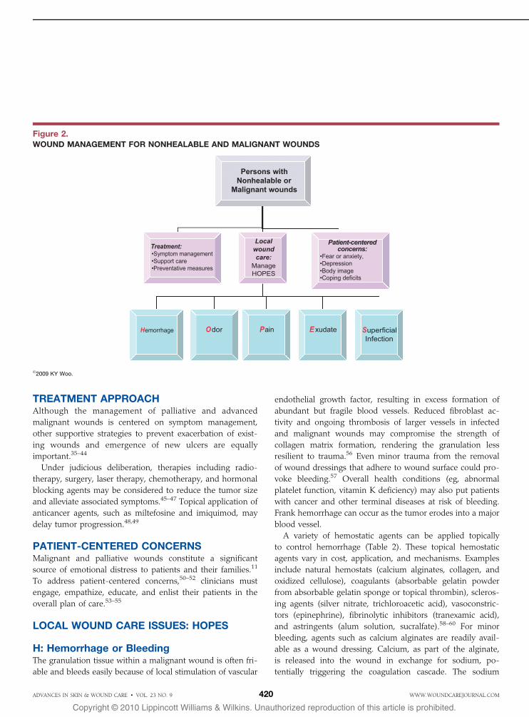
MANAGEMENT OF MALIGNANT WOUNDS:HOPESA systematized and comprehensive approach is required to
manage the complexity of malignant and palliative wounds and
optimize patient outcomes. The plan of care begins with treating
thewound causewhere possible and addressing patient-centered
concerns prior to local wound care. In addition to treating the
cause, based on previous study results,19,33,34 local wound care
must be modified to address several key concerns, including
hemorrhage, odor, pain, exudate, and superficial infection
(HOPES).Woundmanagement formalignantwounds is outlined
in Figure 2.
Figure 1.FUNGATING LESIONS RELATED TO METASTATIC
CERVICAL CANCER
*2008 RG Sibbald and KY Woo.
ADVANCES IN SKIN & WOUND CARE & SEPTEMBER 2010419WWW.WOUNDCAREJOURNAL.COM
Copyright @ 20 Lippincott Williams & Wilkins. Unauthorized reproduction of this article is prohibited.10
TREATMENT APPROACHAlthough the management of palliative and advanced
malignant wounds is centered on symptom management,
other supportive strategies to prevent exacerbation of exist-
ing wounds and emergence of new ulcers are equally
important.35–44
Under judicious deliberation, therapies including radio-
therapy, surgery, laser therapy, chemotherapy, and hormonal
blocking agents may be considered to reduce the tumor size
and alleviate associated symptoms.45–47 Topical application of
anticancer agents, such as miltefosine and imiquimod, may
delay tumor progression.48,49
PATIENT-CENTERED CONCERNSMalignant and palliative wounds constitute a significant
source of emotional distress to patients and their families.11
To address patient-centered concerns,50–52 clinicians must
engage, empathize, educate, and enlist their patients in the
overall plan of care.53–55
LOCAL WOUND CARE ISSUES: HOPES
H: Hemorrhage or BleedingThe granulation tissue within a malignant wound is often fri-
able and bleeds easily because of local stimulation of vascular
endothelial growth factor, resulting in excess formation of
abundant but fragile blood vessels. Reduced fibroblast ac-
tivity and ongoing thrombosis of larger vessels in infected
and malignant wounds may compromise the strength of
collagen matrix formation, rendering the granulation less
resilient to trauma.56 Even minor trauma from the removal
of wound dressings that adhere to wound surface could pro-
voke bleeding.57 Overall health conditions (eg, abnormal
platelet function, vitamin K deficiency) may also put patients
with cancer and other terminal diseases at risk of bleeding.
Frank hemorrhage can occur as the tumor erodes into a major
blood vessel.
A variety of hemostatic agents can be applied topically
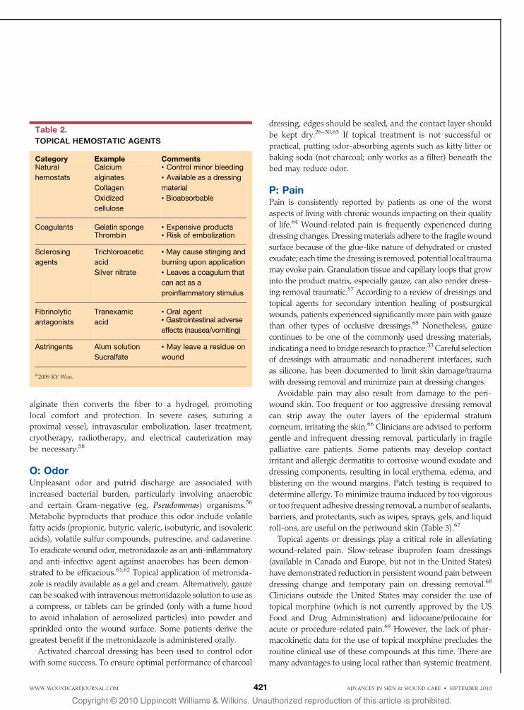
to control hemorrhage (Table 2). These topical hemostatic
agents vary in cost, application, and mechanisms. Examples
include natural hemostats (calcium alginates, collagen, and
oxidized cellulose), coagulants (absorbable gelatin powder
from absorbable gelatin sponge or topical thrombin), scleros-
ing agents (silver nitrate, trichloroacetic acid), vasoconstric-
tors (epinephrine), fibrinolytic inhibitors (tranexamic acid),
and astringents (alum solution, sucralfate).58–60 For minor
bleeding, agents such as calcium alginates are readily avail-
able as a wound dressing. Calcium, as part of the alginate,
is released into the wound in exchange for sodium, po-
tentially triggering the coagulation cascade. The sodium
Figure 2.WOUND MANAGEMENT FOR NONHEALABLE AND MALIGNANT WOUNDS
*2009 KY Woo.
ADVANCES IN SKIN & WOUND CARE & VOL. 23 NO. 9 420 WWW.WOUNDCAREJOURNAL.COM
Copyright @ 20 Lippincott Williams & Wilkins. Unauthorized reproduction of this article is prohibited.10
alginate then converts the fiber to a hydrogel, promoting
local comfort and protection. In severe cases, suturing a
proximal vessel, intravascular embolization, laser treatment,
cryotherapy, radiotherapy, and electrical cauterization may
be necessary.58
O: OdorUnpleasant odor and putrid discharge are associated with
increased bacterial burden, particularly involving anaerobic
and certain Gram-negative (eg, Pseudomonas) organisms.56
Metabolic byproducts that produce this odor include volatile
fatty acids (propionic, butyric, valeric, isobutyric, and isovaleric
acids), volatile sulfur compounds, putrescine, and cadaverine.
To eradicate wound odor, metronidazole as an anti-inflammatory
and anti-infective agent against anaerobes has been demon-
strated to be efficacious.61,62 Topical application of metronida-
zole is readily available as a gel and cream. Alternatively, gauze
can be soakedwith intravenousmetronidazole solution to use as
a compress, or tablets can be grinded (only with a fume hood
to avoid inhalation of aerosolized particles) into powder and
sprinkled onto the wound surface. Some patients derive the
greatest benefit if the metronidazole is administered orally.
Activated charcoal dressing has been used to control odor
with some success. To ensure optimal performance of charcoal
dressing, edges should be sealed, and the contact layer should
be kept dry.26–30,63 If topical treatment is not successful or
practical, putting odor-absorbing agents such as kitty litter or
baking soda (not charcoal; only works as a filter) beneath the
bed may reduce odor.
P: PainPain is consistently reported by patients as one of the worst
aspects of living with chronic wounds impacting on their quality
of life.64 Wound-related pain is frequently experienced during
dressing changes. Dressingmaterials adhere to the fragile wound
surface because of the glue-like nature of dehydrated or crusted
exudate; each time the dressing is removed, potential local trauma
may evoke pain. Granulation tissue and capillary loops that grow
into the product matrix, especially gauze, can also render dress-
ing removal traumatic.57 According to a review of dressings and
topical agents for secondary intention healing of postsurgical
wounds, patients experienced significantly more pain with gauze
than other types of occlusive dressings.65 Nonetheless, gauze
continues to be one of the commonly used dressing materials,
indicating a need to bridge research to practice.33Careful selection
of dressings with atraumatic and nonadherent interfaces, such
as silicone, has been documented to limit skin damage/trauma
with dressing removal and minimize pain at dressing changes.
Avoidable pain may also result from damage to the peri-
wound skin. Too frequent or too aggressive dressing removal
can strip away the outer layers of the epidermal stratum
corneum, irritating the skin.66 Clinicians are advised to perform
gentle and infrequent dressing removal, particularly in fragile
palliative care patients. Some patients may develop contact
irritant and allergic dermatitis to corrosive wound exudate and
dressing components, resulting in local erythema, edema, and
blistering on the wound margins. Patch testing is required to
determine allergy. To minimize trauma induced by too vigorous
or too frequent adhesive dressing removal, a number of sealants,
barriers, and protectants, such as wipes, sprays, gels, and liquid
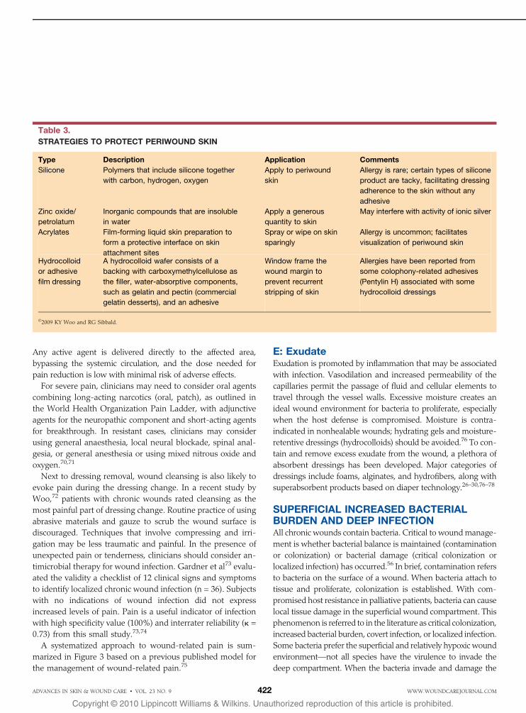
roll-ons, are useful on the periwound skin (Table 3).67
Topical agents or dressings play a critical role in alleviating
wound-related pain. Slow-release ibuprofen foam dressings
(available in Canada and Europe, but not in the United States)
have demonstrated reduction in persistent wound pain between
dressing change and temporary pain on dressing removal.68
Clinicians outside the United States may consider the use of
topical morphine (which is not currently approved by the US
Food and Drug Administration) and lidocaine/prilocaine for
acute or procedure-related pain.69 However, the lack of phar-
macokinetic data for the use of topical morphine precludes the
routine clinical use of these compounds at this time. There are
many advantages to using local rather than systemic treatment.
Table 2.
TOPICAL HEMOSTATIC AGENTS
Category Example CommentsNatural
hemostats
Calcium
alginates
Collagen
Oxidized
cellulose
& Control minor bleeding
& Available as a dressing
material
& Bioabsorbable
Coagulants Gelatin sponge & Expensive productsThrombin & Risk of embolization
Sclerosing
agents
Trichloroacetic
acid
Silver nitrate
& May cause stinging and
burning upon application
& Leaves a coagulum that
can act as a
proinflammatory stimulus
Fibrinolytic
antagonists
Tranexamic
acid
& Oral agent& Gastrointestinal adverse
effects (nausea/vomiting)
Astringents Alum solution
Sucralfate
& May leave a residue on
wound
*2009 KY Woo.
ADVANCES IN SKIN & WOUND CARE & SEPTEMBER 2010421WWW.WOUNDCAREJOURNAL.COM
Copyright @ 20 Lippincott Williams & Wilkins. Unauthorized reproduction of this article is prohibited.10
Any active agent is delivered directly to the affected area,
bypassing the systemic circulation, and the dose needed for
pain reduction is low with minimal risk of adverse effects.
For severe pain, clinicians may need to consider oral agents
combining long-acting narcotics (oral, patch), as outlined in
the World Health Organization Pain Ladder, with adjunctive
agents for the neuropathic component and short-acting agents
for breakthrough. In resistant cases, clinicians may consider
using general anaesthesia, local neural blockade, spinal anal-
gesia, or general anesthesia or using mixed nitrous oxide and
oxygen.70,71
Next to dressing removal, wound cleansing is also likely to
evoke pain during the dressing change. In a recent study by
Woo,72 patients with chronic wounds rated cleansing as the
most painful part of dressing change. Routine practice of using
abrasive materials and gauze to scrub the wound surface is
discouraged. Techniques that involve compressing and irri-
gation may be less traumatic and painful. In the presence of
unexpected pain or tenderness, clinicians should consider an-
timicrobial therapy for wound infection. Gardner et al73 evalu-
ated the validity a checklist of 12 clinical signs and symptoms
to identify localized chronic wound infection (n = 36). Subjects
with no indications of wound infection did not express
increased levels of pain. Pain is a useful indicator of infection
with high specificity value (100%) and interrater reliability (. =
0.73) from this small study.73,74
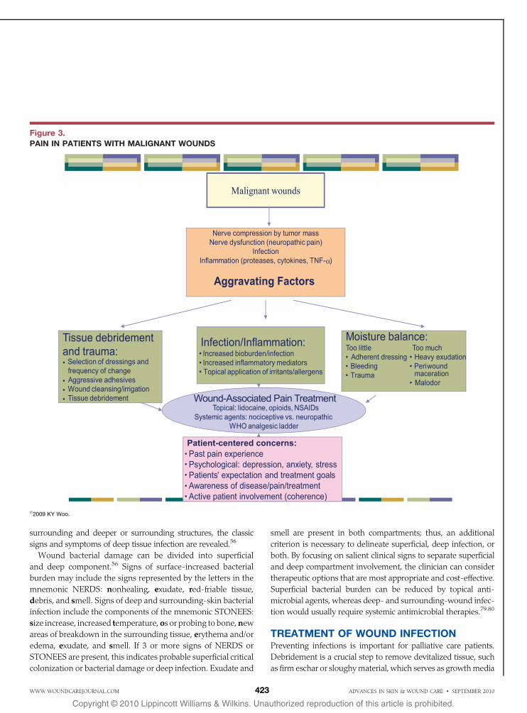
A systematized approach to wound-related pain is sum-
marized in Figure 3 based on a previous published model for
the management of wound-related pain.75
E: ExudateExudation is promoted by inflammation that may be associated
with infection. Vasodilation and increased permeability of the
capillaries permit the passage of fluid and cellular elements to
travel through the vessel walls. Excessive moisture creates an
ideal wound environment for bacteria to proliferate, especially
when the host defense is compromised. Moisture is contra-
indicated in nonhealable wounds; hydrating gels and moisture-
retentive dressings (hydrocolloids) should be avoided.76 To con-
tain and remove excess exudate from the wound, a plethora of
absorbent dressings has been developed. Major categories of
dressings include foams, alginates, and hydrofibers, along with
superabsorbent products based on diaper technology.26–30,76–78
SUPERFICIAL INCREASED BACTERIALBURDEN AND DEEP INFECTIONAll chronic wounds contain bacteria. Critical to woundmanage-
ment is whether bacterial balance is maintained (contamination
or colonization) or bacterial damage (critical colonization or
localized infection) has occurred.56 In brief, contamination refers
to bacteria on the surface of a wound. When bacteria attach to
tissue and proliferate, colonization is established. With com-
promised host resistance in palliative patients, bacteria can cause
local tissue damage in the superficial wound compartment. This
phenomenon is referred to in the literature as critical colonization,
increased bacterial burden, covert infection, or localized infection.
Some bacteria prefer the superficial and relatively hypoxic wound
environmentVnot all species have the virulence to invade the
deep compartment. When the bacteria invade and damage the
Table 3.
STRATEGIES TO PROTECT PERIWOUND SKIN
Type Description Application Comments
Silicone Polymers that include silicone together
with carbon, hydrogen, oxygen
Apply to periwound
skin
Allergy is rare; certain types of silicone
product are tacky, facilitating dressing
adherence to the skin without any
adhesive
Zinc oxide/
petrolatum
Inorganic compounds that are insoluble
in water
Apply a generous
quantity to skin
May interfere with activity of ionic silver
Acrylates Film-forming liquid skin preparation to
form a protective interface on skin
attachment sites
Spray or wipe on skin
sparingly
Allergy is uncommon; facilitates
visualization of periwound skin
Hydrocolloid
or adhesive
film dressing
A hydrocolloid wafer consists of a
backing with carboxymethylcellulose as
the filler, water-absorptive components,
such as gelatin and pectin (commercial
gelatin desserts), and an adhesive
Window frame the
wound margin to
prevent recurrent
stripping of skin
Allergies have been reported from
some colophony-related adhesives
(Pentylin H) associated with some
hydrocolloid dressings
*2009 KY Woo and RG Sibbald.
ADVANCES IN SKIN & WOUND CARE & VOL. 23 NO. 9 422 WWW.WOUNDCAREJOURNAL.COM
Copyright @ 20 Lippincott Williams & Wilkins. Unauthorized reproduction of this article is prohibited.10
surrounding and deeper or surrounding structures, the classic
signs and symptoms of deep tissue infection are revealed.56
Wound bacterial damage can be divided into superficial
and deep component.56 Signs of surface-increased bacterial
burden may include the signs represented by the letters in the
mnemonic NERDS: nonhealing, exudate, red-friable tissue,
debris, and smell. Signs of deep and surrounding-skin bacterial
infection include the components of the mnemonic STONEES:
size increase, increased temperature, os or probing to bone,new
areas of breakdown in the surrounding tissue, erythema and/or
edema, exudate, and smell. If 3 or more signs of NERDS or
STONEES are present, this indicates probable superficial critical
colonization or bacterial damage or deep infection. Exudate and
smell are present in both compartments; thus, an additional
criterion is necessary to delineate superficial, deep infection, or
both. By focusing on salient clinical signs to separate superficial
and deep compartment involvement, the clinician can consider
therapeutic options that are most appropriate and cost-effective.
Superficial bacterial burden can be reduced by topical anti-
microbial agents, whereas deep- and surrounding-wound infec-
tion would usually require systemic antimicrobial therapies.79.80
TREATMENT OF WOUND INFECTIONPreventing infections is important for palliative care patients.
Debridement is a crucial step to remove devitalized tissue, such
as firm eschar or sloughymaterial, which serves as growthmedia
Figure 3.PAIN IN PATIENTS WITH MALIGNANT WOUNDS
*2009 KY Woo.
ADVANCES IN SKIN & WOUND CARE & SEPTEMBER 2010423WWW.WOUNDCAREJOURNAL.COM
Copyright @ 20 Lippincott Williams & Wilkins. Unauthorized reproduction of this article is prohibited.10
for bacteria. Aggressive debridement is not recommended in
malignant wounds in light of the potential risk of causing
pain or bleeding and creating a larger and deeper portal for
bacterial invasion.81 Under judicious deliberation, conserva-
tive debridement of nonhealable wounds may be appropriate
by trimming loose, hanging fibrin to reduce necrotic mass and
associated odor. The purpose of conservative debridement is
to enhance the quality of life and decrease the risk of bacterial
proliferation and infection and not to cut into viable tissue or
facilitate healing. Alternatively, the risk of wound infection has
been demonstrated to decrease by using moisture-retentive
dressings and hydrogel to promote autolytic debridement.
Cleansing solutions, including saline or water, are usually
recommended to remove surface debris because of their low
tissue toxicity. Topical antimicrobial products are available, but
no one product is indicated or suitable for all patients.2 Many
active ingredients in dressings are released into the wound
surface compartment, but they require wound fluid or exudate
to diffuse into the tissue. Despite the lack of randomized
controlled trial evidence of complete wound healing, silver
dressings are one of the most popular topical agents.82 For
silver to exert an antimicrobial effect, it must be activated into
ionic form in the presence of an aqueous wound environment,
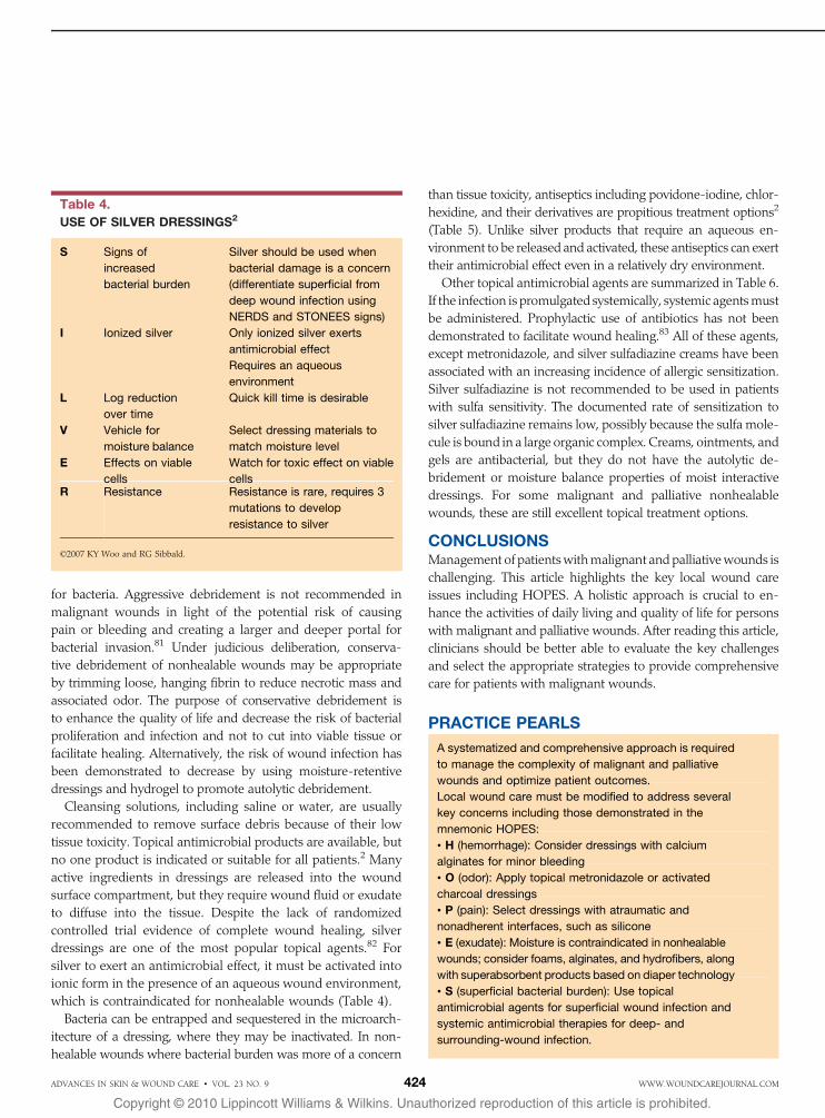
which is contraindicated for nonhealable wounds (Table 4).
Bacteria can be entrapped and sequestered in the microarch-
itecture of a dressing, where they may be inactivated. In non-
healable wounds where bacterial burden was more of a concern
than tissue toxicity, antiseptics including povidone-iodine, chlor-
hexidine, and their derivatives are propitious treatment options2
(Table 5). Unlike silver products that require an aqueous en-
vironment to be released and activated, these antiseptics can exert
their antimicrobial effect even in a relatively dry environment.
Other topical antimicrobial agents are summarized in Table 6.
If the infection is promulgated systemically, systemic agentsmust
be administered. Prophylactic use of antibiotics has not been
demonstrated to facilitate wound healing.83 All of these agents,
except metronidazole, and silver sulfadiazine creams have been
associated with an increasing incidence of allergic sensitization.
Silver sulfadiazine is not recommended to be used in patients
with sulfa sensitivity. The documented rate of sensitization to
silver sulfadiazine remains low, possibly because the sulfa mole-
cule is bound in a large organic complex. Creams, ointments, and
gels are antibacterial, but they do not have the autolytic de-
bridement or moisture balance properties of moist interactive
dressings. For some malignant and palliative nonhealable
wounds, these are still excellent topical treatment options.
CONCLUSIONSManagement of patientswithmalignant and palliativewounds is
challenging. This article highlights the key local wound care
issues including HOPES. A holistic approach is crucial to en-
hance the activities of daily living and quality of life for persons
with malignant and palliative wounds. After reading this article,
clinicians should be better able to evaluate the key challenges
and select the appropriate strategies to provide comprehensive
care for patients with malignant wounds.
PRACTICE PEARLS
A systematized and comprehensive approach is required
to manage the complexity of malignant and palliative
wounds and optimize patient outcomes.
Local wound care must be modified to address several
key concerns including those demonstrated in the
mnemonic HOPES:
& H (hemorrhage): Consider dressings with calcium
alginates for minor bleeding
& O (odor): Apply topical metronidazole or activated
charcoal dressings
& P (pain): Select dressings with atraumatic and
nonadherent interfaces, such as silicone
& E (exudate): Moisture is contraindicated in nonhealable
wounds; consider foams, alginates, and hydrofibers, along
with superabsorbent products based on diaper technology
& S (superficial bacterial burden): Use topical
antimicrobial agents for superficial wound infection and
systemic antimicrobial therapies for deep- and
surrounding-wound infection.
Table 4.
USE OF SILVER DRESSINGS2
S Signs of
increased
bacterial burden
Silver should be used when
bacterial damage is a concern
(differentiate superficial from
deep wound infection using
NERDS and STONEES signs)
I Ionized silver Only ionized silver exerts
antimicrobial effect
Requires an aqueous
environment
L Log reduction
over time
Quick kill time is desirable
V Vehicle for
moisture balance
Select dressing materials to
match moisture level
E Effects on viable
cells
Watch for toxic effect on viable
cellsR Resistance Resistance is rare, requires 3
mutations to develop
resistance to silver
*2007 KY Woo and RG Sibbald.
ADVANCES IN SKIN & WOUND CARE & VOL. 23 NO. 9 424 WWW.WOUNDCAREJOURNAL.COM
Copyright @ 20 Lippincott Williams & Wilkins. Unauthorized reproduction of this article is prohibited.10
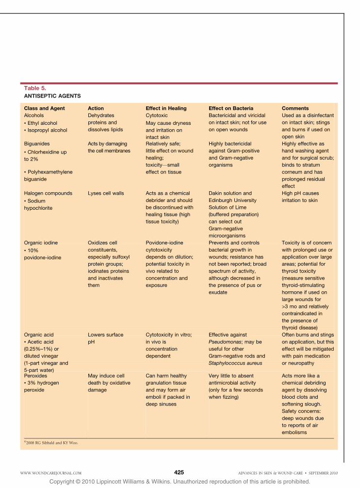
Table 5.
ANTISEPTIC AGENTS
Class and Agent Action Effect in Healing Effect on Bacteria Comments
Alcohols Dehydrates
proteins and
dissolves lipids
Cytotoxic Bactericidal and viricidal
on intact skin; not for use
on open wounds
Used as a disinfectant
on intact skin; stings
and burns if used on
open skin
& Ethyl alcohol May cause dryness
and irritation on
intact skin
& Isopropyl alcohol
Biguanides Acts by damaging
the cell membranes
Relatively safe;
little effect on wound
healing;
toxicityVsmall
effect on tissue
Highly bactericidal
against Gram-positive
and Gram-negative
organisms
Highly effective as
hand washing agent
and for surgical scrub;
binds to stratum
corneum and has
prolonged residual
effect
& Chlorhexidine up
to 2%
& Polyhexamethylene
biguanide
Halogen compounds Lyses cell walls Acts as a chemical
debrider and should
be discontinued with
healing tissue (high
tissue toxicity)
Dakin solution and
Edinburgh University
Solution of Lime
(buffered preparation)
can select out
Gram-negative
microorganisms
High pH causes
irritation to skin& Sodium
hypochlorite
Organic iodine Oxidizes cell
constituents,
especially sulfoxyl
protein groups;
iodinates proteins
and inactivates
them
Povidone-iodine
cytotoxicity
depends on dilution;
potential toxicity in
vivo related to
concentration and
exposure
Prevents and controls
bacterial growth in
wounds; resistance has
not been reported; broad
spectrum of activity,
although decreased in
the presence of pus or
exudate
Toxicity is of concern
with prolonged use or
application over large
areas; potential for
thyroid toxicity
(measure sensitive
thyroid-stimulating
hormone if used on
large wounds for
>3 mo and relatively
contraindicated in
the presence of
thyroid disease)
& 10%
povidone-iodine
Organic acid Lowers surface
pH
Cytotoxicity in vitro;
in vivo is
concentration
dependent
Effective against
Pseudomonas; may be
useful for other
Gram-negative rods and
Staphylococcus aureus
Often burns and stings
on application, but this
effect will be mitigated
with pain medication
or neuropathy
& Acetic acid
(0.25%–1%) or
diluted vinegar
(1-part vinegar and
5-part water)Peroxides
& 3% hydrogen
peroxide
May induce cell
death by oxidative
damage
Can harm healthy
granulation tissue
and may form air
emboli if packed in
deep sinuses
Very little to absent
antimicrobial activity
(only for a few seconds
when fizzing)
Acts more like a
chemical debriding
agent by dissolving
blood clots and
softening slough.
Safety concerns:
deep wounds due
to reports of air
embolisms
*2008 RG Sibbald and KY Woo.
ADVANCES IN SKIN & WOUND CARE & SEPTEMBER 2010425WWW.WOUNDCAREJOURNAL.COM
Copyright @ 20 Lippincott Williams & Wilkins. Unauthorized reproduction of this article is prohibited.10
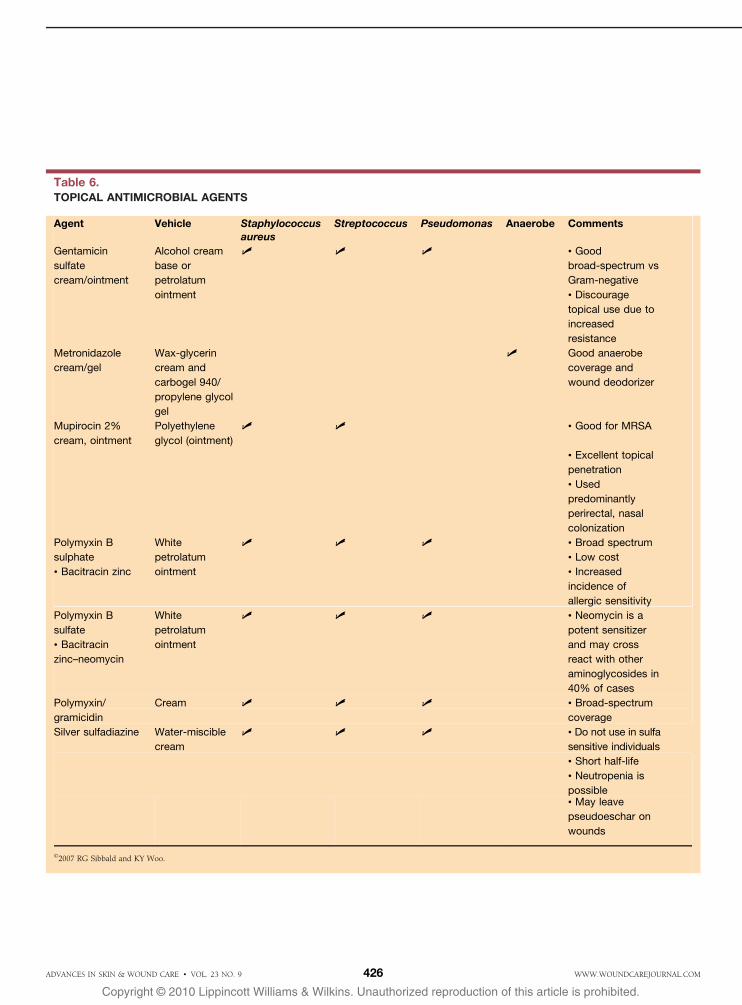
Table 6.TOPICAL ANTIMICROBIAL AGENTS
Agent Vehicle Staphylococcusaureus
Streptococcus Pseudomonas Anaerobe Comments
Gentamicin
sulfate
cream/ointment
Alcohol cream
base or
petrolatum
ointment
( ( ( & Good
broad-spectrum vs
Gram-negative
& Discourage
topical use due to
increased
resistance
Metronidazole
cream/gel
Wax-glycerin
cream and
carbogel 940/
propylene glycol
gel
( Good anaerobe
coverage and
wound deodorizer
Mupirocin 2%
cream, ointment
Polyethylene
glycol (ointment)
( ( & Good for MRSA
& Excellent topical
penetration
& Used
predominantly
perirectal, nasal
colonization
Polymyxin B
sulphate
White
petrolatum
ointment
( ( ( & Broad spectrum
& Bacitracin zinc
& Low cost
& Increased
incidence of
allergic sensitivity
Polymyxin B
sulfate
White
petrolatum
ointment
( ( ( & Neomycin is a
potent sensitizer
and may cross
react with other
aminoglycosides in
40% of cases
& Bacitracin
zinc–neomycin
Polymyxin/
gramicidin
Cream ( ( ( & Broad-spectrum
coverage
Silver sulfadiazine Water-miscible
cream
( ( ( & Do not use in sulfa
sensitive individuals
& Short half-life
& Neutropenia is
possible& May leave
pseudoeschar on
wounds
*2007 R. G. Sibbald and K. Y. Woo.*2007 RG Sibbald and KY Woo.
ADVANCES IN SKIN & WOUND CARE & VOL. 23 NO. 9 426 WWW.WOUNDCAREJOURNAL.COM
Copyright @ 20 Lippincott Williams & Wilkins. Unauthorized reproduction of this article is prohibited.10
REFERENCES1. Woo K, Ayello EA, Sibbald RG. The edge effect: current therapeutic options to advance
the wound edge. Adv Skin Wound Care 2007;20:99-117.
2. Woo KY, Ayello EA, Sibbald RG. SILVER versus other antimicrobial dressings: best
practices! Surg Technol Int 2008;17:50-71.
3. Kantor J, Margolis DJ. A multicentre study of percentage change in venous leg ulcer
area as a prognostic index of healing at 24 weeks. Br J Dermatol 2000;142:960-4.
4. Markuson M, Hanson D, Anderson J, et al. The relationship between hemoglobin
A(1c) values and healing time for lower extremity ulcers in individuals with diabetes.
Adv Skin Wound Care 2009;22:365-72.
5. Sibbald RG, Woo KY, Ayello E. Wound bed preparation: DIM before DIME. Wound
Healing Southern Africa 2008;1(1):29-34.
6. Franz MG, Robson MC, Steed DL, et al. Wound Healing Society. Guidelines to aid
healing of acute wounds by decreasing impediments of healing. Wound Repair Regen
2008;16:723-48.
7. Langemo DK, Brown G. Skin fails too: acute, chronic, and end-stage skin failure. Adv
Skin Wound Care 2006;19:206-11.
8. Sibbald RG, Krasner DL, Lutz J. SCALE: Skin Changes at Life’s End: final consensus
statement: October 1, 2009. Adv Skin Wound Care 2010;23:225-36.
9. O’Brien T, Welsh J, Dunn FG. ABC of palliative care. Non-malignant conditions. BMJ
1998;316(7127):286-9.
10. Liao S, Arnold RM. Wound care in advanced illness: application of palliative care
principles. J Palliat Med 2007;10:1159-60.
11. Ferris FD, Al Khateib AA, Fromantin I, et al. Palliative wound care: managing chronic
wounds across life’s continuum: a consensus statement from the International Palliative
Wound Care Initiative. J Palliat Med 2007;10:37-9.
12. Irwin SA, Rao S, Bower K, et al. Psychiatric issues in palliative care: recognition of
depression in patients enrolled in hospice care. J Palliat Med 2008;11:158-63.
13. Naylor W. Malignant wounds: aetiology and principles of management. Nurs Stand
2002;16(52):45-53.
14. Alexander S. Malignant fungating wounds: epidemiology, aetiology, presentation and
assessment. J Wound Care 2009;18:273-4, 276-8, 280.
15. Iveti O, Lyne PA. Fungating and ulcerating malignant lesions: a review of the literature.
J Adv Nurs 1990;15:83-8.
16. Maida V, Corbo M, Dolzhykov M, Ennis M, Irani S, Trozzolo L. Wounds in advanced
illness: a prevalence and incidence study based on a prospective case series. Int
Wound J 2008;5:305-14.
17. Lookingbill DP, Spangler N, Sexton FM. Skin involvement as the presenting sign of
internal carcinoma. A retrospective study of 7316 cancer patients. J Am Acad Dermatol
1990;22:19-26.
18. Lookingbill DP, Spangler N, Helm KF. Cutaneous metastases in patients with metastatic
carcinoma: a retrospective study of 4020 patients. J Am Acad Dermatol 1993;29(2 Pt
1):228-36.
19. Maida V, Ennis M, Kuziemsky C, Trozzolo L. Symptoms associated with malignant
wounds: a prospective case series. J Pain Symptom Manage 2009;37:206-11.
20. Collier M. Management of patients with fungating wounds. Nurs Stand 2000;15(11):46-
52.
21. Trent JT, Kirsner RS. Wounds and malignancy. Adv Skin Wound Care 2003;16(1):31-4.
22. Esther RJ, Lamps L, Schwartz HS. Marjolin ulcers: secondary carcinomas in chronic
wounds. J South Orthop Assoc 1999;8:181-7.
23. Copcu E. Marjolin’s ulcer: a preventable complication of burns? Plast Reconstr Surg
2009;124(1):156e-64e.24. Schiech L. Malignant cutaneous wounds. Clin J Oncol Nurs 2002;6:305-9.25. Walton A, Broadbent AL. Radiation-induced second malignancies. J Palliat Med 2008;11:
1345-52.26. Grocott P. Care of patients with fungating malignant wounds. Nurs Stand 2007;21(24):
57-8, 60, 62.27. Alvarez OM, Kalinski C, Nusbaum J, et al. Incorporating wound healing strategies to
improve palliation (symptom management) in patients with chronic wounds. J Palliat
Med 2007;10:1161-89.28. McDonald A, Lesage P. Palliative management of pressure ulcers and malignant wounds
in patients with advanced illness. J Palliat Med 2006;9:285-95.29. Dowsett C. Malignant fungating wounds: assessment and management. Br J Community
Nurs 2002;7:394-400.30. Langemo DK, Anderson J, Hanson D, Hunter S, Thompson P. Managing fungating
wounds. Adv Skin Wound Care 2007;20:312-4.
31. Seaman S. Management of malignant fungating wounds in advanced cancer. Semin
Oncol Nurs 2006;22:185-93.
32. Schim SM, Cullen B. Wound care at end of life. Nurs Clin North Am 2005;40:281-94.
33. Probst S, Arber A, Faithfull S. Malignant fungating wounds: a survey of nurses’ clinical
practice in Switzerland. Eur J Oncol Nurs 2009;13:295-8.
34. Schulz V, Triska OH, Tonkin K. Malignant wounds: caregiver-determined clinical problems.
J Pain Symptom Manage 2002;24:572-7.
35. Langemo DK, Black J. National Pressure Ulcer Advisory Panel. Pressure ulcers in
individuals receiving palliative care: a National Pressure Ulcer Advisory Panel white
paper. Adv Skin Wound Care 2010;23:59-72.
36. Searle C, McInerney F. Nurses’ decision-making in pressure area management in the
last 48 hours of life. Int J Palliat Nurs 2008;14:432-8.
37. Vanderwee K, Grypdonck M, Defloor T. Alternating pressure air mattresses as prevention
for pressure ulcers: a literature review. Int J Nurs Stud 2008;45:784-801.
38. Krapfl LA, Gray M. Does regular repositioning prevent pressure ulcers? J Wound
Ostomy Continence Nurs 2008;35:571-7.
39. McInnes E, Bell-Syer SE, Dumville JC, Legood R, Cullum NA. Support surfaces for
pressure ulcer prevention. Cochrane Database Syst Rev 2008;Oct 8(4):CD001735.
40. Chasen MR, Bhargava R. A descriptive review of the factors contributing to nutritional
compromise in patients with head and neck cancer. Support Care Cancer 2009;17:
1345-51.
41. Dorner B, Posthauer ME, Thomas D. National Pressure Ulcer Advisory Panel. The role of
nutrition in pressure ulcer prevention and treatment: National Pressure Ulcer Advisory
Panel white paper. Adv Skin Wound Care 2009;22:212-21.
42. Ellinger S, Stehle P. Efficacy of vitamin supplementation in situations with wound
healing disorders: results from clinical intervention studies. Curr Opin Clin Nutr Metab Care
2009;12:588-95.
43. Beeckman D, Schoonhoven L, Verhaeghe S, Heyneman A, Defloor T. Prevention and
treatment of incontinence-associated dermatitis: literature review. J Adv Nurs 2009;65:
1141-54.
44. Woo KY, Sibbald RG. The ABCs of skin care for wound care clinicians: dermatitis and
eczema. Adv Skin Wound Care 2009;22:230-6.
45. Lee KF, Ennis WJ, Dunn GP. Surgical palliative care of advanced wounds. Am J Hosp
Palliat Care 2007;24:154-60.
46. Ciezki JP, Komurcu S, Macklis RM. Palliative radiotherapy. Semin Oncol 2000;27:90-3.
47. Stephen Haynes J. An overview of caring for those with palliative wounds. Br J Community
Nurs 2008;13(12):S24, S26, S28 passim.
48. Adderley U, Smith R. Topical agents and dressings for fungating wounds. Cochrane
Database Syst Rev 2007;(2):CD003948.
49. Spaner DE, Miller RL, Mena J, Grossman L, Sorrenti V, Shi Y. Regression of lymphomatous
skin deposits in a chronic lymphocytic leukemia patient treated with the Toll-like receptor-7/
8 agonist, imiquimod. Leuk Lymphoma 2005;46:935-9.
50. Alexander S. Malignant fungating wounds: key symptoms and psychosocial issues. J
Wound Care 2009;18:325-9.
51. Lo SF, Hu WY, Hayter M, Chang SC, Hsu MY, Wu LY. Experiences of living with a
malignant fungating wound: a qualitative study. J Clin Nurs 2008;17:2699-708.
52. Piggin C, Jones V. Malignant fungating wounds: an analysis of the lived experience. Int
J Palliat Nurs 2007;13:384-91.
53. Segal ES. Maintaining communication in a time of uncertainty. Arch Fam Med 1995;4:1066-7.
54. Houston RE. The angry dying patient. Prim Care Companion J Clin Psychiatry 1999;1(1):5-8.
55. Kelly VF, Carroll JG. A new model for physician-patient communication. Patient Educ
Couns 1994;23:131-40.
56. Sibbald RG, Woo K, Ayello EA. Increased bacterial burden and infection: the story of
NERDS and STONES. Adv Skin Wound Care 2006;19:447-61.
57. Woo KY, Harding K, Price P, Sibbald G. Minimising wound-related pain at dressing
change: evidence-informed practice. Int Wound J 2008;5:144-57.
58. Harris DG, Noble SI. Management of terminal hemorrhage in patients with advanced
cancer: a systematic literature review. J Pain Symptom Manage 2009;38:913-27.
59. Alexander S. Malignant fungating wounds: managing pain, bleeding and psychosocial
issues. J Wound Care 2009;18:418-25.
60. Haisfield-Wolfe ME, Rund C. Malignant cutaneous wounds: a management protocol.
Ostomy Wound Manage 1997;43(1):56-60, 62, 64-6.
61. Young CV. The effects of malodorous fungating malignant wounds on body image and
quality of life. J Wound Care 2005;14:359-62.
62. Paul JC, Pieper BA. Topical metronidazole for the treatment of wound odor: a review of
the literature. Ostomy Wound Manage 2008;54(3):18-27.
ADVANCES IN SKIN & WOUND CARE & SEPTEMBER 2010427WWW.WOUNDCAREJOURNAL.COM
Copyright @ 20 Lippincott Williams & Wilkins. Unauthorized reproduction of this article is prohibited.10
63. Alexander S. Malignant fungating wounds: managing malodour and exudate. J Wound
Care 2009;18:374-82.
64. Price PE, Fagervik-Morton H, Mudge EJ, et al. Dressing-related pain in patients with
chronic wounds: an international patient perspective. Int Wound J 2008;5:159-71.
65. Ubbink DT, Vermeulen H, Goossens A, Kelner RB, Schreuder SM, Lubbers MJ. Occlusive vs
gauze dressings for local wound care in surgical patients: a randomized clinical trial. Arch
Surg 2008;143:950-5.
66. Woo K, Sibbald G, Fogh K, et al. Assessment and management of persistent (chronic)
and total wound pain. Int Wound J 2008;5:205-15.
67. Woo KY, Sibbald RG, Ayello EA, Coutts PM, Garde DE. Peristomal skin complications
and management. Adv Skin Wound Care 2009;22:522-32.
68. Gottrup F, Jrgensen B, Karlsmark T, et al. Reducing wound pain in venous leg ulcers
with Biatain Ibu: a randomized, controlled double-blind clinical investigation on the
performance and safety. Wound Repair Regen 2008;16:615-25.
69. Evans E, Gray M. Do topical analgesics reduce pain associated with wound dressing
changes or debridement of chronic wounds? J Wound Ostomy Continence Nurs 2005;32:
287-90.
70. Hollinworth H. Nurses’ assessment and management of pain at wound dressing changes.
J Wound Care 1995;4(2):77-83.
71. Queen D, Woo K, Schulz V, Sibbald RG. Managing chronic wound pain in patients
receiving palliative care. Ostomy Wound Manage 2003;49:A4(Suppl):36-41.
72. Woo KY. Meeting the challenges of wound-associated pain: anticipatory pain, anxiety,
stress, and wound healing. Ostomy Wound Manage 2008;54(9):10-2.
73. Gardner SE, Frantz RA, Troia C, et al. A tool to assess clinical signs and symptoms
of localized infection in chronic wounds: development and reliability. Ostomy Wound
Manage 2001;47(1):40-7.
74. Gardner SE, Frantz RA, Park H, Scherubel M. The inter-rater reliability of the Clinical Signs
and Symptoms Checklist in diabetic foot ulcers. Ostomy Wound Manage 2007;53(1):46-51.
75. Woo KY, Sibbald RG. Chronic wound pain: a conceptual model. Adv Skin Wound Care
2008;21:175-88.
76. Okan D, Woo K, Ayello EA, Sibbald G. The role of moisture balance in wound healing.
Adv Skin Wound Care 2007;20(1):39-53.
77. Selby T. Managing exudate in malignant fungating wounds and solving problems for
patients. Nurs Times 2009;105(18):14-7.
78. Cutting KF, White RJ. Avoidance and management of peri-wound maceration of the
skin. Prof Nurse 2002;18:33, 35-36.
79. Alavi A, Woo K, Sibbald RG. Common nail disorders and fungal infections. Adv Skin
Wound Care 2007;20:358-9.
80. Woo KY, Sibbald RG. A cross-sectional validation study of using NERDS and STONEES
to assess bacterial burden. Ostomy Wound Manage 2009;55(8):40-8.
81. Kirshen C, Woo K, Ayello EA, Sibbald RG. Debridement: a vital component of wound bed
preparation. Adv Skin Wound Care 2006;19:506-17.
82. Vermeulen H, van Hattem JM, Storm-Versloot MN, Ubbink DT. Topical silver for treating
infected wounds. Cochrane Database Syst Rev 2007;(1):CD005486.
83. O’Meara SM, Cullum NA, Majid M, Sheldon TA. Systematic review of antimicrobial
agents used for chronic wounds. Br J Surg 2001;88(1):4-21.
For more than 53 additional continuing education articles related to Skin/Wound Care topics, go to NursingCenter.com/CE.
CONTINUING MEDICAL EDUCATION INFORMATION FOR PHYSICIANSLippincott Continuing Medical Education Institute, Inc. is accredited by
the Accreditation Council for Continuing Medical Education to provide
continuing medical education for physicians.
Lippincott Continuing Medical Education Institute, Inc. designates
this educational activity for a maximum of 1 AMA PRA Category 1
CreditTM. Physicians should only claim credit commensurate with the
extent of their participation in the activity.
PROVIDER ACCREDITATION INFORMATION FOR NURSESLippincott Williams & Wilkins, publisher of the Advances in Skin
& Wound Care journal, will award 2.6 contact hours for this continuing
nursing education activity.
LWW is accredited as a provider of continuing nursing education
by the American Nurses Credentialing Center’s Commission on
Accreditation.
This activity is also provider approved by the California Board of
Registered Nursing, Provider Number CEP 11749 for 2.6 contact hours.
Lippincott Williams & Wilkins is also an approved provider of continuing
nursing education by the District of Columbia and Florida #FBN2454.
Your certificate is valid in all states.
The ANCC’s accreditation status of Lippincott Williams & Wilkins
Department of Continuing Education refers only to its continuing nursing
education activities and does not imply Commission on Accreditation
approval or endorsement of any commercial product.
CONTINUING EDUCATION INSTRUCTIONS&Read the article beginning on page 417.
& Take the test, recording your answers in the test answers section (SectionB)of the CE enrollment form. Each question has only one correct answer.
& Complete registration information (Section A) and course evaluation
(Section C).
& Mail completed test with registration fee to: Lippincott Williams &
Wilkins, CE Group, 333 7th Avenue, 19th Floor, New York, NY 10001.
& Within 3 to 4 weeks after your CE enrollment form is received, you will
be notified of your test results.
& If you pass, you will receive a certificate of earned contact hours and an
answer key. Nurses who fail have the option of taking the test again at no
additional cost. Only the first entry sent by physicians will be accepted
for credit.
& A passing score for this test is 12 correct answers.
& Nurses: Need CE STAT? Visit http://www.nursingcenter.com for
immediate results, other CE activities, and your personalized CE
planner tool. No Internet access? Call 1-800-787-8985 for other rush
service options.
& Questions? Contact Lippincott Williams & Wilkins: 1-800-787-8985.
Registration Deadline: September 30, 2012 (nurses); September 30,
2011 (physicians)
PAYMENT AND DISCOUNTS
& The registration fee for this test is $24.95 for nurses; $22 for physicians.
& Nurses: If you take two or more tests in any nursing journal published by
LWW and send in your CE enrollment forms together by mail, you may
deduct $0.95 from the price of each test. We offer special discounts for
as few as six tests and institutional bulk discounts for multiple tests. Call
1-800-787-8985 for more information.
ADVANCES IN SKIN & WOUND CARE & VOL. 23 NO. 9 428 WWW.WOUNDCAREJOURNAL.COM
Copyright @ 20 Lippincott Williams & Wilkins. Unauthorized reproduction of this article is prohibited.10