Embed Size (px)
Citation preview
1
Ozone oil promotes wound healing by increasing the migration of
fibroblasts via PI3K/Akt/mTOR signaling pathway
Weirong Xiao, Hua Tang, Meng Wu, Yangying Liao, Ke Li, Lan Li, Xiaopeng Xu*
Dermatological Department, Hunan People's Hospital, Changsha 410005, P. R. China.
Corresponding author: Dr. Xiaopeng Xu, Dermatological Department, Hunan People's Hospital,
Changsha 410005, P. R. China.
Email: [email protected]
Tel: +86- 0731-82278120
AC
CE
PT
ED
MA
NU
SC
RIP
T
10.1042/BSR20170658. Please cite using the DOI 10.1042/BSR20170658http://dx.doi.org/up-to-date version is available at
encouraged to use the Version of Record that, when published, will replace this version. The most this is an Accepted Manuscript, not the final Version of Record. You are:Bioscience Reports
). http://www.portlandpresspublishing.com/content/open-access-policy#ArchivingArchiving Policy of Portland Press (which the article is published. Archiving of non-open access articles is permitted in accordance with the Use of open access articles is permitted based on the terms of the specific Creative Commons Licence under
2
Abstract
Background: Skin injury affects millions of people via the uncontrolled inflammation and infection.
Many cellular components including fibroblasts and signaling pathways such as TGF-β were
activated to facilitate the wound healing to repair injured tissues.
Methods and Results: C57BL/6 female mice were divided into control and ozone oil treated
groups. Excisional wounds were created on the dorsal skin and the fibroblasts were isolated from
granulation tissues. The skin injured mouse model revealed that ozone oil could significantly
decrease the wound area and accelerate wound healing compared with control group. QPCR and
western blotting assays showed that ozone oil upregulated collagen I, α-SMA and TGF-β1 mRNA
and protein levels in fibroblasts. Wound healing assay demonstrated that ozone oil could increase
the migration of fibroblasts. Western blotting assay demonstrated that ozone oil increased the EMT
process of fibroblasts via upregulating fibronectin, vimentin, N-cadherin, MMP-2, MMP-9,
IGFBP-3, IGFBP5 and IGFBP6 and decreasing epithelial protein E-cadherin and cellular
senescence marker p16 expression. Mechanistically, western blotting assay revealed that ozone oil
increased the phosphorylation of PI3K, Akt and mTOR to regulate the EMT process, while
inhibition of PI3K reversed this effect of ozone oil. At last, the results from Cytometric Bead Array
demonstrated ozone oil significantly decreased the inflammation in fibroblasts.
Conclusion: Our results demonstrated ozone oil facilitated the wound healing via increasing
fibroblast migration and EMT process via PI3K/Akt/mTOR signaling pathway in vivo and vitro.
The cellular and molecular mechanisms we found here may provide new therapeutic targets for the
treatment of skin injury.
Keywords: wound healing, ozone oil, fibroblasts, EMT, PI3K/Akt/mTOR
3
Introduction
Wound is caused by trauma, burn, ulcer, surgery and others. Although most of the wounds will
generally heal well, the failure of wound healing affects millions of people in the world through the
uncontrolled inflammation and infection [1, 2]. Wound healing is composed of many complex
processes which include inflammation response, new tissue formation and tissue remodeling [1, 3].
In the first 48 hours after injury, different immune cells such as neutrophils, monocytes and
lymphocytes work together to prevent the bleeding and remove the dead tissues to balance the
inflammation process and make the appropriate repair of the wound [4, 5]. In the next 2-10 days,
the new tissue formation is followed via cellular proliferation and migration of different cell types
such as fibroblasts, keratinocytes and endothelial cells. In this stage, fibroblasts play very important
roles in the new tissue formation [2]. The wound will attract amount of fibroblasts to the injury sites
to facilitate the wound healing via different mechanisms [6, 7]. For example, wound production can
increase the proliferation and migration of fibroblasts to promote the scar formation [8, 9]. In
addition, fibroblasts can secrete many factors, such as matrix metallopeptidase-14 (MMP-14), basic
fibroblast growth factor (bFGF), fibroblast growth factor-9 (FGF-9) to regulate the collagen
homeostasis, angiogenesis or other important functions to facilitate the wound healing [10-13]. Also,
fibroblasts can differentiate into myofibroblasts [14],which produces extracellular matrix and
ultimately forms the mature scar [15]. In the 2-3 weeks after injury, the tissue remodeling process
happens which may last for a year or more. In this stage, the entire processes activated by injury
will wind down and cease while the activated cells will undergo apoptosis. Different cells
(fibroblasts, macrophages and endothelial cells) will secrete matrix metalloproteinases to remodel
and strengthen the repaired tissues [1, 16]. Through these classic wound healing processes, the
wound will be repaired.
Ozone, made up of 3 oxygen atoms, is a natural gaseous molecule which can be in gaseous and
liquid form. Ozone can react with blood components to affect oxygen metabolism, antioxidant
defense system, cell energy and microcirculation [17]. Furthermore, ozone can activate the
expression of many cytokines which are important for wound healing, antibacterial and antivirus
[18]. Thus, ozone therapy (OT) has been widely used for improving wound healing, antibacterial
4
agent and modulating immune system [19, 20]. In the treatment of tissue injury, OT is getting more
and more attentions. For example, OT can improve the wound healing via enhancing blood
perfusion [17]. And OT induces the expression of vascular endothelial growth factor (VEGF),
transforming growth factor-β (TGF-β) and platelet-derived growth factor (PDGF) to facilitate
wound healing of diabetic foot ulcers [21]. OT also has been applied for the clinical treatment of
tissue injury and has shown very good results [22, 23]. But till now, no research focuses on the
effect of ozone oil, which is produced via bubbling high concentrations of ozone in the oil to retain
the ozone, on wound healing and the underlying mechanisms study.
Epithelial-mesenchymal transition (EMT) is an important physiological process during
embryogenesis and tumorgenesis [24, 25]. EMT is characterized by loss of epithelial proteins, such
as E-cadherin, and increase of several mesenchymal proteins, such as vimentin, fibronectin and
N-cadherin [24, 26]. Many kinds of factors can trigger the EMT process. Among these factors,
TGF-β plays important roles in EMT via diverse downstream pathways, like Smads, RhoA, MAPK
and PI3K [27-30]. Recently, activation of PI3K/Akt/mTOR signaling pathway by TGF-β is critical
for the regulation of EMT [31-33]. The activation of PI3K can activate mTOR via Akt to accelerate
the EMT. Although many studies have found EMT plays critical roles in cancer development [31,
33], EMT has been proved to participate in the wound healing [34, 35]. For example, impairment of
fibroblasts growth and EMT in vinmentin or slug deficient mice demonstrates to slow the wound
healing [36, 37]. Tumor necrosis factor-α (TNF-α) can promote the fibroblast EMT process via
inducting of bone morphogen protein (BMP) 2/4 to facilitate the wound healing [38]. However,
whether ozone oil can promote the wound healing via regulating EMT process through
PI3K/Akt/mTOR signaling pathway is still unknown.
At present, the functional study of the ozone oil drug on wound healing is still lacking and the
underlying mechanisms are still unknown. Furthermore, no studies have been carried out to
elucidate if ozone oil can promote the wound healing via increasing the migration of fibroblasts. In
our study, we found the ozone oil drugs facilitate the wound healing through promoting the
migration of fibroblast via activating PI3K/Akt/mTOR signaling pathway in vitro and in vivo
studies.
5
Materials and Methods
Mice model for wound. C57BL/6 female mice (7 weeks old, n = 24) were separated for two groups,
control and ozone oil treated groups. Ozone oil, which contains 99% ozonide, superoxide and
Camellia oil, was bought from Health Care Technology, Inc, China. These mice were first
anesthetized with chloral hydrate followed by the shaving of the area assigned for wounding under
sterile condition. Excisional wounds with 1 cm diameter were created on the dorsal skin on day 0.
Then the mice were maintained in sterile condition and ozone oil was applied for the treatment from
the day 1. The treatment of wounded skin with 400 μl ozone oil was done with cotton swabs for 12
days (once every 2 days) compared to the control group without ozone oil. The areas were
measured every 2 days to evaluate the therapeutic efficiency. All of the procedures involving
animals and their care in this study were performed in accordance with the Guidelines for the Care
and Use of Laboratory Animals of Provincial People's Hospital in Hunan.
Isolation and culture of fibroblasts from new tissues. The granulation tissues were isolated from
dorsal skin of the injured mice which were cut into 1 mm3 by scissors under sterile condition. The
tissues were digested with 0.25% trypsin (25200056, Gibco) for about 60 min at 37°C. Then,
DMEM medium (11995065, Gibco) containing 10% FBS (16000044, Gibco) was added to stop the
digestion. The solution was passed through 70 μm filters (352360, BD Falcon) to remove the
undigested tissues and large cell aggregates followed by the centrifuged at 1500 rpm for 5 min. The
cell pellet was washed twice with DMEM medium and suspended in DEME medium containing
10% FBS and seeded into 12-well plates (3335, Corning). They were incubated in a humidified
incubator with an atmosphere of 95% air and 5% CO2 at 37°C. The cultured fibroblasts were
further treated with ozone oil in vitro. For every well, 10 μl ozone oil was used for the treatment of
cultured cells. For the treatment of LPS, the cultured fibroblasts were treated with 100 ng/ml LPS
with or without ozone oil for 24 hours. For the treatment of PI3K inhibitor, LY294002 (L9908,
6
Sigma), the cultured fibroblasts were pretreated with 10 μM LY294002 then treated with ozone oil
for 24 hours.
Wound healing assay. For the migration assay of fibroblasts, the fibroblasts were isolated and
cultured from the granulation tissues of injured skin in ozone oil treated and control group. The
fibroblast monolayers were carefully scratched using a 10 μl pipette tip. Then, the cells were further
treated with or without ozone oil for 24 hours. After 24 hours, the wounded area was photographed.
The empty area which indicates the wound region was calculated.
Cytometric Bead Array. The injured fibroblasts isolated from the mice were plated in 24-well
plates with 4×10⁴ cells/well (0.5 ml). After incubation overnight, the fibroblasts were pretreated
stimulation with LPS (100 ng/ml) with or without ozone oil for 24 hours. Cytokines in the culture
medium were quantitatively measured by Cytometric Bead Array (CBA) human inflammation kit
(BD Biosciences, San Jose, CA, USA) according to the manufacturer’s instructions. Data were
acquired using CELLQuest software on the flow cytometer (FACSCalibur; BD Biosciences). The
CBA kit can detect two cytokines including IL-6 and TNF-α with a minimum detectable level of 5
pg/ml.
Real-time PCR. Total RNA was extracted from cultured fibroblasts treated with or without ozone
oil by Trizol (15596018, Invitrogen) according to the manufacturer’s instructions. Complementary
DNA (cDNA) was synthesized from total RNA with the First-Strand Synthesis System (2680A,
Takara) according to the manufacturer’s instructions. Real-time PCR was performed by SYBR
Premix Ex Taq II (DRR081A, Takara) with 7500 Real-Time PCR system (Applied Biosystems) and
normalized to the expression of GAPDH. Relative quantification and statistics were estimated as the
mean of three replicate assays calculated by the 7500 FAST system sequence detection software
Q17 (Applied Biosystems).The primers used are:
Collagen I-F: 5'-GCTCCTCTTAGGGGCCACT-3'
7
Collagen I-R: 5'-CCACGTCTCACCATTGGGG-3'
α-SMA-F: 5'-AGGGAGTAATGGTTGGA ATGG-3'
α-SMA-R: 5'-GGTGATGATGCCGTGTTCTA-3'
TGF-β1-F: 5’-CTCCCGTGGCTTCTAGTGC-3’
TGF-β1-R: 5’-GCCTTAGTTTGGACAGGATCTG-3’
Gapdh-F: 5’-AGGTCGGTGTGAACGGATTTG-3’
Gapdh-R: 5’-TGTAGACCATGTAGTTGAGGTCA-3’
Western blotting. Cells were lysed in RIPA buffer on ice. After centrifugation at 12,000 g for 15
min at 4°C, the supernatant was collected and the concentration was measured using BCA
method.50μg total proteins were separated by SDS-PAGE. Then, proteins were transferred to PVDF
membranes (Millipore). After blocking with TBST containing 5% milk for 1 hour at room
temperature, the membranes were incubated with primary antibodies at 4°C overnight. Next day, the
membranes were washed with TBST for 4 times. Then, the membranes were incubated with
horseradish peroxidase-conjugated secondary antibody (Santa Cruz) for 1 hour at room temperature.
The blots were developed by ECL detection reagents (GE Healthcare). The gray intensity of protein
bands was quantified with ImageJ and normalized to GAPDH. The MMP2, MMP9, Collagen I,
fibronectin, p-Akt (Thr308), Akt, p-PI3K (Tyr458), PI3K, p-mTOR (Ser2481), mTOR, GAPDH
antibodies were purchased from Cell Signaling. IGFBP3, IGFBP5 and IGFBP6 antibodies were
purchased from Abcam. α-SMA, N-cadherin, E-cadherin and p16 antibodies were purchased from
BD Biosciences, and vimentin was purchased from Covance.
Statistical analysis. All data were presented as mean±SD. The difference between two groups was
analyzed by unpaired two-tailed Student’s t-test. For multiple comparisons, the one-way ANOVA
was used to analyze the difference. The significance of all data was calculated with GraphPad Prism
5.0 software. A value of P < 0.05 was considered significant.
8
Results
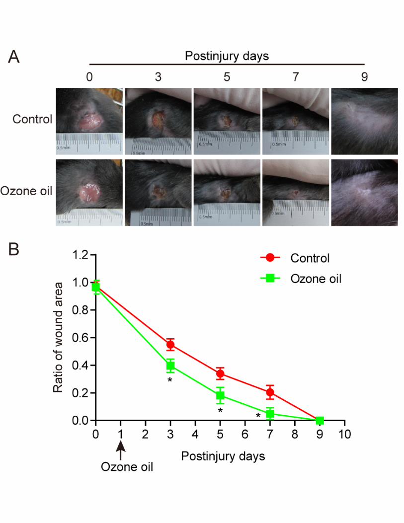
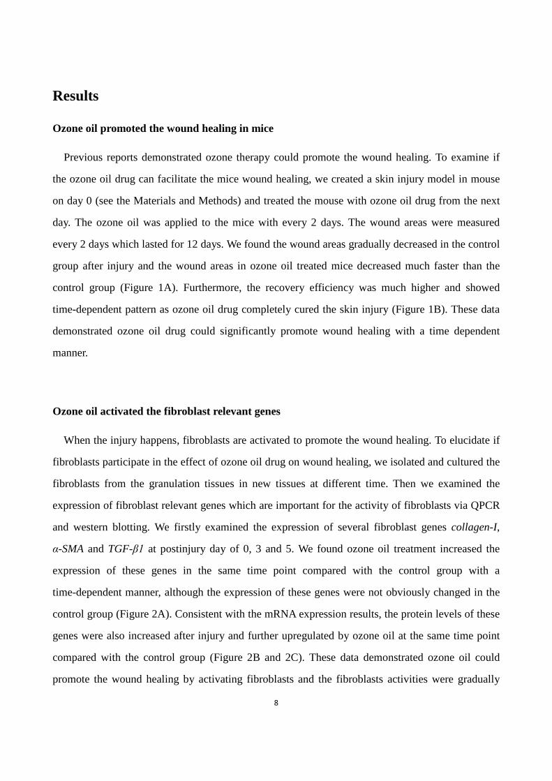
Ozone oil promoted the wound healing in mice
Previous reports demonstrated ozone therapy could promote the wound healing. To examine if
the ozone oil drug can facilitate the mice wound healing, we created a skin injury model in mouse
on day 0 (see the Materials and Methods) and treated the mouse with ozone oil drug from the next
day. The ozone oil was applied to the mice with every 2 days. The wound areas were measured
every 2 days which lasted for 12 days. We found the wound areas gradually decreased in the control
group after injury and the wound areas in ozone oil treated mice decreased much faster than the
control group (Figure 1A). Furthermore, the recovery efficiency was much higher and showed
time-dependent pattern as ozone oil drug completely cured the skin injury (Figure 1B). These data
demonstrated ozone oil drug could significantly promote wound healing with a time dependent
manner.
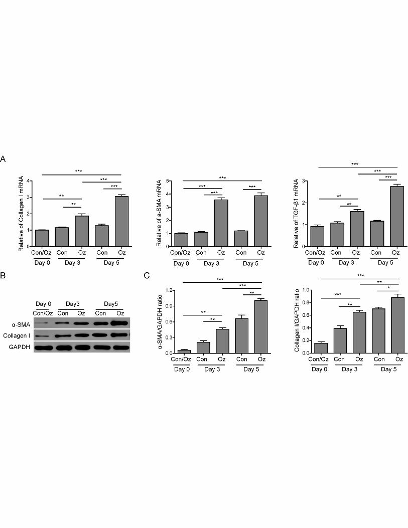
Ozone oil activated the fibroblast relevant genes
When the injury happens, fibroblasts are activated to promote the wound healing. To elucidate if
fibroblasts participate in the effect of ozone oil drug on wound healing, we isolated and cultured the
fibroblasts from the granulation tissues in new tissues at different time. Then we examined the
expression of fibroblast relevant genes which are important for the activity of fibroblasts via QPCR
and western blotting. We firstly examined the expression of several fibroblast genes collagen-I,
α-SMA and TGF-β1 at postinjury day of 0, 3 and 5. We found ozone oil treatment increased the
expression of these genes in the same time point compared with the control group with a
time-dependent manner, although the expression of these genes were not obviously changed in the
control group (Figure 2A). Consistent with the mRNA expression results, the protein levels of these
genes were also increased after injury and further upregulated by ozone oil at the same time point
compared with the control group (Figure 2B and 2C). These data demonstrated ozone oil could
promote the wound healing by activating fibroblasts and the fibroblasts activities were gradually
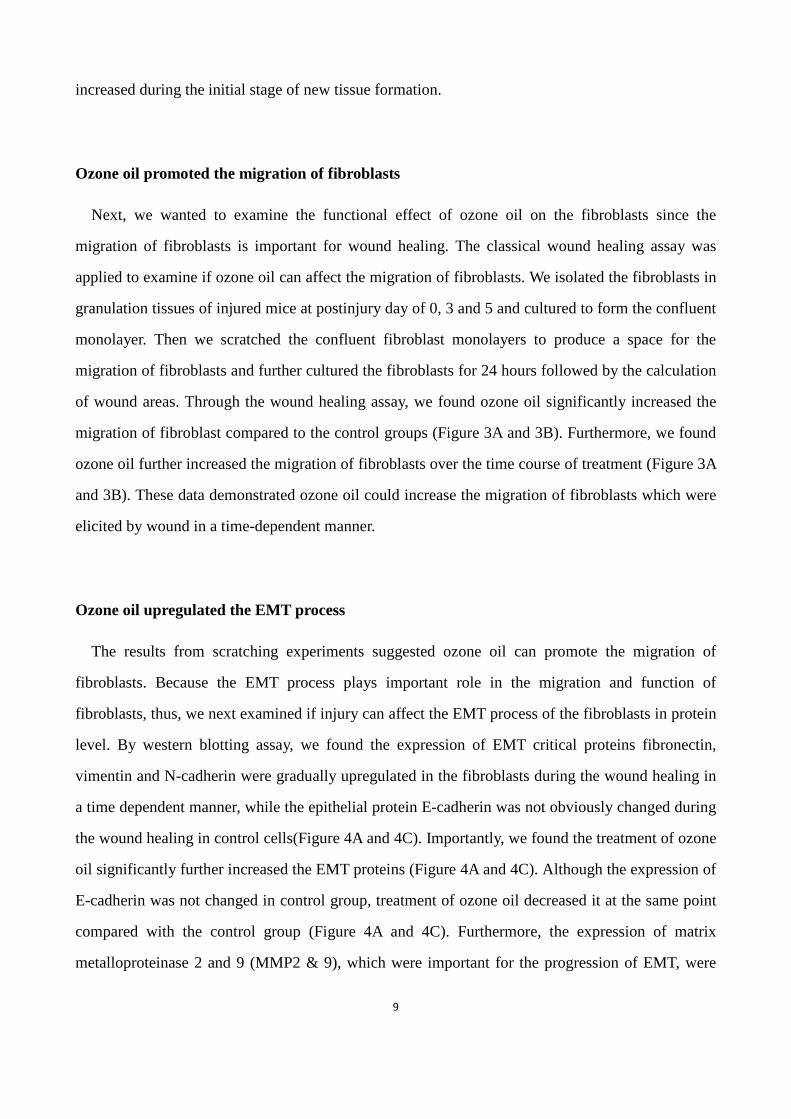
9
increased during the initial stage of new tissue formation.
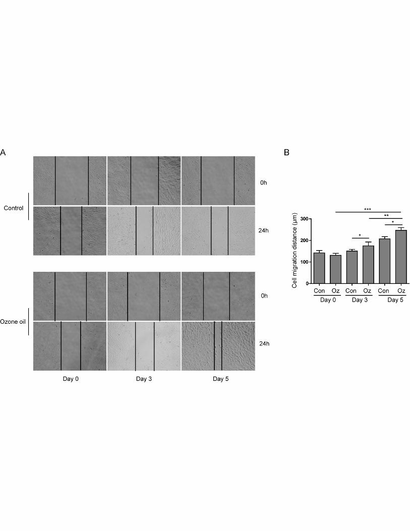
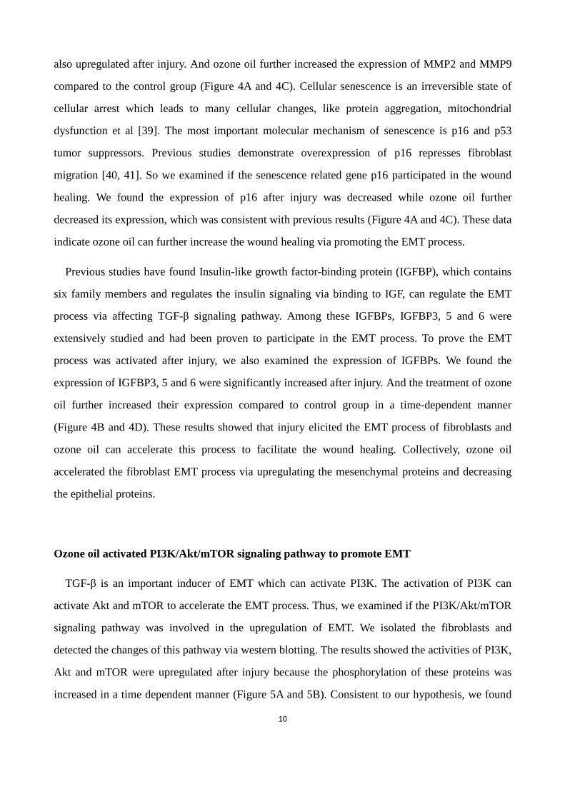
Ozone oil promoted the migration of fibroblasts
Next, we wanted to examine the functional effect of ozone oil on the fibroblasts since the
migration of fibroblasts is important for wound healing. The classical wound healing assay was
applied to examine if ozone oil can affect the migration of fibroblasts. We isolated the fibroblasts in
granulation tissues of injured mice at postinjury day of 0, 3 and 5 and cultured to form the confluent
monolayer. Then we scratched the confluent fibroblast monolayers to produce a space for the
migration of fibroblasts and further cultured the fibroblasts for 24 hours followed by the calculation
of wound areas. Through the wound healing assay, we found ozone oil significantly increased the
migration of fibroblast compared to the control groups (Figure 3A and 3B). Furthermore, we found
ozone oil further increased the migration of fibroblasts over the time course of treatment (Figure 3A
and 3B). These data demonstrated ozone oil could increase the migration of fibroblasts which were
elicited by wound in a time-dependent manner.
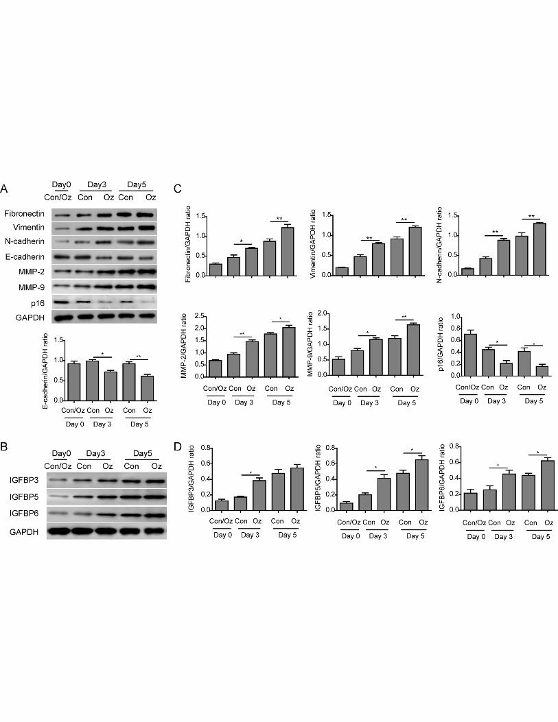
Ozone oil upregulated the EMT process
The results from scratching experiments suggested ozone oil can promote the migration of
fibroblasts. Because the EMT process plays important role in the migration and function of
fibroblasts, thus, we next examined if injury can affect the EMT process of the fibroblasts in protein
level. By western blotting assay, we found the expression of EMT critical proteins fibronectin,
vimentin and N-cadherin were gradually upregulated in the fibroblasts during the wound healing in
a time dependent manner, while the epithelial protein E-cadherin was not obviously changed during
the wound healing in control cells(Figure 4A and 4C). Importantly, we found the treatment of ozone
oil significantly further increased the EMT proteins (Figure 4A and 4C). Although the expression of
E-cadherin was not changed in control group, treatment of ozone oil decreased it at the same point
compared with the control group (Figure 4A and 4C). Furthermore, the expression of matrix
metalloproteinase 2 and 9 (MMP2 & 9), which were important for the progression of EMT, were
10
also upregulated after injury. And ozone oil further increased the expression of MMP2 and MMP9
compared to the control group (Figure 4A and 4C). Cellular senescence is an irreversible state of
cellular arrest which leads to many cellular changes, like protein aggregation, mitochondrial
dysfunction et al [39]. The most important molecular mechanism of senescence is p16 and p53
tumor suppressors. Previous studies demonstrate overexpression of p16 represses fibroblast
migration [40, 41]. So we examined if the senescence related gene p16 participated in the wound
healing. We found the expression of p16 after injury was decreased while ozone oil further
decreased its expression, which was consistent with previous results (Figure 4A and 4C). These data
indicate ozone oil can further increase the wound healing via promoting the EMT process.
Previous studies have found Insulin-like growth factor-binding protein (IGFBP), which contains
six family members and regulates the insulin signaling via binding to IGF, can regulate the EMT
process via affecting TGF-β signaling pathway. Among these IGFBPs, IGFBP3, 5 and 6 were
extensively studied and had been proven to participate in the EMT process. To prove the EMT
process was activated after injury, we also examined the expression of IGFBPs. We found the
expression of IGFBP3, 5 and 6 were significantly increased after injury. And the treatment of ozone
oil further increased their expression compared to control group in a time-dependent manner
(Figure 4B and 4D). These results showed that injury elicited the EMT process of fibroblasts and
ozone oil can accelerate this process to facilitate the wound healing. Collectively, ozone oil
accelerated the fibroblast EMT process via upregulating the mesenchymal proteins and decreasing
the epithelial proteins.
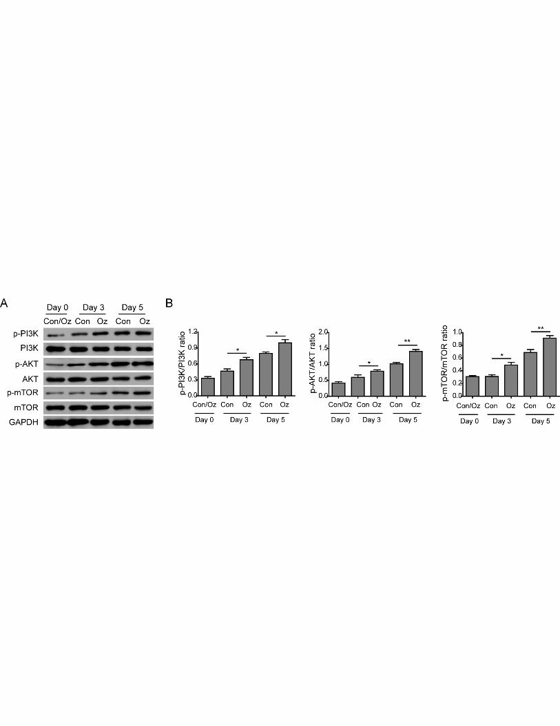
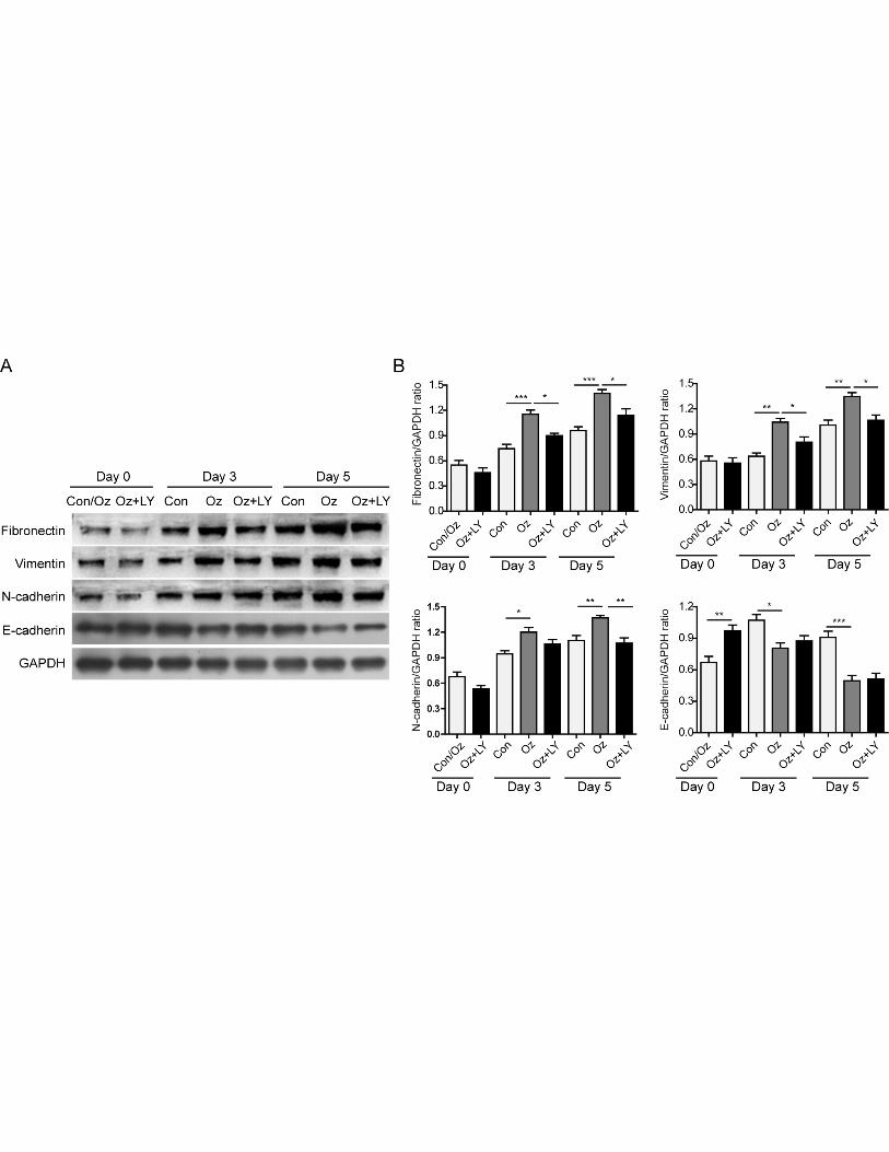
Ozone oil activated PI3K/Akt/mTOR signaling pathway to promote EMT
TGF-β is an important inducer of EMT which can activate PI3K. The activation of PI3K can
activate Akt and mTOR to accelerate the EMT process. Thus, we examined if the PI3K/Akt/mTOR
signaling pathway was involved in the upregulation of EMT. We isolated the fibroblasts and
detected the changes of this pathway via western blotting. The results showed the activities of PI3K,
Akt and mTOR were upregulated after injury because the phosphorylation of these proteins was
increased in a time dependent manner (Figure 5A and 5B). Consistent to our hypothesis, we found
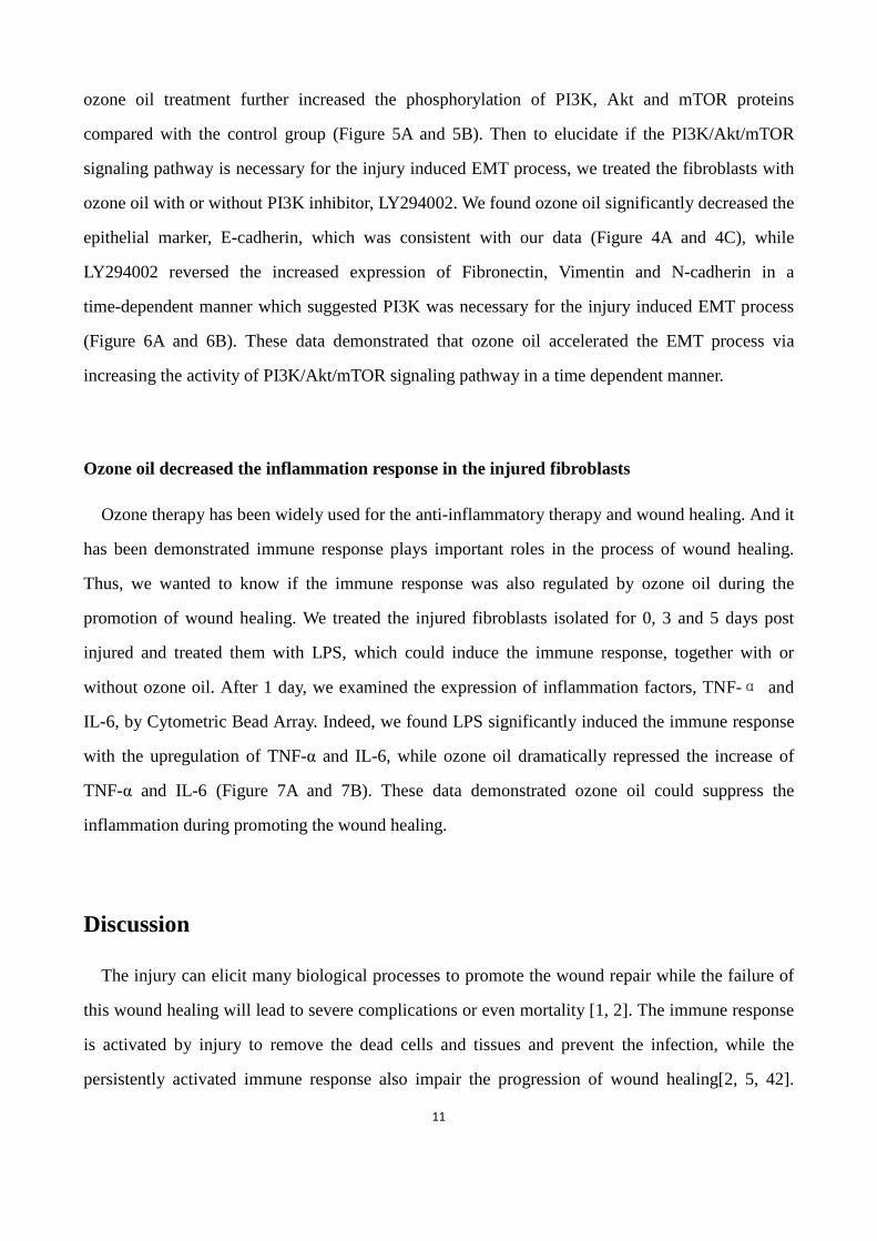
11
ozone oil treatment further increased the phosphorylation of PI3K, Akt and mTOR proteins
compared with the control group (Figure 5A and 5B). Then to elucidate if the PI3K/Akt/mTOR
signaling pathway is necessary for the injury induced EMT process, we treated the fibroblasts with
ozone oil with or without PI3K inhibitor, LY294002. We found ozone oil significantly decreased the
epithelial marker, E-cadherin, which was consistent with our data (Figure 4A and 4C), while
LY294002 reversed the increased expression of Fibronectin, Vimentin and N-cadherin in a
time-dependent manner which suggested PI3K was necessary for the injury induced EMT process
(Figure 6A and 6B). These data demonstrated that ozone oil accelerated the EMT process via
increasing the activity of PI3K/Akt/mTOR signaling pathway in a time dependent manner.
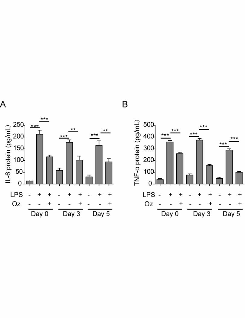
Ozone oil decreased the inflammation response in the injured fibroblasts
Ozone therapy has been widely used for the anti-inflammatory therapy and wound healing. And it
has been demonstrated immune response plays important roles in the process of wound healing.
Thus, we wanted to know if the immune response was also regulated by ozone oil during the
promotion of wound healing. We treated the injured fibroblasts isolated for 0, 3 and 5 days post
injured and treated them with LPS, which could induce the immune response, together with or
without ozone oil. After 1 day, we examined the expression of inflammation factors, TNF-α and
IL-6, by Cytometric Bead Array. Indeed, we found LPS significantly induced the immune response
with the upregulation of TNF-α and IL-6, while ozone oil dramatically repressed the increase of
TNF-α and IL-6 (Figure 7A and 7B). These data demonstrated ozone oil could suppress the
inflammation during promoting the wound healing.
Discussion
The injury can elicit many biological processes to promote the wound repair while the failure of
this wound healing will lead to severe complications or even mortality [1, 2]. The immune response
is activated by injury to remove the dead cells and tissues and prevent the infection, while the
persistently activated immune response also impair the progression of wound healing[2, 5, 42].
12
Previous reports have found fibroblasts which are attracted by the wound play important roles in
wound healing. Fibroblasts can secrete factors to promote angiogenesis and differentiate into
myofibroblasts to form the mature scar [12, 14]. Thus the studies of fibroblasts in wound healing
may provide new therapeutic targets for curing the injury independent on the immune systems.
Furthermore, since ozone therapy is widely applied for the anti-inflammatory therapy, we wanted to
explore the function of ozone oil on the migration of fibroblasts during wound healing. And, we
found ozone oil can promote the wound healing by increasing the activation and migration of
fibroblasts via PI3K/Akt/mTOR signaling pathway.
Ozone therapy has been recognized as an efficient treatment for tissue repair [21-23]. Ozone
therapy can inactivate bacteria via disrupting their cell envelope, inhibit fungi growth and damage
the capsid of virus. Ozone therapy also increases the glycolysis rate of erythrocytes which leads to
the increase of oxygen released to tissues. Ozone therapy also stimulates the production of
prostacyclin and the enzymes which work as cell-wall protectors and free radical scavengers. Also
ozone therapy is applied for tissue repair which may be through inducing the production of different
growth factors VEGF, TGF-β and PDGF. In our study, we found ozone oil can further accelerate the
rate of wound healing compared the untreated group (Figure 1). Also, we found the treatment of
ozone oil activated fibroblasts through increasing the critical genes (collagen-I, α-SMA and TGF-β1)
for fibroblasts (Figure 2). These results demonstrated that fibroblasts activities were gradually
enhanceed during the initial stage of new tissue formation and ozone oil promoted the wound
healing via regulating the fibroblast functions.
As the tissue injury will attract the fibroblast to the injury site to repair the wound, the migration
of fibroblasts is important for tissue repair [7, 8, 37]. Here, we found ozone oil promoted the
migration of fibroblasts which suggested the cure effect of ozone oil on wound healing through
increasing the fibroblast migration (Figure 3). Furthermore, the EMT process in fibroblasts also
plays important roles in the tissue repair. So we examined if the treatment of ozone oil can promote
the EMT process of fibroblasts. We found ozone oil promoted the upregulation of critical proteins
(fibronectin, vimentin, N-cadherin) and the reduction of epithelial protein (E-cadherin) in EMT
(Figure 4). As IGFBPs play important roles in the EMT process, we examined the expression of
these genes and found their expression was further increased by ozone oil (Figure 4). These data
13
demonstrated ozone oil can activate the fibroblasts and promote the migration and EMT process of
fibroblasts to facilitate the wound healing.
The EMT is an important cellular processes during embryogenesis and plays critical roles in
many diseases[25]. Previous studies have found many signaling pathways are involved in the EMT
process, such as TGF-β, EGF, TNF-α, IL-6 and so on [43-45]. Recent studies have shown
PI3K/Akt/mTOR signaling pathway plays important roles in EMT process [46-49]. For example,
miR-206 inhibits HGF-induced EMT process and angiogenesis via PI3K/Akt/mTOR signaling
pathway in non-small cell lung cancer [46].TGF-β2 induces the EMT process via activation of
PI3K/Akt/mTOR signaling pathway in cultured human lens epithelial cells [47].In hepatocellular
carcinoma, TRAF4 regulates migration, invasion and the EMT process via PI3K/Akt/mTOR
signaling pathway [48]. So we examined whether the regulation of ozone oil on EMT processes was
through affecting the PI3K/Akt/mTOR signaling pathway. We found the treatment of ozone oil
significantly increased the activation of PI3K/Akt/mTOR signaling pathway compared to the
untreated control group. The level of p-PI3K, p-Akt and p-mTOR was gradually increased as the
process of wound healing which was further increased by ozone oil (Figure 5). Importantly, we
found inhibition of PI3K with LY294002 reversed the effect of ozone oil on the EMT process,
which suggests PI3K/Akt/mTOR signaling pathway is necessary for the wound healing (Figure 6).
These data suggested ozone oil promoted the EMT process via accelerating the activation of
PI3K/Akt/mTOR signaling pathway to facilitate the wound healing. In the future, the drugs which
target to PI3K/Akt/mTOR signaling pathway may provide new hope for the treatment of injury.
Inflammation is the initial step after injury and plays important roles in the process of wound
healing, while the continuous inflammation will impair the wound healing process. So inhibition of
the inflammation during wound healing is also critical. We found ozone oil significantly suppressed
the inflammation of the injured fibroblasts which treated with LPS (Figure 7). These results
demonstrated ozone oil can facilitate the wound healing via inhibiting the inflammation instead of
promoting the function of fibroblasts and EMT.
In conclusion, we found ozone oil can promote the wound healing via PI3K/Akt/mTOR signaling
pathway. Mechanistically, we found ozone oil can activate the fibroblasts and promote the
14
migration of fibroblasts. Furthermore, ozone oil can further increase the EMT process of fibroblasts
via up-regulating the important proteins for EMT process. Importantly, we found PI3K/Akt/mTOR
signaling pathway was involved in the regulation of ozone oil on the EMT process and wound
healing. Our studies illustrate the cellular mechanisms for the treatment of ozone oil on wound
healing, which will provide new insight and therapeutic targets for the treatment of tissue injury.
15
References
1. Gurtner, G.C., et al., Wound repair and regeneration. Nature, 2008. 453(7193): p. 314-321.
2. Eming, S.A., P. Martin, and M. Tomic-Canic, Wound repair and regeneration: mechanisms, signaling, and
translation. Sci Transl Med, 2014. 6(265): p. 265sr6.
3. S, A., L. MT, and G. GC, Hypertrophic scar formation following burns and trauma: new approaches to
treatment. PLoS Med., 2007. 4(9): p. e234.
4. P, M. and L. SJ, Inflammatory cells during wound repair: the good, the bad and the ugly Trends Cell Biol. , 2005.
15(11): p. 599-607.
5. P, M., et al., Wound healing in the PU.1 null mouse--tissue repair is not dependent on inflammatory cells. Curr
Biol. , 2003. 13(13): p. 1122-1128.
6. Schmidt, B.A. and V. Horsley, Intradermal adipocytes mediate fibroblast recruitment during skin wound
healing. Development, 2013. 140(7): p. 1517-27.
7. Xie, C., et al., MiR-1908 promotes scar formation post-burn wound healing by suppressing Ski-mediated
inflammation and fibroblast proliferation. Cell Tissue Res, 2016. 366(2): p. 371-380.
8. Amin, Z.A., et al., Application of Antrodia camphorata Promotes Rat's Wound Healing In Vivo and Facilitates
Fibroblast Cell Proliferation In Vitro. Evid Based Complement Alternat Med, 2015. 2015: p. 317693.
9. Park, J., et al., Anti-aging effects of Piper cambodianum P. Fourn. extract on normal human dermal fibroblast
cells and a wound-healing model in mice. Clinical Interventions in Aging, 2016. 11: p. 1017-1026.
10. Upadhyay, A., et al., Ixora coccinea Enhances Cutaneous Wound Healing by Upregulating the Expression of
Collagen and Basic Fibroblast Growth Factor. ISRN Pharmacol, 2014. 2014: p. 751824.
11. Zheng, Z., et al., Up-regulation of fibroblast growth factor (FGF) 9 expression and FGF-WNT/beta-catenin
signaling in laser-induced wound healing. Wound Repair Regen, 2014. 22(5): p. 660-5.
12. Nakamichi, M., et al., Basic Fibroblast Growth Factor Induces Angiogenic Properties of Fibrocytes to Stimulate
Vascular Formation during Wound Healing. Am J Pathol, 2016. 186(12): p. 3203-3216.
13. Zigrino, P., et al., Fibroblast-Derived MMP-14 Regulates Collagen Homeostasis in Adult Skin. J Invest Dermatol,
2016. 136(8): p. 1575-83.
14. SR, O. and D. JM, Fibroblast differentiation of bone marrow-derived cells during wound repair. FASEB J, 2005.
19(11): p. 1561-1563.
15. Werner, S., T. Krieg, and H. Smola, Keratinocyte–Fibroblast Interactions in Wound Healing. Journal of
Investigative Dermatology, 2007. 127(5): p. 998-1008.
16. 3rd, L.H., et al., Relative distribution and crosslinking of collagen distinguish fetal from adult sheep wound
repair. J Pediatr Surg, 1999. 34(1): p. 218-223.
16
17. Tasdemir, Z., B.A. Alkan, and H. Albayrak, Effects of Ozone Therapy on the Early Healing Period of
Deepithelialized Gingival Grafts: A Randomized Placebo-Controlled Clinical Trial. J Periodontol, 2016. 87(6): p.
663-71.
18. Patel, P.V., et al., Cytological assessment of healing palatal donor site wounds and grafted gingival wounds
after application of ozonated oil: an eighteen-month randomized controlled clinical trial. Acta Cytol, 2012.
56(3): p. 277-84.
19. Elvis, A.M. and J.S. Ekta, Ozone therapy: A clinical review. J Nat Sci Biol Med, 2011. 2(1): p. 66-70.
20. Holz, O., et al., Validation of the human ozone challenge model as a tool for assessing anti-inflammatory
drugs in early development. J Clin Pharmacol, 2005. 45(5): p. 498-503.
21. Zhang, J., et al., Increased growth factors play a role in wound healing promoted by noninvasive oxygen-ozone
therapy in diabetic patients with foot ulcers. Oxid Med Cell Longev, 2014. 2014: p. 273475.
22. Shah, P., A.K. Shyam, and S. Shah, Adjuvant combined ozone therapy for extensive wound over tibia. Indian J
Orthop, 2011. 45(4): p. 376-9.
23. Degli Agosti, I., et al., Effectiveness of a Short-Term Treatment of Oxygen-Ozone Therapy into Healing in a
Posttraumatic Wound. Case Reports in Medicine, 2016. 2016: p. 1-5.
24. JP, T., et al., Epithelial-mesenchymal transitions in development and disease. Cell, 2009. 139(5): p. 871-890.
25. Chen, T., et al., Epithelial-Mesenchymal Transition (EMT): A Biological Process in the Development, Stem Cell
Differentiation and Tumorigenesis. J Cell Physiol, 2017.
26. Lee, J.M., et al., The epithelial-mesenchymal transition: new insights in signaling, development, and disease. J
Cell Biol, 2006. 172(7): p. 973-81.
27. E, P., et al., TGF-β type I receptor/ALK-5 and Smad proteins mediate epithelial to mesenchymal
transdifferentiation in NMuMG breast epithelial cells. J Cell Sci, 1999. 112(Pt24): p. 4557-4568.
28. Bakin, A.V., et al., Phosphatidylinositol 3-kinase function is required for transforming growth factor
beta-mediated epithelial to mesenchymal transition and cell migration. J Biol Chem, 2000. 275(47): p.
36803-10.
29. NA, B., et al., Transforming growth factor-beta1 mediates epithelial to mesenchymal transdifferentiation
through a RhoA-dependent mechanism. Mol Biol Cell, 2001. 12(1): p. 27-36.
30. L, Y., H. MC, and Z. YE, TGF-beta receptor-activated p38 MAP kinase mediates Smad-independent TGF-beta
responses. EMBO J., 2002. 21(14): p. 3749-3759.
31. Chen, W.C., et al., Curcumin suppresses doxorubicin-induced epithelial-mesenchymal transition via the
inhibition of TGF-beta and PI3K/AKT signaling pathways in triple-negative breast cancer cells. J Agric Food
Chem, 2013. 61(48): p. 11817-24.
32. Yeh, Y.H., et al., Rhapontigenin inhibits TGF-beta-mediated epithelialmesenchymal transition via the
PI3K/AKT/mTOR pathway and is not associated with HIF-1alpha degradation. Oncol Rep, 2016. 35(5): p.
17
2887-95.
33. Baek, S.H., et al., Ginkgolic Acid Inhibits Invasion and Migration and TGF-beta-Induced EMT of Lung Cancer
Cells Through PI3K/Akt/mTOR Inactivation. J Cell Physiol, 2017. 232(2): p. 346-354.
34. Yin, S.Y., et al., The Phytochemical Shikonin Stimulates Epithelial-Mesenchymal Transition (EMT) in Skin Wound
Healing. Evid Based Complement Alternat Med, 2013. 2013: p. 262796.
35. Stone, R.C., et al., Epithelial-mesenchymal transition in tissue repair and fibrosis. Cell Tissue Res, 2016. 365(3):
p. 495-506.
36. Hudson, L.G., et al., Cutaneous wound reepithelialization is compromised in mice lacking functional Slug
(Snai2). Journal Of Dermatological Science, 2009. 56(1): p. 19-26.
37. F, C., et al., Vimentin coordinates fibroblast proliferation and keratinocyte differentiation in wound healing via
TGF-β-Slug signaling. Proc Natl Acad Sci U S A, 2016. 113(30): p. 4320-4327.
38. Yan, C., et al., Epithelial to mesenchymal transition in human skin wound healing is induced by tumor necrosis
factor-alpha through bone morphogenic protein-2. Am J Pathol, 2010. 176(5): p. 2247-58.
39. Rayess, H., M.B. Wang, and E.S. Srivatsan, Cellular senescence and tumor suppressor gene p16. Int J Cancer,
2012. 130(8): p. 1715-25.
40. Al-Ansari, M.M., et al., p16INK4A represses breast stromal fibroblasts migration/invasion and their
VEGF-A-dependent promotion of angiogenesis through Akt inhibition. Neoplasia, 2012. 14(12): p. 1269-77.
41. Al-Ansari, M.M., et al., p16(INK4A) represses the paracrine tumor-promoting effects of breast stromal
fibroblasts. Oncogene, 2013. 32(18): p. 2356-64.
42. Grose, R. and S. Werner, Wound-healing studies in transgenic and knockout mice. Mol Biotechnol, 2004. 28(2):
p. 147-66.
43. Dumitriu, I.E., et al., Human dendritic cells produce TGF-beta 1 under the influence of lung carcinoma cells and
prime the differentiation of CD4+CD25+Foxp3+ regulatory T cells. J Immunol, 2009. 182(5): p. 2795-807.
44. Fernando, R.I., et al., IL-8 signaling plays a critical role in the epithelial-mesenchymal transition of human
carcinoma cells. Cancer Res, 2011. 71(15): p. 5296-306.
45. Yadav, A., et al., IL-6 promotes head and neck tumor metastasis by inducing epithelial-mesenchymal transition
via the JAK-STAT3-SNAIL signaling pathway. Mol Cancer Res, 2011. 9(12): p. 1658-67.
46. Chen, Q.Y., et al., MiR-206 inhibits HGF-induced epithelial-mesenchymal transition and angiogenesis in
non-small cell lung cancer via c-Met /PI3k/Akt/mTOR pathway. Oncotarget, 2016. 7(14): p. 18247-61.
47. Guo, R., et al., TGF-beta2 induces epithelial-mesenchymal transition in cultured human lens epithelial cells
through activation of the PI3K/Akt/mTOR signaling pathway. Mol Med Rep, 2016. 13(2): p. 1105-10.
48. Liu, K., et al., TRAF4 regulates migration, invasion and the epithelial-mesenchymal transition via PI3K/AKT
signaling in hepatocellular carcinoma. Oncol Res, 2017.

18
49. Shen, T., et al., CXCL8 induces epithelial-mesenchymal transition in colon cancer cells via the
PI3K/Akt/NF-kappaB signaling pathway. Oncol Rep, 2017.
19
Figure legends
Figure 1.Ozone oil promoted the wound healing at a time-dependent manner. A.
Representative images showing the treatment of ozone oil promotes the wound healing in injured
mouse model. B. The statistical results showing the wound area gradually decreased in a time
dependent manner which was accelerated by ozone oil. Excisional wounds with 1 cm diameter were
created on day 0. The ozone oil was applied for the treatment from the day 1. Error bars represent
the mean ± SD. *p < 0.05 versus control group respectively.
Figure 2. Ozone oil promoted the expression of fibroblast genes. A. Real-time PCR results
showing ozone oil increases the fibroblast marker genes, collagen I, α-SMA and TGF-β1 after
injury in time-dependent manner. B and C. Western blotting results showing the protein expression
of these fibroblast genes is also increased after injury. Ozone oil further increases the expression of
these genes. Error bars represent the mean ± SD. *p < 0.05, **p < 0.01 and ***p < 0.01.
Figure 3. Ozone oil promoted the migration of fibroblasts. A. Scratching experiment showing
ozone oil increases the migration of fibroblasts elicited by injury with time-dependent manner
compared to the control group. B. The summary of cell migration distance of fibroblasts isolated
from the injury sites of mice with or without the treatment of ozone oil. Error bars represent the
mean ± SD. *p < 0.05, **p < 0.01, ***p < 0.001.
Figure 4. Ozone oil increased the expression of fibroblast EMT proteins. A. Western blotting
results showing the expression of EMT proteins, Fibronectin, Vimentin, N-cadherin, MMP-2 and
MMP-9 is increased in fibroblasts and further increased by ozone oil in a time dependent manner,
while the epithelial marker (E-cadherin) and cellular senescence marker (p16) are decreased. B.
Western blotting results showing the expression of IGFBP3, 5 and 6 increased in fibroblasts and
further increased by ozone oil in a time-dependent manner. C. Summary of the western blotting
results of A. D. Summary of the western blotting results of B. Error bars represent the mean ± SD.
*p < 0.05 and **p < 0.01.
Figure 5. Ozone oil activated the PI3K/Akt/mTOR signaling pathway. A. Western blotting
results showing the expression of important proteins for PI3K/Akt/mTOR signaling pathway is
20
increased in the fibroblasts. Ozone oil further increases the expression of these proteins. B.
Summary of the western blotting results. Error bars represent the mean ± SD. *p < 0.05 and **p <
0.01.
Figure 6. Inhibition of PI3K diminished the effect of ozone oil on the expression of
EMT-related proteins. A. Fibroblasts isolated from injured mice at 0, 3 and 5 post-injury day were
treated with ozone oil with or without PI3K inhibitor, LY294002. The increased expression of
Fibronectin, Vimentin and N-cadherin and the decreased expression of E-cadherin after treatment of
ozone oil were reversed by LY294002. B. Summary of the western blotting results. Error bars
represent the mean ± SD. *p < 0.05 , **p < 0.01 and ***p < 0.001.
Figure 7. Ozone oil inhibited the inflammation of the injured fibroblasts. A. Ozone oil
decreased the IL-6 protein level after the injured fibroblasts were treated with LPS. B. Ozone oil
decreased the TNF-α protein level after the injured fibroblasts were treated with LPS. Error bars
represent the mean ± SD. **p < 0.01 and ***p < 0.001.