Embed Size (px)
Citation preview

Rapid communication OMICS International
Journal of Bioprocessing & BiotechniquesJo
urna
l of B
ioprocessing & Biotechniques
ISSN: 2155-9821
Marasabessy et al., J Bioprocess Biotech 2017, 7:1DOI: 10.4172/2155-9821.1000299
J Bioprocess Biotech, an open access journalISSN:2155-9821 Volume 7 • Issue 1 • 1000299
*Corresponding author: Ahmad Marasabessy, Agency for the Assessment and Application of Technology (BPPT), Jakarta, Indonesia, Tel: +62217563120; Fax: +62 21 7560208; E-mail: [email protected]
Received January 01, 2017; Accepted January 30, 2017; Published January 31, 2017
Citation: Marasabessy A, Rudiyono R, Dewi D (2017) Separation, Purification and Chemical Structure Examination of Antifungal Compound from Streptomyces herbaricolor Biomcc-A.RP-131. J Bioprocess Biotech 7: 299. doi:10.4172/2155-9821.1000299
Copyright: © 2017 Marasabessy A, et al. This is an open-access article distributed under the terms of the Creative Commons Attribution License, which permits unrestricted use, distribution, and reproduction in any medium, provided the original author and source are credited.
Keywords: Streptomyces herbaricolor; Antifungal; Fractionation; Purification; Structure elucidation
Abbreviations and SymbolsBioMCC: Biotechnology Laboratory Microbial Culture Collection
– BPPT Indonesia; ISP-2: Selective media 2 developed by Difco Laboratories for characterization of Streptomyces species; PDA: Potato Dextrose Agar; VEG: Liquid vegetative medium for cell proliferation of Streptomyces; FER: Liquid fermentative medium for Streptomyces; UHPLC-MS: Ultra High Performance Liquid Chromatography - Mass Spectrophotometry; MW: Molecular weight (g.mol-1); NMR: Nuclear Magnetic Resonance; MetOH: Methanol; F1: The first group of fractions collected from the second silica gel column; F2: The second group of fractions collected from the second silica gel column; F3: The third group of fractions collected from the second silica gel column.
IntroductionDevelopment of new antibiotic is a costly effort, while the risk of
failure it brings is higher than those of other types of drugs; consequently only a few leading pharmaceutical companies are currently interested in developing new antibiotics. For example, GlaxoSmithKline reported that within the last 10 years of the 72 candidates of new antibiotic compounds that passed the first phase of clinical trials, only one antibiotic was approved by the FDA (Food and Drug Administrations, USA) [1,2]. In addition, research on antifungal drugs against deadly invasive fungal infections also stagnated in which only a few companies are still involved [1].
In the last 25 years, a steady increase of microbial resistance to antibiotics has become a serious threat to global public health. A search to discover new antibiotics has become an utmost important research program around the world because of the increase in resistance toxicity of some available antibiotics [1,2]. Pharmaceutical industry is now facing a scarcity of new antibiotics; therefore, to ensure the availability of effective drugs in the future, it is important to increase efforts for identification of new antibiotics [3]. Development of new molecules with broad spectrum towards the target pathogen is an urgent need [1,3]. High rates of antibiotic resistance due to inappropriate use of antibiotics have also triggered need of new antibiotics having different chemical structure and mechanism of action to the present antibiotics used in medication.
Filamentous actinomycetes, especially the genus Streptomyces is the producer of various natural metabolites [3-5]. These organisms have the ability to secrete various compounds with different biological activities, such as antibiotics, antitumor, antifungi, antituberculosis, as well as pesticides [4,6]. Although Streptomyces is hardly rivaled by other bacterial groups in terms of the ability to produce natural antifungal metabolites, it is likely that chance to discover a new antifungal molecules from Streptomyces is now very low unless the proper strategy and adequate screening and isolation is applied [5,7,8]. Indonesian nature offers a potential source for isolation of newly Streptomyces derived antifungal compounds.
The majority of natural antibiotic substances are antibacterial and are indispensable to treat bacterial infections in humans, animals and plants [9]. In contrast, antifungal antibiotics for treating fungal infections are relatively few, mainly due to solubility and toxicity problems [9-11]. The increasing incidents of fungal infections in humans have directed attention to a search for novel antifungal antibiotics from actinomycetes having a broad spectrum of activity and lesser toxicity [12].
One of our strains within Streptomyces group was isolated from Indonesian soil and screened for bioactive compound in 2006. This strain was preserved and kept in -80ºC until re-grown in 2015 for further studies of its potential as antifungal producer. In our earlier study, this strain showed broad and strong antifungal activities against
Separation, Purification and Chemical Structure Examination of Antifungal Compound from Streptomyces herbaricolor Biomcc-A.RP-131Ahmad Marasabessy*, Rudiyono Rudiyono and Diana Dewi
Agency for the Assessment and Application of Technology (BPPT), Jakarta, Indonesia
AbstractAn actinobacteria of Indonesia origin identified as Streptomyces herbaricolorBioMCC-a.RP-131 has potential antifungal activities. In our earlier study, this strain exhibited a broad spectrum of in vitro antifungal activity against various type strains of fungus (molds and yeasts). Fermentation of this strain in a complex medium lasted for 8 days to produce a broth having high antifungal activity for separation. Extraction of 2 litre fermentation broth with n-Butanol (1:1) produced 24 grams of dried crude extract. Separation of active compound via two times fractionations of the crude extract in silica gel column chromatography eluted with co-solvent in gradient was able to separate almost all impurities. From the analysis of UHPLC-MS spectrum, we found the final fraction still contained 2-4 components with 12 possible chemical formulas. One of the formula was C39H39N4 (MW 562.3175 g.mol-1) not reported yet in Chemspider database, suggesting it to be a new compound. Further work of separation is required to obtain single antifungal compound for chemical structure elucidation with UHPLC-MS and NMR.

Citation: Marasabessy A, Rudiyono R, Dewi D (2017) Separation, Purification and Chemical Structure Examination of Antifungal Compound from Streptomyces herbaricolor Biomcc-A.RP-131. J Bioprocess Biotech 7: 299. doi:10.4172/2155-9821.1000299
Page 2 of 7
J Bioprocess Biotech, an open access journalISSN:2155-9821 Volume 7 • Issue 1 • 1000299
all of tested mold and yeasts. The molecular identification done in 2015 revealed and designated its taxonomic name as Streptomyces herbaricolor BioMCC-a.RP-131. This paper describes the results of the isolation, purification and chemical structure examination of active antifungal compounds derived from fermentation of Streptomyces herbaricolor BioMCC-a.RP-131.
Materials and MethodsMicrobial strains and materials
Streptomyces herbaricolor BioMCC-a.RP-131 as the producer of antifungal compound was maintained in agar slant ISP-2 (Merck) and Aspergillus niger for bioassay was maintained in agar slant PDA. Both strains were obtained from the stock culture of BioMCC (BPPT Indonesia). Unless otherwise stated, all chemicals used were of analytical grade.
Preparation of broth by shake flask fermentation
The spores of Streptomyces herbaricolor BioMCC-a.RP-131 grown on the agar slant ISP-2 (Difco) was taken 1x loopful and inoculated into 20 ml of vegetative medium, namely VEG, consisting of (per litre): 16 g corn steep liquor, 10 g glucose, 20 g sucrose, 2 g ammonium sulphate, 7 g calcium carbonate and water. After shaking at 28ºC, 150 rpm, 2.5 cm stroke, during 72 h, 5 ml of the vegetative culture was transferred to 50 ml of the fermentation medium, namely FER, in a 500 ml Erlenmeyer flask. The FER consisted of (per litre): 30 g cassava starch, 30 g soybean meal, 40 g dextrin, 2 g ammonium sulphate, 6 g calcium carbonate, 2 ml soybean oil and water. The total volume of the FER prepared was 2 L (this gave 40 flasks, each containing 50 ml of the FER). Fermentation was run for 8 days (192 h) at a temperature of 28ºC, 220 rpm and 2.5 cm stroke. During fermentation, 1 ml broth taken from a flask was centrifuged at 10,000 rpm for 10 minutes to obtain a cell-free filtrate, which was used for measuring antifungal activity by bioassay as described later.
Preparation of crude extract
Fermentation broth (2 L) was extracted with 2 litre n-butanol (1:1) by stirring with orbital shaker for 20 min. After extraction, organic phase (top layer) was separated from the aqueous phase by centrifugation at 10,000 rpm for 20 min. The organic phase was evaporated with a vacuum rotary evaporator at a temperature of 50-55ºC until the organic solvent completely evaporated, leaving a dried crude extract in a rotary evaporator flask. The dried crude extract obtained was 24 gm. The crude extract was stored in -20ºC if not immediately used for the separation.
Isolation and purificationSilica gel (50 g, 200-300 mesh, Merck), mixed with methanol, was
packed in a glass column 2 cm in diameter, resulting in 31 cm bed height. The steps for isolation and purification of the active antifungal compounds were as follows. The pre-weighed crude extract was dissolved in semi-polar solvent (methanol or acetonitrile). Separation of the non-polar components was done by extraction using organic solvent with a low boiling point (<80ºC) immiscible with methanol or acetonitrile, i.e. n-hexane. Antifungal activity in both phases was examined by bioassays to confirm the availability antifungal components in the semi-polar solvent and to check the residual antifungal activity in the non-polar solvent. The active fraction was then fractionated in a gradient elution in the column of silica gel with two organic solvent. This stage could be performed 2-3 times depending on needs. Antifungal activity of each fraction was examined in bioassays to
determine the fractions containing the active compound. The solvent of the active fraction was evaporated to obtain a powder of separated compounds.
Bioassay for antifungal detection
A 20 ml Potato dextrose agar medium (PDA, Merck) (kept liquid at 50ºC) in a tube (diameter 2.5 cm, length 15 cm) was suspended with 0.1 ml Aspergillus niger spores suspension in water (equivalent to 1 × 10⁴ spores) and shaken gently, then poured into a sterile petri dish and allowed to solidify. Bioassay for measuring the antifungal activity was carried out by the so-called agar-plug method, done according to the following procedures. Wells on a PDA plate were made with the help of a rubber cork (metal made) having the diameter 6 mm. If not specified otherwise, as many as 40 µl sample was pipetted into a well. The test plates were incubated at 28ºC for 48-72 hours. The diameter of the inhibition zone (mold inhibition area) formed was measured. The inhibition zone diameter was measured with caliper (Mitutoyo, USA) three times in three different directions, and then averaged. The diameter of inhibition zone of a sample is the average value of two replicates (Duplo).
Examination of the chemical structure of active compound
The pattern of mass spectrum of the active compound was investigated by UHPLC-MS to ensure the molecular formula, molecular weight, and fragments resulted from the ionisation of anti-fungal molecules.
Results and DiscussionFermentation broth preparation
From the bioassay test results, no antifungal activity was detected in the fermentation broth after 5 days, indicating that the culture was still in the logarithmic growth phase. The antifungal activity was detected on the seventh day of fermentation (Figure 1), with the average diameter of inhibition zone was 17 mm. These results showed
Figure 1: Antifungal activities of fermentation at 7th day.
Citation: Marasabessy A, Rudiyono R, Dewi D (2017) Separation, Purification and Chemical Structure Examination of Antifungal Compound from Streptomyces herbaricolor Biomcc-A.RP-131. J Bioprocess Biotech 7: 299. doi:10.4172/2155-9821.1000299
Page 3 of 7
J Bioprocess Biotech, an open access journalISSN:2155-9821 Volume 7 • Issue 1 • 1000299
that long fermentation time (more than 5 days) is required to produce sufficient antifungal compound ready for isolation and purification. Fermentation accomplished for 8 days. The crude extract was prepared from the fermentation broth, has been described in Methods.
Isolation and purification of active compounds
First fractionation with silica gel column: Crude extract (0.75 g) was suspended with 5 ml of acetonitrile and was extracted with 5 ml of n-hexane (solvent immiscible with acetonitrile) for 20 min using a reciprocal shaker to separate non-polar components. The acetonitrile phase (bottom layer) was collected and dried in a vacuum concentrator to obtain 0.70 g dry solid. The solid was re-dissolved in 2 ml methanol for separation with silica gel column. Before sample loading, the silica gel column was firstly saturated with diethyleter-ethylacetat (19:1). The sample was then loaded into the silica gel column and elution in gradient was carried out successively using a mixture of diethylether-ethylacetate in ratio, respectively, 19:1 (50 ml); 9:1 (50 ml); 4:1 (50 ml); 1:1 (50 ml); followed by ethylacetate-methanol in ratio, respectively, 19:1 (50 ml); 9:1 (50 ml); 6:4 (50 ml); 1:9 (50 ml); 0:1 (100 ml); followed by methanol-water in ratio 1:1 (100 ml).
Volume of each fraction collected was 7-8 ml. There were 81 fractions collected. Fraction 1-70 was dried by a vacuum concentrator (45ºC) for 2 h. Fraction 71-81 was dried overnight (16 h) at the same temperature. Visual appearance of the dried solid in the bottom of the tube of fraction is shown in Table 1.
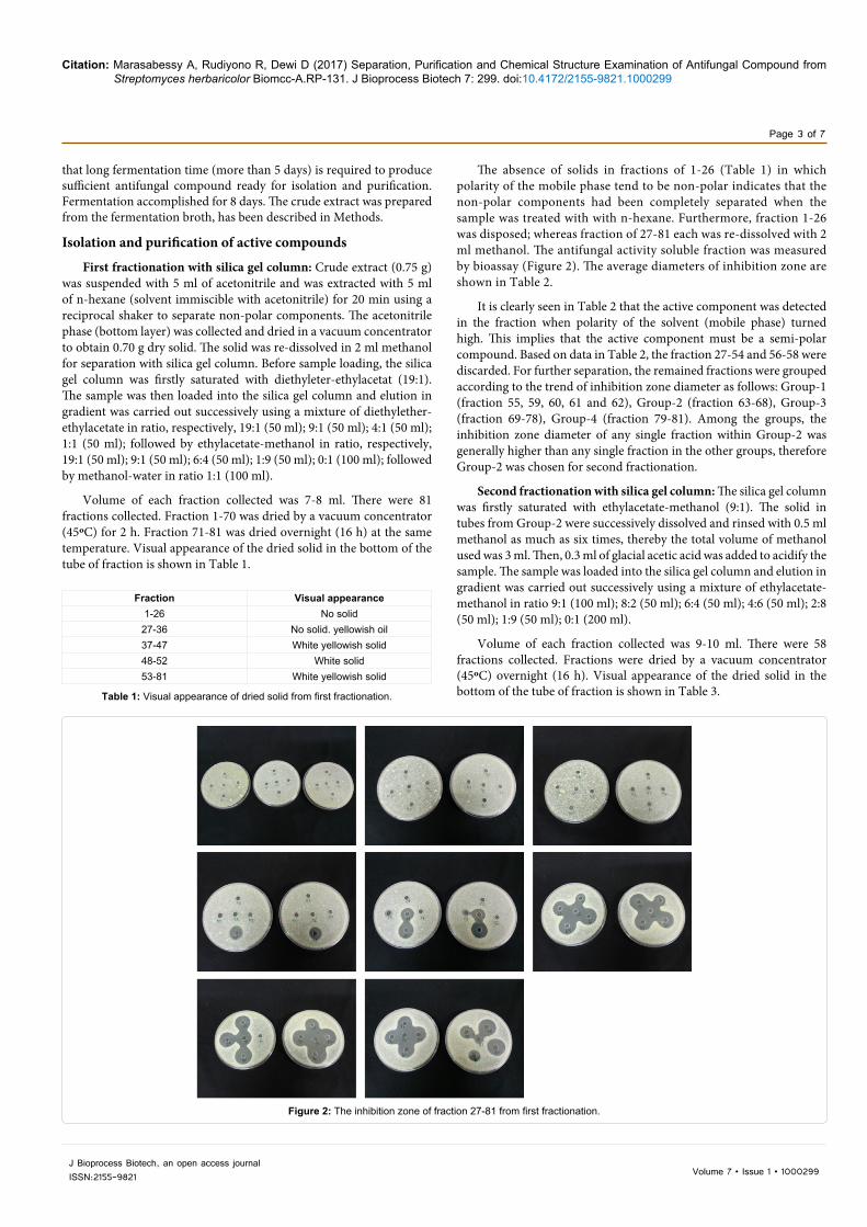
The absence of solids in fractions of 1-26 (Table 1) in which polarity of the mobile phase tend to be non-polar indicates that the non-polar components had been completely separated when the sample was treated with with n-hexane. Furthermore, fraction 1-26 was disposed; whereas fraction of 27-81 each was re-dissolved with 2 ml methanol. The antifungal activity soluble fraction was measured by bioassay (Figure 2). The average diameters of inhibition zone are shown in Table 2.
It is clearly seen in Table 2 that the active component was detected in the fraction when polarity of the solvent (mobile phase) turned high. This implies that the active component must be a semi-polar compound. Based on data in Table 2, the fraction 27-54 and 56-58 were discarded. For further separation, the remained fractions were grouped according to the trend of inhibition zone diameter as follows: Group-1 (fraction 55, 59, 60, 61 and 62), Group-2 (fraction 63-68), Group-3 (fraction 69-78), Group-4 (fraction 79-81). Among the groups, the inhibition zone diameter of any single fraction within Group-2 was generally higher than any single fraction in the other groups, therefore Group-2 was chosen for second fractionation.
Second fractionation with silica gel column: The silica gel column was firstly saturated with ethylacetate-methanol (9:1). The solid in tubes from Group-2 were successively dissolved and rinsed with 0.5 ml methanol as much as six times, thereby the total volume of methanol used was 3 ml. Then, 0.3 ml of glacial acetic acid was added to acidify the sample. The sample was loaded into the silica gel column and elution in gradient was carried out successively using a mixture of ethylacetate-methanol in ratio 9:1 (100 ml); 8:2 (50 ml); 6:4 (50 ml); 4:6 (50 ml); 2:8 (50 ml); 1:9 (50 ml); 0:1 (200 ml).
Volume of each fraction collected was 9-10 ml. There were 58 fractions collected. Fractions were dried by a vacuum concentrator (45ºC) overnight (16 h). Visual appearance of the dried solid in the bottom of the tube of fraction is shown in Table 3.
Fraction Visual appearance1-26 No solid
27-36 No solid. yellowish oil37-47 White yellowish solid 48-52 White solid 53-81 White yellowish solid
Table 1: Visual appearance of dried solid from first fractionation.
Figure 2: The inhibition zone of fraction 27-81 from first fractionation.
Citation: Marasabessy A, Rudiyono R, Dewi D (2017) Separation, Purification and Chemical Structure Examination of Antifungal Compound from Streptomyces herbaricolor Biomcc-A.RP-131. J Bioprocess Biotech 7: 299. doi:10.4172/2155-9821.1000299
Page 4 of 7
J Bioprocess Biotech, an open access journalISSN:2155-9821 Volume 7 • Issue 1 • 1000299
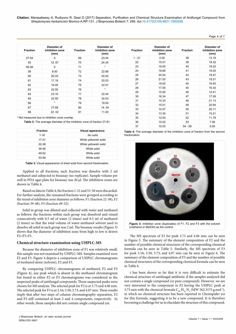
Applied to all fractions, each fraction was dissolve with 2 ml methanol and subjected to bioassay (no replicate). Sample volume per well in PDA agar plate for bioassay was 20 µl. The inhibition zones are shown in Table 4.
Based on data in Table 4, the fraction 1-21 and 53-58 were discarded. For further analysis, the remained fractions were grouped according to the trend of inhibition zone diameter as follows: F1 (fraction 22-38), F2 (fraction 39-48), F3 (fraction 49-52).
Solid in group was diluted and collected with water and methanol as follows: the fractions within each group was dissolved and rinsed consecutively with 0.5 ml of water (2 times) and 0.5 ml of methanol (2 times) so that the total volume of water-methanol solvent used to dissolve all solid in each group was 2 ml. The bioassay results (Figure 3) shows that the diameter of inhibition zone from high to low is drawn F2>F3>F1.
Chemical structure examination using UHPLC-MS
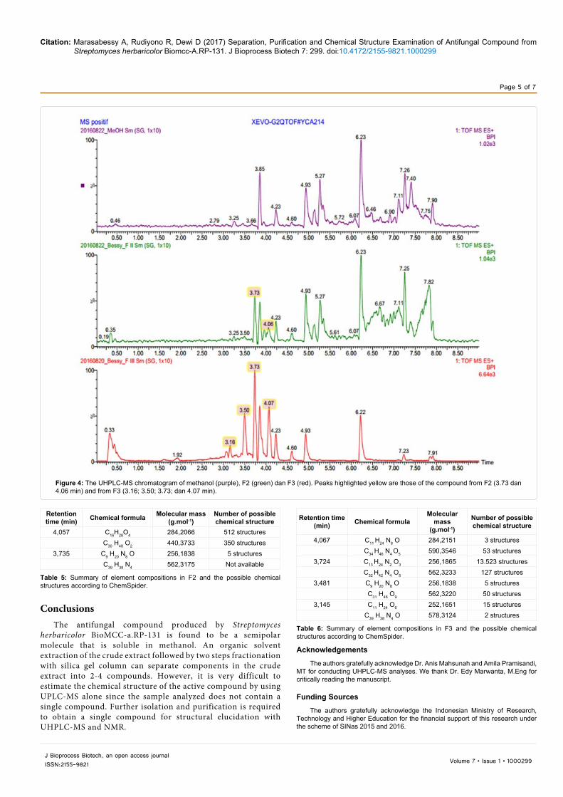
Because the diameter of inhibition zone of F1 was relatively small, this sample was not examined by UHPLC-MS. Samples examined were F2 and F3. Figure 4 depicts a comparison of UHPLC chromatograms of methanol alone (solvent), F2 and F3.
By comparing UHPLC chromatograms of methanol, F2 and F3 (Figure 4), any peak which is absent in the methanol chromatogram but found in either F2 or F3 chromatograms was considered as the suspected peaks of antifungal compounds. Those suspected peaks were chosen for MS analysis. The selected peak for F2 is at 3.73 and 4.06 min. The selected peak for F3 is at 3.16; 3.50; 3.73; and 4.07 min. These results imply that after two steps of column chromatography separation, F2 and F3 still contained at least 2 and 4 components, respectively. In other words, those samples did not contain single compound yet.
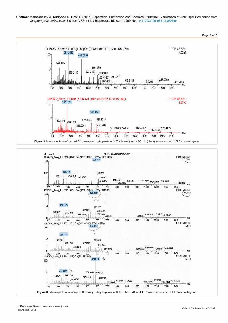
The MS spectrum of F2 for peak 3.73 and 4.06 min can be seen in Figure 5. The summary of the element composition of F2 and the number of possible chemical structures of the corresponding chemical formula can be seen in Table 5. Similarly, the MS spectrum of F3 for peak 3.16; 3.50; 3.73; and 4.07 min can be seen in Figure 6. The summary of the element composition of F3 and the number of possible chemical structures of the corresponding chemical formula can be seen in Table 6.
t has been shown so far that it is very difficult to estimate the chemical structure of antifungal antibiotic if the samples analyzed did not contain a single compound (or pure compound). However, we are very interested to the component in F2 having the UHPLC peak at 3.73 min with the chemical formula C39 H39 N4 (MW 562.3175 g.mol-1), in which no chemical structure has been reported in Chemspider yet for this formula, suggesting it to be a new compound. It is therefore becoming a challenge for us to elucidate the structure of this compound.
FractionDiameter of
inhibition zone (mm)
FractionDiameter of
inhibition zone (mm)
27-54 0 69 23.0455 12. 67 70 24.34
56-58 0 71 *59 9.00 72 22.6660 20.33 73 20.0261 17.18 74 20.0362 16.00 75 22.0163 22.50 76 * 64 23.16 77 22.0465 22.50 78 24.0366 * 79 19.0067 27.68 80 14. 0468 22. 02 81 11.30
* Not measured due to inhibition zone overlapTable 2: The average diameter of the inhibition zone of fraction 27-81.
Fraction Visual appearance1-16 No solid17-21 White yellowish solid 22-38 White yellowish solid39-48 White solid49-52 White solid53-58 White solid
Table 3: Visual appearance of dried solid from second fractionation.
FractionDiameter of
inhibition zone (mm)
FractionDiameter of
inhibition zone (mm)
1 - 21 0.00 38 13.1822 15.01 39 18.3223 19.00 40 18.0224 18.66 41 18.0025 20.04 42 19.4726 21.03 43 19.3127 18.02 44 16.4028 17.05 45 16.3429 15.00 46 12.4130 16.34 47 17.2631 15.33 48 21.1332 15.01 49 20.6433 14.07 50 20.1134 13.35 51 15.2135 12.00 52 11.7936 10.02 53 7.0637 10.03 54 - 58 0.00
Table 4: The average diameter of the inhibition zone of fraction from the second fractionation.
Figure 3: Inhibition zone (duplicates) of F1, F2 and F3 with the solvent (methanol or MetOH) as the control.
Citation: Marasabessy A, Rudiyono R, Dewi D (2017) Separation, Purification and Chemical Structure Examination of Antifungal Compound from Streptomyces herbaricolor Biomcc-A.RP-131. J Bioprocess Biotech 7: 299. doi:10.4172/2155-9821.1000299
Page 5 of 7
J Bioprocess Biotech, an open access journalISSN:2155-9821 Volume 7 • Issue 1 • 1000299
ConclusionsThe antifungal compound produced by Streptomyces
herbaricolor BioMCC-a.RP-131 is found to be a semipolar molecule that is soluble in methanol. An organic solvent extraction of the crude extract followed by two steps fractionation with silica gel column can separate components in the crude extract into 2-4 compounds. However, it is very difficult to estimate the chemical structure of the active compound by using UPLC-MS alone since the sample analyzed does not contain a single compound. Further isolation and purification is required to obtain a single compound for structural elucidation with UHPLC-MS and NMR.
Acknowledgements
The authors gratefully acknowledge Dr. Anis Mahsunah and Amila Pramisandi, MT for conducting UHPLC-MS analyses. We thank Dr. Edy Marwanta, M.Eng for critically reading the manuscript.
Funding Sources
The authors gratefully acknowledge the Indonesian Ministry of Research, Technology and Higher Education for the financial support of this research under the scheme of SINas 2015 and 2016.
Figure 4: The UHPLC-MS chromatogram of methanol (purple), F2 (green) dan F3 (red). Peaks highlighted yellow are those of the compound from F2 (3.73 dan 4.06 min) and from F3 (3.16; 3.50; 3.73; dan 4.07 min).
Retention time (min) Chemical formula Molecular mass
(g.mol-1)Number of possible chemical structure
4,057 C16H28O4 284,2066 512 structuresC30 H48 O2 440,3733 350 structures
3,735 C9 H20 N8 O 256,1838 5 structuresC39 H38 N4 562,3175 Not available
Table 5: Summary of element compositions in F2 and the possible chemical structures according to ChemSpider.
Retention time (min) Chemical formula
Molecular mass
(g.mol-1)
Number of possible chemical structure
4,067 C11 H24 N8 O 284,2151 3 structuresC34 H46 N4 O5 590,3546 53 structures
3,724 C13 H24 N2 O3 256,1865 13.523 structuresC32 H42 N4 O5 562,3233 127 structures
3,481 C9 H20 N8 O 256,1838 5 structuresC31 H46 O9 562,3220 50 structures
3,145 C11 H24 O6 252,1651 15 structuresC39 H38 N4 O 578,3124 2 structures
Table 6: Summary of element compositions in F3 and the possible chemical structures according to ChemSpider.
Citation: Marasabessy A, Rudiyono R, Dewi D (2017) Separation, Purification and Chemical Structure Examination of Antifungal Compound from Streptomyces herbaricolor Biomcc-A.RP-131. J Bioprocess Biotech 7: 299. doi:10.4172/2155-9821.1000299
Page 6 of 7
J Bioprocess Biotech, an open access journalISSN:2155-9821 Volume 7 • Issue 1 • 1000299
Figure 5: Mass spectrum of sampel F2 corresponding to peaks at 3.73 min (red) and 4.06 min (black) as shown on UHPLC chromatogram.
Figure 6: Mass spectrum of sampel F3 corresponding to peaks at 3.16; 3.50; 3.73; and 4.07 min as shown on UHPLC chromatogram.
Citation: Marasabessy A, Rudiyono R, Dewi D (2017) Separation, Purification and Chemical Structure Examination of Antifungal Compound from Streptomyces herbaricolor Biomcc-A.RP-131. J Bioprocess Biotech 7: 299. doi:10.4172/2155-9821.1000299
Page 7 of 7
J Bioprocess Biotech, an open access journalISSN:2155-9821 Volume 7 • Issue 1 • 1000299
Author Contributions
Conceived and designed the experiments: AM, RR. Analysed the data: AM, DD. Wrote the first draft of the manuscript: AM. Contributed to the writing of the manuscript: RR, DD. Agree with manuscript results and conclusions: AM, DD.Jointly developed the structure and arguments for the paper: AM, DD. Made critical revisions and approved final version: DD. All authors reviewed and approved of the final manuscript.
References
1. Jezek A (2013) Antibiotic and Antifungal Drug Transferrable Research andDevelopment Tax Credit. In The Infect Dis Soc America: 1-3.
2. GlaxoSmithKline (2016) Antibiotics research. Brentford, UK: GlaxoSmithKline plc.
3. Munaganti RK, Muvva V, Konda S, Naragani K, Mangamuri UK, et al. (2016)Antimicrobial profile of Arthrobacter kerguelensis VL-RK_09 isolated from Mango orchards. Braz J Microbiol 47: 1030-1038.
4. Goshi K, Uchida T, Lezhava A, Yamasaki M, Hiratsu K, et al. (2002) Cloning and analysis of the telomere and terminal inverted repeat of the linear chromosome of Streptomyces griseus. J Bacteriol 184: 3411-3415.
5. Islam MR, Jeong YT, Ryu YJ, Song CH, Lee YS (2009) Isolation, Identification
and Optimal Culture Conditions of Streptomyces albidoflavus C247 Producing Antifungal Agents against Rhizoctonia solani AG2-2. Mycobiology 37: 114-120.
6. Li-Li C, You-Zhi L, Hong-Jian Z, Yu-Hua L, Bi-Da G (2012) Fractionation andStructural Identification of Antibiotic Activity Substances from Streptomyces herbaricolor HNS2-2. Agri Sci 3: 567-571.
7. Omura S (1986) Philosophy of new drug discovery. Microbiol Rev 50: 259-279.
8. Kumar KS, Haritha R, Mohan YSYVJ, Ramana T (2011) Screening of marineactinobacteria for antimicrobial compounds. Res J Microbiol 6: 385-393.
9. Ouhdouch Y, Barakate M, Finance C (2001) Actinomycetes of Moroccanhabitats: Isolation and screening for antifungal activities. Eur J Soil Biol 37:69-74.
10. Caffrey P, Lynch S, Flood E, Finnan S, Oliynyk M (2001) Amphotericinbiosynthesis in Streptomyces nodosus: deductions from analysis of polyketidesynthase and late genes. Chem Biol 8: 713-723.
11. Vartak A, Mutalik V, Parab RR, Shanbhag P, Bhave S, et al. (2014) Isolation of a new broad spectrum antifungal polyene from Streptomyces sp. MTCC 5680.Lett Appl Microbiol 58: 591.
12. Raytapadar S, Paul AK (2001) Production of an antifungal antibiotic byStreptomyces aburaviensis IDA-28. Microbiol Res 155: 315-323.