Embed Size (px)
Citation preview

ORIGINAL RESEARCH ARTICLE
The HIV Nef protein modulates cellular and exosomalmiRNA profiles in human monocytic cells
Madeeha Aqil1, Afsar Raza Naqvi1#, Saurav Mallik2,Sanghamitra Bandyopadhyay2, Ujjwal Maulik3 and Shahid Jameel1*
1Virology Group, International Centre for Genetic Engineering and Biotechnology, New Delhi, India;2Machine Intelligence Unit, Indian Statistical Institute, Kolkata, India; 3Department of Computer Science andEngineering, Jadavpur University, Kolkata, India
Introduction: The HIV Nef protein is a multifunctional virulence factor that perturbs intracellular membranes
and signalling and is secreted into exosomes. While Nef-containing exosomes have a distinct proteomic
profile, no comprehensive analysis of their miRNA cargo has been carried out. Since Nef functions as a viral
suppressor of RNA interference and disturbs the distribution of RNA-induced silencing complex proteins
between cells and exosomes, we hypothesized that it might also affect the export of miRNAs into exosomes.
Method: Exosomes were purified from human monocytic U937 cells that stably expressed HIV-1 Nef. The
RNA from cells and exosomes was profiled for 667 miRNAs using a Taqman Low Density Array. Selected
miRNAs and their mRNA targets were validated by quantitative RT-PCR. Bioinformatics analyses were used
to identify targets and predict pathways.
Results: Nef expression affected a significant fraction of miRNAs in U937 cells. Our analysis showed 47
miRNAs to be selectively secreted into Nef exosomes and 2 miRNAs to be selectively retained in Nef-
expressing cells. The exosomal miRNAs were predicted to target several cellular genes in inflammatory
cytokine and other pathways important for HIV pathogenesis, and an overwhelming majority had targets
within the HIV genome.
Conclusions: This is the first study to report miRnome analysis of HIV Nef expressing monocytes and
exosomes. Our results demonstrate that Nef causes large-scale dysregulation of cellular miRNAs, including
their secretion through exosomes. We suggest this to be a novel viral strategy to affect pathogenesis and to
limit the effects of RNA interference on viral replication and persistence.
Keywords: exosomes; miRNA; HIV; Nef; inflammatory cytokine
*Correspondence to: Shahid Jameel, Virology Group, ICGEB, Aruna Asaf Ali Marg, New Delhi 110067,
India, Email: [email protected]
To access the supplementary material to this article, please see Supplementary files under Article Tools online.
Received: 21 October 2013; Revised: 28 January 2014; Accepted: 15 February 2014; Published: 25 March 2014
The human immunodeficiency virus (HIV) encodes
prototypic retroviral proteins (Gag, Pol, Env) as
regulatory (Rev, Tat) and accessory (Nef, Vif, Vpr,
Vpu/Vpx) proteins, the last group being dispensable for
virus replication in vitro, but important for persistence
and disease in an immunocompetent host (1). The nef
gene is located at the 3?-end of the viral genome, partially
overlapping the env gene and the U3 region in the 3? long
terminal repeat (LTR). Nef is the largest of the four HIV
accessory proteins, is expressed early in infection and at
far higher levels than the other early proteins, Tat and
Rev (2). It is primarily localized at cellular membranes,
which include endosomal membranes, the perinuclear
region and the inner surface of plasma membrane. Nef is
also released from cells, either in microvesicles (MVs)
(3,4) or as a soluble protein, and has effects on bystander
cells (5�7).
The extracellular MVs are called shedding vesicles
or exosomes depending on their size and origin (8).
The shedding vesicles are 0.1�1 mm and are formed by
outward budding and pinching of the plasma membrane.
Exosomes are 30�100 nm and are formed by inward
#Current Address: Department of Periodontics, College of Dentistry, University of Illinois at Chicago, Chicago, IL 60612, USA
�
Journal of Extracellular Vesicles 2014. # 2014 Madeeha Aqil et al. This is an Open Access article distributed under the terms of the Creative CommonsAttribution-Noncommercial 3.0 Unported License (http://creativecommons.org/licenses/by-nc/3.0/), permitting all non-commercial use, distribution, andreproduction in any medium, provided the original work is properly cited.
1
Citation: Journal of Extracellular Vesicles 2014, 3: 23129 - http://dx.doi.org/10.3402/jev.v3.23129(page number not for citation purpose)

invagination of the multivesicular body (MVB) mem-
brane forming intraluminal vesicles, which are released
in the extracellular medium on fusion of the MVBs with
the plasma membrane. These contain various mRNAs,
miRNAs and proteins that vary depending on the
producer cell type (8,9). Further, since these vesicles can
be taken up by other cells, and the exosomal mRNAs
and miRNAs are shown to be functional in recipient
cells, the MVs are proposed as a novel mode of inter-
cellular communication (10). The modulatory effects of
the exosomal cargo on cellular gene expression are likely
to affect intracellular viral replication, persistence and
pathogenesis.
The first evidence of extracellular Nef came from
reports on the detection of anti-Nef antibodies in the sera
of AIDS patients (11). Later, Nef was also found in
exosome-like vesicles released from HEK293 cells expres-
sing a Nef-green fluorescent protein (Nef-GFP) fusion
protein (3). These vesicles were shown to be taken up by
Jurkat CD4� T cells, in which the Nef-GFP fusion
protein localized mainly to the cytoplasm as punctate
structures (3). The vesicles are likely to enter recipient
cells via endocytosis, which had been reported in other
systems (12). Importantly, Nef exosomes could also fuse
with Nef-deficient HIV-1 virions and restore the infectiv-
ity of mature particles (3).
The mammalian miRNA pathway restricts the replica-
tion of infecting viruses and promotes latency. While miR-
323, miR-491 and miR-654 inhibit the replication of H1N1
influenza virus, miR-24 and miR-93 limit vesicular stoma-
titis virus (VSV) replication, and miR-32 restricts primate
foamy virus type 1 (PFV-1) (13). Human miR-28, miR-
125b, miR-150, miR-223 and miR-382 target the 3?UTRof
HIV-1 transcripts potentially taking productive infection
towards latency (14). Recent advances in deep sequencing
technology have also led to the identification of small viral
RNA species in HIV-1 infected cells (15). Further, the
knockdown of several miRNA processing pathway pro-
teins, including Dicer, Drosha and DGCR8, leads to an
increase in viral replication, and HIV-1 transcripts have
been co-localized with RNAi effector proteins in P-bodies
(16). Thus, cellular miRNAs affect viral replication, either
by targeting viral RNAs or cellular RNAs that encode host
proteins necessary for viral replication (16).
We have studied human monocytic U937 cells that
stably expressed a Nef-EYFP fusion protein and found
the protein in exosomes (17). Transfer of exosomal
miRNAs to other recipient cells is now recognized as a
mode of communication between different cells and
tissues (10). Further, dysregulation of cellular miRNAs
in diseased states also affects their repertoire in exosomes
(9). Nef localizes to MVBs, interacts with Argonaute-2,
redistributes RNA-induced silencing complex (RISC)
components between cells and exosomes, and acts as
a viral suppressor of RNA interference (17). We have
profiled miRNAs in U937/Nef-EYFP cells and exosomes
purified from these cells and have identified the pathways
potentially regulated by these miRNAs. Our results
suggest that Nef mediates the redistribution of miRNAs
between cells and exosomes to aid in viral replication and
persistence.
Materials and methods
Exosome isolation and characterizationThe U937/Nef-EYFP and U937/EYFP cells and their
culture conditions have been described previously (17).
Exosomes were prepared from culture supernatants (18)
and visualized by electron microscopy as described earlier
(17). Exosome quantification was done using Bradford
reagent (BioRad, USA) as described elsewhere (18). For
flow cytometry, purified exosomes (�3�5 mg protein)
were first incubated with 10 ml of aldehyde latex beads for
15 minutes at room temperature. Then PBS was added to
a final volume of 1 ml and incubated on a rotator for 2
hour at room temperature, or overnight at 48C. To this
110 ml of 1 M glycine (final concentration, 100 mM) was
added and beads were incubated at room temperature for
30 minutes. The beads were then centrifuged for 3
minutes at 4,000 rpm at room temperature and washed
twice with 1 ml PBS/0.5% BSA with final re-suspension in
0.5 ml PBS-BSA. For staining with antibodies, 10 ml of
coated beads were incubated with 50 ml of the appropriate
antibody diluted in PBS-BSA for 30 minutes at 48C.
The beads were then washed twice as described above
followed by incubation with 50 ml secondary antibodies
for 30 minutes at 48C. The beads were again washed
twice and were acquired on a Cyan-ADP flow cytometer
(Beckman Coulter). Data were analysed using the Flow
Jo software. For the labelling of exosomal membranes
with PKH26, a fluorescent lipid dye (Sigma) the manu-
facturer’s instructions were followed. About 10�50 mg of
exosomes were used for each labelling reaction. The
excess dye was removed by washing exosomes with 3 ml
of PBS with ultracentrifugation at 100,000�g in a SW60
rotor at 48C. These were visualized using a Nikon A1-R
Confocal Microscope at 100� magnification. The la-
belled exosomes were also bound to aldehyde latex beads
as described above and beads were observed under 60�magnification in the confocal microscope.
Isolation of cellular and exosomal RNAsTotal RNA was isolated from cells or exosomes using
Trizol (Invitrogen) as per manufacturer’s instructions.
Approximately 10 million cells or 50�100 mg exosomes
were re-suspended in 500 ml of Trizol and vortexed
thoroughly. The mixture was kept at room temperature
for 10 minutes. Then 200 ml of chloroform was added and
mixed gently. This was left for 10 minutes at room
temperature and then centrifuged at 13,000 rpm at 48C
Madeeha Aqil et al.
2(page number not for citation purpose)
Citation: Journal of Extracellular Vesicles 2014, 3: 23129 - http://dx.doi.org/10.3402/jev.v3.23129
for 10 minutes. The upper aqueous phase was mixed with
2 volumes of isopropanol and incubated overnight at
�208C. The mixture was then centrifuged at 13,000 rpm
for 10 minutes at 48C. The pellet was washed with 70%
ethanol, air-dried and dissolved in DEPC-treated water.
RNA preparations were treated with DNaseI (New
England Biolabs, USA) according to manufacturer’s
instructions before further use. This RNA was used for
messenger RNA analysis and for the Taqman miRNA
array profiling. The miRNAs were isolated using the
miRNeasy kit (Qiagen, Germany) following the supplier’s
protocol. Briefly, �2�3 million cells or 80�100 mg exo-
somes were harvested and lysed in �750 ml Qiazol. The
cells were vortexed and �200 ml chloroform was added.
The mixture was vortexed for a few seconds and cen-
trifuged at 13,000 rpm for 10 minutes at 48C. An equal
amount of ethanol was added to the supernatant and
loaded on the columns, which were washed with the
buffers provided in the kit, and eluted in �50 ml nuclease-
free water. The RNA amount and quality were estimated
by absorbance values at 260 and 280 nm on a Nanodrop
spectrophotometer.
Profiling of miRNAs in cells and exosomesTotal RNA was isolated as above using Trizol and the
miRNA profiling was carried out at Labindia (Gurgaon,
India) using the Applied Biosystems Taqman Low
Density Array � Human MicroRNA Array Version 2.0,
which covered 667 known human microRNAs, and in-
cluded 6 endogenous miRNA controls � MammU6,
RNU6B, RNU48, RNU44, RNU24 and RNU43. The
small RNA fraction was purified from total RNA pool
using the mirVanaTM miRNA Isolation Kit (Qiagen),
which was converted to cDNAs using MegaplexTM RT
Primers and TaqMan MicroRNA Reverse Transcription
kit. A pre-amplification step was carried out using
MegaplexTM PreAmp Primers and TaqMan† PreAmp
Master Mix. The cDNA was diluted, TaqMan universal
PCR master mix was added and the mixture was loaded
on a 384-well format Taqman Array. The arrays were run
using Applied Biosystems 7900HT Fast Real-Time PCR
System. Data were analysed using Data Assist Software
v3.0.1 with the threshold Ct value as 35. For all analyses,
the levels in control samples were set as 1, and the levels
in the test sample were calculated with the software. The
control miRNAs in the array were set as endogenous
control for both samples.
Quantification of miRNAs and mRNAsOne mg total RNA from U937/Nef-EYFP and U937/
EYFP cells, or 50�100 ng from exosomes was reverse
transcribed with a specific stem-loop (SL) primer (Sup-
plementary file). The reaction mix was prepared as
described above and incubated as follows: 708C for 5
minutes followed by 7 cycles of 168C for 5 minutes and
428C for 5 minutes. After heating at 858C for 5 minutes,
the mixture was either used immediately for quantitative
PCR or stored at �708C. An appropriate amount of
cDNA was first used to optimize the RNU6B signal in
each sample. Subsequently, amplifications of specific
mature miRNAs were carried out using the primers
shown in Supplementary file. For the quantification of
mRNAs, total RNA was converted to cDNA using a kit
(Promega, USA) according to the manufacturer’s proto-
col. Briefly, a reaction mix was prepared using 1 mg total
RNA, 500 ng oligo(dT)20 and DEPC water. The reaction
mixture was incubated at 658C for 5 minutes and chilled
on ice for at least 1 minute. The following components
were then added in the given order: 10 ml of 5X Promega
RT buffer, 0.25 ml RNaseIN (Recombinant RNase In-
hibitor; 40 U/ml), 0.5 ml of MULV RT enzyme (400 U/ml)
and 0.5 ml of dNTPs (10 mM). The reaction was incubated
at 428C for 60 minutes, followed by heating at 708C for 15
minutes and then chilled on ice. A 20-ml reaction mix was
prepared using 2X EVAGreen dye, 1�2 ml of cDNA, and
5�10 pmoles each of forward and reverse primers. The
amplification conditions were as follows: 948C for 10
minutes, followed by 40 cycles of 948C for 30 seconds,
60�628C for 30 seconds, 728C for 30 seconds, and a final
extension at 728C for 2 minutes. The PCR was carried out
in a StepOne Plus thermocycler (Applied Biosystems).
The Ct values of replicates were analysed to calculate
relative fold change by the DDCt method. Data Analysis
was done using the Data Assist Software (Applied
Biosystems). All primer sequences are listed in Supple-
mentary file.
Bioinformatic analysisThe miRWalk database (http://www.umm.uniheidelberg.de/
apps/zmf/mirwalk/) (19) was searched to identify validated
target genes of the selected miRNAs, followed by path-
way analysis using the DAVID Bioinformatics Database
(http://david.abcc.ncifcrf.gov/gene2gene.jsp) (20,21). Micro-
inspector (http://bioinfo.uni-plovdiv.bg/microinspector/)
(22) and RNA Hybrid (http://bibiserv.techfak.uni-bielefeld.
de/rnahybrid/) (23) algorithms were used to identify
miRNAs targeting HIV-1 strains Indie-C1 (Accession
No. AB023804) and NL4-3 (Accession No. AF324493).
All of the 2,578 mature human miRNAs available in
miRBase version 20 were analysed using a free-energy
cut-off of �14 kcal/mole. We also compared the list of
validated targets of miRNAs upregulated or downregu-
lated in U937/Nef-EYFP cells with genes that are
reported in literature to be upregulated or downregulated
by Nef; the latter information is part of the HIV, Human
Protein Interaction Database and is available at http://
www.ncbi.nlm.nih.gov/projects/RefSeq/HIVInteractions/
nef.html.
Western blottingCells were washed twice in cold 1X PBS (137 mM NaCl,
2.7 mM KCl, 10 mM Na2HPO4, 18 mM KH2PO4) and
HIV Nef modulates host miRNA profiles
Citation: Journal of Extracellular Vesicles 2014, 3: 23129 - http://dx.doi.org/10.3402/jev.v3.23129 3(page number not for citation purpose)

lysed in 1X RIPA Buffer (20 mM Tris-HCl (pH 7.5), 150
mM NaCl, 1 mM Na2EDTA, 1 mM EGTA, 1% NP-40,
1% sodium deoxycholate, 2.5 mM sodium pyropho-
sphate, 1 mM b-glycerophosphate, 1 mM Na3VO4, 1
mg/ml leupeptin). Cell lysis was carried out for 45 minutes
at 48C with gentle vortexing every 10 minutes. The lysate
was clarified by centrifugation at 13,000�g for 20
minutes at 48C. The protein concentration of the lysate
was estimated using the Bradford reagent (Bio-Rad, USA).
For western blotting, 100 microgram of cell lysate was
boiled in Laemmli buffer, and the proteins were separated
by sodium dodecyl sulphate-polyacrylamide gel electro-
phoresis (SDS-PAGE). The separated proteins were
transferred to a nitrocellulose membrane (Hybond ECL;
Amersham Biosciences) at constant voltage of 65 V for 60
minutes. The membrane was then blocked with Tris-
buffered saline (TBS) containing 3% Blotto (Bio-Rad)
for 1 hour at room temperature and washed with TBST
(TBS containing 0.1% Tween 20). The membrane was
then incubated overnight at 48C with the primary anti-
body appropriately diluted in TBST�3% Blotto, washed
4 times for 10 minutes each with TBST, and incubated
with horseradish peroxidase-linked secondary antibodies
diluted in TBST�5% Blotto for 1 hour at room tempera-
ture. After the membrane was washed as described above,
chemiluminescent detection of proteins was carried out
using Luminol reagent (Santa Cruz Biotechnology, USA)
according to the supplier’s protocol. Primary antibodies
anti-IL6 (sc7920), anti-IL1b (sc1250), anti-Actin and
HRP-linked secondary antibodies were from Santa Cruz
Biotechnology (USA).
Results
Characterization of exosomes secreted from U937human monocytic cellsExosomes isolated from U937/EYFP and U937/Nef-
EYFP cells using a differential centrifugation protocol
were found by electron microscopy to be of the expected
size of 30�90 nm (Fig. 1A). These were also tested for
the exosomal marker proteins Alix, Tsg101 and CD81,
which were present in purified exosomes as well as in
both types of cells. However, unlike cell lysates we did
not detect voltage-dependent anion channel (VDAC),
Calnexin or Cytochrome C in the exosome prepara-
tion, indicating that it was not contaminated with
Fig. 1. Characterization of exosomes from U937/Nef-EYFP and U937/EYFP cells. (A) Electron microscopy of exosomes from U937/
Nef-EYFP cells; bar 100 nm. (B) Flow cytometry of exosomes for the marker protein CD81. (C) Exosomes were labelled with lipid dye
PKH26; these were (a, b) observed directly at 100X magnification, or (c, d) first bound to 4 mm latex beads and then observed at 100X
magnification. The insets on the right show enlarged views of the images. (D) Amount of exosomes secreted by U937/EYFP and U937/
Nef-EYFP cell lines presented as protein concentration from 1�106 cells. Data is presented as mean9SD from 3 independent
experiments. The p-value calculated using Students t-test is shown.
Madeeha Aqil et al.
4(page number not for citation purpose)
Citation: Journal of Extracellular Vesicles 2014, 3: 23129 - http://dx.doi.org/10.3402/jev.v3.23129
mitochondria, endoplasmic reticulum or apoptotic bodies
(17). We further detected CD81 on the surface of exo-
somes prepared from both cell lines by flow cytometry
(Fig. 1B). The exosomes were also labelled with PKH26,
a red fluorescent lipid dye, and were visualized by con-
focal microscopy either directly (Fig. 1C, panels a and b)
or after binding to aldehyde latex beads (Fig. 1C panels c
and d). This confirmed that exosomes were isolated with
an intact limiting membrane. We also quantified exosome
secretion from the two cell lines by normalizing total exo-
somal protein with the number of cells harvested for exo-
some preparation. The average exosome yields were 0.384
mg/million U937/EYFP cells and 0.615 mg/million U937/
Nef-ETFP cells (Fig. 1D). Thus, the Nef-expressing U937
cells secreted on an average about 60% more exosomes
than control cells.
Differential miRNA expression and secretion inexosomesThe RNA isolated from both cell lines and their exo-
somes was analysed on an Agilent Bioanayzer. The yield
for cellular RNA was 1.177 mg/ml for U937/EYFP cells
and 2.055 mg/ml for U937/Nef-EYFP cells with RIN of
9.4 and 9.1, respectively (Supplementary file). The high
RIN values indicate the extracted RNA to be of high
quality. The yields for exosomal RNAs were 53 and 52
ng/ml for control and Nef exosomes, respectively. The
bioanalyzer profiles showed 2 distinct peaks of ribosomal
RNAs in the cellular RNA pool, which is characteristic
of the 18S and 28S species that were absent in the exo-
somal RNA pool (Supplementary file). On the contrary,
the exosomal RNAs showed a major peak around 25 nt
demonstrating the enrichment of small RNAs in exo-
somes. The miRNA population was then isolated from
both cellular and exosomal RNA pools using the
miRVana miRNA isolation kit and profiling was carried
out using Taqman low density arrays. The miRNAs
detected in U937/Nef-EYFP cellular and exosomal pools
were then compared to their respective control pools to
determine differential cellular expression and exosomal
secretion of miRNAs in Nef-expressing U937 monocytic
cells. The results from cellular and exosomal datasets
were then compared to identify the miRNAs that were pre-
ferentially packaged in or excluded from Nef-containing
exosomes. In any sample, only �50% of the 667 profiled
miRNAs were detected and the rest remained undetected
for each pool. This showed that not all miRNAs were
produced in U937 cells, confirming the cell-specific
expression of miRNAs. The profiling data also suggested
that cellular and exosomal miRNA pools of U937/Nef-
EYFP cells vary considerably from that of control U937/
EYFP cells.
Of the 667 miRNAs analysed, 320 miRNAs (i.e. 48%)
were detected in U937/Nef-EYFP and U937/EYFP cells,
of which 87 (27%) were expressed at 1.4-fold or higher
levels (miNCup) and 67 (21%) were expressed at lower
than 0.8-fold (miNCdown) in U937/Nef-EYFP compared
to U937/EYFP cells; the remaining 166 (52%) miRNAs
were expressed at similar levels in both cell lines (Fig. 2A
and Supplementary file). Analogous to the cellular pool,
�50% of the profiled miRNAs were detected in any of
the exosome samples and the rest remained undetected.
Of the 349 miRNAs detected in Nef exosomes, 311 (89%)
were present at 1.4-fold or higher levels (miNEup) and 28
(8%) were present at lower that 0.8 fold (miNEdown) in
exosomes from U937/Nef-EYFP compared to U937/
EYFP cells; the remaining 10 (3%) were present at
similar levels in exosomes from both cell lines (Fig. 2B
and Supplementary file). While the miRNA profile of
Nef-expressing cells showed almost similar percentages of
upregulated and downregulated miRNAs, the exosomes
from these cells contained increased levels of most of the
detected miRNAs.
We then examined miRNAs upregulated in both cel-
lular and exosomal pools of U937/Nef-EYFP compared
to U937/EYFP cells (miNCupSmiNEup). There were 87
and 311 in Nef-expressing cells and exosomes, respec-
tively, of which 77 miRNAs were common to these
groups (Fig. 2Ci). These 77 miRNAs are likely to be
packaged more in Nef exosomes because their levels are
also higher in Nef-expressing cells. To identify miRNAs
selectively secreted in Nef exosomes (miNEsel) we com-
pared the miRNAs down regulated in Nef-expressing
cells with those that were upregulated in Nef exosomes
(miNCdownSnmiNEup) and found 47 miRNAs in this
category (Fig. 2Cii, Supplementary file). Finally, we
analysed miRNAs that are selectively retained in Nef-
expressing cells (miNCsel) and are not exported out
through exosomes. For this, we compared the group of
miRNAs upregulated in Nef-expressing cells with those
that were downregulated in Nef exosomes (miNCupS
miNEdown), and found only 2 such miRNAs (Fig. 2Ciii,
Supplementary file). Thus, Nef expression leads to the
selective secretion of many more miRNAs in exosomes
compared to those that are selectively retained in Nef-
expressing cells.
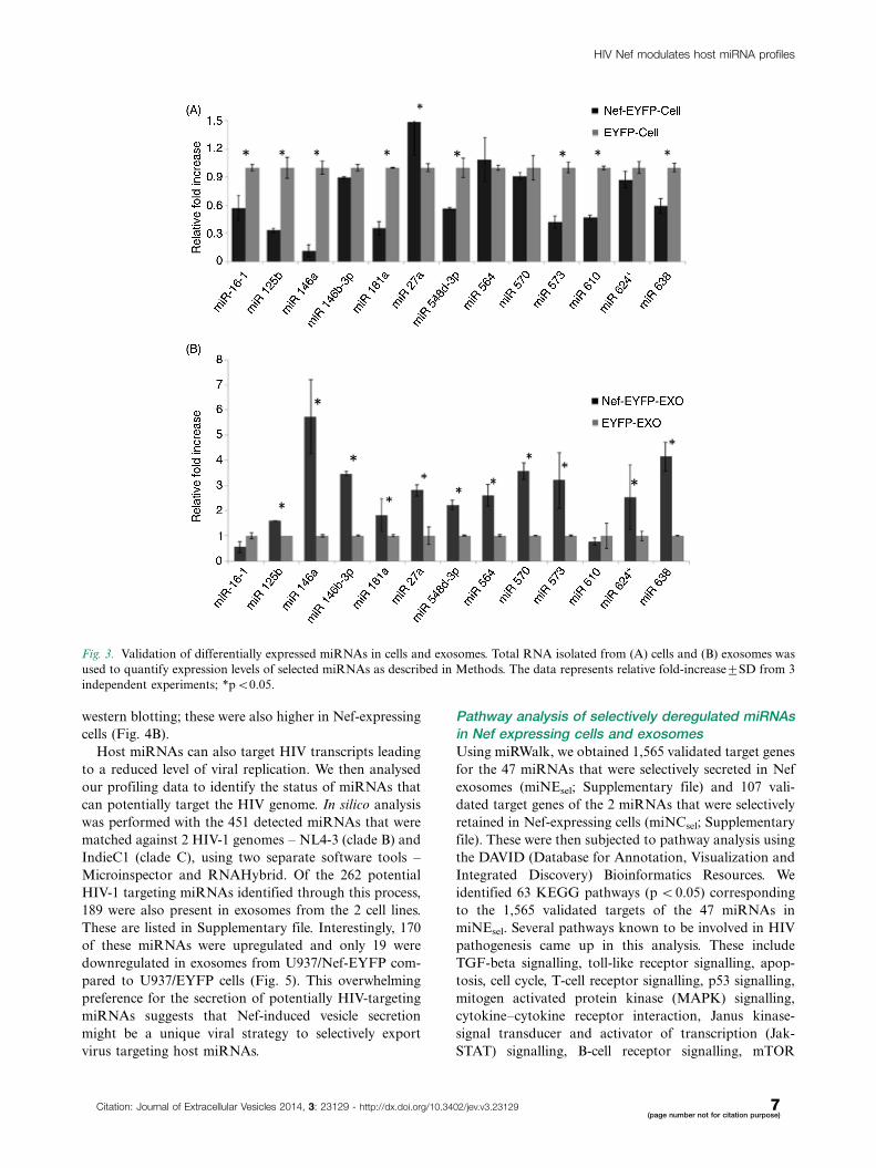
Validation of miRNAs in cells and exosomesTo confirm the profiling data, we selected 13 miRNAs for
validation by stem-loop quantitative RT-PCR in multiple
replicates of cells and exosomes. These include miR-573
and miR-638 (upregulated in Nef cells), miR-548d-3p
and 564 (same levels in Nef and control cells), and miR-
16-1, miR-146a, miR-125b, miR-146b-3p, miR-181a,
miR-27a, miR-570, miR-610 and miR-624* (down regu-
lated in Nef cells). All these miRNAs, except miR-146a,
were upregulated in exosomes in the profiling array data.
As there is no well documented control small RNA for
exosomes, we quantified RNU6 levels in both Nef and
control exosomes, and found these to be almost similar in
HIV Nef modulates host miRNA profiles
Citation: Journal of Extracellular Vesicles 2014, 3: 23129 - http://dx.doi.org/10.3402/jev.v3.23129 5(page number not for citation purpose)

multiple replicates of the 2 populations. The average Ct
values obtained were 23.3 in control exosomes and 23.7 in
Nef exosomes (Supplementary file). Therefore, the exoso-
mal miRNA data was normalized to RNU6. The results
showed most of the miRNA levels to correlate with the
profiling data. In the cells we found 4 miRNAs to show
a trend opposite to the profiling data. These include
miR-27a, which is upregulated, and miR-548d-3p, miR-
573 and miR-638, which are down regulated in Nef-
expressing cells based on qRT-PCR analysis (Fig. 3A).
All the analysed miRNAs showed higher levels in Nef
exosomes, which is consistent with the profiling data
(Fig. 3B). Thus, the qRT-PCR results broadly validate
our profiling data.
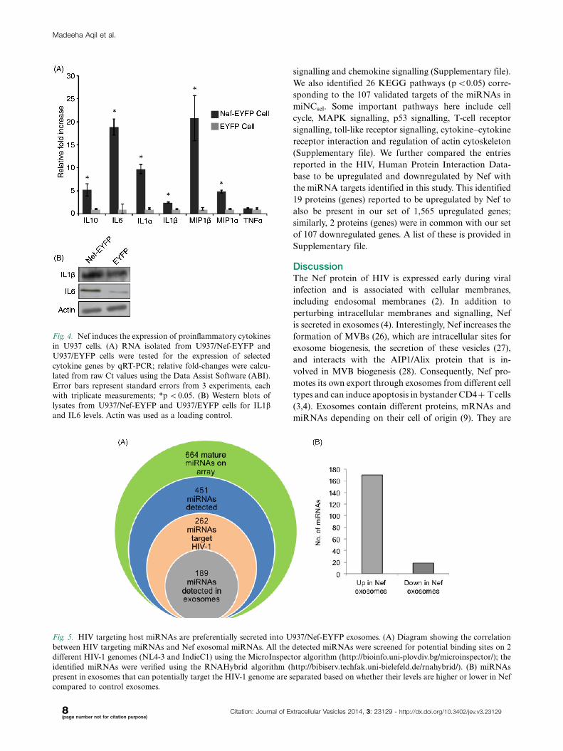
Nef exosomes are enriched in miRNAs thatpotentially target innate immune responses andthe HIV-1 genomeSince each miRNA can potentially target hundreds of
transcripts, changes in their abundance can have sig-
nificant effects on cellular pathways. The validated
targets of miRNAs that were selectively secreted from
Nef-expressing cells were extracted using the miRwalk
software. A majority of these miRNAs were found to
target the expression of cytokines/chemokines (e.g. inter-
leukins), proteins involved in innate immunity (e.g.
interferon, NFKB, STATs), tumour suppressors (PTEN,
RB1), cell survival factors (BCL2, MCL1) and the HIV
restriction factor APOBEC3G. In monocytes, Nef in-
duces the synthesis of proinflammatory cytokines and
chemokines. This effect is observed with Nef expressed
intracellularly as well as that added exogenously to
monocytes (24). We therefore selected 6 of these genes �IL1a, IL1b, IL6, TNFa, MIP1a, MIP1b, which are
induced in the presence of Nef, and observed that
many of the miRNAs that target these cytokines, such
as miR-146a, miR-146b-3p, miR-125b and miR-181a, are
downregulated in Nef-expressing cells, but are selectively
secreted in exosomes from these cells. These 4 miRNAs
regulate proinflammatory cytokine levels, antigen proces-
sing and presentation pathways (25).
We quantified the expression levels of IL1a, IL1b, IL6,
TNFa, MIP1a, MIP1b, and IL10 mRNAs in U937/Nef-
EYFP and U937/EYFP cells. Except for TNFa, which
did not show any significant change, all other mRNAs
were present at much higher levels in Nef-expressing
cells (Fig. 4A). The results showed �10-fold increase for
IL1b, IL6 and MIP1b mRNAs, and about 5-fold increase
for IL10 and MIP1a mRNAs. For 2 of these, IL1b and
IL6, the protein expression levels were also estimated by
Fig. 2. Nef expression modulates cellular and exosomal miRNAs profiles. The miRNAs in (A) cells and (B) exosomes were profiled and
relative fold-changes calculated as described in Methods. (C) The Venn diagrams show the intersection of detected miRNAs as follows:
(i) Upregulated miRNAs in both U937/Nef-EYFP cells and exosomes; (ii) Downregulated miRNAs in U937/Nef-EYFP cells and
upregulated miRNAs in U937/Nef-EYFP exosomes; (iii) Upregulated miRNAs in U937/Nef-EYFP cells and downregulated miRNAs
in U937/Nef-EYFP exosomes.
Madeeha Aqil et al.
6(page number not for citation purpose)
Citation: Journal of Extracellular Vesicles 2014, 3: 23129 - http://dx.doi.org/10.3402/jev.v3.23129
western blotting; these were also higher in Nef-expressing
cells (Fig. 4B).
Host miRNAs can also target HIV transcripts leading
to a reduced level of viral replication. We then analysed
our profiling data to identify the status of miRNAs that
can potentially target the HIV genome. In silico analysis
was performed with the 451 detected miRNAs that were
matched against 2 HIV-1 genomes � NL4-3 (clade B) and
IndieC1 (clade C), using two separate software tools �Microinspector and RNAHybrid. Of the 262 potential
HIV-1 targeting miRNAs identified through this process,
189 were also present in exosomes from the 2 cell lines.
These are listed in Supplementary file. Interestingly, 170
of these miRNAs were upregulated and only 19 were
downregulated in exosomes from U937/Nef-EYFP com-
pared to U937/EYFP cells (Fig. 5). This overwhelming
preference for the secretion of potentially HIV-targeting
miRNAs suggests that Nef-induced vesicle secretion
might be a unique viral strategy to selectively export
virus targeting host miRNAs.
Pathway analysis of selectively deregulated miRNAsin Nef expressing cells and exosomesUsing miRWalk, we obtained 1,565 validated target genes
for the 47 miRNAs that were selectively secreted in Nef
exosomes (miNEsel; Supplementary file) and 107 vali-
dated target genes of the 2 miRNAs that were selectively
retained in Nef-expressing cells (miNCsel; Supplementary
file). These were then subjected to pathway analysis using
the DAVID (Database for Annotation, Visualization and
Integrated Discovery) Bioinformatics Resources. We
identified 63 KEGG pathways (p B0.05) corresponding
to the 1,565 validated targets of the 47 miRNAs in
miNEsel. Several pathways known to be involved in HIV
pathogenesis came up in this analysis. These include
TGF-beta signalling, toll-like receptor signalling, apop-
tosis, cell cycle, T-cell receptor signalling, p53 signalling,
mitogen activated protein kinase (MAPK) signalling,
cytokine�cytokine receptor interaction, Janus kinase-
signal transducer and activator of transcription (Jak-
STAT) signalling, B-cell receptor signalling, mTOR
Fig. 3. Validation of differentially expressed miRNAs in cells and exosomes. Total RNA isolated from (A) cells and (B) exosomes was
used to quantify expression levels of selected miRNAs as described in Methods. The data represents relative fold-increase9SD from 3
independent experiments; *pB0.05.
HIV Nef modulates host miRNA profiles
Citation: Journal of Extracellular Vesicles 2014, 3: 23129 - http://dx.doi.org/10.3402/jev.v3.23129 7(page number not for citation purpose)
signalling and chemokine signalling (Supplementary file).
We also identified 26 KEGG pathways (pB0.05) corre-
sponding to the 107 validated targets of the miRNAs in
miNCsel. Some important pathways here include cell
cycle, MAPK signalling, p53 signalling, T-cell receptor
signalling, toll-like receptor signalling, cytokine�cytokine
receptor interaction and regulation of actin cytoskeleton
(Supplementary file). We further compared the entries
reported in the HIV, Human Protein Interaction Data-
base to be upregulated and downregulated by Nef with
the miRNA targets identified in this study. This identified
19 proteins (genes) reported to be upregulated by Nef to
also be present in our set of 1,565 upregulated genes;
similarly, 2 proteins (genes) were in common with our set
of 107 downregulated genes. A list of these is provided in
Supplementary file.
DiscussionThe Nef protein of HIV is expressed early during viral
infection and is associated with cellular membranes,
including endosomal membranes (2). In addition to
perturbing intracellular membranes and signalling, Nef
is secreted in exosomes (4). Interestingly, Nef increases the
formation of MVBs (26), which are intracellular sites for
exosome biogenesis, the secretion of these vesicles (27),
and interacts with the AIP1/Alix protein that is in-
volved in MVB biogenesis (28). Consequently, Nef pro-
motes its own export through exosomes from different cell
types and can induce apoptosis in bystander CD4� T cells
(3,4). Exosomes contain different proteins, mRNAs and
miRNAs depending on their cell of origin (9). They are
Fig. 4. Nef induces the expression of proinflammatory cytokines
in U937 cells. (A) RNA isolated from U937/Nef-EYFP and
U937/EYFP cells were tested for the expression of selected
cytokine genes by qRT-PCR; relative fold-changes were calcu-
lated from raw Ct values using the Data Assist Software (ABI).
Error bars represent standard errors from 3 experiments, each
with triplicate measurements; *p B0.05. (B) Western blots of
lysates from U937/Nef-EYFP and U937/EYFP cells for IL1band IL6 levels. Actin was used as a loading control.
Fig. 5. HIV targeting host miRNAs are preferentially secreted into U937/Nef-EYFP exosomes. (A) Diagram showing the correlation
between HIV targeting miRNAs and Nef exosomal miRNAs. All the detected miRNAs were screened for potential binding sites on 2
different HIV-1 genomes (NL4-3 and IndieC1) using the MicroInspector algorithm (http://bioinfo.uni-plovdiv.bg/microinspector/); the
identified miRNAs were verified using the RNAHybrid algorithm (http://bibiserv.techfak.uni-bielefeld.de/rnahybrid/). (B) miRNAs
present in exosomes that can potentially target the HIV-1 genome are separated based on whether their levels are higher or lower in Nef
compared to control exosomes.
Madeeha Aqil et al.
8(page number not for citation purpose)
Citation: Journal of Extracellular Vesicles 2014, 3: 23129 - http://dx.doi.org/10.3402/jev.v3.23129

taken up by recipient cells, in which the exosomal mRNA
is translated and the miRNA can post-transcriptionally
regulate gene expression (10), thus supporting the view
that exosomes function as intercellular messengers. Mod-
ulation of exosomal miRNAs is reported in several cancers
as well as viral infections (29).
We have carried out the first miRNome analysis of Nef-
expressing human monocytic cells and their exosomes. We
show that Nef exosomes are enriched in miRNAs that can
target proinflammatory cytokines and other genes in-
volved in key pathways like JAK-STAT signalling, MAPK
signalling and apoptosis. Further, an overwhelming per-
centage of miRNAs that can potentially target HIV-1 are
secreted out of Nef-expressing cells into exosomes. As
reported earlier for other cell types, U937 cells expressing
Nef also showed increased exosome secretion. There was
differential expression of about 50% of detected miRNAs
under the influence of Nef. It was shown recently that
reducing GW182, which is an important component of
Glycine-Tryptophan (GW) bodies, also reduced miRNA
secretion through exosomes (30). This agrees with our
recent findings that Nef-expressing U937 cells have higher
levels of GW182 (17) and display increased secretion of
exosomes and miRNAs reported here.
We observed significant changes in the levels of several
miRNAs that regulate innate immune responses, espe-
cially the proinflammatory cytokines. These include miR-
16, miR-125b, miR-146a, miR-146b-3p and miR-181a,
which are reduced in Nef-expressing U937 cells. Of these,
miR-16 is required for the degradation of transcripts that
contain AU-rich elements in their 3?UTR, which includes
mRNAs for most of the inflammatory mediators. It
regulates IL6, TNFa and IL8, and high levels of miR-16
restrict the production of inflammatory mediators under
non-stimulated conditions (25). Interestingly, miR-16
levels were �50% lower in the Nef cellular pool, suggest-
ing that Nef might regulate proinflammatory cytokines
through this pathway. MiR-125b inhibits HIV replication
by targeting viral transcripts (31). Our profiling data
showed about 3-fold reduction in miR-125b levels in Nef-
expressing U937 cells and a corresponding increase in
their exosomes, suggesting that Nef might also render cells
more permissive for viral replication through miR-125b
modulation.
The miR-146 family comprises 2 members � miR146a
and miR146b, of which the former is also a well-known
inhibitor of innate immune responses (32). Its levels
increase in myeloid cells in response to TLR2, TLR4 or
TLR5, and exposure to inflammatory cytokines like
TNFa or IL1b (33,34); in alveolar epithelial cells it reduces
IL1b levels (33,34). We found miR-146a levels to be
about 5-fold lower in U937/Nef-EYFP cells compared to
U937/EYFP cells, and observed a corresponding increase
in Nef exosomes. In THP-1 monocytic cells, miR-146a
regulates inflammatory cytokines/chemokines, including
IL1b, IL6, TNFa, IL8, IP10, and MCP-1 (34). High levels
of miR-146a correlated with increased numbers of GW
bodies in THP-1 cells, suggesting that GW bodies are mar-
kers for miRNA activity during innate immune signalling
(34). Our data showing reduced levels of miR-146a in
Nef-expressing monocytes, suggests that Nef modulates
key miRNAs that regulate innate immune responses.
Further, miR-181a levels are also �50% lower in U937/
Nef-EYFP cells compared to U937/EYFP cells, and
show a corresponding increase in Nef exosomes. This
miRNA regulates T-cell signalling by down regulating
multiple phosphatases, and is thought to act as a rheostat
that regulates protein phosphorylation levels (25). Thus,
several miRNAs that affect inflammatory cytokines
and innate immune responses are present at reduced
levels in Nef-expressing monocytes and show a corre-
sponding increase in exosomes secreted from these cells.
Correspondingly, the mRNAs and expression of these
cytokines/chemokines are at higher levels in Nef-expressing
cells.
Host miRNAs also target viral transcripts and limit
replication. Naturally then, modulation of host miRNAs
is emerging as a viral counter-strategy for successful
infection (13,35). HIV-1 infection of Jurkat cells down-
regulates the polycistronic miRNA cluster miR-17/92,
which includes miR-17/-18/-20a, miR-19a/-19b and miR-
92a (16). The downregulation of miR-21, miR-26a, miR-
29a, miR-29b and miR-29c was also observed in HIV
patients (31). We found miR-18, miR-19a, miR-20a, miR-
21 and miR-29b to be downregulated in Nef expressing
monocytes. A comparison of miRNA expression patterns
in resting and activated CD4� T cells and use of specific
antagomirs concluded that miR-28, miR-125b, miR-
150, miR-223 and miR-382 target the nef/3?LTR region
and contribute to HIV latency in resting CD4� T cells;
similar data were also reported for monocytes and macro-
phages (14,16). Our profiling demonstrated reduced levels
of miR-125b and miR-223 in Nef-expressing monocytes,
whereas miR-382 was not detected. Based on in silico
analyses, miR-29a, miR-29b, miR-149, miR-324-5p and
miR-378 were reported to target conserved regions of the
HIV-1 genome, including the nef gene (16). Of these, we
found miR-29b to be downregulated in Nef-expressing
cells. A majority of miRNAs that inhibit HIV replication,
including miR-17, miR-19a, miR-19b, miR-20a, miR-
26a, miR-28, miR-29a, miR-29b, miR-29c, miR-92a,
miR-125b, miR-149, miR-150, miR-223, miR-324-5p,
miR-378 and miR-382 were present at 1.5-folds or higher
levels in Nef exosomes. Further, when correlated with our
in silico analysis of miRNA target sites in the HIV
genome, an overwhelming majority of miRNAs that can
potentially target HIV-1 genomes were present at in-
creased levels in exosomes secreted by Nef-expressing
cells. We also found 47 miRNAs to be present at increased
levels in Nef exosomes despite being present at reduced
HIV Nef modulates host miRNA profiles
Citation: Journal of Extracellular Vesicles 2014, 3: 23129 - http://dx.doi.org/10.3402/jev.v3.23129 9(page number not for citation purpose)
levels in Nef-expressing cells. Of these selectively secreted
miRNAs, 21 also had target sites on HIV-1 genomes.
Thus, Nef expression reduces the cellular levels of several
host miRNAs that target innate immune responses and
viral transcripts by exosome-mediated export. This is
likely to modify the host cell environment to favour virus
replication.
The increased formation of MVB membranes (26,28)
and enhanced vesicle secretion (4,27) in the presence of
Nef can favour virus egress and spread. However, recent
reports also show MVBs to be sites for miRNA assembly
and activity (36,37). Thus, the host can limit HIV
replication by targeting viral transcripts to membranes,
which it evolved to exploit. We have recently shown that
Nef attenuates the activity of let-7a, which silences target
genes primarily at MVBs (17). This is attributed to the
ability of Nef to interact with Ago2 through 2 highly
conserved GW motifs (17). We now show that many
miRNAs whose levels are reduced in Nef-expressing cells
accumulate in exosomes secreted from these cells. Thus,
besides employing endosomal membranes for Ago2
interaction, HIV Nef further exploits the exosomal path-
way to alter the cellular and exosomal distribution of
miRNAs. Based on these findings, we propose that Nef
has evolved a 2-pronged suppressor function to inhibit
miRNA-mediated viral silencing and to modulate exo-
some-mediated cell-to-cell communication. How Nef
influences the selective expression and export of miRNAs,
and how the exosomal cargo influences uninfected cells
remain important issues to explore in future studies.
Conflict of interest and fundingThis work was funded by grants from the Department of
Biotechnology (DBT), Government of India and the Indian
Council of Medical Research (ICMR). A Senior Research
Fellowship from the Council for Scientific and Industrial
Research (CSIR), India, supported MA.
References
1. Das SR, Jameel S. Biology of the HIV Nef protein. Indian J
Med Res. 2005;121:315�32.
2. Landi A, Iannucci V, Nuffel AV, Meuwissen P, Verhasselt B.
One protein to rule them all: modulation of cell surface recep-
tors and molecules by HIV Nef. Curr HIV Res. 2011;9:
496�504.
3. Campbell TD, Khan M, Huang MB, Bond VC, Powell MD.
HIV-1 Nef protein is secreted into vesicles that can fuse with
target cells and virions. Ethn Dis. 2008;18:S2�14-9.
4. Lenassi M, Cagney G, Liao M, Vaupotic T, Bartholomeeusen
K, Cheng Y, et al. HIV Nef is secreted in exosomes and
triggers apoptosis in bystander CD4� T cells. Traffic. 2010;11:
110�22.
5. Raymond AD, Campbell-Sims TC, Khan M, Lang M, Huang
MB, Bond VC, et al. HIV type 1 Nef is released from infected
cells in CD45(�) microvesicles and is present in the plasma
of HIV-infected individuals. AIDS Res Hum Retroviruses.
2011;27:167�78.
6. Fujii Y, Otake K, Tashiro M, Adachi A. Soluble Nef antigen
of HIV-1 is cytotoxic for human CD4� T cells. FEBS Lett.
1996;393:93�6.
7. Campbell PE, Isayev O, Ali SA, Roth WW, Huang MB, Powell
MD, et al. Validation of a novel secretion modification region
(SMR) of HIV-1 Nef using cohort sequence analysis and
molecular modeling. J Mol Model. 2012;18:4603�13.
8. Zomer A, Vendrig T, Hopmans ES, van Eijndhoven M,
Middeldorp JM, Pegtel DM. Exosomes: fit to deliver small
RNA. Commun Integr Biol. 2010;3:447�50.
9. de Jong OG, Verhaar MC, Chen Y, Vader P, Gremmels H,
Posthuma G, et al. Cellular stress conditions are reflected
in the protein and RNA content of endothelial cell-derived
exosomes. J Extracell Vesicles. 2012;1:18396, doi: http://dx.doi.
org/10.3402/jev.v1i0.18396
10. Ekstrom K, Valadi H, Sjostrand M, Malmhall C, Bossios A,
Eldh M, et al. Characterization of mRNA and microRNA in
human mast cell-derived exosomes and their transfer to other
mast cells and blood CD34 progenitor cells. J Extracell Vesicles.
2012;1:18389, doi: http://dx.doi.org/10.3402/jev.v1i0.18389
11. Ameisen JC, Guy B, Chamaret S, Loche M, Mach B, Tartar A,
et al. Persistent antibody response to the HIV-1-negative
regulatory factor in HIV-1-infected seronegative persons. N
Engl J Med. 1989;320:251�2.
12. Svensson KJ, Christianson HC, Wittrup A, Bourseau-
Guilmain E, Lindqvist E, Svensson LM, et al. Exosome
uptake depends on ERK1/2-heat shock protein 27 signaling
and lipid Raft-mediated endocytosis negatively regulated by
caveolin-1. J Biol Chem. 2013;288:17713�24.
13. Song L, Gao S, Jiang W, Chen S, Liu Y, Zhou L, et al.
Silencing suppressors: viral weapons for countering host cell
defenses. Protein Cell. 2011;2:273�81.
14. Wang X, Ye L, Hou W, Zhou Y, Wang YJ, Metzger DS, et al.
Cellular microRNA expression correlates with susceptibility of
monocytes/macrophages to HIV-1 infection. Blood. 2009;113:
671�4.
15. Yeung ML, Bennasser Y, Watashi K, Le SY, Houzet L, Jeang
KT. Pyrosequencing of small non-coding RNAs in HIV-1
infected cells: evidence for the processing of a viral-cellular
double-stranded RNA hybrid. Nucleic Acids Res. 2009;37:
6575�86.
16. Sun G, Li H, Wu X, Covarrubias M, Scherer L, Meinking K,
et al. Interplay between HIV-1 infection and host microRNAs.
Nucleic Acids Res. 2012;40:2181�96.
17. Aqil M, Naqvi AR, Bano AS, Jameel S. The HIV-1 Nef protein
binds argonaute-2 and functions as a viral suppressor of RNA
interference. PLoS One. 2013;8:e74472.
18. Thery C, Amigorena S, Raposo G, Clayton A. Isolation and
characterization of exosomes from cell culture supernatants
and biological fluids. Curr Protoc Cell Biol. 2006;Chapter 3:
Unit 3 22.
19. Dweep H, Sticht C, Pandey P, Gretz N. miRWalk � database:
prediction of possible miRNA binding sites by ‘‘walking’’ the
genes of three genomes. J Biomed Inform. 2011;44:839�47.
20. Huang da W, Sherman BT, Lempicki RA. Bioinformatics
enrichment tools: paths toward the comprehensive functional
analysis of large gene lists. Nucleic Acids Res. 2009;37:1�13.
21. Huang da W, Sherman BT, Lempicki RA. Systematic and
integrative analysis of large gene lists using DAVID bioinfor-
matics resources. Nat Protoc. 2009;4:44�57.
22. Rusinov V, Baev V, Minkov IN, Tabler M. MicroInspector: a
web tool for detection of miRNA binding sites in an RNA
sequence. Nucleic Acids Res. 2005;33:W696�700.
23. Rehmsmeier M, Steffen P, Hochsmann M, Giegerich R. Fast
and effective prediction of microRNA/target duplexes. RNA.
2004;10:1507�17.
Madeeha Aqil et al.
10(page number not for citation purpose)
Citation: Journal of Extracellular Vesicles 2014, 3: 23129 - http://dx.doi.org/10.3402/jev.v3.23129

24. Olivetta E, Percario Z, Fiorucci G, Mattia G, Schiavoni I,
Dennis C, et al. HIV-1 Nef induces the release of inflammatory
factors from human monocyte/macrophages: involvement of
Nef endocytotic signals and NF-kappa B activation. J
Immunol. 2003;170:1716�27.
25. Lindsay MA. microRNAs and the immune response. Trends
Immunol. 2008;29:343�51.
26. Stumptner-Cuvelette P, Jouve M, Helft J, Dugast M,
Glouzman AS, Jooss K, et al. Human immunodeficiency
virus-1 Nef expression induces intracellular accumulation of
multivesicular bodies and major histocompatibility complex
class II complexes: potential role of phosphatidylinositol
3-kinase. Mol Biol Cell. 2003;14:4857�70.
27. Muratori C, Cavallin LE, Kratzel K, Tinari A, De Milito A,
Fais S, et al. Massive secretion by T cells is caused by HIV Nef
in infected cells and by Nef transfer to bystander cells. Cell
Host Microbe. 2009;6:218�30.
28. Costa LJ, Chen N, Lopes A, Aguiar RS, Tanuri A, Plemenitas
A, et al. Interactions between Nef and AIP1 proliferate multi-
vesicular bodies and facilitate egress of HIV-1. Retrovirology.
2006;3:33.
29. Turchinovich A, Samatov TR, Tonevitsky AG, Burwinkel B.
Circulating miRNAs: cell�cell communication function? Front
Genet. 2013;4:119.
30. Yao B, La LB, Chen YC, Chang LJ, Chan EK. Defining a new
role of GW182 in maintaining miRNA stability. EMBO Rep.
2012;13:1102�8.
31. Witwer KW, Watson AK, Blankson JN, Clements JE. Re-
lationships of PBMC microRNA expression, plasma viral
load, and CD4� T-cell count in HIV-1-infected elite suppres-
sors and viremic patients. Retrovirology. 2012;9:5.
32. Chan EK, Ceribelli A, Satoh M. MicroRNA-146a in auto-
immunity and innate immune responses. Ann Rheum Dis.
2013;72:ii90�5.
33. Nahid MA, Pauley KM, Satoh M, Chan EK. miR-146a is
critical for endotoxin-induced tolerance: implication in innate
immunity. J Biol Chem. 2009;284:34590�9.
34. Pauley KM, Satoh M, Pauley BA, Dominguez-Gutierrez PR,
Wallet SM, Holliday LS, et al. Formation of GW/P bodies as
marker for microRNA-mediated regulation of innate immune
signaling in THP-1 cells. Immunol Cell Biol. 2010;88:205�12.
35. Tan Gana NH, Onuki T, Victoriano AF, Okamoto T.
MicroRNAs in HIV-1 infection: an integration of viral and
cellular interaction at the genomic level. Front Microbiol.
2012;3:306.
36. Gibbings DJ, Ciaudo C, Erhardt M, Voinnet O. Multivesicular
bodies associate with components of miRNA effector com-
plexes and modulate miRNA activity. Nat Cell Biol. 2009;11:
1143�9.
37. Lee YS, Pressman S, Andress AP, Kim K, White JL, Cassidy
JJ, et al. Silencing by small RNAs is linked to endosomal
trafficking. Nat Cell Biol. 2009;11:1150�6.
HIV Nef modulates host miRNA profiles
Citation: Journal of Extracellular Vesicles 2014, 3: 23129 - http://dx.doi.org/10.3402/jev.v3.23129 11(page number not for citation purpose)