Embed Size (px)
Citation preview
MOLECULAR MECHANISMS OF HIV NEF-INDUCED SRC KINASE ACTIVATION AND SURVIVAL SIGNALING IN MYELOID CELLS
by
Hyun-Jung Choi
B.Sc (Pharm), Ewha Womans University, 1994
MS, Seoul National University, 1998
Submitted to the Graduate Faculty of
School of Medicine, Department of Molecular Genetics and Biochemistry in partial fulfillment
of the requirements for the degree of
Doctor of Philosophy
University of Pittsburgh
2004
UNIVERSITY OF PITTSBURGH
FACULTY OF SCHOOL OF MEDICINE
This dissertation was presented
by
Hyun-Jung Choi
It was defended on
Oct. 18, 2004
and approved by
Toshiaki Kodama, DVM, PhD.
Edward V. Prochownik, MD, PhD.
Baskaran Rajasekaran, PhD.
Todd A. Reinhart, ScD.
Martin C. Schmidt, PhD.
Thomas E. Smithgall, PhD. Dissertation Director
ii
MOLECULAR MECHANISMS OF HIV NEF-INDUCED SRC KINASE ACTIVATION AND SURVIVAL SIGNALING IN MYELOID CELLS
Hyun-Jung Choi, PhD
University of Pittsburgh, 2004
The Nef protein unique to the primate lentiviruses HIV and SIV is essential for high-titer
viral replication and AIDS progression. Despite its essential role, the molecular mechanisms by
which Nef functions in HIV pathogenesis are not fully understood. Since Nef lacks intrinsic
catalytic activity, research has been focused on analyzing interactions between Nef and cellular
proteins in an attempt to understand the many functions attributed to Nef. Nef binds to the
macrophage-specific Src family member Hck through its SH3 domain with the highest affinity
known for an SH3-mediated protein-protein interaction. Previous studies from our laboratory
have shown that Nef-Hck interaction results in constitutive Hck kinase activation capable of
transforming Rat-2 fibroblasts. Nef-Hck interaction may be crucial to M-tropic HIV replication
and AIDS pathogenesis, identifying this virus-host protein complex as a rational target for anti-
HIV drug discovery.
To investigate whether interaction with Hck is a common feature of Nef alleles from
different strains of HIV-1, we compared the ability of four different HIV-1 Nef alleles to induce
Hck activation and transformation in our Rat-2 fibroblast model. We found that not all HIV-1
Nef alleles have a similar affinity for Hck, despite strong conservation of the PxxPxR motif and
hydrophobic pocket residues identified in the crystal structure as part of the SH3 interface.
Further characterization of the interface of the Hck SH3-Nef complex revealed additional critical
residues in the Nef hydrophobic pocket responsible for the differential interaction of HIV-1 Nef
iii
alleles to Hck. This study provides the first evidence that the HIV-1 Nef hydrophobic pocket is
critical for SH3-mediated Hck activation in vivo and identifies the pocket as a rational target for
drug design to selectively disrupt Nef-Hck signaling in HIV infected cells.
Suppression of cell death of HIV-infected cells allows time for viral replication prior to
cell lysis, facilitating productive viral replication indirectly. Recently Nef has been demonstrated
to be an important factor in promoting the survival of HIV-infected T cells by affecting
mediators of apoptosis. Since macrophages serve as HIV viral reservoirs and play a critical role
in persistent virus infection, we were interested in the role of Nef in survival of macrophages.
Previous work from our laboratory has shown that Nef promotes cytokine independent
proliferation of the macrophage precursor cell line, TF-1, through a mechanism that requires the
Stat3 transcription factor. Studies presented in this dissertation demonstrate that Nef suppresses
apoptosis in this cell line by selectively upregulating the anti-apoptosis gene, Bcl-XL. This
signaling induction by Nef is dependent on Erk MAPK activation but not Stat3. This Nef-
induced survival signal is the first to show that Nef generates anti-apoptosis signals in cells of the
myelomonocytic lineage and adds important evidence to the hypothesis that Nef may contribute
to the establishment and maintenance of an HIV reservoir by conferring a survival advantage on
HIV-infected macrophages.
iv

TABLE OF CONTENTS PREFACE...................................................................................................................................... ix 1. Chapter I - Introduction .......................................................................................................... 1
1.1. Primate Lentivirus Genome ............................................................................................ 2 1.1.1. HIV-1 Auxiliary Proteins........................................................................................ 2
1.2. Nef : General Properties.................................................................................................. 4 1.3. Structure of Nef............................................................................................................... 4 1.4. Posttranslational Modification of Nef............................................................................. 6
1.4.1. Myristylation........................................................................................................... 6 1.4.2. Phosphorylation ...................................................................................................... 7 1.4.3. Proteolytic Processing............................................................................................. 9
1.5. Oligomerization of Nef ................................................................................................. 10 1.6. Functions of Nef ........................................................................................................... 12
1.6.1. Early Nef studies................................................................................................... 12 1.6.2. Positive role in viral replication............................................................................ 13 1.6.3. Enhancement of the infectivity of virus particles ................................................. 15 1.6.4. Downregulation of CD4 and MHC I Cell-Surface Expression ............................ 17 1.6.5. Modulation of internal cell signaling.................................................................... 22
1.7. Cellular partners of Nef ................................................................................................ 28 1.7.1. Cellular Receptors and trafficking proteins .......................................................... 28 1.7.2. Serine Kinases....................................................................................................... 32 1.7.3. Src Family Tyrosine Kinases and Nef .................................................................. 36
1.8. Macrophages in HIV infection...................................................................................... 51 1.9. Nef in HIV-infected macrophages ................................................................................ 52
2. Chapter II .............................................................................................................................. 55 Dissertation Hypothesis and Specific Aims.................................................................................. 55 3. Chapter III............................................................................................................................ 58
3.1. Abstract ......................................................................................................................... 59 3.2. Introduction................................................................................................................... 60 3.3. Materials and Methods.................................................................................................. 63
3.3.1. Retroviral Expression Constructs ......................................................................... 63 3.3.2. Expression of Nef and Src Kinases in Sf9 Cells and GST-Nef Binding Assay ... 63 3.3.3. GST-SH3 fusion protein binding assay ................................................................ 64 3.3.4. Transformation assays .......................................................................................... 65 3.3.5. Co-immunoprecipitation assay and immunoblot analysis .................................... 65
3.4. Results........................................................................................................................... 67 3.4.1. Analysis of the interaction of Nef variants from different HIV-1 alleles with Src family kinases in vitro........................................................................................................... 67 3.4.2. Allelic variation in Nef-induced Hck activation in vivo....................................... 72 3.4.3. Interaction of chimeric SF2-ELI Nef proteins with Hck ...................................... 75
v
3.4.4. Tyr 120 is required for high affinity binding of SF2 Nef to Hck in vitro and Hck activation in vivo................................................................................................................... 79 3.4.5. Multiple substitutions are required to restore ELI Nef interaction with Hck ....... 83 3.4.6. Analysis of HIV-1 Nef alleles from long-term non-progressors (LTNPs) reveals sequence heterogeneity at amino acid positions 83, 116, and 120 ....................................... 87
3.5. Discussion..................................................................................................................... 89 4. Chapter IV............................................................................................................................. 94
4.1. Abstract ......................................................................................................................... 95 4.2. Introduction................................................................................................................... 96 4.3. Materials and methods .................................................................................................. 98
4.3.1. Materials ............................................................................................................... 98 4.3.2. Cell Culture........................................................................................................... 98 4.3.3. Nef, Nef-ER, and Stat3YF expression constructs................................................. 98 4.3.4. Cell lysis and Western blot analysis ..................................................................... 99 4.3.5. Apoptosis Assay.................................................................................................. 100 4.3.6. RNase protection assay ....................................................................................... 100 4.3.7. Detection of Nef-ER dimers ............................................................................... 101 4.3.8. Electrophoretic Mobility Shift Assay ................................................................. 101
4.4. Results......................................................................................................................... 103 4.4.1. HIV-1 Nef inhibits apoptosis induced by cytokine deprivation in the human myeloid leukemia cell line, TF-1 ........................................................................................ 103 4.4.2. Induction of Bcl-XL expression in TF-1 cells expressing Nef............................ 105 4.4.3. Nef survival signaling in TF-1 cells requires MAPK but not PI3K activation... 107 4.4.4. The Erk MAPK pathway regulates Bcl-XL induction by Nef ............................ 111 4.4.5. Fusion of Nef to the estrogen receptor (ER) hormone binding domain creates a conditional Nef protein (Nef-ER) capable of inducible survival signaling ........................ 113 4.4.6. Inducible stabilization and dimerization of Nef-ER in TF-1 cells...................... 114 4.4.7. Cell survival and Stat3 activation by Nef-ER requires Erk activation ............... 117 4.4.8. Dominant-negative Stat3 does not affect survival or Bcl-XL induction by Nef-ER 121
4.5. Discussion................................................................................................................... 123 5. Chapter V - Discussion ....................................................................................................... 128 BIBLIOGRAPHY....................................................................................................................... 137
vi

LIST OF TABLES Table 1 Variation in Nef amino acid sequences from long-term non-progressors (LTNPs) at
positions 83, 116, and 120 .................................................................................................... 88
vii
LIST OF FIGURES Figure 1. Model for Nef-induced MHC-I and CD4 down-modulation. ....................................... 21 Figure 2. The structure of Hck as a representative of Src family kinases. ................................... 39 Figure 3. A Model for Src Regulation. ......................................................................................... 41 Figure 4. Structure of Nef–SH3 Complex. ................................................................................... 49 Figure 5. Macrophages in HIV infection. ..................................................................................... 54 Figure 6. Sequence alignment of HIV-1 Nef alleles SF2, ELI, LAI, and Consensus (Con). ....... 69 Figure 7. Differential binding of Nef variants to Src family kinases............................................ 70 Figure 8. Binding of Nef variants to Src family kinase SH3 domains. ........................................ 71 Figure 9. Activation of Hck by HIV-1 Nef variants in vivo......................................................... 74 Figure 10. Binding of SF2-ELI Nef C-terminal chimeras to Hck. ............................................... 77 Figure 11. Binding of SF2-ELI Nef N-terminal chimeras to Hck. ............................................... 78 Figure 12. Tyr 120 is required for SF2 Nef binding to Hck in vitro............................................. 81 Figure 13. Hck activation by SF2 Nef requires Tyr 120 in vivo. ................................................. 82 Figure 14. Full recovery of ELI binding to Hck requires substitution of Ala 83, His 116, and Tyr
120......................................................................................................................................... 85 Figure 15. Activation of Hck by ELI Nef mutants. ...................................................................... 86 Figure 16. Expression of HIV-1 Nef suppresses apoptosis induced by cytokine deprivation in the
human GM-CSF-dependent myeloid leukemia cell line, TF-1. ......................................... 104 Figure 17. HIV-1 Nef selectively induces upregulation of anti-apoptotic gene, Bcl-XL............ 106 Figure 18. The Mek inhibitor PD98059 blocks the anti-apoptotic effect of Nef in TF-1 cells. . 109 Figure 19. The PI3K inhibitor LY294002 does not suppress Nef-induced survival signaling in
TF-1 cells. ........................................................................................................................... 110 Figure 20. Induction of Bcl-XL expression by HIV Nef requires the Erk MAPK pathway. ...... 112 Figure 21. Fusion of Nef to the estrogen receptor hormone-binding domain (Nef-ER) allows for
4-HT-dependent survival signaling..................................................................................... 115 Figure 22. Treatment with 4-HT stabilizes the Nef-ER protein and induces its dimerization in
TF-1/Nef-ER cells............................................................................................................... 116 Figure 23. Survival signaling from Nef-ER requires the Erk MAPK pathway in TF-1 cells..... 119 Figure 24. Inducible activation of Stat3 by Nef-ER requires Erk activity.................................. 120 Figure 25. Dominant-negative Stat3 does not affect Nef-induced survival or Bcl-XL RNA levels.
............................................................................................................................................. 122
viii
PREFACE
I would first like to thank my advisor, Dr. Tom Smithgall for his guidance and support
over the past five years of my Ph.D. study. I feel extremely fortunate to have him as my mentor
and role model. He is not only a gifted scientist but also inspiring teacher. I am grateful to have
the opportunity to learn from him. His encouragement and understanding helped me get through
difficulties and frustration. There are no words that seem to be able to express my appreciation
enough. I wish to thank my committee members, Dr. Toshiaki Kodama, Dr. Edward V.
Prochownik, Dr. Baskaran Rajasekaran Dr. Todd A. Reinhart, and Dr. Martin C. Schmidt for
their time, assistance, and guidance throughout my time in graduate school.
I would also like to thank all the past and current members of the Smithgall lab, who have
provided me incredible support and offered their help and suggestions whenenver I needed it: Dr.
Frank Delfino, Theodora Pene Dumitrescu, Dr. Charles Laurent, Dr. Edwina Lerner, Dr. Tony
Meyn, Dr. Linda O’Reilly, Jerrod Poe, Jonathan Schaffer, Anthoy Schiavone, Ron Trible,
Matthe Wilson, Huihui Ye.
I would like to thank my family and friends for their love and support over these years for
my work and study. I would like to extend my special thanks to my mom, Yang-Ja Chung.
Without her, I would never have made it this far. I will be forever grateful for all of her love,
support, and encouragement. I thank my husband, Jin-Wan for endless love, support, and
patience. His sacrifice for my study and career are beyond expressing in words. I am so grateful
to have him as a partner who spends with the rest of my life.
Finally, I dedicate this dissertation to my father, Jae-Wan Choi. Although he is not here to
see the completion of my work, I have no doubt that he was smiling at me in spirit when I
finished.
ix
1. Chapter I - Introduction
1
1.1. Primate Lentivirus Genome
Primate Lentiviruses are a subfamily of retroviruses, which are associated with chronic
diseases of the immune system in their host (1, 2). This virus family includes human
immunodeficiency virus type 1 and 2 (HIV-1, HIV-2) and simian immunodeficiency virus (SIV).
In particular, HIV-1 is the etiological agent of the global AIDS epidemic. The genomes of
lentiviruses are more complex than retroviruses in that they encode six regulatory and accessory
genes in addition to the three structural genes gag (group-specific core antigen), pol
(polymerase), and env (envelop), which are common to all replication competent retroviruses.
The auxiliary genes from the prototype lentivirus, HIV-1 consist of the regulatory genes tat
(trans-activation of transcription) and rev (regulatory of virion protein expression), and the
accessory genes nef (negative factor), vif (viral infectivity factor), vpr (viral protein R), and vpu
(viral protein U) (3, 4, 5). HIV-2 and SIVmac lack the vpu gene, but contain a related gene called
vpx (viral protein X) (3, 4).
1.1.1. HIV-1 Auxiliary Proteins
While the regulatory proteins (Tat and Rev) were demonstrated to be essential for viral
replication, the accessory proteins (Nef, Vif, Vpr and Vpu) were initially considered to be
dispensable for virus replication since they can be deleted without destroying virus replication in
many culture settings (3, 6). It soon emerged, however, that accessory proteins contribute to
efficient lentivirus replication and pathogenesis in vivo.
Tat is a potent transcriptional activator of the HIV-1 long terminal repeat (LTR) promoter
element and regulates high-level HIV-1 transcription from the integrated DNA form of the virus
2
(the provirus) by acting mostly at the level of transcriptional elongation (7, 8). Rev is a
sequence-specific nuclear RNA export factor and is able to induce efficient nuclear export, and
hence expression, of the various incompletely spliced viral transcripts encoding late viral
proteins (8, 9).
Vif enhances virus infectivity in many culture settings, including primary lymphocytes (10,
11). Vpr mediates the nuclear import of the preintegration complex (PIC) into the nucleus of
particularly nondividing cells such as primary macrophages, enhancing HIV-1 replication in
these cells (12). Vpr also arrests infected cells in the G2 phase of the cell cycle, which facilitates
viral transcription since the HIV-1 promoter is more active in G2-arrested cells (13). Two
independent functions of Vpu unique to HIV-1 life cycle have been reported: enhancement of
virion release from infected cells (14) and the selective degradation of CD4 in the endoplasmic
reticulum (ER) (15).
The central role of Nef in HIV replication and AIDS pathogenesis in vivo was originally
demonstrated by the study of Kestler et al. (16). They showed that inactivation of the nef gene in
a pathogenic strain of SIV caused a dramatic decrease of both viral loads and pathogenic
potential in infected monkeys. Some long-term non-progressors of HIV-positive patients were
found to be infected with Nef-defective HIV, further suggesting that Nef is a key determinant for
disease progression (17, 18). Nef, therefore, rapidly became a subject of intense scientific
analysis. The molecular mechanisms of Nef interactions with host cell signaling pathways are the
major focus of this dissertation.
3
1.2. Nef : General Properties
Nef is the largest auxiliary protein encoded by HIV-1 (206 amino acids in length, ~27
kDa in molecular weight) and is expressed as a cytoplasmic protein anchored to membranes
through N-terminal myristylation (19). The open reading frame encoding Nef begins within or
immediately following the 3’ end of the env gene and partially overlaps with the U3 region of the
3’ long terminal repeat (LTR) (20, 21, 22). HIV-1 Nef can also be translated from an internal
AUG 57 base pairs downstream from the initiating AUG, leading to the production of a
truncated, non-myristylated 25 kDa protein (23). The Nef proteins of HIV-2 and SIV are slightly
longer, containing approximately 250 amino acids (24). Nef is produced abundantly during the
early stage of viral replication and localizes itself to the plasma membrane, cytosol, and nucleus,
and is also incorporated into virus particles. Quantitative analysis revealed Nef to be
incorporated on the order of 10% of reverse transcriptase incorporation, which corresponds to 5
to 10 molecules of Nef per virion (25).
The Nef sequence is relatively well conserved among the HIV-1 Nef alleles, while more
sequence variation exists between HIV and SIV Nef. Nef has a number of well-defined motifs
particularly in the core region, and those motifs are critical for several Nef functions. The N- and
C-terminal regions have more variable and flexible structures. Additional structural and
functional features of Nef are discussed in detail below.
1.3. Structure of Nef
The crystal structure of the core of HIV-1 Nef complexed with a Src family SH3 domain
has been solved (26). In addition, the solution structure of Nef alone and of Nef bound to a
peptide from the cytoplasmic tail of CD4 have been determined by nuclear magnetic resonance
(NMR) spectroscopy (27, 28, 29). These studies revealed that the N-terminal region of the core
4
of HIV-1 Nef consists of a type II polyproline helix (aa 70-77), which contains the main binding
site for the Src homology 3 (SH3) domain of Src family kinases. This domain is followed by two
anti-parallel α helices (aa 81-120, αA and αB), which are packed against a four-stranded anti-
parallel β sheet (aa 121-186). The C-terminal region consists of two short additional α helices
(aa 187-203), and these helices pack on the other side of the β strands. Residues 60-71 and 149-
180 form flexible solvent-exposed loops.
These structural studies show that the first two anti-parallel α helices are connected by a
relatively long linker and are separated to form a hydrophobic and solvent-accessible crevice
between the helices. The co-crystal structure of the Nef core and SH3 domain shows that this
crevice is occupied at one end by the specificity-determining isoleucine side chain of the SH3
domain, but is otherwise vacant in the structure of the complexes. This finding suggests that this
cavity may be accessible to other molecules, which could disrupt the interaction between Nef
and the SH3 domain. The studies presented in this dissertation demonstrate the biological
significance of this Nef hydrophobic pocket in terms of recruitment and activation of
macrophage-specific Src family member, Hck (Chaper III).
The structure of the four anti-parallel β strands is irregular, and does not form a
contiguous four-stranded β sheet. Instead they are separated into two distinct anti-parallel pairs
of strands. Only two hydrogen bonds are formed between βB and βC, and the presence of two
strictly conserved proline residues, one (136) in βB and the other (147) in βC appears to hold
apart the two strands. The two short α helices of the C-terminal region of Nefcore only partially
cover the distal surface of the β strands.
5
1.4. Posttranslational Modification of Nef
1.4.1. Myristylation
Nef is posttranslationally modified by the irreversible attachment of the 14 carbon saturated
fatty acid, myristic acid to its N-terminus, which targets Nef to the cellular membrane (30, 31).
Nef contains a consensus sequence for the myristylation, Met-Gly-X-X-X-Ser/Thr, at the N-
terminus (32, 33, 34). Covalent linkage of myristate via an amide bond to the N-terminal glycine,
Gly 2 is mediated by the soluble enzyme N-myristyl transferase after the initial methionine is
removed cotranslationally by methionine amino-peptidase. The same posttranslational
modification occurs in a number of viral proteins as well as cellular proteins which can be found
in the plasma membrane or other intracellular membranes in eukaryotic cells. The reduction or
loss of membrane binding was observed by mutation of Gly 2 to Ala, indicating the requirement
of Gly 2 for myristylation-mediated membrane binding (33, 35).
Although myristylation is necessary for membrane binding of some proteins, it is not
sufficient for anchoring proteins in the membrane. An additional signal is therefore required for
efficient membrane binding of myristylated proteins. A polybasic cluster of amino acids or a
palmitate moiety has been identified as a second signal for membrane binding of N-myristylated
proteins. The ‘myristate plus basic’ motif allows the myristylated proteins to bind the membrane
stably by both hydrophobic and electrostatic interactions. The HIV-1 Nef protein contains the
myristylation signal sequence as well as the basic motif, and these membrane anchoring regions
are almost absolutely conserved in Nef sequences (36).
The roles of myristylation in Nef functions have been proposed in several studies and
demonstrated to be required for virtually all of its biological activities. Harris et al. found that
Nef was myristylated at the N-terminus when it was expressed as a GST-fusion protein in the
baculovirus system (37). Cellular proteins that bound to Nef in a myristylation-dependent
6
manner were identified by pull-down assay using the myristylated Nef-GST and a myristylation
defective Nef-GST mutant. Cellular partners that bound Nef-GST in a myristylation-dependent
manner were the HIV receptor CD4, the Src family kinase Lck, and a component of non-clathrin
coated vesicles β-COP (35, 38). Other work has shown that the interaction of Nef with a
cytoskeleton protein, actin, also requires myristylation of Nef (39). In their study, a recombinant
HIV-1 Nef non-covalently associated with actin and this interaction was dependent on N-
terminal myristylation of Nef. Chowers et al. showed that missense mutation at the myristylation
signal sequences reduced viral replication in CEM cells, a T-lymphocyte cell line, demonstrating
that myristylation of Nef is required for optimal replication (40). Deletion of the myristylation
signal and basic residues from Nef completely abrogated both CD4 and MHC I downregulation,
suggesting that the myristylaion of Nef is critical for these functions as well (41). These studies
indicate that N-myristylation of Nef is critical not only for membrane targeting but also for
interacting with cellular proteins localized in the membrane, and for optimal Nef functioning.
1.4.2. Phosphorylation
Another posttranslational modification of Nef is phosphorylation on serine, threonine, and
tyrosine residues. In early studies, the Nef protein of HIV-1 strain BRU was demonstrated to be
phosphorylated on the threonine residue at position 15 (Thr 15) by protein kinase C (PKC) (42).
The phosphorylation on Thr 15 of Nef expressed in hamster kidney cells, BHK-21 was induced
by 12-O-tetradecanoyl-phorbol-13-acetate (TPA), a potent PKC activator. The involvement of
PKC in Nef phosphorylation was subsequently confirmed by use of protein kinase C inhibitors.
This notion was further supported by the demonstration that purified PKC is also able to
phosphorylate Nef in vitro. When a recombinant HIV-1 Nef–GST fusion protein was incubated
7
with Jurkat cell lysates, cytoplasmic extracts from a number of other human cell lines, or protein
kinase C, Ser/Thr phosphorylation was observed (43). Nef Ser/Thr phosphorylation in both HeLa
and Jurkat cells occurred in vivo when stimulated by phorbol ester treatment and was reduced by
an inhibitor of protein kinase C (44, 45). Phosphorylation of a nonmyristylated Nef mutant was
impaired, suggesting that membrane targeting of Nef is required for phosphorylation. The
significance of Thr phosphorylation in Nef is unclear since some HIV-1 isolates of Nef (SF2 and
BH10) have substitution of Thr 15 with Ala, and phosphorylation predominantly occurs on
serine residues in this case. Although serine phosphorylation of Nef has been reported to
correlate with an increase in virion infectivity (46), the role of Ser/Thr phosphorylation in
functions of Nef remains to be determined.
Tyrosine phosphorylation of Nef has been studied mainly on SIV Nef since SIV Nef but not
HIV Nef contains the potential SH2 domain binding sites. Indeed, some of the pathogenic forms
SIV Nef were reported to be highly phosphorylated on tyrosine residues in a cell culture system
(47). The significance of Nef tyrosine phosphorylation has been demonstrated in animal studies.
The replacement of two residues Arg-Gln (RQ) with Tyr-Glu (YE) converts Nef from
SIVmac239 to a similar phenotype of an acutely lethal strain of SIV, SIVpbj14. This substitution
introduces a second potential SH2 binding site which results in a virus (SIVmac239/YE-Nef)
that can cause acute disease in rhesus and Pig-tailed monkeys and replicate in lymphocytes from
peripheral blood mononuclear cells (PBMC) without prior lymphocyte activation. YE-Nef can
also transform NIH 3T3 fibroblasts when transfected into cells. Furthermore, the high level of
tyrosine phosphorylation of YE-Nef was detected when co-transfected in COS1 cells with the
Src tyrosine kinase, although direct phosphorylation on tyrosine residues was not observed in
8
cells isolated from the infected monkeys. These findings implicate tyrosine phosphorylation of
Nef in SIV viral replication and pathogenesis.
Whereas SIV Nef has been found to be phosphorylated on tyrosine residues in putative SH2
binding sites, HIV Nef has not been successfully demonstrated to be phosphorylated on tyrosine
residues. One study showed that HIV Nef is tyrosine phosphorylated in Nef-expressing cells
stimulated with PMA, and this phosphorylation was proposed to facilitate the stable interaction
of Nef with Src family kinase, Lck (48). However no subsequent studies confirmed this result.
Whether or not HIV Nef is tyrosine-phosphorylated in cells, and if so, the importance of HIV
Nef tyrosine phosphorylation has yet to be determined.
1.4.3. Proteolytic Processing
In addition to myristylation and phosphorylation, Nef can also be post-translationally
modified by proteolytic cleavage. Nef is cleaved specifically by the viral protease within the
virion. The main HIV-1 cleavage site is located between W57 and L58 (ACAW*LEAQ) and
determines the modular organization of Nef, separating it into N-terminal anchor domain and C-
terminal core domain (49, 50). While the N-terminal domain has a more extended conformation
and is likely to be located at the surface of the protein, the C-terminal domain has a compactly
folded core and is stable in the absence of the anchor domain. N-terminal myristylation does not
appear to have a major influence on the proteolytic cleavage. Recombinant Nef protein with
additional amino acids MPARS in front of the first methionine, thus preventing myristylation,
was still cleaved by viral protease (51). Recombinant purified Nef protein of HIV-1, as well as
Nef protein derived from extracts of HIV-1-infected glioblastoma cells and monocytes, were
specifically cleaved by the HIV-1 protease, giving rise to the same molecular weight fragments
9
(50). A large proportion (60-80%) of virion-associated Nef is the large fragment (18kDa) derived
from protease cleavage, while Nef is detected as a full-length 27 kDa protein in infected cells.
This observation suggests that cleavage is most likely to occur concomitantly with viral
structural proteins during maturation of virus particles. Nef cleavage in particle preparations was
completely abolished by a specific inhibitor of HIV-1 protease, indicating that the Nef cleavage
is not due to nonspecific proteolytic activity of particle preparations (25). The six residues in the
cleavage site are all very well conserved; Trp 57 in particular is almost absolutely conserved. In
addition to their role as a target site for the HIV-1 protease, residues that compose the cleavage
site, in particular W57 and L58 have been shown to participate in an intramolecular interaction
with the hydrophobic groove formed by two α-helices in the Nef core domain.
Despite the conservation of Nef proteolytic cleavage, it remains unclear whether this
process is of functional relevance (52, 53, 54). Chen et al. found no clear correlation between the
level of Nef cleavage and the ability to stimulate virion infectivity (52). SIV Nef, which lacks the
cleavage sequence and thus is not cleaved by viral protease, efficiently stimulates the infectivity
of Nef-defective HIV-1, suggesting that the proteolytic process is not required for enhancement
of infectivity. Furthermore, they also showed that mutation of residues around the cleavage site
of Nef decreased its proteolytic cleavage by HIV protease, but did not affect the enhancement of
viral infectivity. Further studies need to be done to verify this observation since it could be
argued that mutations around the HIV-1 protease cleavage site may affect critical regions or sites
in Nef necessary for enhanced infectivity.
1.5. Oligomerization of Nef
A possibility of Nef oligomerization was initially suggested by the observation that a
conserved leucine zipper-like repeat is present in the core region of the Nef proteins of HIV-1,
10
HIV-2, and SIV (55). Subsequent studies demonstrated that HIV-1 and HIV-2 Nef proteins
expressed in bacteria and eukaryotic cells are able to form homo-oligomers (56). Fujii et al. has
also reported homomeric Nef dimers, trimers, and higher oligomers formed on the cell surface as
well as the cytosol of infected HeLa CD4+ cells (57). Nef oligomers were observed under
reducing and non-reducing conditions, suggesting that these oligomers could be formed non-
covalently without stabilization of disulfide bonds. Grzesiek et al. have shown Nef core domain
dimerization using NMR spectroscopy (29). In addition, the structures of two crystal forms of
HIV-1 Nef reveal dimeric and trimeric packing (26).
Studies by Arold et al. using chemical cross-linking, dynamic light scattering,
equilibrium analytical ultracentrifugation, and NMR spectroscopy, further characterized the self-
association property of HIV-1 Nef (58). In this study, a Nef core mutant (∆1-56, ∆206; Nef ∆1-56
is the core region cleaved by viral protease in virion, C206 is the only solvent accessible
cysteine) was used in chemical cross-linking experiment to preclude oligomerization by disulfide
bond formation. Monomeric, dimeric, and trimeric forms of the Nef core mutant were detected
by SDS-PAGE analysis, supporting the idea that Nef oligomerization does not require disulfide
bridges. The oligomeric state of Nef core was shown to be concentration dependent. They also
predicted residues that are involved in homologous contacts, which include R105, D108, I109,
L112, Y115, H116, F121, P122, and D123. These same amino acids in both space groups have
been shown to establish the contacts in the crystal structure. These residues are conserved among
HIV-1 isolates and form a hydrophobic core flanked by charged amino acids. Although Liu et al.
demonstrated that mutation of a conserved residue, D123 abolishes Nef dimer formation, the
contribution of other residues to Nef oligomerization (59), remains to be determined. The
11
biological significance of Nef oligomerization in infected cells also requires further
investigation.
1.6. Functions of Nef
1.6.1. Early Nef studies
The Nef protein was discovered as a 3’ ORF protein product of HTLV-III (HIV) in 1985 by
Allan et al. (20). It was first considered to be a GTPase because of its limited sequence
homology to the GTP-binding sites of G-proteins. Early studies of Guy et al. demonstrated
recombinant Nef proteins partially purified from E.coli and then renatured bind guanine
nucleotide, hydrolyze GTP, and autophosphorylate at Thr 15 (42). However, subsequent studies
have failed to reproduce all three of these findings. The results of Guy et al. are likely artifacts
derived from the contamination of the recombinant Nef with a bacterial GTPase since the Nef
used was only 70% pure, isolated from inclusion bodies and renatured. Moreover, it was later
revealed that Nef does not have a highly conserved GTP-binding domain by closer sequence
analysis (60).
Initial studies on the role of Nef in the HIV-1 replication cycle demonstrated that Nef is not
required for viral growth and that mutation of the nef ORF leads to more efficient viral
replication (61, 62, 63). These studies were soon followed by reports suggesting that the negative
effect on viral replication results from inhibition of transcription from the proviral long terminal
repeat (LTR) by the Nef protein (64, 65). Because of these negative effects on transcription and
replication, the 3’ orf was renamed Nef - an acronym for “negative factor”. However, subsequent
studies have failed to confirm the negative effect of Nef on viral growth. In addition, a
significant body of work has also refuted prior work suggesting that Nef inhibits viral replication
in culture (see below). Although the name Nef still is used, it is now clear that Nef is in fact
12
critical for enhanced viral replication in tissue culture and in animals as well as progression to
full-blown AIDS.
1.6.2. Positive role in viral replication
In contrast to the initial studies suggesting the negative influence of Nef on HIV replication
and transcription, Nef has clearly been demonstrated to have a positive role in viral replication
and infectivity both in vivo and in cell culture. In addition, Nef was demonstrated to be a critical
determinant in AIDS pathogenesis in several animal models (66, 67, 68). A number of studies
have shown the positive relationship of Nef and viral growth and elucidated the mechanism of
activity of Nef to enhance the viral growth since it was discovered (69, 70).
In 1989, Kim et al. presented the first results arguing against a role for Nef as a suppressor
of HIV replication. They observed no negative effect of Nef on viral replication upon infection
of the human T lymphocyte cell lines H9 and CEM-SS, primary T cells enriched for CD4+ cells,
or the human monocytic cell lines U937 and THP-1 with Nef positive (Nef+) viruses (71). They
also demonstrated that Nef+ viruses replicated more efficiently in some cell types than Nef-
viruses. Subsequent studies also showed that Nef does not inhibit transcription from the HIV
LTR in several cell types including primary T lymphocytes, and Jurkat, U937, and COS cell
lines (72).
Subsequently, a positive role of the nef gene in disease pathogenesis was documented by
the study of rhesus monkeys infected with molecular clones of SIV containing mutated nef
sequences. Nef-deleted virus replicates at relatively low levels compared to the wild-type viruses
in infected monkeys, resulting in 1000-fold lower viral loads than wild-type (16). Nef-deleted
SIV was incapable of causing AIDS-like diseases in infected monkeys, while a mutant with a
13
premature stop codon in the Nef sequence was virulent following reversion in vivo back to the
wild-type ORF (16). This observation indicates a strong selective pressure for maintaining a
functional nef gene in vivo, and implies a critical role for Nef in disease pathogenesis.
Other important studies showing the positive role for Nef in viral replication in vitro were
done by Spina et al. and Miller et al. They used acute virus infection of purified, quiescent CD4
lymphocytes to establish a nonproductive infection, and subsequent induction of viral replication
through mitogen activation of the T cell proliferation cascade. It is believed that this system
reflects more accurately virus-cell interactions as they occur in vivo. A significant positive effect
of Nef on HIV-1 replication, demonstrated by 10-500 fold differences in replication between
Nef+ and Nef- viruses was shown in these more “in vivo-like” conditions (69, 70). An
enhancement of viral replication by Nef was also observed in virus-infected primary
macrophages (69). However, the positive growth advantage conferred to HIV by Nef was not
significant for viral infection of fully proliferating CD4 cells. The positive Nef function for viral
cycles is also most pronounced when the lowest M.O.I. was used, indicating a potential
infectivity difference between Nef+ and Nef- virus (69). These studies imply that the lack of a
positive “Nef-associated replication phenotype” in immortalized T cell lines is possibly due to
the greater ease with which HIV, even the less infectious Nef- HIV, can spread in these highly
permissive culture conditions.
Kirchhoff et al. and Deacon et al. established indirect evidence that Nef is an important
factor for AIDS progression in HIV infection in vivo (17, 18). They found that some patients
with long-term nonprogressive HIV-1 infection carry Nef-defective viruses. Interestingly, long-
term nonprogressive HIV-1 infection of these patients was strikingly similar to the course of
infection of rhesus monkeys inoculated with Nef-defective pathogenic SIV described in the study
14

of Kestler et al. For example they had extremely low viral burdens, normal CD4+ lymphocyte
counts, and no signs of disease progression (18). Of note, the predominant deletion was located
in the Nef-unique portion of the genome, removing a highly conserved acidic domain and a
highly conserved PxxP motif and placing downstream sequences out of frame. These data
suggest that survival after HIV infection can be determined by the particular HIV-1 viral genome
sequence and support the importance of Nef in determining the pathogenecity of HIV-1.
A study of Hanna et al. in 1998 showed that Nef expression in CD4 T lymphocytes and
macrophages was sufficient to cause AIDS-like symptoms in transgenic mice (66). They first
showed that expression of a complete HIV proviral sequence in these cell compartments of mice
resulted in the development of severe AIDS-like pathology. They then investigated the effect of
each individual HIV-1 gene in CD4 T lymphocytes and macrophages. No other gene products
except Nef were sufficient to cause the AIDS-like disease in transgenic mice, suggesting that Nef
is a critical determinant for HIV-1 pathogenesis in this model. Taken together, all of these
studies strongly suggest that Nef is an important factor for efficient viral replication as well as
for pathogenesis of HIV.
1.6.3. Enhancement of the infectivity of virus particles
As discussed above, Nef-mutated virions showed a severe growth defect in macrophages
and primary blood lymphocytes infected while resting and activated afterwards. Miller et al.
demonstrated that the positive effect of Nef on viral replication is due to the increased infectivity
of Nef+ viral particles (69). Studies using single-round infection and end-point dilution
demonstrated that Nef- viruses are less infectious than Nef+ viruses (40, 69, 70). Subsequent
studies of Aiken et al. and Schwartz et al. found that the lower infectivity of HIV particles with
15
defective Nef can be complemented by expression of Nef in trans during virus production but
not by ectopic expression of Nef in the target cells of infection (73, 74). This finding implies that
the positive effect of Nef on infectivity is determined at the stage of virus particle formation.
While Nef- and Nef+ virions do not differ in their cell-free reverse transcriptase activity,
genomic RNA amount or ability to enter the target cells, once in the cell Nef- viruses are unable
to synthesize viral DNA completely (74, 75, 76). Successful completion of viral DNA synthesis
in the Nef+ virus-infected cells may account for enhanced infectivity. Nef proteins from HIV-2
and SIVmac239 were also shown to enhance the infectivity of Nef-defective HIV-1 using a
trans-complementary assay, indicating that this is a very well conserved Nef function and is
likely to play an important role in vivo (73). However, how the Nef+ virion creates an
environment for complete viral DNA synthesis is currently unknown and still under
investigation.
Several mechanisms have been suggested to explain how the expression of Nef during virus
production could result in more infectious virus particles. In early studies, Nef incorporation into
the virion itself was hypothesized to have an effect on the virus infectivity, since Nef molecules
were shown to be present in each virus particle (25, 36, 54, 73, 77, 78), and most of these
incorporated Nef proteins (60-80 %) are cleaved by viral protease within the particles as
discussed in the previous section (25). The C-terminal core region cleaved from the membrane
linked N-terminal residues was hypothesized to serve a specific function in the increase of viral
infectivity. However, subsequent mutational studies of the conserved cleavage sequence of Nef
revealed that the proteolytic cleavage is not critical for enhanced viral infectivity (52).
Alternatively, it has been suggested that Nef might not have a direct role in the virus
particle per se, and its incorporation within the virus particles may play a role in recruiting
16
cellular proteins into the virion. Subsequently, cellular proteins packed within the virus particles
may affect the viral assembly and maturation. Supporting this scenario, it was shown that Nef
enhances serine phosphorylation of the viral matrix proteins (79), and that the alteration in the
status of phosphorylation of matrix proteins influences viral infectivity (80). In addition,
serine/threonine kinases such as Erk MAPK were found in the particles (81) and thought to be
responsible for serine phosphorylation of the matrix proteins. Nef associates with
serine/threonine kinases including Erk and Pak (82, 83, 84). Whereas a Nef-induced change in
the phosphorylation state of matrix could certainly affect virion infectivity, whether or not Nef
coordinates these events remains to be determined.
1.6.4. Downregulation of CD4 and MHC I Cell-Surface Expression
The best characterized property of Nef is its ability to downregulate CD4 from the cell
surface by accelerating its endocytosis (Figure 1). CD4 is a cell surface glycoprotein expressed
on thymocytes, helper T lymphocytes and macrophage/monocyte lineage cells. It is a
component of the T cell receptor in mature helper T cells and plays a key role in maturation of T
cells as well. Importantly, it serves as the primary cellular receptor for HIV and SIV. Normally,
CD4 is endocytosed after dissociation from the Src family protein-tyrosine kinase p56 Lck,
which is bound to the cytoplasmic tail of CD4 in an inactive state. Lck release triggered by serine
phosphorylation of the cytoplamic domain of CD4 upon TCR stimulation or phorbol ester
treatment results in the exposure of a dileucine motif of CD4, which is a recognition signal of the
endocytosis machinery that transports CD4 to the lysosomal compartment (85).
Nef appears to alter the normal route of CD4 downregulation in HIV infected cells. Nef acts
as a connector protein between the CD4 receptor and components of the cellular endocytic
17
machinery, leading to CD4 endocytosis (86). Nef binds to the dileucine motif located in the
membrane proximal region of the cytoplasmic tail of CD4 (27, 87). The corresponding CD4
binding site of Nef is centered around residues 57-59, and includes the proximal region of the
core (27). A conserved dileucine-based signal in HIV-1 Nef itself in turn acts as a lysosomal
targeting signal and was shown to interact with components of cell protein trafficking apparatus
(88, 89). Nef-induced CD4 downregulation does not require other viral proteins and occurs in
both Lck-positive and Lck-negative cells (90). It is also independent of serine phosphorylation of
CD4 (91).
Since Nef downregulates CD4 by accelerating endocytosis, the interaction of Nef with
endocytic machinery has been intensively studied to elucidate the mechanism of CD4
downregulation. The adaptor protein complex (AP) of clathrin coated pits (CCP) was identified
as a main downstream binding partner for Nef in CD4 downregulation (88, 92, 93) (see section
1.7.1. for more details on AP complexes). Clathrin-associated adapter complexes regulate the
assembly of clathrin-coated pits and recruit clathrin to various membrane proteins containing
endocytosis signals (94). The constitutive interaction of Nef and the AP complex recruits the
CD4 receptor to the clathrin coated pit and induces endocytosis.
An additional downstream binding partner of Nef is the COP I coatomer (95), which has
been implicated in endosomal sorting events. The interaction of Nef with COP I may direct CD4
from early or recycling endosomes to late degradation compartments (see 1.7.1. and Figure 1).
While the significance of the downregulation of CD4 by Nef in vivo has not been
clarified so far, several functional roles of CD4 downregulation in viral replicative cycle have
been suggested. One major benefit of Nef-induced CD4 downmodulation may be the enhanced
release of virus particles since CD4 was shown to interfere with the budding of viral progeny
18
(96, 97). High levels of CD4 expression on the surface of HIV-producing cells also inhibits the
infectivity of released virions by trapping the viral envelope. Another benefit of virus
downregulation of CD4 by Nef would be to enhance viral replication by preventing potentially
lethal superinfection events (98). Finally, Nef may alter TCR signaling events in infected cells to
the advantage of the virus because CD4 is involved in antigen-driven TCR signaling events. It is
controversial as to whether CD4 downregulation by Nef enhances or decreases the activation of
infected T cells (see 1.6.5. for more detail).
MHC I is another cell surface receptor downregulated by Nef (Figure 1). Although Nef
induces MHC class I downregulation less efficiently than CD4 and the mechanism of this
regulation is less well understood, it is clear that Nef increases the endocytosis of MHC I from
the cell surface without affecting its synthesis or transport through the endoplasmic reticulum
(ER) and cis-Golgi apparatus (99). In the presence of Nef, surface MHC I receptors are rapidly
internalized toward the endosomal pathway for protein degradation. Furthermore, upon budding
from the trans-Golgi network, Nef redirects MHC I to clathrin-coated vesicles. The critical
residues for this cellular response to Nef are the tyrosine residues found in the C-terminal tail
region of HLA-I, HLA-B but not of HLA-C. Although the Nef dileucine residues (LL165) that
target the AP complex in CD4 regulation are dispensable for the downmodulation of MHC I
(100), Nef appears to interact with AP complexes to regulate MHC I expression on the cell
surface via a different mechanism (92). However, in the regulation of MHC I, Nef does not seem
to act as an connector between the receptor and the endocytotic apparatus and it rather functions
by facilitating the exposure of the critical tyrosine residues of MHC I as a signal for the binding
to AP-1 or AP-2 (92).
19
The pathophysiological significance of the downregulation of MHC I can be inferred from
the normal role of MHC I in physiology. The role of MHC I is to present antigenic epitopes on
the cell surface and permit the recognition and destruction of the cells expressing foreign
proteins by cytotoxic T lymphocytes (CTL). Many pathogenic viruses including HIV develop
ways to escape this immune surveillance by downregulating the MHC I molecules from the cell
surfaces. Nef plays a major role in downregulating MHC I, thus allowing HIV-infected cells to
evade CTL (101). One important thing is that only HLA-A, B, but not C have the critical
tyrosine residues for endocytosis and binding to AP complexes. Since cells without any
expression of MHC I can be attacked by Natural Killer (NK) cells, the selective downregulation
of MHC I permits the virus infected cells to avoid this immune response (102).
20
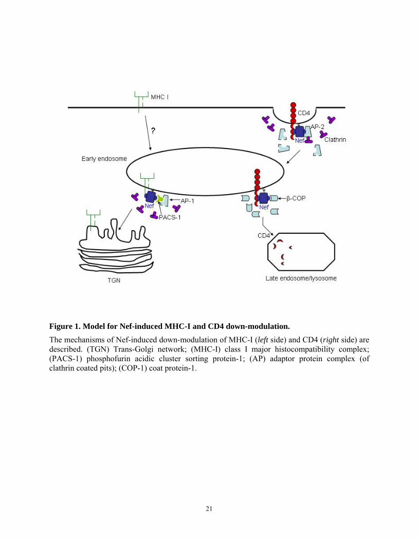
Figure 1. Model for Nef-induced MHC-I and CD4 down-modulation. The mechanisms of Nef-induced down-modulation of MHC-I (left side) and CD4 (right side) are described. (TGN) Trans-Golgi network; (MHC-I) class I major histocompatibility complex; (PACS-1) phosphofurin acidic cluster sorting protein-1; (AP) adaptor protein complex (of clathrin coated pits); (COP-1) coat protein-1.
21
1.6.5. Modulation of internal cell signaling
In addition to its ability to downregulate cell surface receptors, Nef appears to alter
intracellular signaling by interacting with proteins which mediate signal transduction in CD4+ T
cells and macrophages. A large and growing number of studies suggest that Nef manipulates
multiple signaling pathways, affecting T cell activation. Nef also regulates cell signaling to kill
or protect cells from apoptosis. In addition, other signaling pathways involved in cytoskeletal
rearrangement or cytokine production are also affected by Nef expression.
T cell activation
Whether Nef promotes or interferes with the activation of T-lymphocytes has been
controversial. Initially, Nef has been suggested to disturb the activation of T cells by not only
downregulating CD4 receptors but also by altering the intracellular signaling pathways activated
by TcR stimulation. The downregulation of CD4 by Nef releases p56Lck from the cytoplasmic
tail of CD4. The association between p56 Lck and CD4 is necessary for the proximal localization
of Lck with TcR-CD3 complex upon the activation of T-cells through the TcR or anti-CD3
cross-linking. A Src family kinase, Lck is required for phosphorylation of the zeta chain of the
TcR, which in turn facilitates the recruitment and binding of ZAP-70 leading to subsequent up-
regulation of sensitive genes, such as IL-2 (104). IL-2 up-regulation is an important marker of T-
cell activation. Without the proximal localization of Lck to TcR complexes these signaling
cascades in T cells are unlikely to occur. In this scenario, Nef suppresses the activation of T
cells, leading to the development of the quiescent latently infected T cell populations. Supporting
this, early studies showed that Nef expression in the Jurkat T-cell line results in inhibition of up-
regulation of IL-2 mRNA in response to either PMA, PHA, or anti-CD3 cross-linking (105).
22
Furthermore, Nef downregulates the transcription factors NF-κB and AP-1 in cells stimulated
either by mitogens or by antibodies to the TcR-CD3 complex through effects on the TcR
signaling cascades (65, 106).
Conflicting results to the previous negative effect of Nef on T-cell activation have soon
emerged. Rhee and Marsh reported that the expression of Nef in a T-cell hybridoma
downmodulates the CD4 receptor from the cell surface and enhances T-cell activation stimulated
by TcR (68). In vivo studies further demonstrated the positive effect of Nef on T-cell activation.
For example, expression of HIV-1 Nef in T lymphocytes caused T cell hyperactivation in the
thymus, with CD4 depletion and increased activation of the rare mature CD4 T cells in the
peripheral lymphoid tissues of transgenic mice (67). In the same study, Nef-expressing
thymocytes of the transgenic mice also displayed elevated mitogenic and calcium responses to T
cell receptor (TcR) stimulation with a CD3-specific antibody. Consistent with this result, the Nef
transgenic mice created by Hanna et al. showed similar T-cell phenotypes, including increased
sensitivity of CD4 T cells to CD3 and TcR-induced activation (66). A microarray study of the
overall gene regulation in Nef-expressing T lymphocytes showed almost identical gene
expression profiles (97%) to that of control T cells activated by CD3 stimulation (107).
One possible explanation for these conflicting experimental results comes from the study
of Baur et al. Their study showed that the positive or negative effect of Nef on T-cell activation
depends on its intracellular localization (108). They expressed a hybrid CD8-Nef protein in
Jurkat cells. When expressed in the cytoplasm, the chimera inhibited early signaling events from
the T cell antigen receptor, whereas the chimera expressed on the cell surface activated the same
signaling events in T cells. These effects of subcellular localization may reconcile the opposing
phenotypes of Nef and suggest a key role for N-myristylation as well. The inconsistency in
23

results regarding the effect of Nef on T-cell activation could also be explained by the use of
different cells, cell culture conditions, and allelic variation in Nef sequences. Positive and
negative effects of Nef on T-cell activation actually could benefit the virus replication in infected
cells in different ways. While the activation of T lymphocytes by Nef may create a cellular
environment favorable for viral replication, the inhibition of T cells may lead to the latency of
infected cells and thus facilitate immune escape and the establishment of a chronic infection.
Apoptosis
To ensure survival of an infected cell until the replicated virus particles are ready to leave
and spread to other target cells, viruses deploy multiple strategies to protect the infected cells
from self-destruction (apoptosis). When infected with virus, cells start the programs to kill
themselves to save the host. For example, Fas ligand is up-regulated in HIV-infected T cells,
thereby killing the infected cells in an autocrine fashion through Fas ligation (109, 110). Another
example is the up-regulation of membrane-bound TNF on macrophages by the binding of HIV
gp120 to CXCR4 receptor (111). The induced TNF triggers cell death via TNFR in CD8 T cells
and potentially also in infected cells. From the prospect of the virus, maintaining the viability of
the infected cells while inducing the apoptosis of the bystander cells is paramount.
To prevent apoptosis in response to viral infection, HIV takes advantage of the ability of
Nef to interact with host cell survival signaling molecules. Geleziunas et al. showed that Nef
protects infected T cells from apoptosis triggered by CD95 (Fas) and TNF-alpha receptor via
inhibition of the apoptosis signal regulating kinase (ASK1) (112). ASK1 links both cell death
receptor (Fas and TNFR)-mediated signals to the downstream JNK/p38 pathways. ASK1 kinase
activity is inhibited by an association with thioredoxin (Trx), a redox regulatory factor. Nef
24
blocks the dissociation of ASK1-Trx, which is normally induced by TNF-α. This study is
supported by another report, showing that Nef blocks the Fas signaling pathway through
inhibition of caspase-3 and caspase-8 activation (113).
Another example of Nef inhibition of apoptosis signaling pathways comes from a study by
Wolf and co-workers. They found that Nef blocks cell death mediated by pro-apoptotic Bcl-2
family members inside cells (114). Control of apoptosis by mitochondria is partly regulated by
the balance of Bcl-2 family members. Pro-apoptotic members (Bad, Bax, Bak, Bid, and others)
of this family form heterodimers with and thereby inactivate anti-apoptotic members of the same
family (Bcl-2, Bcl-XL, Bcl-w, and others) (115, 116). Nef inhibits Bad proapoptotic activity by
inducing the serine phosphorylation of Bad, a pro-apoptotic protein, and facilitates release of the
anti-apoptotic partner, Bcl-2. Normally, Bad inactivation induced by growth factor or cytokine
stimulation is mediated by phosphatidylinositol-3-kinase (PI3K) via the downstream Ser/Thr
kinase, Akt. However, Nef-induced Bad phosphorylation is not mediated by Akt. Nef binds and
activates PI-3 kinase not to stimulate Akt but to activate the Nef-associated kinase, PAK. The
Nef-PI3K-PAK complex phosphorylates Bad, consequently blocks apoptosis in T cells induced
by serum starvation or more importantly, HIV replication. In this way, Nef seems to serve to
counter balance the apoptosis-inducing effect of HIV-1. The anti-apoptotic signaling of Nef
indeed enhanced viral particle release, supporting that this Nef function is an important
mechanism in viral replication.
Nef was also reported to physically bind to p53 via its N-terminal 57-residue fragment (1-
57) and thereby inhibit p53-dependent apoptosis (117). The inhibition of p53-mediated apoptosis
by Nef is likely due to the decreased half-life of p53. Consequently decreased p53 DNA binding
activity and transcription activity were observed in Nef expressing cells. Importantly, all these
25
events correlated with the binding ability of Nef to p53. Together, these results suggest that the
protective effect of Nef may augment HIV replication by prolonging the viability of infected
cells.
Recent work from our laboratory demonstrated that HIV Nef also promotes a Stat3-
dependent proliferation of the macrophage progenitor cell line, TF-1 (118). Nef-induced TF-1
proliferation requires the myristylation signal sequence and polyproline motif, since mutation of
these sequences abolished the proliferative phenotypes of Nef-expressing cells. This result
suggests that Nef not only protects infected cells from apoptosis but also produces a signal of cell
proliferation, promoting an environment for viral replication. The studies presented in this thesis
also demonstrate that HIV-1 Nef protects TF-1 cells from apoptosis by inducing Bcl-XL in an
Erk-dependent manner (Chapter IV). This is the first evidence that Nef is involved in survival in
macrophage-lineage cells, a key target of HIV infection.
Other Nef-associated signaling pathways
Several lines of evidence suggest that the host cell actin cytoskeleton plays a role in the
entry steps of the infectious pathway utilized by HIV-1. It has been suggested that actin
microfilaments facilitate the co-localization of receptors during virion fusion to the host cell, and
this clustering of receptors is required for virus entry into the cell (119, 120). Furthermore, the
actin cytoskeleton contributes to the establishment of a functional reverse transcriptase complex
(121). HIV-1 virion trafficking in a microtubule-independent manner in the periphery of the
cytoplasm after entry into the cytoplasm has been observed with live-cell microscopy (122). This
movement is likely due to an interaction with the actin cytoskeleton in the host cell cytoplasm.
26
Nef has been suggested to interact with the actin cytoskeleton early in infection. A
myristylation-dependent association of HIV-1 Nef with actin has been shown in B and T cells,
and this interaction affects its subcellular localization (39). Nef has also been reported to interact
with a number of proteins associated with actin microfilament reorganization. Particularly, Nef
binds and activates the Rho GTPase exchange factor Vav, inducing rearrangements of
cytoskeleton (123). In addition, Nef also interacts with members of the p21-activated kinase
(PAK) family, resulting in kinase activation (67, 124, 125). PAK family kinases have been
implicated in cytoskeletal organization and rearrangement in mammalian cells (126, 127).
Moreover, the residues of Nef involved in cytoskeletal association overlap the regions critical for
enhancement of viral infectivity, suggesting that the interaction with actin cytoskeleton might be
important in Nef’s ability to increase infectivity.
Since Nef also associates with the viral core (77, 128), the significance of Nef interaction
with the actin cytoskeleton might be to facilitate a post-fusion trafficking of the core via a
mechanism that involves the actin cytoskeleton. Recently, Campbell et al. found that disruption
of the actin cytoskeleton by treatment of two drugs, Cytochalasin (CytD) and latrunculin B
(LatB) restores the infectivity of Nef deficient virions to that of wild type virus control (129).
Since this complementation failed with HIV virion pseudotyped to enter cells via endocytosis,
the ability of actin disruption to complement Nef infectivity defect was specific to the native
HIV envelope which fuses at the cell surface. These results indicate that Nef may function to
facilitate the penetration of the HIV genome through the cortical actin network, a known barrier
for intracellular parasitic organisms, allowing the virus to infect the host cell more efficiently.
27
1.7. Cellular partners of Nef
Since Nef lacks intrinsic enzymatic activity, the positive role of Nef in viral replication,
infectivity and AIDS pathogenesis is attributed to its ability to interact with several cellular
proteins. The binding partners of Nef can be divided into two groups, the first of which includes
proteins that lack kinase activity. This group of proteins includes actin cytoskeletal regulators
such as Vav, the CD4 receptor, MHC I, and the protein components of the endocytotic
machinery. The second group is composed of the protein kinases which mediate cellular
signaling pathways. Serine/threonine kinases and Src family kinases constitute this group. The
significance of these interactions for HIV replication and pathogenesis has been described above.
What follows are more detailed descriptions of the molecular interactions.
1.7.1. Cellular Receptors and trafficking proteins
CD4 and MHC I cell surface receptors
CD4 constitutes the primary receptor of primate lentiviruses (130) and is rapidly
downregulated in HIV infected cells via a Nef-mediated endocytosis mechanism (42, 87). CD4 is
a type I integral membrane protein that is mainly expressed in immune cells (131, 132), and its
physical interaction with Nef has been reported in several experimental systems. For example,
Harris et al. demonstrated the binding of HIV Nef to CD4 in insect cells overexpressing both
proteins (38). They also showed that binding is dependent on N-terminal myristylation of Nef.
Subsequent studies detected the interaction of Nef with CD4 in the yeast-two hybrid system
(133) and in a CD4 capture assay using immobilized Nef as the bait (134). Furthermore the
interaction of these two proteins was demonstrated in an NMR study using recombinant Nef and
a CD4-derived peptide (27). In this study, they identified the di-leucine residues in the membrane
28
proximal region of the CD4 receptor tail, which normally mediate its interaction with the
endocytosis apparatus upon TcR or mitogen stimulation, as the binding site for Nef.
Through this interaction, Nef accelerates the endocytosis of CD4 from the HIV infected
cell surface by directly connecting CD4 to the adaptor complex of cellular transport machinery
(see section 1.6.4. and Figure 1). Nef also redirects some CD4 from the endosomes to lysosomes
and inhibits its recycling to the cell surface, targeting it instead to the lysosomal degradation
compartment (135). The significance of the association of CD4 with Nef was discussed in the
earlier section (1.6.4). It prevents deleterious superinfection and releases the inhibition of virion
budding by cell-surface CD4.
Functional class I MHC complexes composed of the class I MHC heavy chain, β2-
microglobulin and a peptide are assembled in the ER and possibly in the cis-Golgi. MHC class I
is responsible for presenting viral antigens on the surface of infected cells to cytotoxic T
lymphocytes (CTL). Because of this function, downregulation of MHC I by Nef allows the
infected cells to escape host immune surveillance, particularly CTL (101), as described in section
1.6.4. Nef binding and downregulation of MHC class I utilize a distinct mechanism from the
interaction with CD4. The binding site of Nef to MHC I is also genetically separable from the
regions required for CD4 binding (136). Mutation of residues critical for CD4 downregulation
was shown to have no effects on the binding and downregulation of MHC I by Nef. In addition,
the N-terminal α-helix, proline repeat, and acidic cluster all of which are dispensable for CD4
downregulation by Nef, were required for MHC I downmodulation (136, 137). However, the
mechanism of MHC I downregulation by Nef is less clear. Recently, Nef has been reported to
induce MHC I downregulation by promoting the retrieval of MHC I molecules from the cell
29
surface to trans-Golgi via PACS-1 (138). Swann et al. suggested that HIV-1 Nef inhibits the
transport of MHC I molecules to the cell surface through a PI3K dependent mechanism (139).
Protein components of the cellular trafficking machinery
An important downstream partner of Nef in interaction and downregulation of CD4 is the
clathrin-associated adaptor protein complex (AP). Adaptor complexes are heterotetrameric
structures which recruit clathrin to the cytoplasmic tail of receptors containing endocytosis
signals. Three classes of APs have been reported so far. AP-1 and AP-2 play a role in protein
sorting in the trans-Golgi network (TGN) and at the plasma membrane, respectively (140). AP-3
mediates a direct transport from the Golgi to the lysosomes (141, 142). All APs contain two large
subunits (Mr 100,000; γβ′ for AP-1, αβ for AP-2, and β3δ for AP-3), one medium chain (µ1 for
AP-1, µ2 for AP-2, and µ3 for AP-3), and one small chain (Mr 19,000; σ1, σ2, and σ3
respectively). Normally, APs recognize a tyrosine-based motif (YXXφ, where Y is a tyrosine, X
is any amino acid, and φ is a hydrophobic residue) or a dileucine motif (LL, where L is a leucine
or equivalent amino acid with a bulky hydrophobic side chain) on the cytoplasmic tail of
receptors (94, 143, 144). While the medium chain (µ) has been shown to bind directly to a
tyrosine-based motif in the yeast two hybrid system and in vitro (145), the binding component
for the dileucine motif was less clear, although there are a few reports to show the binding of this
motif to both µ and β′ subunit of AP complexes (146, 147). Of note, the interaction of Nef and µ
chain was detected in the yeast-two hybrid system and by using recombinant proteins (92). The
association of Nef with other subunits of APs has not been demonstrated in these and other
systems. HIV-2 and SIV Nef interact with the µ chain of APs via its tyrosine-based motif located
30
in the N-terminal region. HIV-1 Nef binds to µ with a weaker affinity than HIV-2 or SIV Nef,
and the di-leucine motif in the C-terminal region of HIV-1 Nef is required for this binding (89).
The requirement of AP binding for Nef-induced CD4 internalization was demonstrated
using a mutant Nef protein defective for AP recruitment. This mutant was unable to accelerate
CD4 endocytosis, either in trans or when fused to the extracellular and transmembrane domain
of CD4 receptor (88). This and other studies demonstrate that adaptor complexes are the major
downstream partners of Nef for CD4 downregulation. The interaction of endocytosis machinery
is further facilitated by a subunit of the ATPase associated with Nef.
A second Nef-binding partner involved in CD4 downregulation is COP-I coatomer. This
non-clathrin-coated-vesicle mediates protein sorting in ER-Golgi transport as well as from early
to late endosomes within the endocytosis pathway. The role of interaction of Nef with COP I has
been suggested to block the recycling of CD4 to the cell membrane by targeting the protein for
degradation to lysosome. Nef was shown to physically interact with the β subunit of COP I in
vitro and in the yeast-two hybrid system (95). A diacidic (EE155) motif in the C-terminal tail of
HIV-1 Nef is critical for this interaction (135). The binding was strongly enhanced by the
addition of COP-depleted cytosolic extracts, indicating that additional cellular proteins may be
involved in regulation of this interaction of Nef with the COP I coatomer (135). Recently, ARF1
was identified as a regulator of Nef association with β-COP and Nef-induced lysosomal targeting
of CD4 (148). However, another study showed that the mutation of diacidic motif did not affect
the binding of Nef to β-COP, and this motif is poorly conserved among HIV isolates (149).
Additional studies are therefore required to evaluate the contribution of the Nef-COP I
interaction to CD4 downregulation.
31
The downstream binding partners of Nef for MHC class I downmodulation are not as well
characterized as compared to CD4 downregulation. PACS-1, a molecule that controls the TGN
(Trans Golgi Network) localization of the cellular protein furin, was recently identified as a
binding partner of Nef in the function of MHC I downregulation (138). The acidic cluster and
other Nef regions important for MHC I regulation are highly similar to the PACS-1 binding,
TGN-retrieval motif of furin. Nef interacts with PACS-1 and redirects MHC-I molecules to the
TGN via the retrieval pathway. Because PACS-1 is a sorting protein that connects membrane
proteins to AP-1 to allow the membrane proteins to be retrieved from the endosome to the Golgi
in clathrin-coated pits, AP-containing clathrin-coated-pits seem to participate in Nef-induced
MHC I downregulation.
1.7.2. Serine Kinases
p21-activated kinase
A 62 kDa phosphoprotein that co-immunoprecipitates with Nef has been observed in
many earlier studies and is often referred to as Nef-associated kinase activity (NAK) (83).
Subsequent studies revealed that this protein belongs to the family of p21-activated kinase
(PAK) (150) and is most likely identical to PAK2.
Several regions of Nef are critical for interaction with PAK2, including the second
arginine residue of a diarginine motif (RR) within the core region (46, 83, 151, 152) and the
conserved PxxP motif (124). Leu 112 and Phe 121 on the NL4-3 HIV-1 were also identified as
the residues necessary for interaction between Nef and PAK2 (124). Mutation of the RR motif
also interfered with the ability of Nef to downregulate CD4, to increase viral infectivity and to
alter TcR-initiated signal transduction, suggesting that interaction with PAK may be important in
Nef function. However, it remains to be determined whether all of these defects in Nef function
32
are due solely to the inability to bind to PAK2. The PxxP motif, a well known motif of binding
partners to SH3 domain, is required for the interaction of Nef with PAK2. As PAK2 does not
contain a SH3 domain, it has been suggested that another intermediary protein which contains a
SH3 domain may facilitate the Nef-PAK2 complex formation in a PxxP-dependent manner. This
hypothesis was supported by the finding that the total Nef associated PAK2 activity was not
increased by the ectopic overexpression of PAK2 (153).
Although the association of Nef with PAK is highly conserved among HIV and SIV
isolates, the functional significance is not clear. A requirement for this interaction in Nef
functions such as the enhancement of the viral infectivity and replication kinetics in primary cell
culture has been demonstrated by Wiskerchen et al. (76). Mutation of the Nef residues
responsible for Pak association decreased the infectivity of viruses compared to wild type. The
lower levels of proviral DNA synthesis were also observed in cells infected with mutant viruses
that are defective in the ability of their Nef proteins to associate with the serine kinase activity.
Furthermore, in the Sawai study, a Nef mutant disrupting the association with the activated PAK
was reverted to prototype Nef sequence and function in SIV-infected macaques (152). The
interaction between Nef and PAK correlated with the induction of high viral loads and
pathogenesis of infected hosts, suggesting that this interaction is important for the Nef function
in vivo.
The PAK family plays a central role in the organization of the cytoskeleton (126, 127),
and, in addition, PAK2 has also been implicated in apoptosis signaling (154, 155). PAK2 also
mediates the signals from the plasma membrane to the nucleus, leading to activation of
transcription factors such as serum response factor (SRF) via MAPK cascade (126, 156). Nef
may positively affect some steps of the HIV life cycle by altering these PAK-mediated processes
33
of host cells. For example, the interaction of Nef with PAK may facilitate lipid raft formation via
the effect of PAK2 on cytoskeletal rearrangement. This effect of Nef on lipid raft formation is
likely to impact positively the HIV virus particle budding since HIV-1 has been shown to bud
from lipid rafts (157, 158). Also, Nef-PAK2 interaction may modulate the sensitivity to
apoptosis of the HIV infected cells. As PAK2 has been shown to be cleaved and activated by the
apoptotic caspases (159), the binding of Nef to PAK2 may affect apoptotic processes, which
favors HIV replication (see section 1.6.5.).
Other Nef-assoicated serine kinases
Other serine/threonine kinases which do not belong to the PAK family also have been
reported to bind to Nef. These include members of protein kinase C (PKC) and mitogen-
activated protein kinase (MAPK) families. The 80 kDa θ form of PKC was reported to co-
precipitate with a GST-Nef fusion protein by Smith et al. (84). The binding site of PKC θ for Nef
appears to be different from the substrate binding site of PKC since the Nef-PKC θ interaction
was not affected by a PKC pseudosubstrate peptide. In addition, the association of PKC with Nef
impeded its usual relocation from the cytosol to the particular cellular fraction in Jurkat cells
upon PMA/PHA stimulation, implying that Nef-PKC interaction may regulate the enzymatic
activity of PKC. The PKC family has also been shown to phosphorylate the serine/threonine
residues in the very N-terminus of Nef (44, 45). The phosphorylation of Nef at its N-terminus
may act as an electrostatic switch to alter association of Nef with plama membrane by
introducing negative charges. Basic N-terminal residues cooperate with myristylation signal in
anchoring the Nef protein to the plasma membrane (see section 1.4.1.). Negative charges
introduced by phosphoryation thus may repel Nef from the membrane. This dynamic of
34
membrane binding may explain the large distribution of the myristylated Nef protein in the
cytoplasm (45) and its interaction with a wide variety of cellular proteins located at the plasma
membrane, cytoplasm, and even the nucleus. Nevertheless, the significance and role of Nef-PKC
interaction in Nef-mediated functions in infected cells remains to be determined.
Erk MAPK interaction with Nef was found in binding assays using a recombinant GST-
Nef fusion protein to probe cytoplasmic extracts of MT-2 T-cell line and PBMC (134). The
interaction between Nef and Erk requires the Nef PxxP motif, although Erk proteins do not
contain SH3 domains. As for Pak, Nef-Erk interaction may require another protein with an SH3
domain to mediate or stabilize this interaction. However, Nef was also demonstrated to directly
bind and inactivate MAPK in vitro assay using recombinant proteins (82) Contradictory to this
result, Nef was shown to increase Erk1/2 activity initiated by T cell receptor stimulation in
primary CD4+ T cells obtained from peripheral blood (160). Enhanced Erk activity may be
responsible for increased T cell activation by Nef, which facilitates HIV replication in this target
cell type. Erk also mediates the induction of the AP-1 transcription factor by Nef in
macrophages. The induction of AP-1 may contribute to the activated phenotype of Nef-
transfected and HIV-1-infected macrophages. Further investigation is required to address
whether these effects of Nef on MAPK are mediated by direct interaction.
Finally, Nef has been shown to associate with c-Raf1 kinase, which is a central player in
coordinating the Ras-Raf-MAPK pathway. Hodge et al. showed that c-Raf1 can be co-
precipitated from CEM T-cells together with Nef (161). This interaction was also demonstrated
with recombinant Nef and Raf produced in E. coli, suggesting that complex formation is direct.
A highly conserved acidic sequence at the carboxy-terminal region of Nef was mapped as the c-
Raf binding site and contains the core sequence Asp-Asp-X-X-X-Glu (position 174-179). This
35
binding site shares striking similarity with an acidic consensus Raf-binding motif within the Ras
effector region. The same acidic motif previously has been shown to be important for
downregulation of cell surface CD4 expression by Nef (162, 163). However, additional studies
are required to determine the significance of the Nef-Raf interaction in terms of regulation of the
signaling pathway and cell surface CD4 expression. The study presented in this thesis
demonstrates that Nef induces survival signaling in an Erk-dependant manner (see section 3.4.).
The interaction of Nef and Raf may mediate the activation of Erk and consequent survival
signaling by Nef (Nef-Raf-Erk), which requires further investigation.
1.7.3. Src Family Tyrosine Kinases and Nef
The c-src gene was the first to be identified as a protooncogene, encoding a protein capable
of inducing cell transformation (164). Cellular src and related genes encode protein-tyrosine
kinases, which activate multiple cellular signaling pathways linked to normal growth control and
cancer. The members of the Src family can be divided into two classes: those with a broad
expression range (Fyn, Lyn, Src and Yes) and those with expression patterns restricted, primarily
to hematopoietic cells in some cases (Blk, Fgr, Hck, Lck, and Yrc). Several of these latter
members have been shown to interact with HIV-1 and SIV Nef.
Structure of Src Family Tyrosine Kinases
The proteins of the Src family range in molecular mass from 52 to 62 kDa and have a
similar structure, which consists of several distinct functional domains: N-terminal SH4 domain,
a unique region, SH3, SH2, an SH2-kinase linker region, and SH1 domain followed by a C-
terminal negative regulatory tail (Figure 2) (165).
36
The SH4 domain is a region located at the extreme N-terminus, and contains a signal
sequence for posttranslational modification with fatty acids such as myristate or palmitate (33,
166, 167). As for HIV Nef, myristylation at Gly 2 is required for membrane localization of Src
family proteins. In addition to myristylation signals, the Src SH4 domain contains basic residues
which could be the substrate for posttranslational palmitoylation. Palmitoylation occurs only
when the molecules are myristylated and this additional modification may stabilize membrane
association. As palmitylation is a reversible process, it has been thought to be a mechanism for
modulating Src family kinase localization. Following the SH4 domain, is a unique region
specific for each Src family protein. Although this region contains a site bound and
phosphorylated by protein kinase C for some family members, the functional significance of this
event is unclear.
The SH3 domain follows the unique domain, and is approximately 60 residues in length.
This domain mediates protein-protein interactions through proline-rich motifs (PxxP) which
form polyproline type II helices (168, 169, 170, 171). The SH3 domain also binds
intramolecularly to the SH2-kinase linker region, regulating catalytic activity, protein
localization in the cell, and association with protein targets (168). Binding of other proteins to
the SH3 domain (e.g. Nef) can cause kinase activation by displacing this negative regulatory
interaction (see below).
Adjacent to the SH3 domain is an SH2 domain, which also mediates protein-protein
interactions. The SH2 domain consists of approximately 90 amino acids and binds to short amino
acid sequences containing phosphotyrosine (169, 170, 171). The specificity of each SH2 domain
is determined by the three to five residues C-terminal to the phosphotyrosine (172). In the case of
Src and Lck, the SH2 domain forms a deep hydrophobic pocket which accommodates amino
37
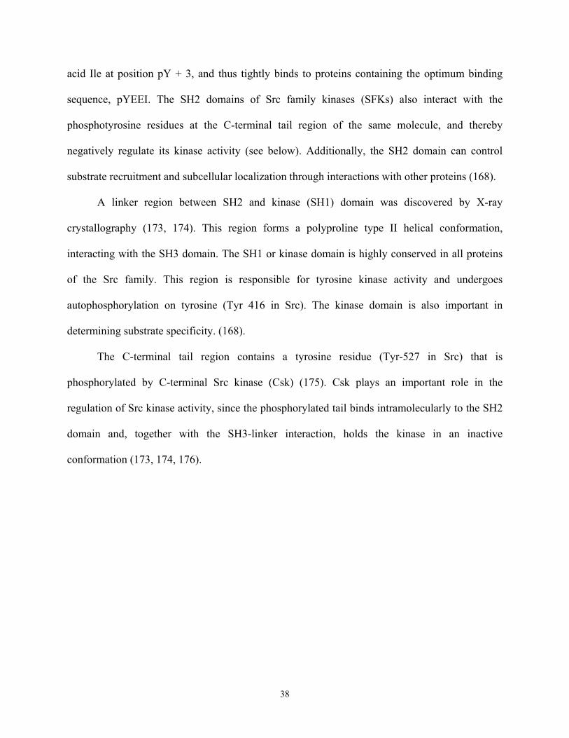
acid Ile at position pY + 3, and thus tightly binds to proteins containing the optimum binding
sequence, pYEEI. The SH2 domains of Src family kinases (SFKs) also interact with the
phosphotyrosine residues at the C-terminal tail region of the same molecule, and thereby
negatively regulate its kinase activity (see below). Additionally, the SH2 domain can control
substrate recruitment and subcellular localization through interactions with other proteins (168).
A linker region between SH2 and kinase (SH1) domain was discovered by X-ray
crystallography (173, 174). This region forms a polyproline type II helical conformation,
interacting with the SH3 domain. The SH1 or kinase domain is highly conserved in all proteins
of the Src family. This region is responsible for tyrosine kinase activity and undergoes
autophosphorylation on tyrosine (Tyr 416 in Src). The kinase domain is also important in
determining substrate specificity. (168).
The C-terminal tail region contains a tyrosine residue (Tyr-527 in Src) that is
phosphorylated by C-terminal Src kinase (Csk) (175). Csk plays an important role in the
regulation of Src kinase activity, since the phosphorylated tail binds intramolecularly to the SH2
domain and, together with the SH3-linker interaction, holds the kinase in an inactive
conformation (173, 174, 176).
38
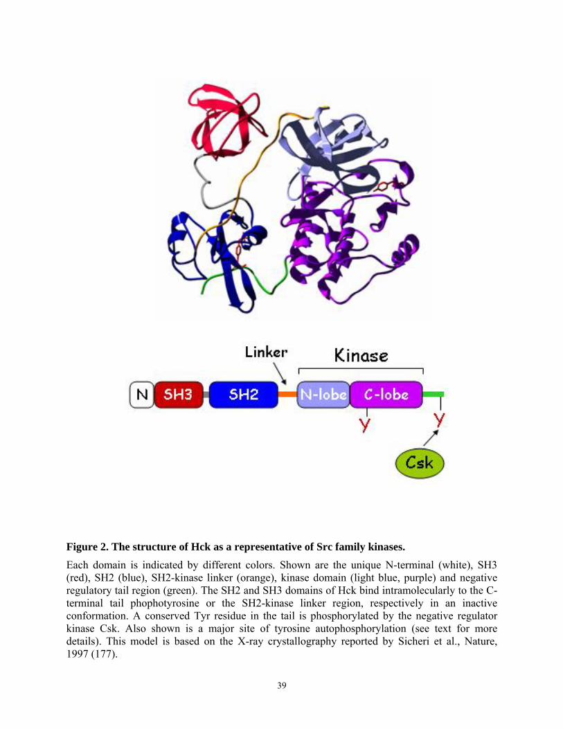
Figure 2. The structure of Hck as a representative of Src family kinases. Each domain is indicated by different colors. Shown are the unique N-terminal (white), SH3 (red), SH2 (blue), SH2-kinase linker (orange), kinase domain (light blue, purple) and negative regulatory tail region (green). The SH2 and SH3 domains of Hck bind intramolecularly to the C-terminal tail phophotyrosine or the SH2-kinase linker region, respectively in an inactive conformation. A conserved Tyr residue in the tail is phosphorylated by the negative regulator kinase Csk. Also shown is a major site of tyrosine autophosphorylation (see text for more details). This model is based on the X-ray crystallography reported by Sicheri et al., Nature, 1997 (177).
39
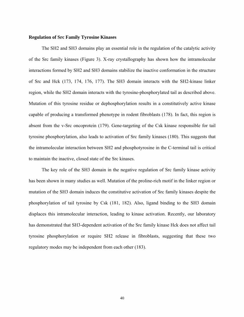
Regulation of Src Family Tyrosine Kinases
The SH2 and SH3 domains play an essential role in the regulation of the catalytic activity
of the Src family kinases (Figure 3). X-ray crystallography has shown how the intramolecular
interactions formed by SH2 and SH3 domains stabilize the inactive conformation in the structure
of Src and Hck (173, 174, 176, 177). The SH3 domain interacts with the SH2-kinase linker
region, while the SH2 domain interacts with the tyrosine-phosphorylated tail as described above.
Mutation of this tyrosine residue or dephosphorylation results in a constitutively active kinase
capable of producing a transformed phenotype in rodent fibroblasts (178). In fact, this region is
absent from the v-Src oncoprotein (179). Gene-targeting of the Csk kinase responsible for tail
tyrosine phosphorylation, also leads to activation of Src family kinases (180). This suggests that
the intramolecular interaction between SH2 and phosphotyrosine in the C-terminal tail is critical
to maintain the inactive, closed state of the Src kinases.
The key role of the SH3 domain in the negative regulation of Src family kinase activity
has been shown in many studies as well. Mutation of the proline-rich motif in the linker region or
mutation of the SH3 domain induces the constitutive activation of Src family kinases despite the
phosphorylation of tail tyrosine by Csk (181, 182). Also, ligand binding to the SH3 domain
displaces this intramolecular interaction, leading to kinase activation. Recently, our laboratory
has demonstrated that SH3-dependent activation of the Src family kinase Hck does not affect tail
tyrosine phosphorylation or require SH2 release in fibroblasts, suggesting that these two
regulatory modes may be independent from each other (183).
40
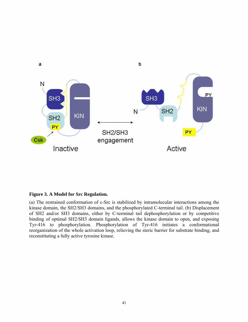
Figure 3. A Model for Src Regulation. (a) The restrained conformation of c-Src is stabilized by intramolecular interactions among the kinase domain, the SH2/SH3 domains, and the phosphorylated C-terminal tail. (b) Displacement of SH2 and/or SH3 domains, either by C-terminal tail dephosphorylation or by competitive binding of optimal SH2/SH3 domain ligands, allows the kinase domain to open, and exposing Tyr-416 to phosphorylation. Phosphorylation of Tyr-416 initiates a conformational reorganization of the whole activation loop, relieving the steric barrier for substrate binding, and reconstituting a fully active tyrosine kinase.
41
Different mechanisms of Src kinase activation may generate distinct output signals because of
different accessibility of SH2 or SH3 domain to substrates. When Src kinases are activated by
disruption of intramolecular interactions, the conserved tyrosine residue (Tyr 416 in c-Src) at the
activation loop in the catalytic domain becomes autophosphorylated. The regulatory
phosphorylation of two important tyrosine residues leads to opposite results. While
autophosphorylation of Tyr 416 in the activation loop of the kinase domain activates the enzyme,
phosphorylation of C-terminal tail Tyr 527 by Csk causes its inactivation.
Hck
Hematopoietic cell kinase (Hck) is a member of the Src kinase family which is expressed
predominantly in macrophages, monocytes, and granulocytes (184, 185, 186). Two isoforms
with molecular weights 59 kDa and 61 kDa in humans arise from a single mRNA by utilization
of alternative translational initiation codons (187, 188, 189). Two murine Hck isoforms (56 kDa
and 59 kDa) are generated by the same mechanism. These two Hck isoforms have different N
termini and are differentially modified by myristylation and palmitoylation (189). Depending on
the acylation at the N-terminus, these two isoforms are differentially distributed between
membranes and cytoplasm. The smaller form in both species is posttranslationally modified by
both myristylation and palmitoylation, resulting in its strong association with the cellular
membrane. In contrast, the larger Hck protein is only myristylated, and thus is present both in
membranes and cytosol.
Although the precise function of Hck in myeloid cells is unclear, it serves as a transducer
of growth factor and adhesion signals from the membrane to the interior of the cell. Early studies
demonstrated that Hck plays a role in mediating the cellular effect of cytokines. Hck was
42
reported to be involved in signaling of cytokines such as GM-CSF, IL-3, and LIF in
hematopoietic cells and embryonic stem cells. Hck expression was found to correlate with GM-
CSF-induced proliferation of HL-60 cells. In these cells, overexpression of Hck functionally re-
coupled the GM-CSF receptor to downstream signaling components, implying its key role as a
mediator of GM-CSF signaling (190). Hck was activated by IL-3 stimulation in 32Dcl3 cells and
was additionally demonstrated to physically bind to the β subunit of the IL-3 receptor, implying
the possible contribution of Hck to the IL-3 signaling in myeloid cells (191). Hck is also
expressed in undifferentiated embryonic stem (ES) cells and plays a role in LIF signaling, which
is important in suppressing stem cell differentiation in vitro (192). When Hck was constitutively
activated by replacement of a conserved C-terminal tail tyrosine with phenylalanine by gene
targeting in ES cells, the targeted ES cells required ~15 times less LIF than parental ES cells to
maintain the undifferentiated state. LIF stimulation induced a rapid and transient increase in Hck
tyrosine kinase activity in ES cells, and gp130, a component of the LIF receptor, physically
bound to Hck. This suggests that Hck takes part in signal transduction from the LIF receptor to
biological responses.
As the macrophage is the major cell type to express Hck, the involvement of Hck in the
function of macrophages has been reported in several studies. First, Hck was shown to be
upregulated when macrophages were activated upon LPS stimulation (193, 194). Subsequently,
the study of English et al. suggested that Hck is an important component of macrophage
activation pathway stimulated by LPS (195). Overexpression of Hck in a murine macrophage
cell line increased tumor necrosis factor (TNF) production in response to LPS. Inhibition of
endogenous Hck via antisense RNA impeded LPS-mediated TNF synthesis, suggesting that Hck
plays a role in mediating the LPS-stimulated signal for TNF production. Furthermore, Hck was
43
shown to bind physically to the high affinity immunoglobulin G receptor FcγRI, which is
expressed constitutively on monocytes and macrophages (196). The phosphorylation and kinase
activity of Hck were rapidly increased after cross-linking of this receptor, demonstrating that
Hck contributes to FcγRI receptor signaling functionally as well. As the cellular effects initiated
by ligand binding to FcγRI include phagocytosis, cytotoxicity against IgG-coated target cells,
and expression of proinflammatory cytokine genes, the physical and functional association of
Hck with FcγRI suggests the potential role of Hck in macrophage physiological functions. Hck
also seems to play a role in neutrophil functions such as migration and degranulation. For
example, the human p59 form of Hck was found to be located in the secretory granules of human
neutrophiles, implicating Hck in the degranulation process (197). Moreover, Bohuslav et al. have
shown the association between Hck and the urokinase plasminogen activator receptor, a key
molecule involved in migration of leukocytes (198), suggesting that Hck may contribute to the
migration of neutrophils as well.
Several lines of evidence supporting the notion that Hck may be involved in macrophage
functions come from in vivo studies using mouse genetics. Mice with a null mutation of the hck
gene underwent normal hematopoiesis, but phagocytosis by macrophages was slightly impaired.
Macrophages cultured from Hck knock-out mice retain many other normal functional properties,
suggesting that the deficiency of Hck is complemented by other Src family members. The
specific activity of the Lyn protein kinase is increased in hck-/- macrophages, implying that Lyn
may compensate for the loss of Hck. The functional redundancy of the Src family kinases was
demonstrated by generation of null mice defective in multiple Src kinases. Double knock-out
mice lacking both Hck and Fgr showed an increased susceptibility to infection with Listeria
monocytogenes, indicating that either Hck or Fgr is required to maintain a normal immune
44
response (199). The neutrophils isolated from these doubly mutant animals were defective in
adhesion-dependent degranulation, which is important in the development of an inflammatory
reaction. In addition, macrophages isolated from Hck-/-Fgr-/- double knockout mice showed
impaired integrin-mediated signaling, an abnormal morphology with reduced numbers of
filopodia, and reduced motility (200). Similarly, neutrophils from these mice also showed
impairment in β-intergrin mediated response and reduced migration, resulting in an inflammation
defect. Together, these data implicate Hck and Fgr in regulation of critical events in integrin
signaling and actin cytoskeleton organization in neutrophils and macrophages. Another double
mutant (hck-/-src-/-) was generated, but two thirds of them died at birth, although the single
mutant of each molecule have deficiency in only specific cell types (201). Surviving animals
developed a severe form of osteopetrosis that is also present in src-/- single mutants, resulting in
extreme levels of splenic extramedullary hematopoiesis, anemia, and leukopenia. In addition,
normal murine osteoclasts express abundant amounts of Src and Hck, and the Hck expression
level is upregulated in osteoclasts isolated from src-/- mice. These observations imply that Hck
and Src serve partially overlapping functions in osteoclasts and increased Hck expression
compensates for the functional deficiency of Src in osteoclasts.
An important role of Hck in HIV-infected cells has been suggested through its strong
interaction with the HIV Nef protein. The characteristics of this interaction will be discussed
below. Although Hck associates with Nef with unusually high affinity, the functional
significance of this interaction in vivo had not been clear until the study of Nef transgenic mice
emerged. Transgenic mice expressing Nef in CD4 T lymphocytes and macrophages developed
severe AIDS-like pathologies: failure to thrive/weight loss, diarrhea, wasting, premature death,
thymus atrophy, loss of CD4+ T cells, interstitial pneumonitis, and tubulo-interstitial nephritis.
45
Breeding of Tg mice expressing wild-type Nef on an hck-/- (knockout) background delayed the
development of these AIDS-like symptoms, implicating Hck in triggering the onset of disease in
vivo (202). Furthermore, a requirement for Hck in the replication of M-tropic HIV in M-CSF-
treated macrophages has recently been reported (203). Treatment of macrophages with antisense
oligonucleotide for Hck (AS-Hck) not only suppressed the expression of Hck, but also inhibited
viral replication. Together, these results strongly suggest that Hck plays a critical role in HIV
replication and pathogenesis possibly via its physical and functional association with Nef.
Src family kinase associtation with Nef
The identification of a proline-rich (PxxP) motif in HIV Nef suggested the possibility that
Nef may interact with Src family kinases. This polyproline type II helix forming sequence is
highly conserved in Nef proteins of different HIV-1, HIV-2, and SIV isolates. The first
identification of Hck as a binding partner for HIV Nef was from the study of Saksela et al. (204).
Using a filter binding assay, they showed that the recombinant Hck and Lyn SH3 domains bound
to GST-fusions with a Nef tetraproline peptide, (PxxP)4. They also showed high affinity binding
between full-length Hck and Nef, and that this association is disrupted by substitution of the
internal two proline residues within the tetraproline sequence with alanines (AxxA). The affinity
of Nef-Hck SH3 interaction has a KD value of approximately 250 nM representing one of the
tightest SH3-ligand interactions reported so far.
Subsequently, several studies demonstrated SH3-mediated Hck-Nef co-
immunoprecipitation in Nef transfected cells. Previous studies from our laboratory have shown
that Nef-Hck interaction results in constitutive Hck kinase activation capable of transforming
Rat-2 fibroblasts (205). This Nef-induced activation of Hck can be explained by the recent
46
structural studies of Hck, showing that Hck is negatively regulated by two intramolecular
interactions similar to other members of the Src kinase family (see the regulation of Src family
kinase, section 1.7.3.). High affinity binding of Nef to the SH3 domain of Hck displaces the
negative regulatory interaction of SH3 from the SH2-kinase linker, leading to kinase activation.
Although the PxxP motif of Nef is known to play a key role in its interaction with the
Hck SH3 domain, the X-ray crystal structure of Nef-Hck complex identified another binding site
outside of the Nef polyproline helix, which consists of Phe 90 and Trp 113 in the cleft region of
hydrophobic pocket (26). These residues contact Ile 96 in the RT loop of the SH3 domain
(Figure 4). In support of this finding, a peptide comprising only the polyproline helix bound Hck
SH3 domain poorly when compared to full-length Nef, suggesting that additional interactions
outside of PxxP motif have an important role. This interaction between the RT loop of the Hck
SH3 domain and a hydrophobic pocket in the Nef core was shown to contribute to the high
affinity binding (206). Lyn is the only other Src family member (besides Hck), that has Ile
residue at this position of the RT loop of the SH3 domain. Because of the conservation of this
residue, Lyn binds to Nef as strongly as Hck. Despite the similar affinity binding of Hck and Lyn
to Nef, only Hck is constitutively activated and transforms Rat 2 fibroblasts in our focus forming
assay (207). The significance and role of Lyn in Nef-mediated AIDS pathogenesis remains to be
determined.
The interaction of other members of the Src kinase family with Nef has been
controversial. Saksela et al. and others failed to detect the binding of other Src SH3 domains to
Nef using in vitro binding assay and co-immunoprecipitated. Whereas Collette et al. and
Greenway et al. detected the binding between HIV-1 Nef and Lck in vitro and in T cells (48, 82).
Arold et al. determined the binding affinity of interaction between the SH3 domains of Fyn, Lck,
47
and Src and the Nef PxxP motif using isothermal titration calorimetric (ITC) analysis, and found
that these SH3 domains bind to Nef PxxP with low affinity (Kd=15.8, 10.6, and 14.3 µM
respectively) compared to the SH3 domain of Hck (0.25 µM) (208). This difference in binding
affinity may be attributed to the single amino acid located at position 96 in RT loop of SH3
domain. While Hck and Lyn have the isoleucine residue at this position, other src family
members contain either arginine or serine residues. Lee et al. demonstrated that Fyn could be
converted to the high affinity binding partner by substituting the arginine residue with isoleucine
in the RT-loop. Whether the interaction between the Ile of the RT loop and hydrophobic pocket
contributes to Src family binding and activation in vivo has not been determined. Studies
presented in chapter III address this important question.
48
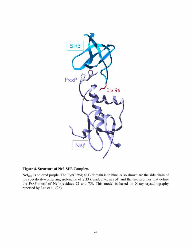
Nef
PxxP
Ile 96
SH3
Figure 4. Structure of Nef–SH3 Complex. Nefcore is colored purple. The Fyn(R96I) SH3 domain is in blue. Also shown are the side chain of the specificity-conferring isoleucine of SH3 (residue 96, in red) and the two prolines that define the PxxP motif of Nef (residues 72 and 75). This model is based on X-ray crystallography reported by Lee et al. (26).
49
Significance of the Nef PxxP motif
The strict conservation of the polyproline PxxP motif in HIV and SIV Nef isolates and its
characteristic interaction with Hck and other proteins with SH3 domains suggest that this motif
may play an important role in Nef functions, contributing to AIDS pathogenesis. Supporting this
idea, the PxxP motif has been shown to be required for several Nef functions including increased
viral replication, enhancement of viral infectivity, MHC I downregulation, and other Nef-
mediated signaling pathways in vitro. However, CD4 downregulation is unaffected by the
mutation of this motif.
Additional evidence that this motif is important for Nef function comes from in vivo
studies using primates as well as transgenic mice. When a Nef PxxP-mutated SIV clone was used
for infection of rhesus macaques, the mutated sequence (AxxA) reverted to PxxP in isolates
recovered from animals as early as eight weeks after infection. In addition, the increase of
revertant viruses correlated with the progression of the course of infection, leading to the death
of two out of five infected animals. These results support an important role for the conserved
PxxP motif for Nef function involved in HIV replication and pathogenesis. However, conflicting
results regarding the importance of this proline-rich motif in disease induction were observed by
Lang et al. (209). They reported that this motif in SIV Nef is dispensable for progression to fatal
simian AIDS in monkeys. The monkeys infected with PxxP- and AxxA-containing viruses both
showed high-level viral loads and rapid disease progression. Furthermore, no correlation
between the reversion rate of AxxA to PxxP and disease progression was observed in their study.
Although the reason for the discrepancy between these two studies has not been determined, it
may be due to the number of monkeys used and the initial virus loads used for inoculation.
50
Despite this controversy, Hanna et al. clearly demonstrate the necessity of the polyproline
motif for disease progression using transgenic mice (202). The same group reported previously
that Nef expression alone in the T lymphocyte and macrophage compartments could induce
dramatic AIDS-like symptoms in transgenic mice. This AIDS-like disease was abolished by
mutation of Nef PxxP motif, suggesting that the interaction of one or several cellular factors
through this Nef region is required for the induction of immunodeficiency disease.
1.8. Macrophages in HIV infection
Although cells of macrophage lineage represent a key target of HIV infection in addition
to CD4 T lymphocytes, the role of macrophages in AIDS pathogenesis is relatively less studied.
The significant contributions of infected macrophages to disease pathogenesis have been
demonstrated in vivo. The non-cytopathic macrophage-tropic strains of HIV, but not highly
cytopathic T-cell tropic strains, were shown to cause extensive T cell depletion in severe
combined immunodeficient mice transplanted with human peripheral blood leukocytes (hu-PBL-
SCID mouse model) (210). Schuitemaker et al. reported that monkeys infected with HIV, which
does not infect simian monocytes/macrophages, exhibited no signs of disease and failed to show
accelerated T-cell apoptosis, implying that macrophage infection is important in disease
progression.
The peculiar dynamics of HIV replication in macrophages, their long-term survival after
HIV infection, and their ability to spread virus particles to bystander CD4 lymphocytes may
allow macrophages to function as a reservoir of viruses, leading to persistent infection in the
host. While a large majority of activated CD4+ lymphocytes are rapidly killed by HIV-1
infection, monocytes/macrophages are poorly affected by the cytopathic effect of HIV-1 (211).
Macrophages can also produce and release high levels of HIV-1 particles for very long periods
51
of time. The role of macrophages as agents for virus dissemination has been demonstrated in
several studies, including that of Crowe et al., in which productively-infected macrophages can
fuse with CD4+ T lymphocytes and transfer the virus to these cells (212). Furthermore, infected
macrophages were demonstrated to recruit and activate CD4 T lymphocytes through the
production of chemokines, which is mediated by Nef (see below) (Figure 5). Macrophages can
also trigger the apoptosis of uninfected T cells, astrocytes, and neurons without direct infection
of these cells by HIV. This finding may explain the observation that apoptosis occurs
predominantly in the bystander cells of the lymph nodes of seropositive individuals.
1.9. Nef in HIV-infected macrophages
Since macrophages are the major cell type to express the Hck protein and the interaction
between HIV Nef and Hck has been well established, the contribution of Nef to macrophage
function in AIDS progression has been studied. Nef has been reported to be a major player of
infected macrophages in recruiting T cells and disseminating virus particles to them. Swingler et
al. showed that macrophages expressing Nef produced two CC-chemokines, macrophage
inflammatory proteins 1α (MIP-1α) and 1β (MIP-1β), that recruit resting T lymphocytes (213).
The supernatant of macrophages expressing HIV-1 Nef also promoted the activation of resting T
lymphocytes to allow productive HIV infection. However, this activating effect of the
supernatant was not dependent on the two chemokines identified as chemotaxis agents for T
lymphocytes (213). This study suggests that Nef may also play a role in lymphocyte activation
through macrophage infection and promoting the environment that facilitates virus dissemination
from infected macrophages to T lymphocytes. In a subsequent study, the same group found that
the mechanism of release of the chemokines and induction of T cell permissivity was likely due
to intersection of Nef and the CD4 ligand signaling pathway. Two soluble proteins, CD23 and I-
52
CAM that are produced from macrophages expressing Nef or via CD4L stimulation were
identified to mediate the induction of T lymphocyte permissivity (214). This study implicates
Nef in expanding the cellular reservoir of HIV-1 by permitting the infection of resting T
lymphocytes.
While Nef generates the signal to protect HIV infected cells from apoptosis, Nef also
plays a role in inducing the apoptosis of uninfected bystander T cells. Nef was shown to induce
upregulation of death ligands, such as FasL in infected T lymphocytes, causing the death of
uninfected bystander cells. However, the effect of Nef on regulation of apoptosis in macrophages
has not been demonstrated, and one of my thesis projects is to investigate the role of HIV Nef in
anti-apoptosis signaling of macrophage lineage cells.
53
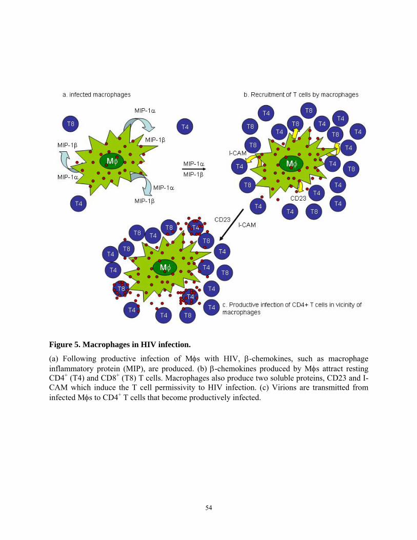
Figure 5. Macrophages in HIV infection.
(a) Following productive infection of Mφs with HIV, β-chemokines, such as macrophage inflammatory protein (MIP), are produced. (b) β-chemokines produced by Mφs attract resting CD4+ (T4) and CD8+ (T8) T cells. Macrophages also produce two soluble proteins, CD23 and I-CAM which induce the T cell permissivity to HIV infection. (c) Virions are transmitted from infected Mφs to CD4+ T cells that become productively infected.
54
2. Chapter II
Dissertation Hypothesis and Specific Aims
55

Hypothesis
HIV Nef affects viral replication, infectivity, CD4 surface expression, and target cell
signaling pathways by interacting with many cellular proteins. Most studies of Nef function have
been performed with the key HIV target cell, the CD4+ T lymphocyte. However, the functions of
Nef in macrophages are less understood. Previous work from our laboratory demonstrated that
Nef promoted cell proliferation in the macrophage lineage cell line, TF-1, and that Nef-induced
proliferation was dependent on Stat3 activation. Nef has recently been shown to affect apoptotic
signaling pathways in T cells and protect infected T cells from infection-induced apoptosis.
Based on these findings, we hypothesized that HIV Nef induces survival in cells of the
monocyte/macrophage lineage, the other HIV-1 relevant cell type, by altering cellular signaling
pathways. One group of important binding targets for Nef is the Src family kinases. Particularly,
Hck binds to Nef through a SH3-PxxP interaction with very high affinity. The association of Nef
with Hck has been a main subject of investigation in our laboratory since this high affinity
interaction was discovered. Previously, we have shown that Nef interaction with Hck causes Hck
activation capable of transforming Rat-2 fibroblasts. This assay is very useful in terms of
assessing the biological activity of Hck bound by HIV Nef. We wanted to further characterize
the association of Nef with Hck using this system and to identify the residues important for the
Nef-Hck interaction. The following aims were designed to address these issues.
56
Specific Aims
Aim 1: Determine if Hck binding and activation is a common property of HIV-1 Nef
proteins, and if not, identify the residues responsible for the differential interaction of Hck
and Nef variants in vivo. Comparative analysis of the ability of four different HIV-1 Nef alleles
to bind to Hck was conducted in vitro. Computer-aided molecular modeling and sequence
alignment identified the residues of Nef that contribute to the differential interaction with Hck.
The functional relevance of identified residues in the hydrophobic pocket outside of the PxxP
motif in recruitment and activation of Hck was evaluated by the Rat-2 fibroblast model. The
biological significance of these residues was assessed by sequence analysis of published full-
length Nef isolates from long-term non-progressors (LTNPs) of HIV-1 infection.
Aim 2: Investigate whether Nef promotes the survival of macrophage lineage cells and how
it affects the anti-apoptotic signal transduction pathways in this HIV-1 target cell type.
Apoptosis measurement revealed that Nef protects cells of the myelomonocytic lineage from
apoptosis. Determining gene expression to correlate with cell survival is a key to characterize
Nef-induced anti-apoptosis signaling pathways. We found an anti-apoptotic protein Bcl-XL to be
up-regulated in Nef-expressing cells. The Nef-induced survival signaling was further
characterized by using an inducible Nef expression system (Nef-ER). Cell survival and Bcl-XL
induction by Nef was dependent on Erk MAPK activation.
57
3. Chapter III
Conserved Residues in the HIV-1 Nef Hydrophobic Pocket are Essential for Recruitment
and Activation of the Hck Tyrosine Kinase
Hyun-Jung Choi and Thomas E. Smithgall
From the Department of Molecular Genetics and Biochemistry, University of Pittsburgh School of Medicine, E1240 Biomedical Science Tower, Pittsburgh, PA 15261
This work has been accepted for publication to Journal of Molecular Biology
58
3.1. Abstract
The Nef protein of the primate lentiviruses HIV and SIV is essential for high-titer viral
replication and AIDS progression. Nef binds to the macrophage-specific Src family member Hck
through its SH3 domain, resulting in constitutive kinase activation capable of transforming
rodent fibroblasts. Nef-Hck interaction may be essential for M-tropic HIV replication and AIDS
pathogenesis, identifying this virus-host protein complex as a rational target for anti-HIV drug
discovery. Here we investigated whether interaction with Hck is a common feature of Nef alleles
from different strains of HIV-1. We compared the ability of four different laboratory HIV-1 Nef
alleles (SF2, LAI, ELI, and Consensus) to induce Hck activation and transformation in our Rat-2
fibroblast model. While SF2, LAI and Consensus Nef all bound and activated Hck, ELI Nef
failed to bind to the Hck SH3 domain in vitro and did not cooperate with Hck in fibroblast
transformation. Molecular modeling identified three residues in the core region of SF2 Nef (Ala
83, His 116, and Tyr 120) which are substituted in ELI with Glu, Asn, and Ile, respectively. Two
of these residues (Ala 83, Tyr 120) form part of the hydrophobic pocket that contacts Ile 96 in
the RT loop of the Hck SH3 domain in the Nef-SH3 crystal structure. Substitution of SF2 Nef
Tyr 120 with Ile completely abolished Hck recruitment and activation. In a complementary
experiment, substitution of ELI Ile 120 with Tyr partially restored ELI Nef-induced Hck
activation and transformation in Rat-2 cells. Hck activation increased further by substitution of
ELI Glu 83 with Ala and Asn 116 with His, suggestive of a supportive role for these residues in
Hck binding. This study provides the first biological evidence that the HIV-1 Nef hydrophobic
pocket is critical to Hck recruitment and activation in vivo. Targeting the Nef hydrophobic
pocket with a small molecule may be sufficient to disrupt Nef signaling through Hck in HIV-
infected macrophages, slowing disease progression.
59
3.2. Introduction
The nef gene of human and simian immunodeficiency viruses (HIV-1, HIV-2, SIV)
encodes a small membrane-associated protein essential for high-titer viral replication and AIDS
pathogenesis (215, 216, 217). The contribution of Nef to AIDS progression was first
demonstrated in rhesus monkeys infected with Nef-defective SIV. These animals developed
simian AIDS only rarely and exhibited low viral load (16). Long-term non-progressive HIV
infection is often associated with Nef-defective HIV strains, providing further support for a role
for Nef in AIDS progression (17, 18, 218). In addition, expression of Nef in the T-cell and
macrophage compartments of transgenic mice was sufficient to cause AIDS-like disease in the
absence of expression of other viral genes (66), supporting the notion that Nef is a critical
determinant of AIDS. Nonetheless, the molecular mechanism of Nef action in HIV replication
and disease pathogenesis is still unclear.
Nef has no known catalytic activity and functions by interacting with multiple host cell
pathways involved in immune recognition, cellular activation, and survival (215, 216, 217). The
sequence of Nef is highly conserved, especially the short amino acid motifs in the core region
required for interactions with infected cell proteins (216). This conserved core mediates binding
to a diverse array of signaling proteins, including several members of the Src protein-tyrosine
kinase family (219). In particular, the SH3 domains of Src family kinases are known to bind to
Nef via its highly conserved proline-rich motif (PxxPxR) and a hydrophobic pocket defined by
the three-dimensional fold of the core (26). The PxxPxR sequence is also required for several
Nef functions including alteration of cellular signaling pathways (124), optimal viral replication
(204), and infectivity (202, 220). Src family kinases have drawn great attention as Nef PxxPxR
effector proteins because of their normal roles in signal transduction in lymphoid and myeloid
60
cells, which are the targets for HIV infection. Of particular interest is Hck, a Src family member
expressed strongly in macrophages that binds to Nef with unusually high affinity (204, 206).
Recent work has shown that Hck is required for HIV replication in cultured macrophages (203),
suggesting that its interaction with Nef may play a role in supporting the replication of HIV in
this cell type. AIDS-like disease development by Nef was delayed in the absence of Hck in the
transgenic mouse model, suggesting the Nef-Hck complex formation contributes to AIDS
progression at the whole animal level (202).
Previous studies from our laboratory have shown that Nef-Hck interaction results in
constitutive Hck kinase activation capable of transforming Rat-2 fibroblasts (205). Structural
studies suggest a mechanism for Nef-induced Hck activation (177). Hck and other Src family
kinases are negatively regulated by intramolecular interactions in which the SH2 and SH3
domains interact with a conserved phosphotyrosine in the C-terminal tail and a polyproline type
II helix formed by the SH2-kinase linker region, respectively. These internal ligands keep the
kinase in a closed, inactive state (173, 174, 176, 177). In the case of the SH3 domain, disruption
of SH3-linker interaction by mutagenesis or by partner protein binding leads to kinase activation
(181, 182, 221, 222). High-affinity binding of Nef to the SH3 domain also displaces the linker,
leading to constitutive kinase activation both in vitro and in vivo (182, 205).
Nef-Hck interaction involves the hydrophobic surface of the Hck SH3 domain and the Nef
proline-rich sequence (PxxPxR) described above (26). The PxxPxR consensus sequence is highly
conserved among primary HIV-1 Nef alleles from AIDS patients (223) and is also found in HIV-
2 and SIV. However, a peptide comprising only a consensus Nef polyproline helix bound the
Hck SH3 domain poorly when compared to full-length Nef (206), suggesting that additional
interactions outside of PxxPxR motif have an important role. Structural studies show that
61
interactions between the RT loop of the Hck SH3 domain and a hydrophobic pocket in the Nef
core also play an important role in high affinity binding (26, 206, 208). In particular, the crystal
structure identified Phe 90 and Trp 113 in the cleft region of hydrophobic pocket, which contact
Ile 96 in the RT loop of SH3 domain. While these residues are all strongly conserved in HIV-1
Nef (Figure 6), the contribution of the hydrophobic pocket to Hck binding and activation have
not been explored biologically.
In this study, we investigated the role of residues outside of the PxxPxR motif in the
recruitment and activation of Hck. Using in vitro binding assays and the Rat-2 fibroblast
transformation model, we compared the ability of Nef proteins from four laboratory strains of
HIV-1 (SF2, LAI, ELI, Consensus) to bind and activate Hck. All four of these Nef sequences
have nearly identical PxxPxR motifs as well as hydrophobic pocket residues Phe 90 and Trp 113.
Despite these sequence similarities, differences in Hck binding and activation were observed.
Most notably, ELI Nef failed to bind and activate Hck. This difference was largely due to
substitution of another hydrophobic pocket residue, Tyr 120, with Ile in ELI Nef. Substitution of
Tyr 120 in SF2 Nef, a strong Hck binding partner, with Ile almost completely blocked Hck
recruitment and activation. Conversely, introduction of Tyr at position 120 in ELI Nef partially
restored Hck binding and activation, with more complete effects observed following additional
substitutions at positions 83 and 116 with the corresponding SF2 residues. Analysis of SF2-ELI
chimeras revealed that sequence variation in the N- and C-terminal regions of Nef had little
impact on Hck binding, suggesting that the conserved core is most important for interaction with
full-length Hck. This study provides the first direct evidence that the HIV-1 Nef hydrophobic
pocket is critical for SH3-directed Hck activation in vivo. Small molecules directed to this Nef
region may selectively disrupt Nef-Hck signaling in HIV-infected macrophages.
62
3.3. Materials and Methods
3.3.1. Retroviral Expression Constructs
Open reading frames encoding the Nef alleles from the SF2, LAI, and ELI strains of HIV-
1 (224, 225) were graciously provided by Dr. Mario Stevenson, University of Massachusetts
Medical School. The Consensus Nef clone (226) was generously provided by the NIH AIDS
Research and Reference Reagent Program. Mutant forms of Nef were generated by standard
PCR-based techniques and subcloned into the retroviral vector pSRαMSVtkneo (205). The
chimeric forms of SF2 and ELI Nef were generated by swapping unique restriction fragments in
the N- or C-terminal region. High-titer stocks of recombinant retroviruses were generated by co-
transfection of 293T cells with the retroviral vectors and an ecotropic packaging vector as
described elsewhere (118, 181, 205, 207). A control retrovirus was produced using the retroviral
vector pSRα-GFP, which confers resistance to G418 and expresses green fluorescent protein
(227).
3.3.2. Expression of Nef and Src Kinases in Sf9 Cells and GST-Nef Binding Assay
Nef coding sequences were amplified by PCR and subcloned into the baculovirus transfer
vector pVL-GST, downstream and in-frame with the coding sequence of glutathione S-
transferase (228). The resulting transfer vectors were used to produce recombinant baculoviruses
in Sf9 insect cells using Baculogold baculovirus DNA (BD-Pharmingen) as described elsewhere
(207). Production of recombinant Hck, Lyn, Fyn, and Src baculoviruses has been described
previously (207, 222).
To investigate binding of GST-Nef to Src kinases, Sf9 cells were co-infected with the Src
family kinase baculoviruses and either the GST-Nef wild type, mutant, or the GST control
63
baculoviruses. Forty-eight hours after infection, cells were lysed in Hck lysis buffer (50 mM
Tris-HCl, pH 7.4, 50 mM NaCl, 1 mM EDTA, 10 mM MgCl2, 1% Triton X-100) containing 2
mM PMSF, 25 µg/ml leupeptin, 10 µg/ml aprotinin, 25 mM NaF, and 2 mM Na3VO4.
Glutathione-agarose beads were added and reactions were incubated for 2 h at 4°C. Following
incubation, the beads were washed with RIPA buffer, and bound proteins were eluted by heating
in SDS-PAGE sample buffer. Nef-associated Src family kinases were detected by
immunoblotting using commercial antibodies (Santa Cruz Biotechnology). As a positive control
for kinase expression, aliquots of the cell lysates were analyzed on the same immunoblot.
Expression of GST and GST-Nef were confirmed on duplicate immunoblots with an anti-GST
antibody (Santa Cruz Biotechnology).
3.3.3. GST-SH3 fusion protein binding assay
Construction of pGEX-2T vectors and expression of the SH3 domain of Src family
kinases as GST fusion proteins is described elsewhere (222). Briefly, E. coli DH5α was
transformed with pGEX-SH3 constructs and GST fusion protein expression was induced with
IPTG. Following induction, recombinant fusion proteins were isolated from clarified cell extracts
with glutathione-agarose beads. The concentration of each protein was determined by
densitometry of Coomassie-stained gels using BSA as a standard. For the binding reaction, Nef
variants were expressed as GFP-fusion proteins in Sf9 insect cells using recombinant
baculoviruses as described elsewhere (229). Immobilized GST fusion proteins (20 µg) or GST
alone as a negative control were added to 1 ml aliquots of clarified Sf9 cell lysates and rotated at
4°C for 2 hours. Fusion proteins were pelleted by centrifugation and washed once with 1.0 ml
Hck lysis buffer followed by three washes with RIPA buffer (50 mM Tris-HCl, pH 7.4, 50 mM
64
NaCl, 1 mM EDTA, 10 mM MgCl2, 1% Triton X-100, 1% sodium deoxycholate, 0.1% SDS).
Precipitated proteins were solubilized in SDS-PAGE sample buffer and associated GFP-Nef
proteins were detected by immunoblotting with an anti-GFP antibody (BD-Clontech).
3.3.4. Transformation assays
Rat-2 fibroblasts were obtained from the ATCC and growth in Dulbecco’s modified
Eagle’s medium (DMEM) containing 5% fetal bovine serum and 50 µg/ml gentamycin. For
transformation assays, Rat-2 fibroblasts (2 x 10⁴) were plated in 6-well tissue culture plates one
day prior to infection with the Hck or GFP control retroviruses. Polybrene was added (4 µg/ml
final), and plates were centrifuged at 1000 g for 4 h at 18° C to enhance infection efficiency
(205). After infection, virus-containing medium was aspirated and replaced with 5 ml of fresh
medium. The next day, the cells were re-infected with the Nef or control retroviruses using the
same procedure. Two days later, cells were trypsinized and equally divided into four 60 mm
dishes, and 5 ml of medium containing G418 (800 µg/ml) was added. G418-containing medium
was replaced every 3 days for 14 days. At day 14, transformed foci were visualized by Wright-
Giemsa staining and counted from a scanned image using Quantity One software (BioRad).
Duplicate plates from the same experiment were used to verify Nef, GFP and Hck protein
expression, Nef-Hck complex formation, and to assess kinase activity as described in the next
section.
3.3.5. Co-immunoprecipitation assay and immunoblot analysis
To detect Hck-Nef protein complexes, cells were lysed in 0.5 ml Hck lysis buffer and
clarified supernatants were incubated with 1 µg anti-Hck antibody and 20 µl protein G-
65
Sepharose (50% slurry; AP-Pharmacia Biotech, Inc.) for 2 h at 4o C. Immunoprecipitates were
washed three times with 1 ml of RIPA buffer. Immunoprecipitates were heated in SDS-PAGE
sample buffer, resolved by SDS-PAGE, transferred to polyvinylidene difluoride (PVDF)
membranes, and probed for associated Nef by immunoblotting with anti-Nef monoclonal
antibodies. For analysis of protein-tyrosine phosphorylation and protein expression, the clarified
cell lysates were separated by SDS-PAGE, transferred to PVDF membranes, and immunoblotted
with antibodies to Hck, Nef, GFP or phosphotyrosine. Immunoreactive bands were visualized
colorimetrically using alkaline phosphatase-conjugated secondary antibodies and nitroblue
tetrazolium/5-bromo-4-chloro-3-indolyl phosphate as substrate.
66
3.4. Results
3.4.1. Analysis of the interaction of Nef variants from different HIV-1 alleles with Src
family kinases in vitro
In previous work, we found strong binding of HIV-1 Nef to Hck through its SH3 domain,
resulting in constitutive Hck activation in both fibroblasts and myeloid cells (118, 205). Nef-
induced activation appeared to be limited to Hck, although binding was observed with Lyn and
Src (207). All of these previous studies were done with a single Nef allele, SF2. To determine
whether Nef proteins from other HIV-1 strains interact with Src family kinases in a similar
manner, three additional Nef proteins were compared to SF2 Nef in Src family kinase binding
assays: LAI, ELI, and Consensus (Cons). An alignment of all four Nef sequences is presented in
Figure 6. LAI Nef was chosen because its sequence is nearly identical to NL4.3, which was used
for the SH3-Nef crystal structure (26). ELI was chosen because it has a more divergent amino
acid sequence from SF2 Nef, while Consensus Nef was created to represent sequences common
to most Nef alleles (226). All four forms of Nef were expressed as GST-fusion proteins in Sf9
insect cells together with the Src family kinases Hck, Lyn, Fyn, and Src. GST-Nef proteins were
precipitated with glutathione-agarose and the presence of associated Src kinases was analyzed by
immunoblotting. As shown in Figure 7, GST-SF2 Nef bound to all four Src family kinases
tested, although there was some variation in the extent of binding. SF2 Nef bound strongly to
Hck and Lyn, which may be explained by the fact that these two kinases are the only Src family
members with Ile at position 96 of the SH3 domain RT loop (206). The binding of SF2 Nef to
Src was moderate while Fyn binding was weak, consistent with previous work from our group
(207). LAI and Cons Nef bound to these Src family kinases in a manner similar to SF2 Nef. In
contrast, ELI Nef bound very weakly to Hck and Lyn and failed to bind Src and Fyn. This result
67
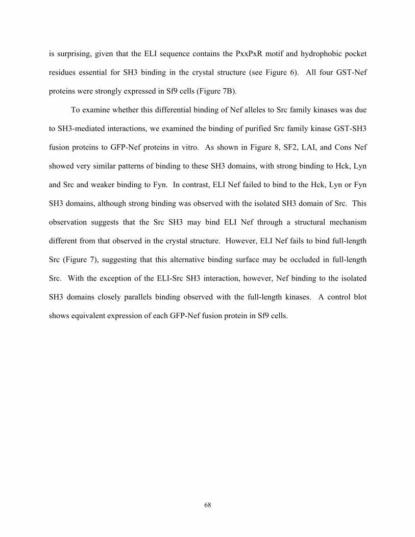
is surprising, given that the ELI sequence contains the PxxPxR motif and hydrophobic pocket
residues essential for SH3 binding in the crystal structure (see Figure 6). All four GST-Nef
proteins were strongly expressed in Sf9 cells (Figure 7B).
To examine whether this differential binding of Nef alleles to Src family kinases was due
to SH3-mediated interactions, we examined the binding of purified Src family kinase GST-SH3
fusion proteins to GFP-Nef proteins in vitro. As shown in Figure 8, SF2, LAI, and Cons Nef
showed very similar patterns of binding to these SH3 domains, with strong binding to Hck, Lyn
and Src and weaker binding to Fyn. In contrast, ELI Nef failed to bind to the Hck, Lyn or Fyn
SH3 domains, although strong binding was observed with the isolated SH3 domain of Src. This
observation suggests that the Src SH3 may bind ELI Nef through a structural mechanism
different from that observed in the crystal structure. However, ELI Nef fails to bind full-length
Src (Figure 7), suggesting that this alternative binding surface may be occluded in full-length
Src. With the exception of the ELI-Src SH3 interaction, however, Nef binding to the isolated
SH3 domains closely parallels binding observed with the full-length kinases. A control blot
shows equivalent expression of each GFP-Nef fusion protein in Sf9 cells.
68
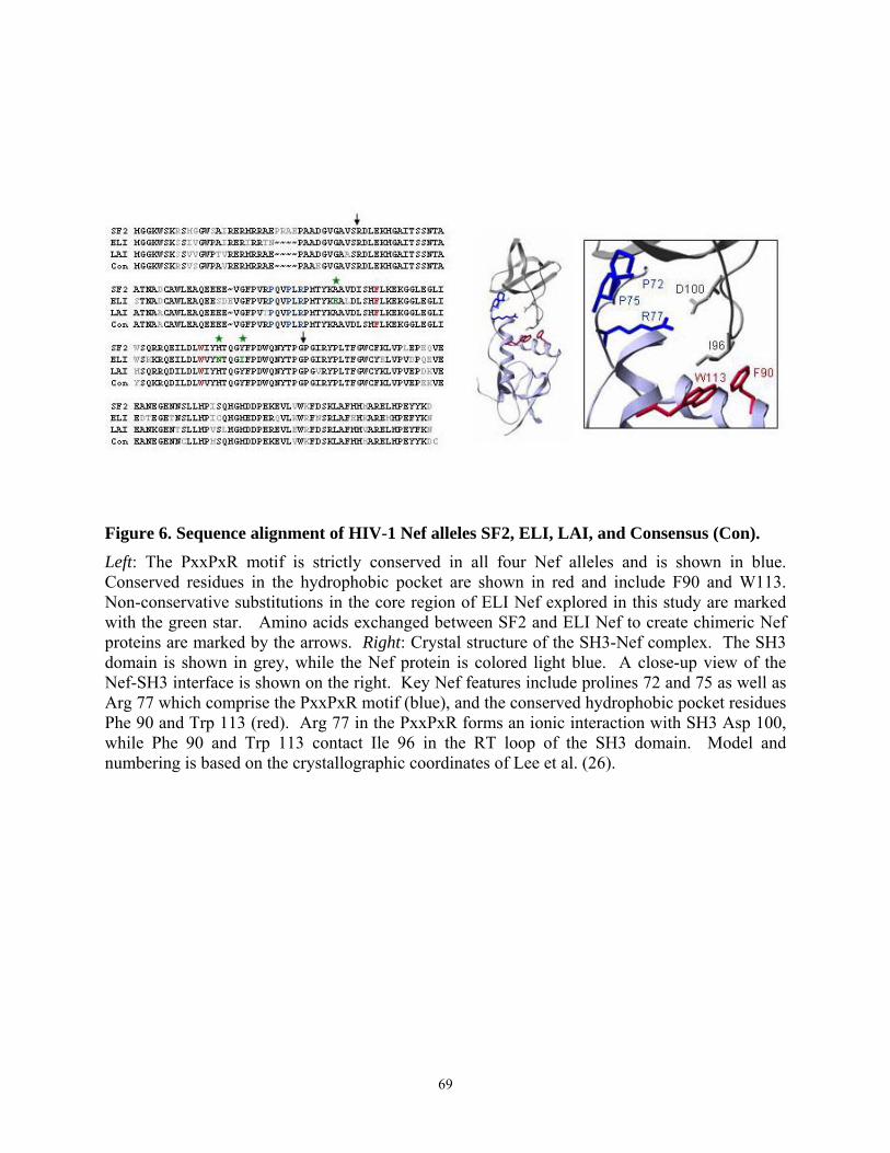
Figure 6. Sequence alignment of HIV-1 Nef alleles SF2, ELI, LAI, and Consensus (Con). Left: The PxxPxR motif is strictly conserved in all four Nef alleles and is shown in blue. Conserved residues in the hydrophobic pocket are shown in red and include F90 and W113. Non-conservative substitutions in the core region of ELI Nef explored in this study are marked with the green star. Amino acids exchanged between SF2 and ELI Nef to create chimeric Nef proteins are marked by the arrows. Right: Crystal structure of the SH3-Nef complex. The SH3 domain is shown in grey, while the Nef protein is colored light blue. A close-up view of the Nef-SH3 interface is shown on the right. Key Nef features include prolines 72 and 75 as well as Arg 77 which comprise the PxxPxR motif (blue), and the conserved hydrophobic pocket residues Phe 90 and Trp 113 (red). Arg 77 in the PxxPxR forms an ionic interaction with SH3 Asp 100, while Phe 90 and Trp 113 contact Ile 96 in the RT loop of the SH3 domain. Model and numbering is based on the crystallographic coordinates of Lee et al. (26).
69
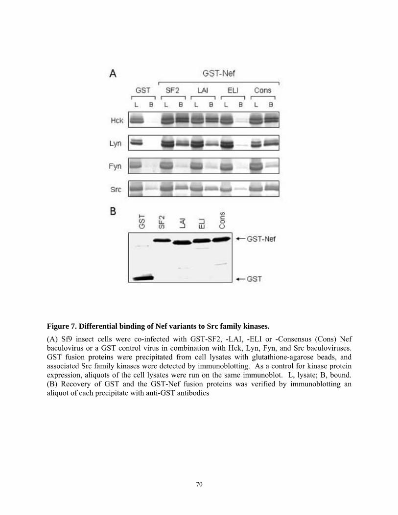
Figure 7. Differential binding of Nef variants to Src family kinases.
(A) Sf9 insect cells were co-infected with GST-SF2, -LAI, -ELI or -Consensus (Cons) Nef baculovirus or a GST control virus in combination with Hck, Lyn, Fyn, and Src baculoviruses. GST fusion proteins were precipitated from cell lysates with glutathione-agarose beads, and associated Src family kinases were detected by immunoblotting. As a control for kinase protein expression, aliquots of the cell lysates were run on the same immunoblot. L, lysate; B, bound. (B) Recovery of GST and the GST-Nef fusion proteins was verified by immunoblotting an aliquot of each precipitate with anti-GST antibodies
70
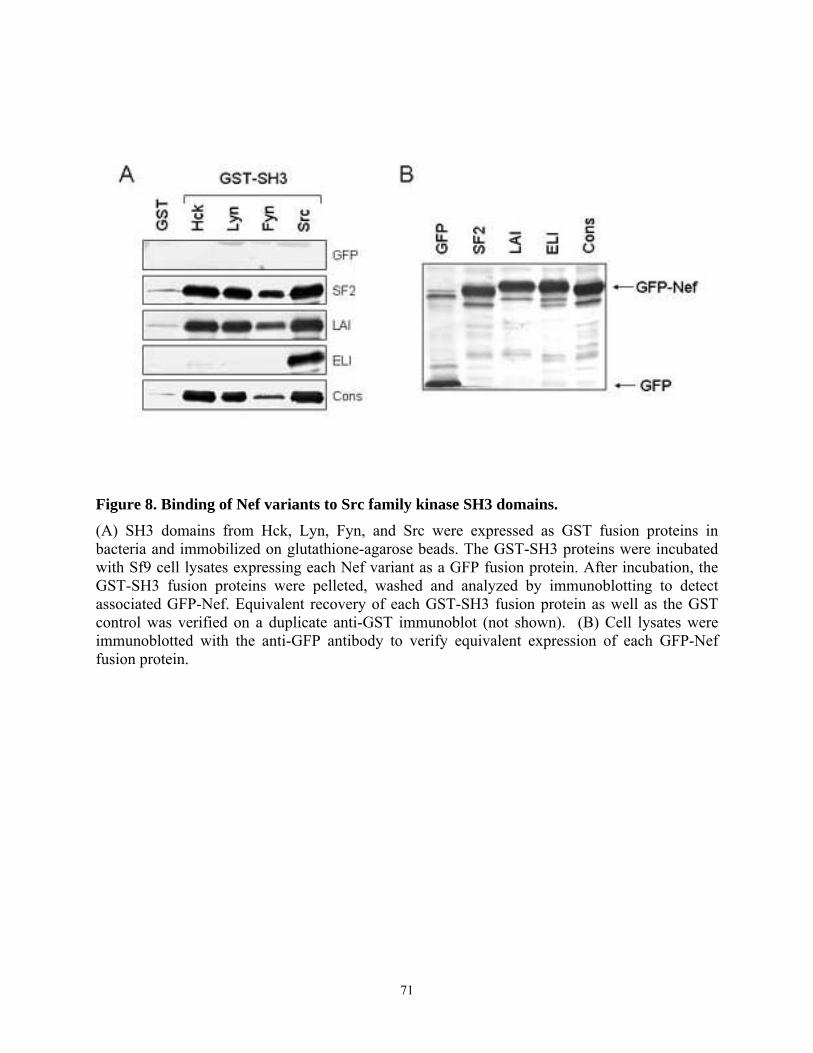
Figure 8. Binding of Nef variants to Src family kinase SH3 domains. (A) SH3 domains from Hck, Lyn, Fyn, and Src were expressed as GST fusion proteins in bacteria and immobilized on glutathione-agarose beads. The GST-SH3 proteins were incubated with Sf9 cell lysates expressing each Nef variant as a GFP fusion protein. After incubation, the GST-SH3 fusion proteins were pelleted, washed and analyzed by immunoblotting to detect associated GFP-Nef. Equivalent recovery of each GST-SH3 fusion protein as well as the GST control was verified on a duplicate anti-GST immunoblot (not shown). (B) Cell lysates were immunoblotted with the anti-GFP antibody to verify equivalent expression of each GFP-Nef fusion protein.
71
3.4.2. Allelic variation in Nef-induced Hck activation in vivo
Previous work from our laboratory has shown that SF2 Nef constitutively activates Hck
kinase through its SH3 domain and induces a strong transformation signal in Rat-2 fibroblasts
(183, 205). Because fibroblasts do not express endogenous Hck, they provide a convenient
model to address the functional consequences of intracellular Nef-Hck interaction. To determine
if other Nef alleles also activate Hck in this system, SF2, LAI, ELI, and Cons Nef were co-
expressed with Hck using recombinant retroviruses. Parallel cultures were infected with the Nef
retroviruses and a control virus expressing GFP for a negative control. Fibroblasts expressing
Hck-2PA, a mutant of Hck activated by substitution of proline residues in the SH2-kinase linker,
served as a positive control (181). As shown in Figure 9A, cells co-expressing SF2 Nef and Hck
produced a similar number of foci as the Hck-2PA mutant, in agreement with our previous
results (181). LAI and Cons Nef also cooperated with Hck to induce foci formation in
fibroblasts, although the number of foci was reduced compared to SF2. The lower activity of
LAI towards Hck may relate to substitution of Thr for Arg at position 71 (immediately N-
terminal to the PxxPxR motif), which has been shown to impact SH3 binding in vitro (204). In
contrast, co-expression of ELI Nef and Hck produced very few transformed foci (less than 10%
of that observed with SF2), consistent with the in vitro binding data (Figures 7 and 8). No
evidence of cellular transformation was observed in cells expressing wild-type Hck alone or
following co-expression of each Nef variant with the GFP control. These results demonstrate that
allelic variation exists in the ability of Nef to activate Hck in a defined system.
We next investigated the interaction of Hck with the Nef variants in Rat-2 fibroblasts. Hck
was immunoprecipitated from cell lysates, and associated Nef was detected by immunoblotting.
As shown in Figure 9B, SF2 Nef was readily detected in Hck immunoprecipitates, while LAI
72
and Cons Nef showed an intermediate level of interaction with Hck in Rat 2 cells. In contrast,
Hck-ELI Nef complexes were not detected in Rat-2 fibroblasts, in agreement with the results of
transformation assay.
We next determined whether the differences in transformation and Hck-Nef complex
formation correlated with Hck kinase activity. Previous work from our laboratory has
demonstrated tyrosine phosphorylation of endogenous proteins following transformation by co-
expression of Hck and Nef as well as constitutively activated Hck mutants (181, 183, 205, 207).
The most prominent of these is a 40 kDa tyrosine phosphoprotein (pp40) which represents a
convenient endogenous transformation-associated marker. As shown in Figure 9B, the level of
p40 tyrosine phosphorylation closely correlates with both focus forming activity and Hck-Nef
complex formation. SF2 Nef strongly activated Hck, while LAI and Cons Nef activated Hck to
an intermediate level. No detectable tyrosine phosphorylation of p40 was observed in fibroblasts
expressing Hck and ELI Nef. Taken together, these results show that Nef proteins from different
HIV-1 strains recruit and activate Hck to different extents despite the high degree of sequence
homology in regions associated with SH3 binding.
73
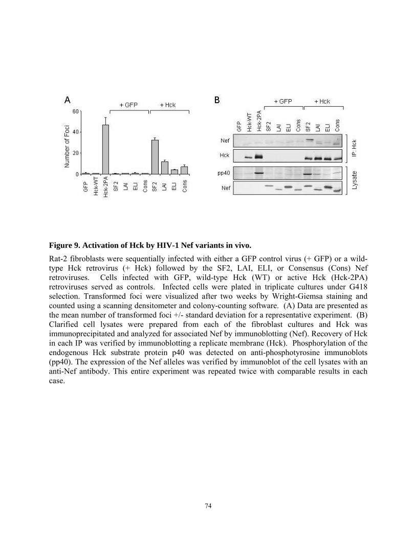
Figure 9. Activation of Hck by HIV-1 Nef variants in vivo. Rat-2 fibroblasts were sequentially infected with either a GFP control virus (+ GFP) or a wild-type Hck retrovirus (+ Hck) followed by the SF2, LAI, ELI, or Consensus (Cons) Nef retroviruses. Cells infected with GFP, wild-type Hck (WT) or active Hck (Hck-2PA) retroviruses served as controls. Infected cells were plated in triplicate cultures under G418 selection. Transformed foci were visualized after two weeks by Wright-Giemsa staining and counted using a scanning densitometer and colony-counting software. (A) Data are presented as the mean number of transformed foci +/- standard deviation for a representative experiment. (B) Clarified cell lysates were prepared from each of the fibroblast cultures and Hck was immunoprecipitated and analyzed for associated Nef by immunoblotting (Nef). Recovery of Hck in each IP was verified by immunoblotting a replicate membrane (Hck). Phosphorylation of the endogenous Hck substrate protein p40 was detected on anti-phosphotyrosine immunoblots (pp40). The expression of the Nef alleles was verified by immunoblot of the cell lysates with an anti-Nef antibody. This entire experiment was repeated twice with comparable results in each case.
74

3.4.3. Interaction of chimeric SF2-ELI Nef proteins with Hck
Results presented in Figure 9 show that ELI Nef fails to bind and activate Hck in vivo.
Inspection of the alignment of the SF2 and ELI Nef sequences shows that the PxxPxR motif
required for SH3 binding is intact, and many of the residues that form the hydrophobic pocket
are also conserved (Figure 6). However, considerable sequence variation exists in the N- and C-
terminal regions, which have been implicated in other studies as having an indirect effect on the
Nef core structure and target protein binding (208, 215). To explore the contribution of N- and
C-terminal Nef sequence variation to Hck binding, we created chimeric forms of Nef using the
SF2 and ELI sequences, which represent two extremes of Hck binding and activation.
In the first experiment, we swapped the C-terminal variable region between SF2 and ELI
Nef. The region exchanged is marked by an arrow in the sequence alignment shown in Figure 6
and the cartoon in Figure 10A. Chimeric Nef proteins were expressed as GST-fusion proteins in
Sf9 insect cells and tested for binding to full-length Hck. As shown in Figure 10B, the chimeric
Nef protein SF2-ELI-C, comprising the N-terminal 131 amino acids of SF2 and the C-terminal
region of ELI, still bound strongly to Hck. In contrast, the complementary mutant (ELI-SF2-C)
showed no detectable Hck binding. These results indicate that C-terminal sequence variation
does not influence Nef-Hck interaction in vitro.
We then created a second pair of chimeric Nef proteins, in which the N-terminal 34 amino
acids were exchanged between SF2 and ELI (Figure 11A) and tested for Hck binding in Sf9
cells. As shown in Figure 11B, fusion of the N-terminal SF2 Nef sequence to ELI (SF2-ELI-N)
did not confer Hck binding activity on ELI Nef. The reciprocal chimera containing the N-
terminal region of ELI fused to the SF2 core and C-terminal regions (ELI-SF2-N) bound to Hck
as efficiently as wild type SF2 Nef. Together with the data in Figure 10, these results
75
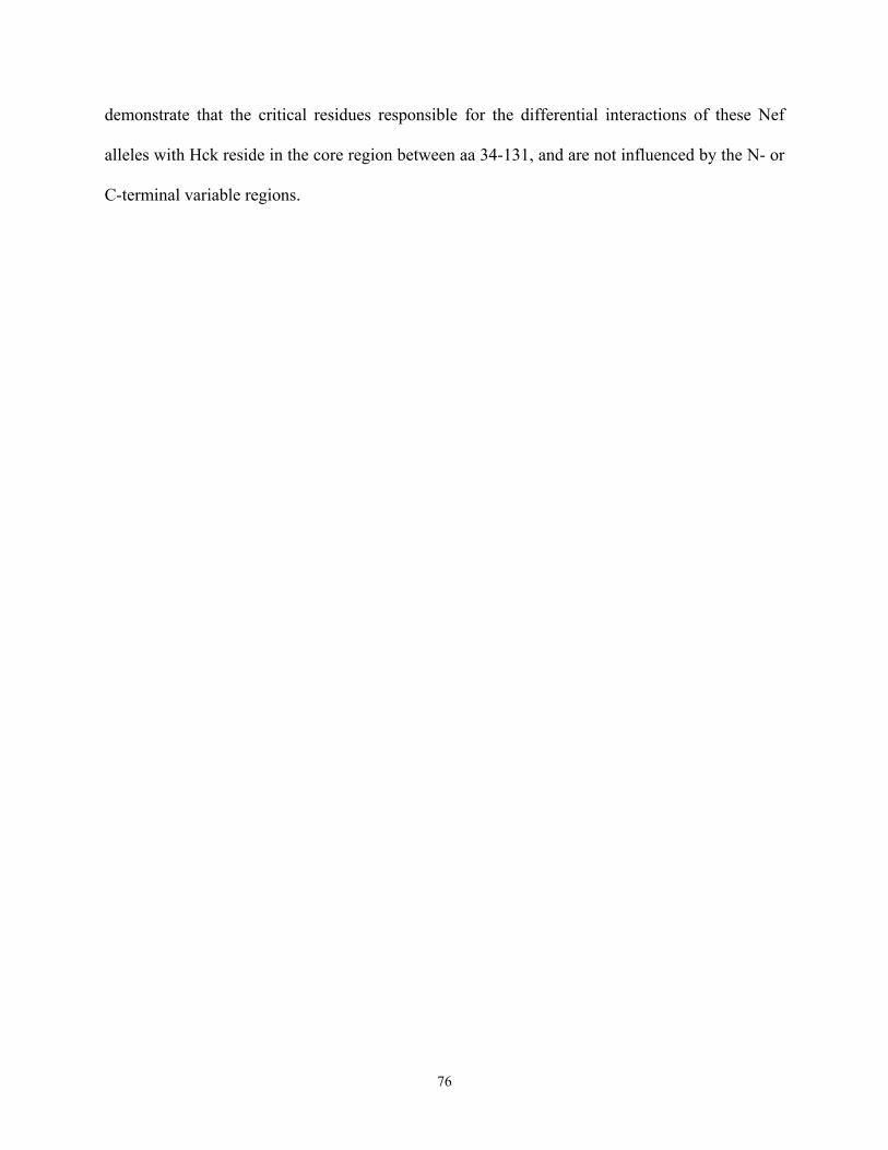
demonstrate that the critical residues responsible for the differential interactions of these Nef
alleles with Hck reside in the core region between aa 34-131, and are not influenced by the N- or
C-terminal variable regions.
76
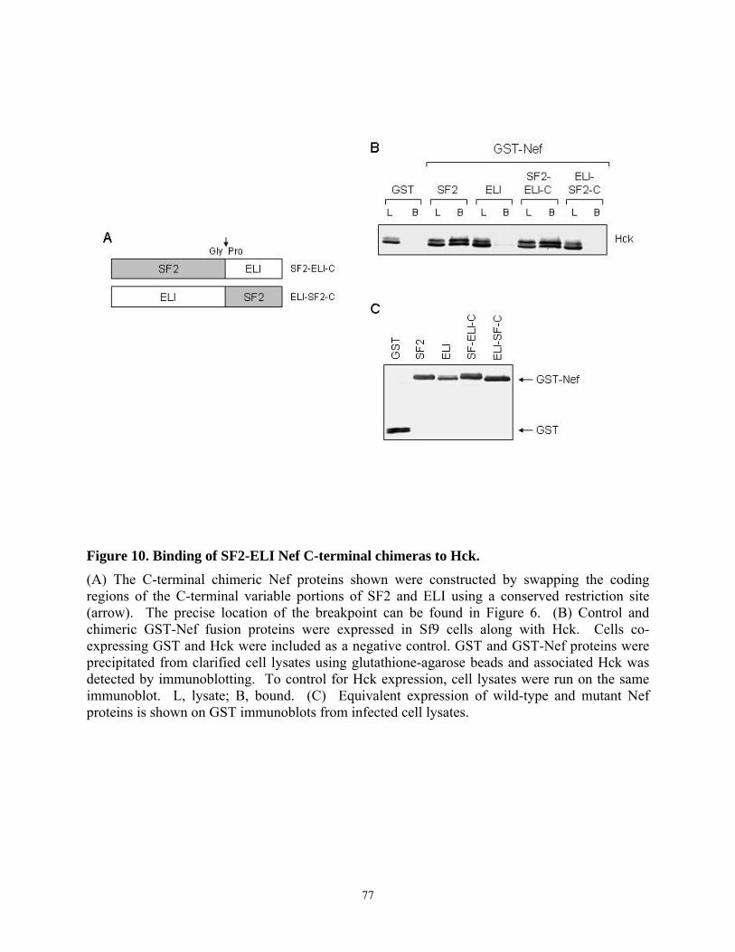
Figure 10. Binding of SF2-ELI Nef C-terminal chimeras to Hck. (A) The C-terminal chimeric Nef proteins shown were constructed by swapping the coding regions of the C-terminal variable portions of SF2 and ELI using a conserved restriction site (arrow). The precise location of the breakpoint can be found in Figure 6. (B) Control and chimeric GST-Nef fusion proteins were expressed in Sf9 cells along with Hck. Cells co-expressing GST and Hck were included as a negative control. GST and GST-Nef proteins were precipitated from clarified cell lysates using glutathione-agarose beads and associated Hck was detected by immunoblotting. To control for Hck expression, cell lysates were run on the same immunoblot. L, lysate; B, bound. (C) Equivalent expression of wild-type and mutant Nef proteins is shown on GST immunoblots from infected cell lysates.
77
Figure 11. Binding of SF2-ELI Nef N-terminal chimeras to Hck. (A) The N-terminal chimeric Nef proteins shown were constructed by swapping the coding regions of the N-terminal variable portions of SF2 and ELI using a conserved restriction site (arrow). The precise location of the breakpoint can be found in Figure 6. (B) Control and chimeric GST-Nef fusion proteins were expressed in Sf9 cells along with Hck. Cells co-expressing GST and Hck were included as a negative control. GST and GST-Nef proteins were precipitated from clarified cell lysates using glutathione-agarose beads and associated Hck was detected by immunoblotting. To control for Hck expression, cell lysates were run on the same immunoblot. L, lysate; B, bound. (C) Equivalent expression of wild-type and mutant Nef proteins is shown on GST immunoblots from infected cell lysates.
78

3.4.4. Tyr 120 is required for high affinity binding of SF2 Nef to Hck in vitro and Hck
activation in vivo
Alignment of the SF2 and ELI Nef core sequences reveals that SF2 Ala 83, His 116, and
Tyr 120 are replaced with Glu, Asn, and Ile in ELI, respectively (Figure 6). Molecular modeling
based on the Nef-SH3 crystal structure shows that two of these amino acids (Ala 83; Tyr 120)
are located in the Nef hydrophobic pocket that contacts Ile 96 of the SH3 domain RT loop
(Figure 12A). In addition, modeling suggests that substitution of Tyr 120 with Ile will remove
contacts with SH3 Ile 96 as a possible explanation for the lack of ELI binding to Hck through its
SH3 domain. Substitution of Ala 83 with Glu increases steric bulk at this position, which may
also interfere with binding to the RT loop. To determine whether Ala 83 and Tyr 120 are crucial
for SF2 Nef binding to Hck, we replaced these residues with the corresponding residues of ELI
Nef (Glu, Ile) and tested for Hck binding in the Sf9 cell system. As shown in Figure 12B,
substitution of Ala with Glu at position 83 did not substantially affect binding of SF2 Nef to Hck.
In contrast, the replacement of Tyr 120 with Ile dramatically reduced complex formation.
Furthermore, when both residues were mutated, the interaction between SF2 Nef and Hck was
completely abolished. This result indicates that Tyr 120 is critical to establish the interaction of
Nef and Hck in vitro, while Ala 83 has a lesser role.
Results shown in Figure 12 demonstrate a requirement for Nef Tyr 120 in Hck binding.
To assess whether this residue is also essential for Hck recruitment and activation in vivo, the
SF2 Nef Tyr 120 mutant (Y120I) was co-expressed with Hck in the fibroblast focus-forming
assay. As shown in Figure 13, this mutation reduced focus-forming activity by more than 90%
compared to wild-type Nef. SF2 Y120I also failed to complex with Hck in fibroblasts and did
79
not stimulate its kinase activity, consistent with the transformation result. These data show that
Tyr 120 in the Nef hydrophobic pocket is essential for Hck recruitment and activation in vivo.
80
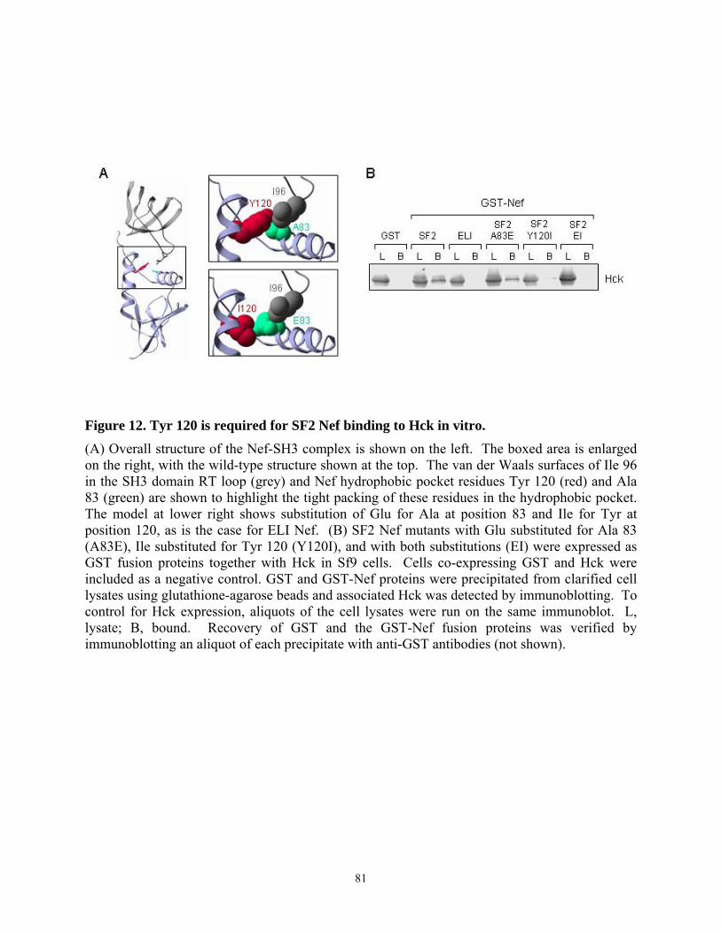
Figure 12. Tyr 120 is required for SF2 Nef binding to Hck in vitro. (A) Overall structure of the Nef-SH3 complex is shown on the left. The boxed area is enlarged on the right, with the wild-type structure shown at the top. The van der Waals surfaces of Ile 96 in the SH3 domain RT loop (grey) and Nef hydrophobic pocket residues Tyr 120 (red) and Ala 83 (green) are shown to highlight the tight packing of these residues in the hydrophobic pocket. The model at lower right shows substitution of Glu for Ala at position 83 and Ile for Tyr at position 120, as is the case for ELI Nef. (B) SF2 Nef mutants with Glu substituted for Ala 83 (A83E), Ile substituted for Tyr 120 (Y120I), and with both substitutions (EI) were expressed as GST fusion proteins together with Hck in Sf9 cells. Cells co-expressing GST and Hck were included as a negative control. GST and GST-Nef proteins were precipitated from clarified cell lysates using glutathione-agarose beads and associated Hck was detected by immunoblotting. To control for Hck expression, aliquots of the cell lysates were run on the same immunoblot. L, lysate; B, bound. Recovery of GST and the GST-Nef fusion proteins was verified by immunoblotting an aliquot of each precipitate with anti-GST antibodies (not shown).
81
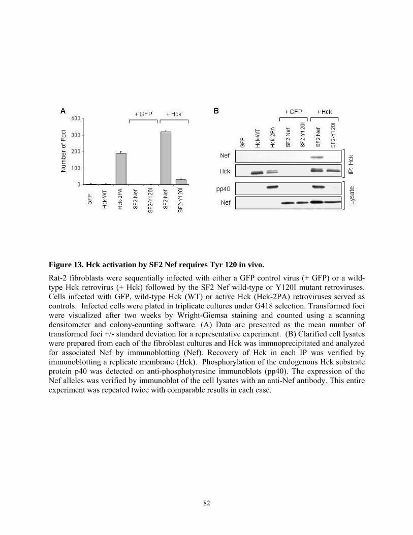
Figure 13. Hck activation by SF2 Nef requires Tyr 120 in vivo. Rat-2 fibroblasts were sequentially infected with either a GFP control virus (+ GFP) or a wild-type Hck retrovirus (+ Hck) followed by the SF2 Nef wild-type or Y120I mutant retroviruses. Cells infected with GFP, wild-type Hck (WT) or active Hck (Hck-2PA) retroviruses served as controls. Infected cells were plated in triplicate cultures under G418 selection. Transformed foci were visualized after two weeks by Wright-Giemsa staining and counted using a scanning densitometer and colony-counting software. (A) Data are presented as the mean number of transformed foci +/- standard deviation for a representative experiment. (B) Clarified cell lysates were prepared from each of the fibroblast cultures and Hck was immnoprecipitated and analyzed for associated Nef by immunoblotting (Nef). Recovery of Hck in each IP was verified by immunoblotting a replicate membrane (Hck). Phosphorylation of the endogenous Hck substrate protein p40 was detected on anti-phosphotyrosine immunoblots (pp40). The expression of the Nef alleles was verified by immunoblot of the cell lysates with an anti-Nef antibody. This entire experiment was repeated twice with comparable results in each case.
82
3.4.5. Multiple substitutions are required to restore ELI Nef interaction with Hck
In a complementary series of experiments, hydrophobic pocket residues Glu 83 and Ile
120 of ELI Nef were substituted with the corresponding amino acids (Ala, Tyr) of SF2 Nef. The
resulting single and double mutants were tested for Hck binding in the Sf9 cell assay. As shown
in Figure 14B, replacement of ELI Ile 120 with Tyr partially reconstituted complex formation
with Hck, whereas substitution of Glu 83 with Ala was without effect. Substitution of both Glu
83 and Ile 120 (AY mutant) also failed to restore interaction completely, suggesting that residues
outside of the hydrophobic pocket may be important. Molecular modeling suggests that amino
acid 116 may have an indirect effect on Hck binding, because this residue is positioned to
influence the flexibility of the PxxP motif (Figure 14A). In SF2 Nef, this position is occupied by
His, but is substituted with Asn in ELI Nef. Asn 116 of ELI Nef was replaced with His in the
WT, I120Y and AY forms of ELI Nef and the resulting mutants were tested for Hck binding
(Figure 14C). The single substitution of Asn with His at position 116 in ELI Nef did not restore
binding to Hck. However, combining the His 116 substitution with the changes at Tyr 120 and
Ala 83 restored the binding of ELI Nef to the same level as SF2, suggesting that position 116,
while not in direct contact with the SH3 domain, can impact Nef-SH3 interaction.
We next investigated whether the replacement of ELI Nef residues Glu 83, Asn 116, and
Ile 120 with the corresponding residues of SF2 (Ala 83, His 116, Tyr 120) was sufficient to
restore Hck recruitment and activation in the Rat-2 fibroblast transformation assay. As shown in
Figure 15A, ELI Nef proteins bearing a single substitution of Ile 120 with Tyr or Asn 116 with
His produced only a small number of transformed foci upon co-expression with Hck in Rat-2
fibroblasts. However, the double mutant of ELI Nef with SF2 substitutions at hydrophobic
pocket positions 83 and 120 (ELI-AY) did cooperate with Hck to transform Rat-2 fibroblasts,
83
producing approximately 40% of the foci observed with SF2 Nef. Addition of the His 116
substitution (AHY triple mutant) increased transforming activity further, to 60% of SF2 levels.
This result is consistent with the binding data and supports a role for His 116 in the regulation of
Hck recruitment and activation in cells.
In a final series of experiments, we correlated transforming activity with Hck-Nef
complex formation and kinase activity for this panel of ELI mutants. As shown in Figure 15B,
complexes of the double (AY) and triple (AHY) ELI Nef mutants with Hck were readily
detected in lysates from Rat-2 cells. Tyrosine phosphorylation of p40 was also observed in cells
transformed by these mutants, consistent with Hck activation. However, the extent of both
complex formation and p40 tyrosine phosphorylation was reduced for these mutants relative to
SF2 Nef, consistent with the results of the focus-forming assay. Taken together, these results
show that sequence variation in the hydrophobic pocket and allosteric effects of other residues
can influence Hck activation in vivo.
84
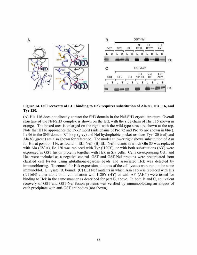
Figure 14. Full recovery of ELI binding to Hck requires substitution of Ala 83, His 116, and Tyr 120. (A) His 116 does not directly contact the SH3 domain in the Nef-SH3 crystal structure. Overall structure of the Nef-SH3 complex is shown on the left, with the side chain of His 116 shown in orange. The boxed area is enlarged on the right, with the wild-type structure shown at the top. Note that H116 approaches the PxxP motif (side chains of Pro 72 and Pro 75 are shown in blue). Ile 96 in the SH3 domain RT loop (grey) and Nef hydrophobic pocket residues Tyr 120 (red) and Ala 83 (green) are also shown for reference. The model at lower right shows substitution of Asn for His at position 116, as found in ELI Nef. (B) ELI Nef mutants in which Glu 83 was replaced with Ala (E83A), Ile 120 was replaced with Tyr (I120Y), or with both substitutions (AY) were expressed as GST fusion proteins together with Hck in Sf9 cells. Cells co-expressing GST and Hck were included as a negative control. GST and GST-Nef proteins were precipitated from clarified cell lysates using glutathione-agarose beads and associated Hck was detected by immunoblotting. To control for Hck expression, aliquots of the cell lysates were run on the same immunoblot. L, lysate; B, bound. (C) ELI Nef mutants in which Asn 116 was replaced with His (N116H) either alone or in combination with I120Y (HY) or with AY (AHY) were tested for binding to Hck in the same manner as described for part B, above. In both B and C, equivalent recovery of GST and GST-Nef fusion proteins was verified by immunoblotting an aliquot of each precipitate with anti-GST antibodies (not shown).
85
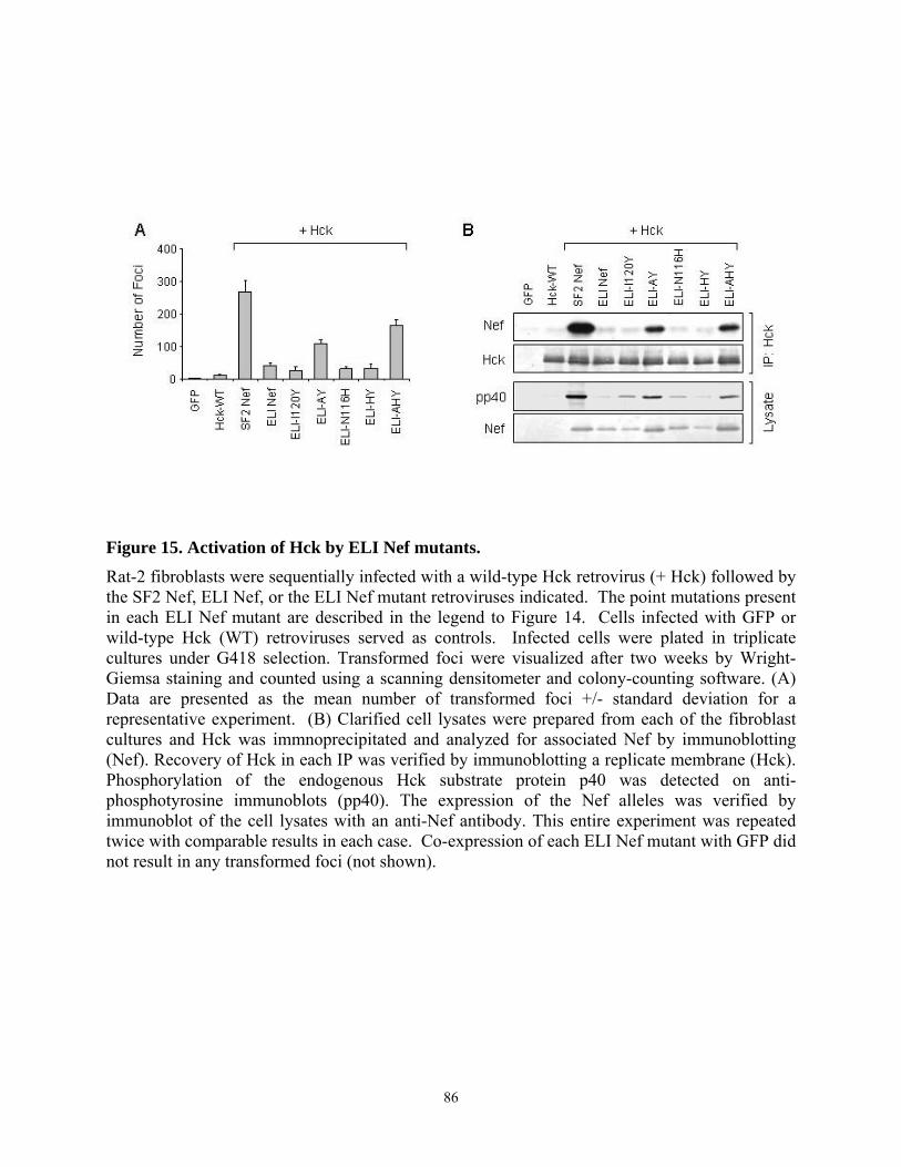
Figure 15. Activation of Hck by ELI Nef mutants. Rat-2 fibroblasts were sequentially infected with a wild-type Hck retrovirus (+ Hck) followed by the SF2 Nef, ELI Nef, or the ELI Nef mutant retroviruses indicated. The point mutations present in each ELI Nef mutant are described in the legend to Figure 14. Cells infected with GFP or wild-type Hck (WT) retroviruses served as controls. Infected cells were plated in triplicate cultures under G418 selection. Transformed foci were visualized after two weeks by Wright-Giemsa staining and counted using a scanning densitometer and colony-counting software. (A) Data are presented as the mean number of transformed foci +/- standard deviation for a representative experiment. (B) Clarified cell lysates were prepared from each of the fibroblast cultures and Hck was immnoprecipitated and analyzed for associated Nef by immunoblotting (Nef). Recovery of Hck in each IP was verified by immunoblotting a replicate membrane (Hck). Phosphorylation of the endogenous Hck substrate protein p40 was detected on anti-phosphotyrosine immunoblots (pp40). The expression of the Nef alleles was verified by immunoblot of the cell lysates with an anti-Nef antibody. This entire experiment was repeated twice with comparable results in each case. Co-expression of each ELI Nef mutant with GFP did not result in any transformed foci (not shown).
86
3.4.6. Analysis of HIV-1 Nef alleles from long-term non-progressors (LTNPs) reveals
sequence heterogeneity at amino acid positions 83, 116, and 120
To begin to approach the clinical relevance of our findings, we asked the question of
whether the Nef amino acids at positions 83, 116 and 120 showed heterogeneity in full-length
Nef isolates from HIV-1-infected individuals that do not progress to clinical disease (long-term
non-progressors or LNTPs). A search of the literature revealed five studies reporting full-length
Nef sequences from LTNPs, and a total of 60 sequences were analyzed for substitutions at
positions 83, 116 and 120. As shown in Table 1, sequence variation was observed at all three of
these Nef residues. Position 83 showed the greatest variation, with Ala (50%) or Gly (47%)
occupying this position in the majority of the variants. Interestingly, isolates from one patient
showed Glu at this position, as observed for ELI Nef, while another was substituted with Ser. In
the Nef-SH3 crystal structure, Ala 83 packs against Tyr 120 and influences its orientation
(Figure 12). Substitution of this position with Gly may make this residue more flexible and
affect its influence on interaction with Ile 96 or other SH3 residues. At position 116, 17% of
LTNPs showed substitution of Asn for His, which was also observed for ELI and found to
influence Hck binding and activation even though it does not contact SH3 directly in the crystal
structure (Figure 14). Tyr was strongly conserved at position 120 (88%), although conservative
substitution with Phe was observed in 12% of the LTNPs. Whether or not Phe substitution at
this postion will impact binding or recruitment of Hck through its SH3 domain will require
further analysis. Note that all sixty sequences had a consensus PxxPxR motif as well as
hydrophobic pocket residues Phe 90 and Trp 113, which are essential components of Hck SH3
binding (Figure 6).
87
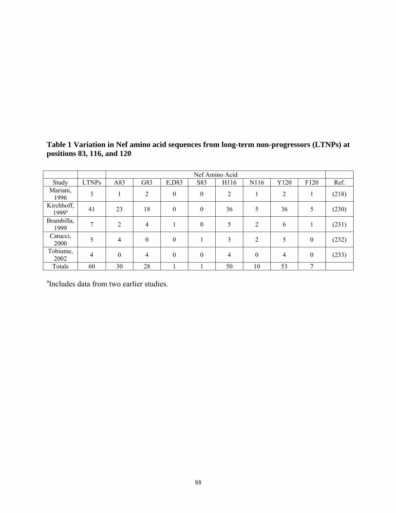
Table 1 Variation in Nef amino acid sequences from long-term non-progressors (LTNPs) at positions 83, 116, and 120
Nef Amino Acid Study LTNPs A83 G83 E,D83 S83 H116 N116 Y120 F120 Ref.
Mariani, 1996 3 1 2 0 0 2 1 2 1 (218)
Kirchhoff, 1999a 41 23 18 0 0 36 5 36 5 (230)
Brambilla, 1999 7 2 4 1 0 5 2 6 1 (231)
Catucci, 2000 5 4 0 0 1 3 2 5 0 (232)
Tobiume, 2002 4 0 4 0 0 4 0 4 0 (233)
Totals 60 30 28 1 1 50 10 53 7
aIncludes data from two earlier studies.
88
3.5. Discussion
A growing body of evidence supports a central role for the Hck-Nef complex in AIDS
progression. Recently Komura et al. have shown that Hck is required for M-tropic HIV
replication in cultured macrophages (203, 215). AIDS-like disease induced by expression of Nef
in a transgenic mouse model also required Hck (202), identifying the Hck:Nef complex as a
target for anti-HIV therapy. Nef interacts with Hck through its SH3 domain, and key residues
involved in the SH3-Nef interface have been identified by structural analysis (26, 28). The
functional relevance of residues outside of the PxxP motif that contribute to Nef-SH3 complex
formation and their impact on Hck activity and signal transduction have not been evaluated. Our
study is the first to compare allelic variants of HIV-1 Nef in terms of Hck binding and activation
in vivo, and to examine the contribution of residues outside of the conserved PxxP motif in these
molecular events. We found that not all HIV-1 Nef alleles have a similar affinity for Hck or
other Src family kinases, despite strong conservation of the PxxPxR motif and hydrophobic
pocket residues identified in the crystal structure as part of the SH3 interface. These
observations suggest that simply aligning HIV-1 Nef sequences may not be predictive of effects
on Hck or other Src family kinases in vivo.
In the first set of experiments, we compared the binding of several well-studied laboratory
Nef alleles to Hck and other Src family kinases using a Sf9 insect cell co-expression system.
This approach allowed us to compare binding interactions in a background devoid of endogenous
Src family kinases or other proteins that may compete for binding. All four of the Nef proteins
tested in these experiments (SF2, ELI, LAI and Consensus) have conserved PxxP motifs and
hydrophobic pocket residues predicted in the crystal structure to contribute to high affinity
binding to the Hck SH3 domain (Figure 6). SF2, LAI and Cons Nef proteins all bound strongly
to Hck and Lyn (Figure 7), which are unique among Src family kinases in that their SH3
89
domains have an Ile residue at position 96 in the RT loop. Previous studies have identified this
residue as a critical determinant of high affinity Nef-SH3 complex formation (206, 208, 215).
Binding of these three isolates to c-Src was moderate, while Fyn binding was weak, consistent
with the lack of the key RT loop Ile residue in their SH3 domains. In contrast, ELI Nef failed to
bind to Hck or any of the other Src family kinases examined despite the presence of a PxxP motif
and conserved hydrophobic pocket residues Phe 90 and Trp 113. This difference was due to
substitution of additional residues in the Nef hydrophobic pocket (particularly at position 120)
and elsewhere in the core region (see below).
Binding of each of the Nef isoforms to the isolated SH3 domain of these Src family
kinases was consistent with the result of Nef binding to the corresponding full-length proteins in
most cases (Figure 8). The one exception was ELI Nef, which bound strongly to the c-Src SH3
domain but not to full-length c-Src. This finding suggests that other regions of Src may
negatively affect the interaction of its SH3 domain with ELI Nef. One possibility is that the
SH2-kinase linker region competes with ELI Nef for SH3 binding. Another possibility is that
interaction of SH3 with other regions of the Src protein affects the conformation of SH3 domain,
which in turn influences the stability of the SH3:ELI Nef complex. This observation is
consistent with our earlier finding that SH3-Nef binding interactions in vitro are not always
predictive of Nef interactions with full-length Src family kinases in vivo (207).
Among the Nef hydrophobic pocket residues examined in the present study, Tyr 120
appears to be most important for high affinity binding to Hck. In the co-crystal structure of the
high-affinity Nef-SH3 complex, Tyr 120 contributes to a network of hydrogen bonds between
the hydrophobic pocket and the SH3 domain RT loop (26). This residue is 90% conserved across
HIV-1, -2 and SIV and 98% conserved within HIV-1 Nef (223). Mutation of Tyr 120 to Ile
90
nearly abolished the interaction of SF2 Nef with Hck in vitro and in vivo (Figures 12 and 13),
supporting the functional importance of this residue in binding and activation of Hck. This
finding is also important from a drug discovery perspective, as it supports the prediction that
targeting the hydrophobic pocket with a small molecule may be sufficient to disrupt Nef-SH3
interaction (26).
Although Tyr 120 is required for SF2 Nef interaction with Hck, replacement of Ile 120 of
ELI Nef with Tyr restored only partial Hck binding in vitro and failed to restore cooperation with
Hck in the fibroblast transformation assay. However, substitution of positions 83, 116, and 120
of ELI with the corresponding residues of SF2 Nef restored binding and biological activities to
60% of those observed with SF2 Nef (Figure 14 and 15). Substantial though incomplete
activation by the triple mutant relative to SF2 suggests that other sequence variations either
directly or indirectly govern binding and activation.
While our study focused on differences in residues within or close to the hydrophobic
pocket, several other sequence differences exist between the SF2 and ELI Nef core regions that
may affect Hck binding. One possibility is a serine inserted within the ELI Nef acidic cluster
(EEEE; Figure 6). This motif has been suggested to mediate Nef binding to PACS-1, a molecule
that controls intracellular protein trafficking from the endosome to the Golgi (234). Nef-PACS-1
interaction leads to the retention of MHC class I in the trans-Golgi, resulting in downregulation
of MHC I from the cell surface (235, 236), an event that is dependent upon the Nef PxxP motif.
Although this acidic cluster is not directly involved in the interaction of Nef and Src family
kinases, this insertion may affect the conformation of Nef, which in turn may influence binding
to Hck.
91

In contrast to the Nef core region, there is considerable sequence variation in the N- and
C-terminal regions (Figure 6), which may contribute to differences in interactions with Src
family kinases. The N-terminal flexible region or C-terminal disordered loop could interact with
the core domain, and thus conceal or expose PxxP and other motifs responsible for Src family
kinase recruitment. Contact with other binding partners could also trigger conformational
changes, indirectly affecting the complex stability. For example, the presence of the flexible N-
terminal arm has been shown to influence binding to SH3 domains in vitro (208, 215).
Nevertheless, we were unable to show an effect of the ELI N- or C-terminal regions on high
affinity binding of SF2 Nef to Hck (Figures 10 and 11). Conversely, replacing the N- or C-
terminal regions of ELI Nef with those of SF2 failed to restore binding, suggesting that the core
region of Nef has the dominant role in the interaction with Hck.
Other studies have shown that Nef proteins derived from HIV-1, HIV-2 or SIV bind to Src
family kinases through distinct mechanisms (237). For example, HIV-1 Nef preferentially binds
to Hck among other Src family kinases via the SH3-dependent mechanism described above (204,
207, 238), whereas HIV-2 Nef has a much weaker affinity for Hck (238, 239). SIV Nef binds to
Src much more strongly than to Hck. The interaction of SIV Nef and Src appears to be
predominantly SH3-independent, as the PxxP motif of SIV Nef is dispensable for Src binding
(237, 238). These observations suggest that although the interaction of Nef proteins of divergent
HIV/SIV strains with Src family kinases appears to be a conserved Nef function, the specific
molecular mechanisms responsible for the interactions differ among these strains.
Our data may help to explain reports of Nef sequences from long-term non-progressors
(LTNPs) with intact Nef alleles. Analysis of Nef sequences from 60 such individuals revealed
two remarkable features. First, all 60 had intact PxxPxR motifs as well as Phe 90 and Trp 113,
92
which are essential for SH3 binding in the crystal structure. Second, sequence variations were
observed at positions 83, 116 and 120 (Table 1), the same residues that accounted for most of the
inactivity of ELI Nef towards Hck in the fibroblast assay (Figures 14 and 15). These
observations further support the idea that sequence variations outside of the core motifs involved
in SH3 binding may have an impact on interaction with Hck and disease progression as a
consequence. Future studies will address this question in the context of a whole animal model of
AIDS.
93
4. Chapter IV
HIV-1 Nef Promotes Survival of TF-1 Macrophages by Inducing Bcl-XL Expression in an Erk-dependent Manner
Hyun-Jung Choi and Thomas E. Smithgall
Department of Molecular Genetics and Biochemistry, University of Pittsburgh School of Medicine, E1240 Biomedical Science Tower, Pittsburgh, PA 15261
This work has been accepted for publication in Journal of Biological Chemistry
94
4.1. Abstract
The Nef protein of HIV-1 is essential for the progression from human and simian
immunodeficiency virus (HIV/SIV) infection to full-blown AIDS. Recent studies indicate that
Nef generates anti-apoptotic signals in HIV-infected T-cells, suppressing cell death early in
infection to allow productive viral replication. Previous work from our laboratory has shown
that Nef also promotes proliferation of myeloid cells through a Stat3-dependent pathway. Here
we demonstrate that Nef suppresses cell death induced by cytokine deprivation in the human
macrophage precursor cell line, TF-1. Nef selectively induced up-regulation of Bcl-XL, an anti-
apoptotic gene that is also regulated by GM-CSF in this cell line. Activation of the Erk MAPK
pathway also correlated with the survival of TF-1/Nef cells. Using the selective Mek inhibitor
PD98059, we found that Nef-induced Erk signaling is essential for Bcl-XL up-regulation and cell
survival. In contrast, expression of Bcl-XL and TF-1 survival was not affected by dominant-
negative Stat3. These data suggest that Nef produces survival signals in myeloid cells through
Erk-mediated Bcl-XL induction, a pathway distinct from Nef survival pathways recently reported
in T-lymphocytes.
95
4.2. Introduction
Nef is an accessory protein unique to human and simian immunodeficiency viruses (HIV-
1, HIV-2, and SIV), and is an essential AIDS progression factor (216, 240). Nef plays a crucial
role in the maintenance of high viral loads and subsequent development of AIDS-like disease in
rhesus monkeys infected with SIV (16). Similarly, long-term survivors of AIDS are often
infected with Nef-defective HIV strains (17, 18). Recent development of a transgenic mouse
model for AIDS demonstrated that expression of Nef alone is sufficient to cause AIDS-like
pathology (66, 202). Although these and other studies implicate Nef as a major progression
factor in AIDS, the mechanism by which it contributes to HIV replication and pathogenesis is
still unclear.
A 25-30 kDa cytosolic protein with an N-terminal myristoylation signal, Nef has no
known catalytic activity. Instead, Nef associates with diverse cellular proteins and modifies their
functions in host cells (216, 217). Nef downmodulates cell-surface CD4 and major
histocompatibility complex I (MHC I) receptors, which contributes to evasion of host immune
surveillance. Protein kinases and other signaling molecules have also been found to interact with
Nef. For example, Nef binds directly to multiple Src family kinases, including Hck, Lck and Fyn
(217, 241). Genetic evidence suggests that Nef-Hck interaction may be essential for AIDS
progression (202). Nef also interacts with several Ser/Thr kinases, including Pak, PKC, and the
Erk MAPKs (217, 241). Nef was shown to increase Erk1/2 activity initiated by T cell receptor
stimulation in primary CD4+ T cells obtained from peripheral blood (160). Enhanced Erk
activity may be responsible for increased T cell activation by Nef, which facilitates HIV
replication in this target cell type. Erk is also involved in induction of the AP-1 transcription
factor by Nef in macrophages, another major target cell for HIV infection (242). The induction
96
of AP-1 may contribute to the activated phenotype of Nef-transfected and HIV-1-infected
macrophages.
Several recent reports have suggested that Nef plays a role in longevity of HIV infected T-
cells by affecting mediators of apoptosis. Death receptor mediated apoptosis, which is critical to
the host cell immune reaction, is blocked by Nef through ASK1 (Apoptosis signal regulating
kinase 1) inhibition (112). In addition, Nef suppresses T-cell apoptosis initiated by serum
starvation or HIV replication. In this case, Nef was shown to induce serine phosphorylation of
Bad, the mitochondrial pro-apoptotic mediator, through a previously described p21-activated
protein kinase (Pak) known to associate with Nef (114).
Recent work from our laboratory has shown that Nef promotes cytokine independent
proliferation of the macrophage precursor cell line, TF-1, through a mechanism that requires the
Stat3 transcription factor (118). In the present report, we show that Nef suppresses apoptosis in
this cell line by selectively up-regulating the mitochondrial anti-apoptosis gene, Bcl-XL (116).
Experiments with pharmacological inhibitors indicate that cell survival and Bcl-XL induction by
Nef are dependent on the Erk MAPK pathway, but independent of PI3K and Akt activation.
Surprisingly, dominant-negative Stat3 did not impact Nef-induced cell survival or Bcl-XL
induction, despite the identification of Bcl-XL as a Stat3 target gene in other systems (243, 244).
These results show for the first time that Nef generates anti-apoptotic signals in cells of the
myelomonocytic lineage, through a pathway distinct from that observed in T-cells.
97
4.3. Materials and methods
4.3.1. Materials
The anti-Nef antibody (EH-1, mouse monoclonal) was generously provided by the NIH
AIDS Research and Reference Reagent Program. Anti-Erk1/2 and anti-phosho-Erk antibodies
were purchased from Santa Cruz Biotechnology. The anti-Stat3 antibody, anti-Akt and phospho-
Akt antibodies were obtained from Cellular Signaling. The anti-pStat3 antibody to p-Tyr 705
was purchased from Upstate Biotechnology. The Mek-specific inhibitor, PD98059, and the PI3K
inhibitor, LY294002, were purchased from Calbiochem.
4.3.2. Cell Culture
The human myeloid leukemia cell line TF-1 (245)was obtained from the American Type
Culture Collection (ATCC) and grown in RPMI 1640 medium supplemented with 10% fetal
bovine serum, 50 µg/ml gentamycin, and 1 ng/ml human GM-CSF (Calbiochem).
4.3.3. Nef, Nef-ER, and Stat3YF expression constructs
To create the Nef-ER fusion construct, the coding sequence of HIV-1 Nef (SF2 strain)
was amplified by PCR and subcloned into the mammalian expression vector, pCDNA3.1
(InVitrogen). The coding sequence of the murine ER ligand-binding domain (amino acids 281 to
599) was PCR-amplified from the plasmid pANMerCreMer (246). A point mutation in this ER
sequence (G525R) abrogates estrogen-binding activity while retaining high affinity for the
synthetic estrogen 4-hydroxytamoxifen (4-HT) (247). The ER segment was subcloned
downstream and in-frame of the Nef C-terminus to generate pCDNA3.1-Nef-ER. Nef and Nef-
ER constructs were subcloned into the retroviral expression vector pSRαMSVtkneo, which
98
carries a G418 resistance marker (248). The resulting constructs were used to generate high-titer
stocks of recombinant retroviruses by co-transfection of 293T cells (249) with an amphotropic
packaging vector as described elsewhere (118). The Stat3 dominant-negative mutant carries a
Phe substitution for Tyr 705 in the C-terminal tail. A pSRαMSVtkneo construct containing this
mutant was used to prepare recombinant retrovirus for infection of TF-1/Nef-ER cells as
described elsewhere (118).
4.3.4. Cell lysis and Western blot analysis
TF-1 cells were lysed in modified RIPA buffer (50 mM Tris-HCl, pH 7.4, 50 mM NaCl, 1
mM EDTA, 10 mM MgCl2, 1% Triton X-100, 1% sodium deoxycholate, 0.1% SDS)
supplemented with the protease inhibitors aprotinin (25 µg/ml), leupeptin (25 µg/ml) and PMSF
(1 mM) and the phosphatase inhibitors NaF (10 mM), and Na3VO4 (1 mM). TF-1 cell lysates
were clarified by centrifugation at 13,000 rpm for 10 min at 4°C. To detect phosphorylation of
Erk and Akt, cell lysates were immunoprecipitated with anti-Erk1, Erk2, or Akt antibodies,
respectively. Immunoprecipitates were resolved on 10% SDS-polyacrylamide gels, transferred
to PVDF membranes and blotted with phosphospecific antibodies to the active forms of Erk1/2
or Akt. Immunoreactive bands were visualized with secondary antibodies conjugated to alkaline
phosphatase with NBT-BCIP as colorimetric substrate (Southern Biotechnology Associates) or
with the CDP star detection system (Tropix).
99
4.3.5. Apoptosis Assay
Apoptosis was assessed as cell-surface Annexin V-FITC binding according to
manufacturer's protocol (BD Pharmingen). Briefly, 106 cells were centrifuged at 1,000 x g for 5
min, and washed twice with cold PBS. Cells were then resuspended in Annexin V binding
buffer and 105 cells were pelleted and incubated with 5 µl Annexin V-FITC and 5 µl propidium
iodide (50 µg/ml) in a final volume of 100 µl for 15 min. Apoptosis was measured using a FACS
Calibur flow cytometer (Becton-Dickinson) set for two-color acquisition, and data were analyzed
using CellQuest software. For inhibitor experiments, cells were treated with 30 µM PD98059
(250), 10 µM LY294002 (251) or 0.15% DMSO vehicle control for 16 h at 37° C prior to
Annexin V-FITC staining and flow cytometry.
4.3.6. RNase protection assay
TF-1 cells (107) were treated for 16 h with either 30 µM PD98059 or 10 µM LY294002.
Following incubation, total cellular RNA was isolated from each cell line using the Totally RNA
kit (Ambion Inc.) according to the manufacturer's instructions. To measure expression of Bcl-
XL, a multi-probe set containing cDNA template of nine Bcl-2 family members as well as
GAPDH and L32 as internal controls was utilized according to the manufacturer's protocol (BD
Biosciences/Pharmingen). Briefly, 32P-labeled riboprobes of defined length were generated
using T7 RNA polymerase and 50 ng of the DNA template in the presence of 150 µCi of [α-32P]
UTP (New England Nuclear). Template DNA was digested with RNase-free DNase, followed by
precipitation of labeled RNA. Five µg of total cellular RNA was mixed with 6.2 x 10⁵ cpm of the
32P-labeled riboprobe in hybridization buffer (40 mM PIPES, 1 mM EDTA, 0.4 M NaCl, and
80% formamide) and incubated for 5 min at 90o C followed by 12 h at 56o C. The hybridized
100
RNA duplexes were then treated with RNase A and RNase T1, followed by proteinase K
digestion. RNase-resistant RNA duplexes were extracted with phenol and precipitated by the
addition of equal volumes of 4 M ammonium acetate and 2 volumes of ethanol. Labeled RNA
samples were resolved on 6% urea denaturing gels and visualized by autoradiography, and the
relative RNA signals were quantitated by densitometry. The relative expression level of Bcl-XL
was normalized to that obtained with the GAPDH control for each sample.
4.3.7. Detection of Nef-ER dimers
TF-1/Nef-ER cells (106) were treated for 24 h with or without 1 µM 4-HT, followed by
incubation in the presence or absence of GM-CSF for 16 h. For crosslinking, cells were washed
twice with PBS and incubated in 2 mM DSS for 30 min at room temperature. The reaction was
terminated by adding 1M Tris⋅HCl (pH 7.5) and incubating for 15 min. The cells were washed
once with PBS and then lysed in 200 µl lysis buffer (50 mM Tris⋅HCl, pH 7.4, 50 mM NaCl, 10
mM MgCl2, 1 mM EDTA, 1% Triton X-100, 25 µg/ml aprotinin, 50 µg/ml leupeptin, 0.5 mM
PMSF, 20 mM NaF, and 1 mM Na3VO4). The protein concentration of the cell lysate was
determined using the Coomassie Plus Protein Assay Reagent (Pierce) and bovine serum albumin
as standard. Cell lysates containing 30 µg of protein were resolved on 10 % SDS-
polyacrylamide gels, transferred to PVDF membranes, and blotted with a Nef antibody.
4.3.8. Electrophoretic Mobility Shift Assay
TF-1 cells (107) were centrifuged, washed once with PBS, and nuclear extracts were made
using the Nuclear Extract Kit (Active Motif) and the manufacturer’s protocol. The protein
concentrations of nuclear extracts were determined using BCA protein assay reagents (Pierce)
101
and bovine serum albumin as standard. The probe used for the Stat3 gel-shift assay is based on
the sis-inducible element (SIE) (118). Complementary SIE oligonucleotides (20 pmol) were
annealed in 10 µl of TE buffer (10 mM Tris-HCl, pH 8.0, 1 mM EDTA) and labeled by
combining 1 µl of annealed oligonucleotide with 4 µl of Labeling Mix-dATP (Pharmacia), 2 µl
of [α-32P] dATP (10 mCi/ml; Amersham), and 1 µl of the Klenow fragment of DNA polymerase
(Gibco-BRL, 3.7 units/µl) in a final volume of 20 µl. Gel-shift reactions contained 10 µg of TF-
1 cell nuclear extract and 40,000 cpm of labeled SIE probe in a final volume of 20 µl as
described elsewhere (222). The mixture was incubated at 37° C for 1 h and unincorporated
nucleotides were removed using a G-25 Sephadex spin column (Millipore). Reactions were
incubated at 30° C for 30 min and run on 5 % polyacrylamide gels in 0.25X Tris-Borate-EDTA
buffer. Gels were fixed with 10% acetic acid/10% methanol, rinsed with water, dried and
exposed to BioMax film (Kodak).
102
4.4. Results
4.4.1. HIV-1 Nef inhibits apoptosis induced by cytokine deprivation in the human
myeloid leukemia cell line, TF-1
Previous work from our laboratory has shown that HIV-1 Nef induces cytokine-
independent proliferation of the human macrophage precursor cell line, TF-1 (118). This cell
line is dependent on GM-CSF for growth, and undergoes apoptosis following cytokine
withdrawal. To investigate whether the GM-CSF-independent proliferation induced by Nef
results from suppression of apoptosis in TF-1 cells, we first expressed HIV-1 Nef in TF-1 cells
by retroviral transduction. Cells were then plated in the presence or absence of GM-CSF, and
apoptosis was measured as cell-surface Annexin V staining four days later. As shown in Figure
16, 60% of the control cells infected with empty vector underwent apoptosis upon GM-CSF
withdrawal, demonstrating the cytokine requirement for survival of this cell line. In contrast,
only 25% of TF-1 cells expressing Nef underwent apoptosis following GM-CSF withdrawal,
demonstrating that Nef suppresses apoptosis induced by cytokine deprivation in cells of myeloid
lineage.
Although only 25% of TF-1/Nef cells underwent apoptosis following GM-CSF
withdrawal, this number was reduced to 10% in the presence of GM-CSF (Figure 16). However,
immunofluorescent staining of the Nef-expressing cell population and flow cytometry revealed
that only 60-70% of the cells express detectable levels of the Nef protein (data not shown). This
heterogeneity in Nef expression in the TF-1 cell population may account for the incomplete
survival effect. As described in a later section, experiments with clonal populations of TF-1 cells
expressing a conditional form of Nef show responses equivalent to GM-CSF.
103
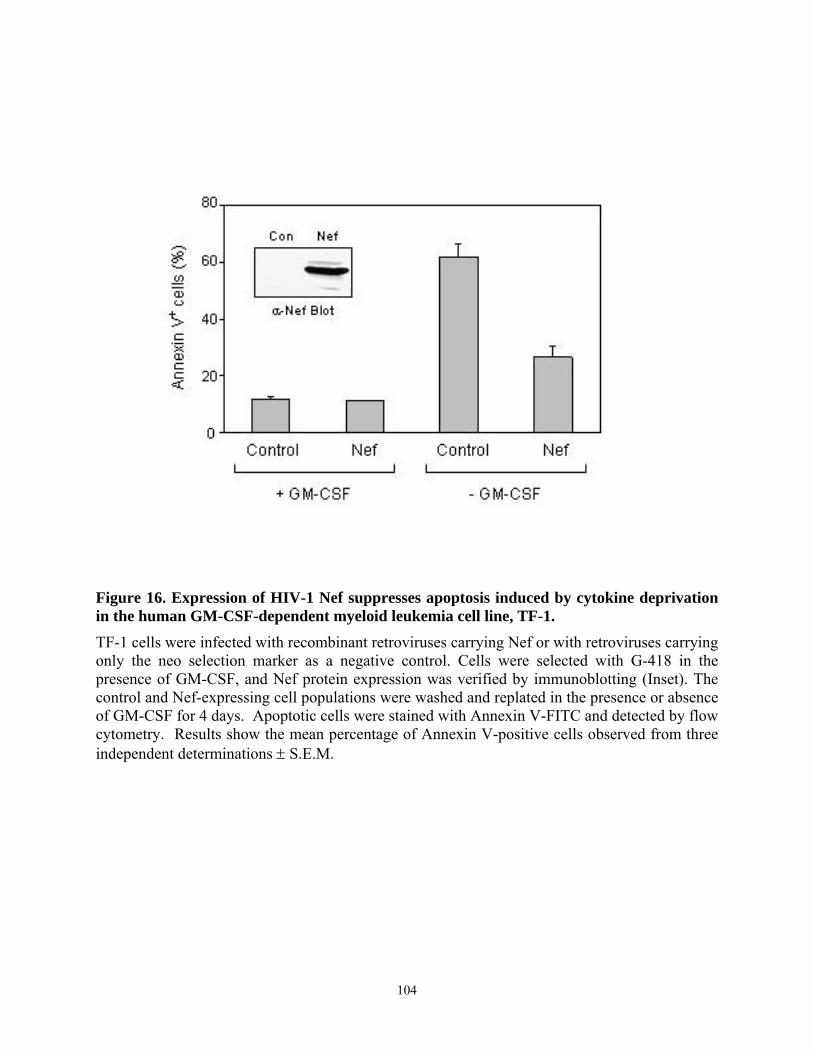
Figure 16. Expression of HIV-1 Nef suppresses apoptosis induced by cytokine deprivation in the human GM-CSF-dependent myeloid leukemia cell line, TF-1. TF-1 cells were infected with recombinant retroviruses carrying Nef or with retroviruses carrying only the neo selection marker as a negative control. Cells were selected with G-418 in the presence of GM-CSF, and Nef protein expression was verified by immunoblotting (Inset). The control and Nef-expressing cell populations were washed and replated in the presence or absence of GM-CSF for 4 days. Apoptotic cells were stained with Annexin V-FITC and detected by flow cytometry. Results show the mean percentage of Annexin V-positive cells observed from three independent determinations ± S.E.M.
104
4.4.2. Induction of Bcl-XL expression in TF-1 cells expressing Nef
Nef-induced proliferation of TF-1 cells requires the Stat3 transcription factor (118), which
has been implicated in many growth factor and cytokine signaling pathways controlling cell
proliferation, differentiation and survival (252). Stat3-mediated survival signaling has been
linked to Bcl-XL expression in tumor cells and other systems (243, 244). Bcl-XL is an anti-
apoptotic member of the Bcl-2 family of mitochondrial apoptotic regulators (116, 253). The
relative levels of the pro- and anti-apoptotic Bcl-2 family proteins are key determinants in the
regulation of cell death and survival. Therefore, we investigated Bcl-2 family gene expression in
Nef-expressing TF-1 cells using an RNase protection assay. As shown in Figure 17, Bcl-XL
transcript levels from TF-1/Nef cells were selectively increased 2.5-fold relative to control cells
in the absence of GM-CSF. The steady-state levels of Bcl-XL transcripts in TF-1/Nef cells are
very close to those observed in parental cells grown in the presence of GM-CSF, suggesting that
the level of Bcl-XL gene expression maintained by Nef is sufficient to support survival. In
contrast, expression of other Bcl-2 family members was unaffected by Nef, including the genes
encoding the anti-apoptotic proteins Bcl-2, Bcl-w, Mcl-1 and Bfl-1 and the pro-apoptotic
proteins Bax, Bak, Bad, and Bik. These results identify Bcl-XL as a candidate downstream target
gene that may contribute to the anti-apoptotic effects of Nef in this myeloid cell context.
105

Figure 17. HIV-1 Nef selectively induces upregulation of anti-apoptotic gene, Bcl-XL. TF-1 cells expressing Nef or GFP as a negative control were incubated in the presence or absence of GM-CSF for 16 h. A) Total RNA was extracted and analyzed for expression of Bcl-2 family members by RNase protection assay as described under “Experimental Procedures”. Yeast tRNA was included in the reaction as a negative control, while HeLa cell RNA served as a positive control. Labeled RNA bands were visualized by autoradiography. The position of each undigested RNA probe is indicated on the left, with a line indicating the position of the smaller protected fragments that result from RNase treatment. The position of the protected Bcl-XL fragment is indicted by the arrow. B) The relative level of Bcl-XL transcripts in each sample were normalized to GAPDH expression and are presented as the fold increase relative to the GFP control. Results represent the mean ± S.E.M. of three independent experiments.
106
4.4.3. Nef survival signaling in TF-1 cells requires MAPK but not PI3K activation
The Erk MAPK pathway is involved in proliferative and survival signaling from a wide
variety of growth factors and cytokines (254, 255). Previous studies have shown that Nef
activates the AP-1 transcription factor in macrophages through a pathway involving Hck and Erk
(242). Other work has shown that Erk activation is involved in the GM-CSF-induced
proliferation of TF-1 cells (256), suggesting that Nef may mimic this aspect of GM-CSF
signaling in this cell line. To test the requirement for Erk activation in Nef-induced cellular
survival, we treated TF-1/Nef cells as well as control cells with the Mek inhibitor PD98059 (250)
in the presence or absence of GM-CSF. Apoptosis was measured as Annexin V staining four
days post-treatment. As shown in Figure 18A, PD98059 completely reversed Nef-induced cell
survival. In contrast, PD98059 produced only a partial reversal of GM-CSF-induced TF-1 cell
survival, indicating that the Nef but not the GM-CSF survival signal is highly dependent upon
Erk activation.
To test the ability of Nef to activate Erk/MAPK signaling in TF-1 cells, control and Nef-
expressing cells were incubated in the presence or absence of GM-CSF for 16 hours. Erk1 and
Erk2 were then immunoprecipitated followed by immunoblotting with phosphospecific
antibodies. As shown in Figure 18B, Nef expression dramatically increased both Erk1 and Erk2
activation in the absence of GM-CSF. The extent of Nef-induced Erk activation was similar to
that observed with GM-CSF treatment. PD98059 suppressed Erk1/2 activation by both Nef and
GM-CSF. Control immunoblots for Erk proteins displayed no differences, demonstrating that
the Nef-mediated increase in Erk1/2 activity was not due to changes in protein levels.
Recent work has shown that anti-apoptotic signaling by HIV-1 Nef involves the PI3K
pathway in T cells (114). To examine whether PI3K also contributes to Nef survival signaling in
107
myeloid cells, we tested the effect of a specific PI3K inhibitor, LY294002 (251), on cell survival
induced by Nef in the TF-1 model system. In contrast to the Mek inhibitor, LY294002 had little
effect on the Nef anti-apoptotic signal in TF-1 cells (Figure 19A). We next investigated whether
the anti-apoptotic effects of Nef involved the phosphorylation and activation of PKB/Akt, a well
known effector of PI3K/survival signaling (257). Control and TF-1/Nef cells were incubated in
the presence or absence of GM-CSF, followed by immunoprecipitation of Akt and
immunoblotting with phosphospecific antibodies. Unlike Erk1/2, very low levels of active Akt
were observed in TF-1/Nef cells in the presence or absence of GM-CSF (Figure 19B), suggesting
that PI3K is not critical to anti-apoptotic signaling by Nef in cells of myeloid lineage.
108
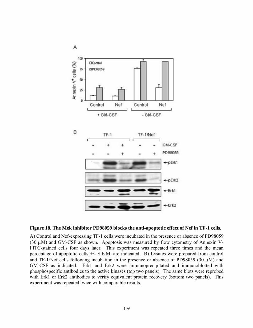
Figure 18. The Mek inhibitor PD98059 blocks the anti-apoptotic effect of Nef in TF-1 cells. A) Control and Nef-expressing TF-1 cells were incubated in the presence or absence of PD98059 (30 µM) and GM-CSF as shown. Apoptosis was measured by flow cytometry of Annexin V-FITC-stained cells four days later. This experiment was repeated three times and the mean percentage of apoptotic cells +/- S.E.M. are indicated. B) Lysates were prepared from control and TF-1/Nef cells following incubation in the presence or absence of PD98059 (30 µM) and GM-CSF as indicated. Erk1 and Erk2 were immunoprecipitated and immunoblotted with phosphospecific antibodies to the active kinases (top two panels). The same blots were reprobed with Erk1 or Erk2 antibodies to verify equivalent protein recovery (bottom two panels). This experiment was repeated twice with comparable results.
109
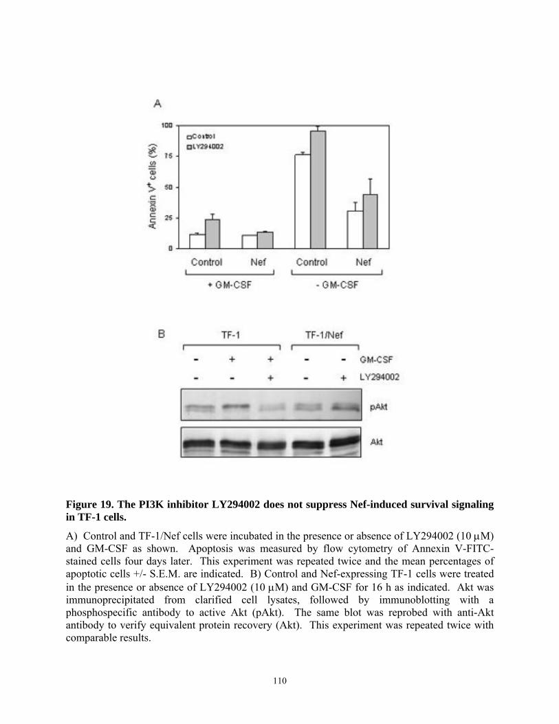
Figure 19. The PI3K inhibitor LY294002 does not suppress Nef-induced survival signaling in TF-1 cells.
A) Control and TF-1/Nef cells were incubated in the presence or absence of LY294002 (10 µM) and GM-CSF as shown. Apoptosis was measured by flow cytometry of Annexin V-FITC-stained cells four days later. This experiment was repeated twice and the mean percentages of apoptotic cells +/- S.E.M. are indicated. B) Control and Nef-expressing TF-1 cells were treated in the presence or absence of LY294002 (10 µM) and GM-CSF for 16 h as indicated. Akt was immunoprecipitated from clarified cell lysates, followed by immunoblotting with a phosphospecific antibody to active Akt (pAkt). The same blot was reprobed with anti-Akt antibody to verify equivalent protein recovery (Akt). This experiment was repeated twice with comparable results.
110
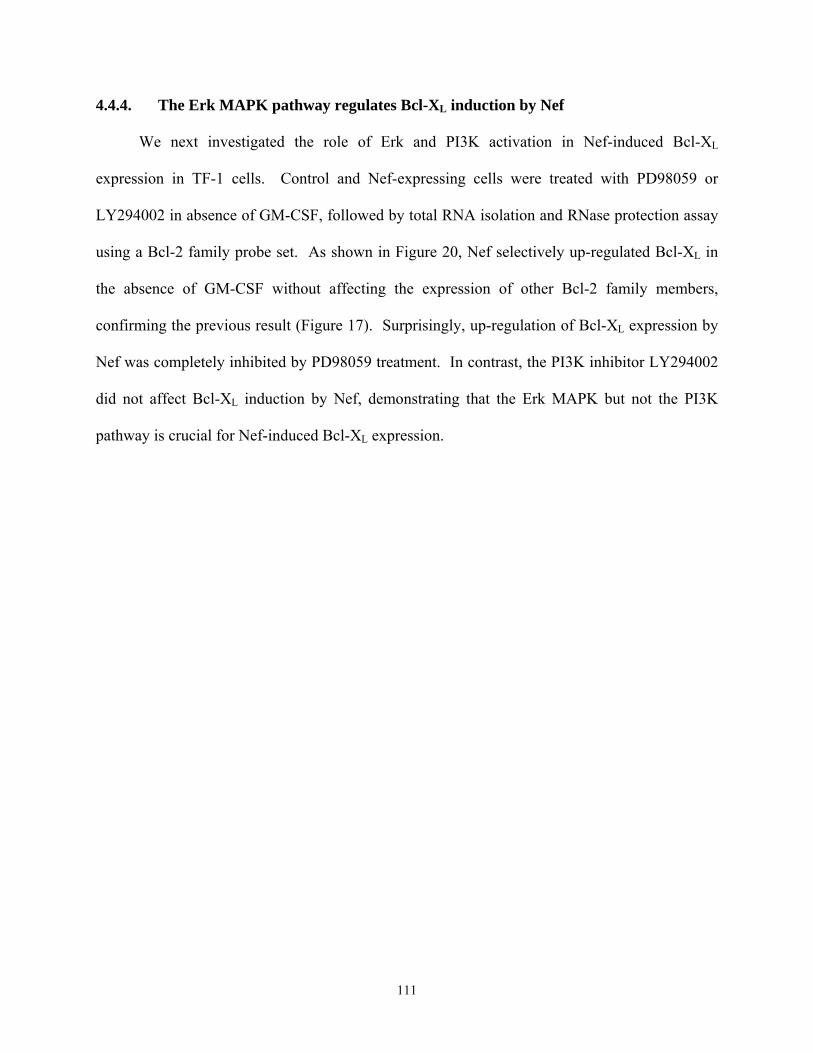
4.4.4. The Erk MAPK pathway regulates Bcl-XL induction by Nef
We next investigated the role of Erk and PI3K activation in Nef-induced Bcl-XL
expression in TF-1 cells. Control and Nef-expressing cells were treated with PD98059 or
LY294002 in absence of GM-CSF, followed by total RNA isolation and RNase protection assay
using a Bcl-2 family probe set. As shown in Figure 20, Nef selectively up-regulated Bcl-XL in
the absence of GM-CSF without affecting the expression of other Bcl-2 family members,
confirming the previous result (Figure 17). Surprisingly, up-regulation of Bcl-XL expression by
Nef was completely inhibited by PD98059 treatment. In contrast, the PI3K inhibitor LY294002
did not affect Bcl-XL induction by Nef, demonstrating that the Erk MAPK but not the PI3K
pathway is crucial for Nef-induced Bcl-XL expression.
111
Figure 20. Induction of Bcl-XL expression by HIV Nef requires the Erk MAPK pathway.
A) Nef-expressing TF-1 cells were treated with the Mek inhibitor, PD98059 (30 µM) or the PI3K inhibitor, LY294002 (10 µM) for 16 h in the absence of GM-CSF. Total RNA was extracted and the RNase protection assay was performed as described in the legend to Fig.2. As a control, RNA was prepared from TF-1 cells expressing the G418 selection marker and incubated with or without GM-CSF for 16 h. A representative autoradiogram is shown. B) The relative level of Bcl-XL transcripts in each sample were normalized to GAPDH expression and are presented as the fold increase relative to the control. Results represent the mean ± S.E.M. of three independent experiments.
112
4.4.5. Fusion of Nef to the estrogen receptor (ER) hormone binding domain creates a
conditional Nef protein (Nef-ER) capable of inducible survival signaling
All of the experiments described so far have been conducted with a retrovirally-
transduced TF-1 cell population in which the expression of Nef varies from one cell to another.
In addition, survival of TF-1/Nef cells may be influenced by G418 selection of the infected cell
population, and not due to a direct signal from Nef itself. To address these issues, we created a
conditionally active Nef protein by fusing it to the hormone binding domain of the estrogen
receptor (ER) using a strategy similar to one originally reported by Walk, et al. (258). Structural
and biochemical studies have shown that Nef can form dimers and higher-order oligomers (26,
28, 58, 259) which may be important for some signaling functions (258). One effect of ER
fusion is to allow regulated dimerization, which may in turn control the functional activity of Nef.
Fusion to ER may also render the protein inactive due to the steric hindrance caused by binding
of cellular chaperones, such as Hsp 90. Treatment with the cell-permeable synthetic estrogen 4-
hydroxytamoxifen (4-HT) induces Nef-ER dimer formation, as well as release of bound
chaperones and protein stabilization (see below). A similar Nef-ER construct has been shown to
induce CD4 downregulation and to associate with the Pak kinase activity in a 4-HT-dependent
fashion (258).
TF-1 cells were infected with Nef-ER retroviruses and cloned by limiting dilution under
G418 selection in the presence of GM-CSF. Cell clones were then screened for expression of the
Nef-ER fusion protein (~60 kDa) by anti-Nef immunoblotting of cell lysates. Four of eleven TF-
1 cell clones tested strongly positive for Nef-ER expression (Figure 21A). To determine whether
Nef-ER activation protects TF-1 cells from apoptosis in response to GM-CSF deprivation, cells
were pretreated with 4-HT for 24 hours followed by GM-CSF withdrawal. Following 16 hours of
113
GM-CSF starvation, cells were analyzed for initiation of apoptosis by Annexin V staining. As
shown in Figure 21B, 4-HT suppressed programmed cell death following cytokine withdrawal in
the TF-1/Nef-ER cell lines, confirming that the survival signal is directly driven by Nef and is
not a secondary effect of selection. Furthermore, Nef-ER suppressed apoptosis in TF-1 cells in
the presence of 4-HT almost as effectively as GM-CSF. This result is in contrast to the partial
effect observed in the bulk TF-1/Nef cultures (Figure 16), which may reflect heterogeneity of
Nef expression in this cell population as described above. TF-1/Nef-ER cell clone #10 was
studied further because its responsiveness to 4-HT in terms of cell survival was indistinguishable
from that of GM-CSF. TF-1 cell clones negative for Nef-ER expression were not protected from
apoptosis by 4-HT following GM-CSF withdrawal (data not shown).
4.4.6. Inducible stabilization and dimerization of Nef-ER in TF-1 cells
We next investigated the mechanism by which 4-HT induced Nef-ER activation in TF-1
cells. Based on work in other systems, we expected to observe 4-HT-dependent formation of
Nef-ER dimers (258). To test this idea, TF-1 cells were incubated in the presence or absence of
4-HT and GM-CSF, followed by treatment with the homobifunctional cross-linker DSS to
stabilize the dimers prior to cell lysis. Nef-ER proteins were then separated by SDS-PAGE,
transferred to PVDF and probed with a Nef-specific antibody. As shown in Figure 22, 4-HT-
dependent Nef-ER dimerization was detected regardless of the presence of GM-CSF.
Interestingly, the Nef-ER protein level was also higher in the presence of 4-HT, suggesting that
ligand binding may stabilize the Nef-ER protein in addition to promoting dimerization.
114
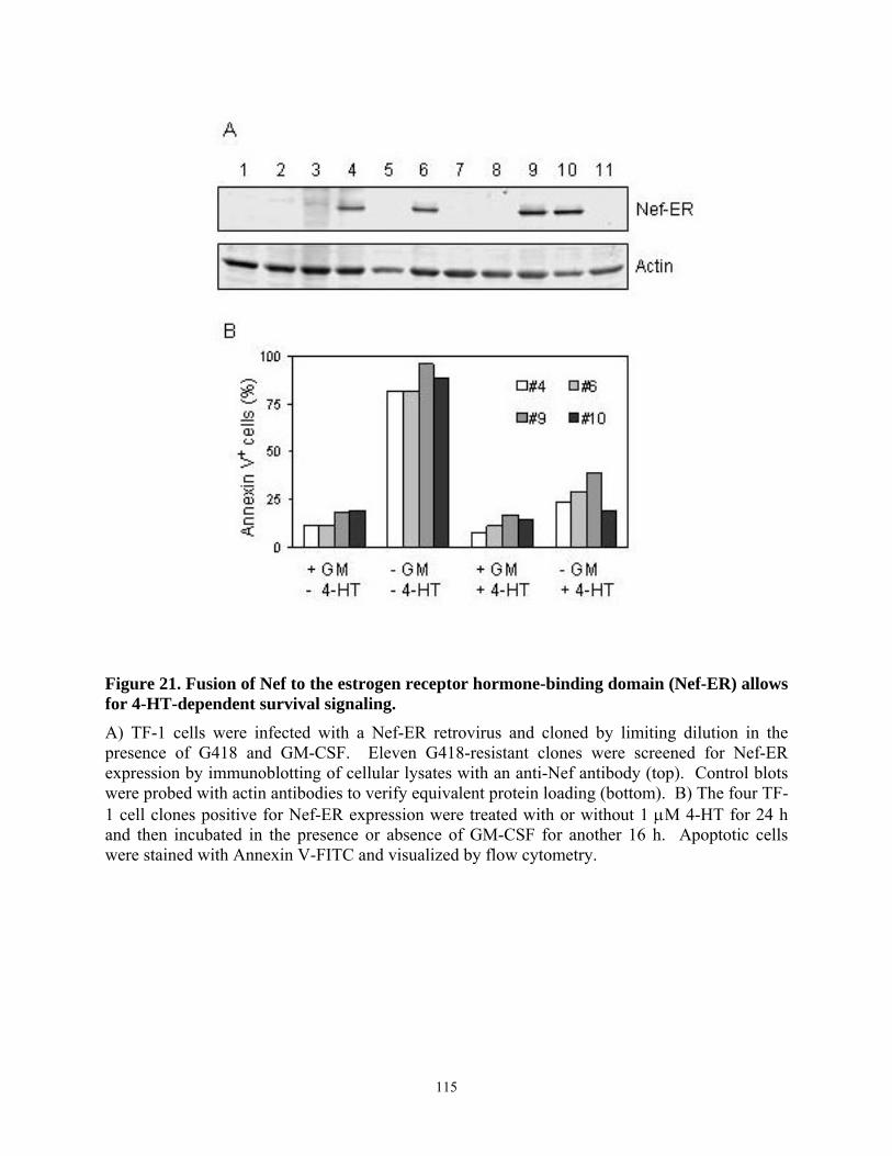
Figure 21. Fusion of Nef to the estrogen receptor hormone-binding domain (Nef-ER) allows for 4-HT-dependent survival signaling. A) TF-1 cells were infected with a Nef-ER retrovirus and cloned by limiting dilution in the presence of G418 and GM-CSF. Eleven G418-resistant clones were screened for Nef-ER expression by immunoblotting of cellular lysates with an anti-Nef antibody (top). Control blots were probed with actin antibodies to verify equivalent protein loading (bottom). B) The four TF-1 cell clones positive for Nef-ER expression were treated with or without 1 µM 4-HT for 24 h and then incubated in the presence or absence of GM-CSF for another 16 h. Apoptotic cells were stained with Annexin V-FITC and visualized by flow cytometry.
115
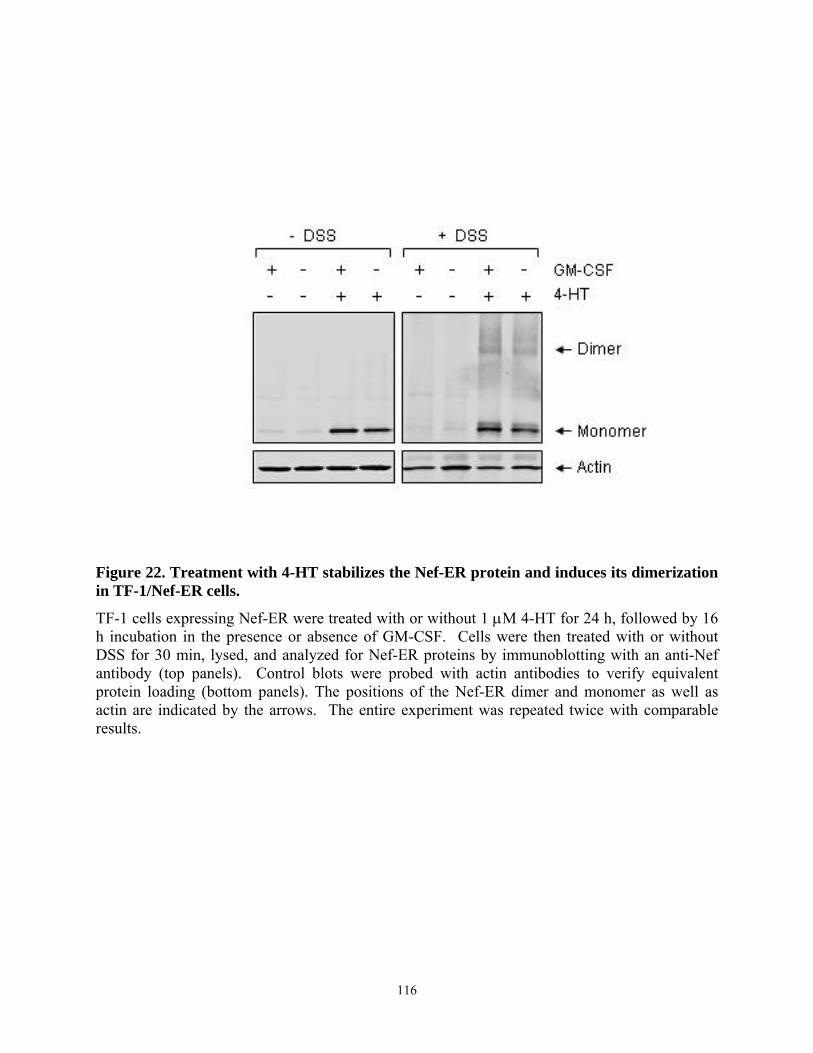
Figure 22. Treatment with 4-HT stabilizes the Nef-ER protein and induces its dimerization in TF-1/Nef-ER cells.
TF-1 cells expressing Nef-ER were treated with or without 1 µM 4-HT for 24 h, followed by 16 h incubation in the presence or absence of GM-CSF. Cells were then treated with or without DSS for 30 min, lysed, and analyzed for Nef-ER proteins by immunoblotting with an anti-Nef antibody (top panels). Control blots were probed with actin antibodies to verify equivalent protein loading (bottom panels). The positions of the Nef-ER dimer and monomer as well as actin are indicated by the arrows. The entire experiment was repeated twice with comparable results.
116
Similar observations have been reported following expression of Nef in T-cells (258), and
provide an additional level of control over Nef activity.
4.4.7. Cell survival and Stat3 activation by Nef-ER requires Erk activation
Constitutive expression of Nef in TF-1 cells promotes survival in an Erk-dependent
manner (Figure 18). To test whether the inducible survival effect of Nef-ER also requires Erk
activation, we treated TF-1/Nef-ER cells with the Mek specific inhibitor, PD98059. As shown in
Figure 23, PD98059 completely blocked the protective effect of activated Nef-ER following
withdrawal of GM-CSF. In contrast, PD98059 did not affect cell survival in the presence of GM-
CSF, indicating that GM-CSF survival signals are not Erk-dependent in this cell line. These
results are consistent with those obtained from the TF-1/Nef cell populations (Figure 18).
Using Stat3 dominant negative mutants, we observed previously that constitutive Stat3
activation is required for GM-CSF-independent growth of TF-1/Nef cells in soft agar (118). Here
we show that Erk signaling is also essential, suggesting a possible connection between the two
pathways. To investigate cross-talk between Stat3 and Erk downstream of Nef, we tested the
effect of PD98059 on Nef-ER-driven Stat3 signaling. PD98059 was added to control and Nef-
ER-expressing cells pretreated with 4-HT in the presence or absence of GM-CSF. Stat3
activation was then assayed using an electrophoretic mobility shift (gel-shift) assay with an
oligonucleotide probe corresponding to the sis-inducible element (SIE), which is strongly bound
by active Stat3. As shown in Figure 24, nuclear extracts prepared from 4-HT-treated TF-1/Nef-
ER cells revealed strong SIE-binding activity, which agrees with our previous findings in TF-
1/Nef cell populations (118). Surprisingly, the levels of the Stat3/SIE complex were
substantially reduced in the presence of PD98059, suggesting a requirement for Erk in Stat3
117
activation by Nef-ER. In contrast, SIE-protein complex formation induced by GM-CSF in
control TF-1 cells was unaffected by PD98059 treatment, indicating that the Erk requirement is
unique to Nef-dependent Stat3 activation. Competition with unlabeled SIE completely blocked
Stat3-SIE complex formation in each case, indicating that this interaction is specific.
To determine whether Stat3 DNA-binding activity induced by Nef correlates with Stat3
tyrosine phosphorylation, nuclear extracts from the control and TF-1/Nef-ER cells were
immunoblotted with anti-phospho-Stat3 antibodies. Figure 24B shows that Stat3 Tyr 705 is
strongly phosphorylated following Nef-ER activation by 4-HT, consistent with the gel-shift
result. In contrast, nuclear extracts from TF-1/Nef-ER cells treated with PD98059 showed
greatly diminished levels of Stat3 Tyr 705 phosphorylation, indicating that Nef-induced tyrosine
phosphorylation of Stat3 also requires Erk activation. In contrast, Stat3 Tyr 705 phosphorylation
by GM-CSF was unaffected by PD98059. Control immunoblots show that approximately
equivalent levels of Stat3 protein were expressed in each culture. Taken together, these results
provide strong evidence that Nef-mediated activation of Stat3 requires active Erk and that the
mechanism of Stat3 activation by Nef is distinct from that of GM-CSF.
118

Figure 23. Survival signaling from Nef-ER requires the Erk MAPK pathway in TF-1 cells. A) TF-1 cells expressing Nef-ER (right) and parental TF-1 cells (left) were incubated in the presence or absence of 4-HT for 24 h. The cells were then washed to remove GM-CSF and replated in the presence or absence of GM-CSF and 30 µM PD98059 as indicated. Apoptotic cells were stained with Annexin V-FITC and detected by flow cytometry. Results show the mean percentage of apoptotic cells observed in three independent determinations ± S.E.M.
119
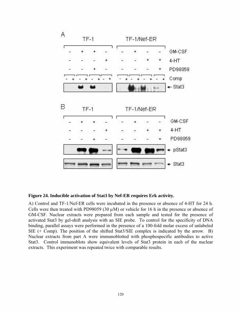
Figure 24. Inducible activation of Stat3 by Nef-ER requires Erk activity.
A) Control and TF-1/Nef-ER cells were incubated in the presence or absence of 4-HT for 24 h. Cells were then treated with PD98059 (30 µM) or vehicle for 16 h in the presence or absence of GM-CSF. Nuclear extracts were prepared from each sample and tested for the presence of activated Stat3 by gel-shift analysis with an SIE probe. To control for the specificity of DNA binding, parallel assays were performed in the presence of a 100-fold molar excess of unlabeled SIE (+ Comp). The position of the shifted Stat3/SIE complex is indicated by the arrow. B) Nuclear extracts from part A were immunoblotted with phosphospecific antibodies to active Stat3. Control immunoblots show equivalent levels of Stat3 protein in each of the nuclear extracts. This experiment was repeated twice with comparable results.
120
4.4.8. Dominant-negative Stat3 does not affect survival or Bcl-XL induction by Nef-ER
In a final series of experiments, we investigated the requirement for Stat3 activation in
Nef-induced survival signaling and induction of Bcl-XL gene expression. For these experiments,
we used a retrovirus carrying a dominant-negative form of Stat3 in which the tyrosine
phosphorylation site at position 705 is replaced with phenylalanine (Stat3YF). Previous studies
have shown that this mutant blocks GM-CSF-independent growth of TF-1/Nef cells in soft-agar
colony assays, suggesting that Stat3 may contribute to Nef-induced suppression of apoptosis
resulting from cytokine withdrawal in this cell line (118). TF-1/Nef-ER cells were infected with
the Stat3YF retrovirus or with a control virus carrying only the drug selection marker. Forty-
eight hours later, 4-HT was added and cells were washed free of GM-CSF and incubated for an
additional 16 hours. As shown in Figure 25A, activation of Stat3 by Nef-ER in the presence of
4-HT or by GM-CSF was dramatically suppressed in cells infected with the Stat3YF retrovirus
but not the control virus. Surprisingly, Stat3YF did not affect the suppression of apoptosis by
active Nef-ER (Figure 25B), nor did it impact expression of Bcl-XL (Figure 25C). These data
show that while Nef strongly activates Stat3 in TF-1 cells, this pathway may not be required for
Bcl-XL induction or survival signaling.
121
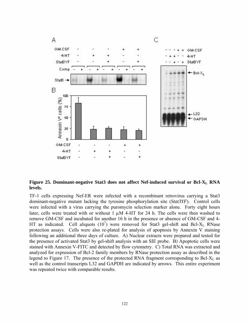
Figure 25. Dominant-negative Stat3 does not affect Nef-induced survival or Bcl-XL RNA levels. TF-1 cells expressing Nef-ER were infected with a recombinant retrovirus carrying a Stat3 dominant-negative mutant lacking the tyrosine phosphorylation site (Stat3YF). Control cells were infected with a virus carrying the puromycin selection marker alone. Forty eight hours later, cells were treated with or without 1 µM 4-HT for 24 h. The cells were then washed to remove GM-CSF and incubated for another 16 h in the presence or absence of GM-CSF and 4-HT as indicated. Cell aliquots (107) were removed for Stat3 gel-shift and Bcl-XL RNase protection assays. Cells were also re-plated for analysis of apoptosis by Annexin V staining following an additional three days of culture. A) Nuclear extracts were prepared and tested for the presence of activated Stat3 by gel-shift analysis with an SIE probe. B) Apoptotic cells were stained with Annexin V-FITC and detected by flow cytometry. C) Total RNA was extracted and analyzed for expression of Bcl-2 family members by RNase protection assay as described in the legend to Figure 17. The presence of the protected RNA fragment corresponding to Bcl-XL as well as the control transcripts L32 and GAPDH are indicated by arrows. This entire experiment was repeated twice with comparable results.
122
4.5. Discussion
Previous work from our laboratory has shown that Nef promotes the cytokine-
independent proliferation of the human myeloid progenitor cell line TF-1 through a mechanism
dependent upon the Stat3 transcription factor (118). Here we show for the first time that Nef
protects TF-1 cells from apoptosis following cytokine withdrawal and selectively up-regulates
expression of the anti-apoptotic gene, Bcl-XL (Figures 16 and 17). Several studies have
established that Bcl-XL, a member of Bcl-2 family of mitochondrial apoptotic regulators, is
directly induced by the Stat3 transcription factor. For example, Catlett-Falcone et al. (244)
reported constitutive Stat3 activation and upregulated Bcl-XL expression in human multiple
myeloma cells. Using a dominant-negative form of Stat3, they also demonstrated that functional
Stat3 is required for Bcl-XL induction and the anti-apoptotic response in this system. Using a
similar dominant-negative approach, we tested the requirement for Stat3 in Bcl-XL induction and
survival signaling by Nef in TF-1 cells. Surprisingly, expression of a well-established dominant-
negative mutant of Stat3 (Stat3YF) did not affect Bcl-XL upregulation or suppression of
apoptosis by Nef (Figure 25), suggesting that Nef induces Bcl-XL and promotes survival through
a Stat3-independent mechanism involving Erk activation (see below). While Nef-driven survival
and Bcl-XL expression were not affected by Stat3YF, this mutant was able to completely block
cytokine-independent soft-agar colony formation by TF-1/Nef cells in previous studies (118). In
light of work presented here, this observation suggests that the Nef-Stat3 pathway may affect
other Stat3-dependent responses in myeloid cells such as adhesion or migration (260).
The Erk kinases are members of the MAP kinase family and regulate a wide variety of cell
functions, including the survival response to cytokines and other soluble factors (254). Relevant
to our study is recent work by Kolonics et al. (256) which showed that GM-CSF-induced
survival of TF-1 cells correlates with Erk1/2 activation and Bcl-XL induction, consistent with our
123
findings. GM-CSF-dependent changes in Bcl-XL proteins levels in this study correlate closely
with changes in transcript levels reported here (Figure 17). In related studies, Kinoshita et al.
(261) reported that activation of the Ras-MAPK pathway may be essential for anti-apoptotic
signaling by the GM-CSF receptor. They found that a mutant GM-CSF receptor uncoupled from
MAP kinase activation was unable to protect transfected Ba/F3 cells from apoptosis in the
presence of GM-CSF. Expression of active Ras complemented the signaling defect from the
mutant receptor and supported long-term proliferation of the cells, suggesting that GM-CSF
prevents apoptosis of some hematopoietic cells by activating Ras-MAPK signaling. Here we
show that Nef mimics this effect of GM-CSF, and that Nef-induced Erk activation is required for
TF-1 cell survival in cytokine-free medium. Supporting this hypothesis, inhibition of Nef-
induced Erk activation with PD98059 completely abolished the survival of Nef-expressing cells
following cytokine withdrawal (Figures 18 and 23). In addition to the role of Erk in the Nef-
induced anti-apoptotic effect described here, Erk MAPK activity has been shown to increase
transcription from the HIV proviral long terminal repeat and HIV replication in T-cells (262,
263). Furthermore, expression of constitutively activated forms of Ras, Raf, or Mek, as well as
activation of Erk signaling with serum or phorbol esters enhanced the infectivity of HIV virions
in T-cells (263). Conversely, virus infectivity was reduced by treatment of cells with PD98059
or with antisense oligonucleotides directed against Erks (263). Erk MAPKs have also been
identified as HIV virion-associated kinases by density gradient fractionation (81). In this study,
viral infectivity was increased by stimulation of virion-associated Erk activity with phorbol
esters and impaired by specific inhibitors of the MAPK cascade. Taken together, these studies
support the conclusion that HIV takes advantage of the Erk MAPK pathway to enhance viral
124
replication as well as promote survival of infected cells, and that HIV Nef plays an essential role
in these processes.
Results presented here show for the first time that Nef induces Bcl-XL induction as a
mechanism for cytokine-independent survival of myeloid cells (Figure 17). Although Nef-
induced Bcl-XL induction depends upon Erk activation (Figure 20), the mechanism by which this
MAPK pathway contributes to Bcl-XL regulation is less clear. One possibility is that Erks
regulate Bcl-XL directly through AP-1 induction. Nef has been found to activate the AP-1
transcription factor through Hck and Erk in macrophages (242), and a consensus motif for AP-1
binding has been identified in the promoter region of Bcl-XL (264). However, whether or not
AP-1 binds to Bcl-XL promoter region and directly induces transcription of Bcl-XL remains to be
examined.
Work presented here also suggests that Nef induces cross-talk between the Erk and Stat3
signaling pathways. This hypothesis is supported by the surprising observation that
pharmacological inhibition of Erk signaling suppresses the tyrosine phosphorylation and DNA
binding activity of Stat3 downstream of Nef-ER (Figure 24). Erks have been implicated in
phosphorylation of Stat3 at Ser 727, which may be required for full transcriptional activity (265)
but has a more controversial role in the regulation of DNA binding (266, 267). Using
phosphospecific antibodies, we did not detect changes in the phosphorylation status of Stat3 Ser
727 in response to Nef expression in TF-1 cells (data not shown). However, Nef-induced Erk
activation is linked to activation of Rsk and other Ser/Thr kinases related to survival of
hematopoietic cells through phosphorylation of CREB and other transcription factor targets
(254). Whether or not these kinases can also influence the activity of Stat3 by phosphorylation
of sites other than Ser 727 remains to be tested. Another possibility is that Nef-induced Erk
125
activation may affect dephosphorylation of active Stat3 by SHP2 or other protein-tyrosine
phosphatases. Erk1 has been shown to phosphorylate SHP2 and inhibit its activity in vitro,
consistent with this possibility (268).
HIV and other pathogenic viruses have developed protective mechanisms to suppress
apoptosis of infected host cells, to allow time for viral replication prior to cell lysis (240).
Recent studies have shown that Nef inhibits death receptor-mediated apoptosis by interacting
with ASK1 and blocking its activity in T-cells (112, 240). More recently, it has been
demonstrated that HIV-1 Nef also suppresses apoptosis by stimulating Akt-independent Bad
phosphorylation through PI3K and PAK pathways in T-lymphocytes (114). In contrast to these
studies, we found that Nef-induced survival does not require PI3K activity (Figure 20) or involve
Bad phosphorylation (data not shown) in cells of myeloid lineage. Instead, Nef appears to
suppress cell death in this macrophage precursor cell line through a distinct mechanism
involving the Erk MAPK pathway and Bcl-XL induction. A growing number of studies
implicate HIV-infected macrophages as key elements in AIDS pathogenesis (269). For example,
in severe combined immunodeficient mice transplanted with human peripheral blood leukocytes
(hu-PBL-SCID mouse model), extensive CD4+ T-cell depletion is induced by non-cytopathic,
macrophage-tropic strains of HIV. Surprisingly, HIV strains that are highly cytopathic towards
T-cells in vitro showed little activity in the hu-PBL-SCID mouse model (210). Also, monkeys
infected with HIV-1, which does not infect simian monocytes/macrophages, showed no signs of
disease and fail to show accelerated T-cell apoptosis (270). The anti-apoptotic signal generated
by HIV Nef described here may allow for persistence of HIV-infected macrophages, promoting
viral replication and spread to uninfected T cells. Our findings support the hypothesis that Nef
126
may contribute to the establishment and maintenance HIV reservoirs by conferring a survival
advantage on HIV-infected macrophages.
127

5. Chapter V - Discussion
128
While the Nef protein of HIV and SIV has been suggested as a key molecule in AIDS
pathogenesis for over a decade, the molecular mechanisms of Nef action in HIV replication and
disease progression are still elusive. Elucidation of the interactions of Nef with cellular signaling
proteins is central to understanding its role in disease progression in vivo, since Nef has no
known catalytic activity. In support of this idea, the physical and functional association of Nef
with a large number of cellular proteins including members of the Src protein-tyrosine
kinasefamily has been demonstrated.
Previous studies from our laboratory have shown that HIV Nef strongly binds and
activates the macrophage-specific Src family member Hck, leading to cellular transformation in
Rat-2 fibroblasts. Genetic evidence suggests that Nef-Hck interaction may be essential for AIDS
progression (202). The interaction between Nef and Hck has been intensively studied and the
interface of this association has been well-defined in structural studies. In these studies, we have
focused our efforts on the further characterization of the molecular mechanism specific to HIV-1
Nef interaction with Hck in a well-defined model system (Rat-2 fibroblasts).
The interaction of Nef with other cellular proteins, particularly those involved in signal
transduction, has also been demonstrated to affect viral life cycle and disease progression. One
way to facilitate the viral replication is to promote survival of infected cells. Supporting this
notion, recent studies have shown that Nef protects CD4+T lymphocytes from the death
receptor-mediated and virus infection-induced apoptosis. Our laboratory has previously
demonstrated that Nef also induces the proliferation of macrophage lineage cells, which are the
other key target of HIV infection. Based on these findings, we set out to investigate the survival
signaling induced by HIV Nef in macrophage precursor cells. Studies presented in this
129
dissertation add significantly to our understanding of Nef function as a signaling molecule in
HIV-infected cells.
Chapter III: Conserved Residues in the HIV-1 Nef Hydrophobic Pocket are Essential for
Recruitment and Activation of the Hck Tyrosine Kinase. The purpose of this study was to identify
the residues responsible for the differential interaction of Hck and Nef variants and evaluate the
functional relevance of those residues in the hydrophobic pocket in recruitment and activation of
Hck in vivo. Nef binds to Hck through its SH3 domain with the unusually high affinity. To
investigate whether interaction with Hck is a conserved property of Nef alleles of different
strains of HIV-1, the binding of several well-studied laboratory HIV-1 Nef alleles (SF2, LAI,
ELI, and Consensus) to Hck and other Src family kinases was first compared in vitro and in
cells. Surprisingly, not all four of the Nef proteins tested in these experiments have a similar
affinity for Hck even though they all contain the PxxPxR motif and hydrophobic pocket residues
essential for SH3 binding in the crystal structure. While SF2 Nef had the strongest affinity for
Hck, ELI Nef had the weakest binding. Sequence alignment and molecular modeling of these
two Nef alleles identified Ala 83 and Tyr 120 in the hydrophobic pocket as critical residues for
establishing stable interaction between Nef and Hck in vivo as well as in vivo. We also found the
important role His 116 in facilitating the stable PxxP-Hck SH3 interaction, which is located
outside of hydrophobic pocket. This finding suggests the importance of the Nef hydrophobic
pocket in activation of Hck. In addition, this study provides evidence that allosteric effect of
other residues of Nef which do not directly contact the SH3 domain in the crystal structure (e.g.
His 116) can also influence Hck activation. The clinical relevance of this study was addressed by
comparative analysis of the Nef sequences of long-term non-progressors (LTNP) which have
130
been reported in the literature. Some sequence heterogeneity at position 83, 116 and 120 was
found in LTNPs with a full-length nef gene. In contrast, consensus PxxPxR motifs as well as
hydrophobic pocket residues identified in the crystal structure as an essential part of the SH3
interface were strictly conserved. These findings support the hypothesis that the presence of
these highly conserved features alone does not assure interaction with Hck, and sequence
variation outside of these core motifs involved in SH3 binding impact interaction with Hck and
may affect disease progression as a consequence.
This study is the first to assign a necessary biological function to the Nef hydrophobic
pocket, in terms of recruitment and activation of the Hck tyrosine kinase in living cells. In
addition to that, these results provide a rationale for the design of small molecules targeted to the
pocket, which could disrupt Nef signaling through Hck in HIV-infected macrophages in vivo.
Such agent is predicted to delay disease progression based on genetic studies from other group
(202).
Future studies. To assess the physiological relevance of the differential recruitment and
activation of Hck by HIV-1 Nef alleles and the functional importance of the specific interaction
of Nef and Hck in HIV infection and pathogenesis, SF2 and ELI Nef could be compared in their
capacity to promote replication of an M-tropic HIV-1 strain in cultured macrophages. Since Hck
was recently demonstrated to be required for HIV replication in macrophage culture (203), the
comparison of role for SF2 and ELI Nef in viral replication would determine whether the
essential role for Hck in HIV replication is mediated through its interaction with Nef in
macrophages. To determine the effect of each identified sequence variation of Nef on viral
replication, the SF2 mutants with substitutions at position 83, 116, and/or 120 could be used.
Whether or not these substitutions alone can account for non-progressor status could be tested in
131
a whole-animal (mouse) model by generating transgenic mice expressing these same Nef
mutants. Previously, Hanna et al. observed the AIDS-like disease development in transgenic
mice expressing Nef in CD4 T lymphocytes and macrophages (66). It would also be interesting
to investigate the effect of these substitutions on binding to T cell specific Src tyrosine kinases.
While Hck kinase is expressed mainly in macrophages, other Src family kinases such as Lck and
Fyn are expressed in T lymphocytes and play an important role in TcR signaling. Since both cell
types are key targets for HIV infection, the differential effect of these substitutions at positions
83, 116, and/or 120 on Nef interaction with other Src family kinases may contributes to viral
tropism. Similarly, the Nef sequences of macrophage tropic- or T cell tropic-HIV isolates could
be compared for correlations with specificity for T-cell vs macrophage-specific Src family
members.
Chapter IV: HIV-1 Nef Promotes Survival of TF-1 Macrophages by Inducing Bcl-XL Expression
in an Erk-dependent Manner. The goal of this study was to investigate the mechanism of
survival signaling induced by the HIV-1 Nef protein in cells of the myelomonocytic lineage.
HIV Nef has been demonstrated to generate anti-apoptotic signals in CD4+ T lymphocytes,
protecting infected cells from immune cell bound death receptor- or virus infection-induced
apoptosis to allow productive viral replication. Previous work from our laboratory has shown
that Nef promotes proliferation of myeloid cells through a Stat3-dependent pathway. Based on
these findings we hypothesized that the HIV Nef protein also suppresses cell death by altering
signal transduction in macrophages. The present study supports this hypothesis by showing that
Nef protects the human macrophage progenitor cell line, TF-1 from apoptosis induced by
cytokine deprivation. This cell survival phenotype induced by Nef correlated with upregulation
132
of Bcl-XL, an anti-apoptotic gene. We additionally found cell survival and up-regulation of Bcl-
XL by Nef requires Erk MAPK activation since the inhibition of Erk MAPK pathway with a
specific antagonist suppressed cell survival and Bcl-XL induction by Nef. However, in contrast to
previous findings which suggested a requirement for Stat3 in the Nef-induced proliferation
signal, the Nef anti-apoptotic pathway was independent of Stat3 in TF-1 cells. Dominant-
negative of Stat3 did not impact the survival of Nef-expressing cells or Bcl-XL up-regulation.
These observations suggest that Nef may use distinctive pathways to induce either cell
proliferation or anti-apoptotic signals in macrophages .
Since Hck is a well-known binding partner of Nef as discussed in Chapter III and
expressed mostly in macrophages/monocytes, we hypothesized Nef may exert its survival
signaling through its binding and activation of Hck in TF-1 cells. However, we were unable to
demonstrate a requirement for Hck kinase activity in Nef-induced survival signaling in TF-1
cells. We tested the role of Hck kinase activity in Nef-induced survival signaling using the Src
kinase-specific inhibitors PP2 and A-419259 (271). No effect of these inhibitors on Nef-induced
cell survival or Bcl-XL upregulation was observed, although the inhibition of Src family kinase
activity by these drugs was confirmed in TF-1 cells. This finding suggests that Hck kinase
activity may not be necessary for anti-apoptosis signaling pathway of Nef in macrophages.
However, we still cannot exclude the possibility that the Hck protein itself may be required for
this Nef-induced survival. Because Hck contains SH2 and SH3 domains, it may function as an
adaptor to link Nef with other cellular targets. To test this possibility, several methods can be
used, such as an antisense RNA or siRNA to knockdown the total protein level of Hck in these
cells. Hck kinase activity may also play an important role in other Nef-induced signal
transduction pathways, which requires further investigation.
133
One unexpected finding in this study was the involvement of the Erk MAPK pathway in
Stat3 activation in Nef-expressing TF-1 cells. Inhibition of Erk MAPK activity by a specific Erk
blocker suppressed not only the survival of Nef-expressing cells but also Stat3 activation.
Although Stat3 was not required for Nef-induced survival signal, activation of Stat3 was
essential for proliferative phenotype of Nef-expressing TF-1 cells, which was demonstrated by
soft-agar assay in our previous work (118). These observations suggest that Erk may contribute
to both the survival signal by up-regulating Bcl-XL and proliferation featured by Stat3 activation
in TF-1 cells. It is, however, mysterious how Erk mediates Stat3 activation, particularly the
aspects of DNA-binding activity and tyrosine phosphorylation (Figure 24). Erk has been shown
to mediate the serine phosphorylation of Stat3 which is required for full transcriptional activity
of Stat3. However, the role of serine phosphorylation in DNA binding activity of Stat3 is still
controversial. The mechanism by which this MAPK pathway contributes to Bcl-XL regulation
has also to be determined. Although the exact mechanism of Erk functions in Nef-induced
signaling remain unclear, Erk has been implicated in enhanced viral infectivity in T cells. Erk
increases transcription from the HIV proviral long terminal repeat and replication in T cells (69,
73) and is identified as a virion-associated kinase (81), which may contribute to serine
phosphorylation of matrix proteins. The phosphorylation of matrix correlates with the increased
viral infectivity (80). However, the role of Erk in viral replication in macrophages has been
elusive. Although the direct effect of Erk on viral replication in macrophages has not been shown
in this study, Erk appears to play an important role in prolonging the survival of macrophages,
which may indirectly contribute to viral replication in this cell type possibly through facilitating
the formation of a viral reservoir.
134
Significance of this study includes the following. (1) This study is the first to show the
survival signaling generated by Nef in macrophage lineage cells. Macrophages serve as a
reservoir of HIV and disseminate viral particles to the other target cells. Despite this important
role, the mechanism of anti-apoptosis in HIV-infected macrophages to secure the productive
viral replication for a long period has not been studied in detail. Our study supports the role of
macrophages as a potential viral reservoir of HIV. (2) We are the first to observe the Bcl-2
family regulation by Nef in macrophage lineage cells. Although Wolf et al. (114) showed that
Nef inhibits T cell death by phosphorylating and thus inactivating Bad, a proapoptotic Bcl-2
family member, the same events have not been demonstrated to occur in macrophages. While
regulating the posttranslational modification of Bad in T lymphocytes, Nef regulates
transcriptionally the expression level of Bcl-XL. (3) Our study demonstrates for the first time the
Erk MAPK requirement for Nef-induced survival signal and Bcl-XL up-regulation in
macrophage lineage cells.
Future studies. The anti-apoptotic effect of Nef should be assessed in primary macrophage
culture, particularly HIV-infected macrophages. Since macrophages express membrane bound
cell death ligands/receptors which play an important role in killing infected cells by autocrine or
cell-cell contacts, a suppressive effect of Nef on death-receptor mediated apoptosis of primary
macrophages or macrophage lineage cells would suggest direct relationship to disease
progression. It would also be interesting to see whether HIV-1 Nef alleles used in the previous
chapter (Chapter III) induce differentially the survival signal in TF-1 cells. Although the
requirement for Hck kinase activity in Nef-induced survival was not observed in TF-1 cells, it is
certainly possible that other Nef alleles differentially bind to Hck, resulting in distinctive
outcomes. Another interesting follow-up study would be to examine the effect of Erk inhibition
135
on viral replication in macrophages. The role of Erk MAPK in HIV life cycle has been suggested
in T cells but not in macrophages. Since the prolonged cell survival facilitates the viral
replication, the survival effect of Nef-induced Erk activation on infected macrophages may
indirectly enhance the viral replication. This hypothesis could be tested by blocking Erk kinase
activity using a specific inhibitor. Also, since we were unable to show the direct interaction of
Nef and endogenous Hck in TF-1 cells, it is possible that other signaling molecules directly
binding to Nef may mediate these signaling events. One potential candidate to transduce the
survival signal from Nef to Erk activation downstream may be PAK family kinases. PAK
kinases have been well documented to bind to Nef and mediate the signal to MAPK kinases.
Particularly, PAK2 kinase is also involved in apoptosis signaling as discussed in the introduction
(see section 1.7.2.). Whether or not Nef binds and activates PAK kinases in this cell type could
be tested by immunoprecipitation and in vitro kinase assay. Finally, understanding the
requirement of specific motifs, such as PxxP motif or N-terminal myristylation for survival
signaling in macrophages could provide an important clue about the binding partners of Nef
mediating these signaling events in macrophages.
In summary, there are two major conclusions from this dissertation: (1) Residues in Nef
hydrophobic pocket play a critical role for recruitment and activation of the Hck tyrosine kinase,
which may be important in disease progression. Targeting the Nef hydrophobic pocket with a
small molecule may be sufficient to prevent recruitment and activation of Hck in vivo, delaying
disease progression. (2) Nef promotes survival of macrophage lineage cells by upregulating Bcl-
XL expression. Cell survival and Bcl-XL induction by Nef require Erk MAPK pathway. This anti-
apoptotic signal generated by Nef may ensure the persistence of HIV-infected macrophages,
facilitating the establishment and maintenance of HIV reservoirs.
136
BIBLIOGRAPHY
137
1. Tang H, Kuhen KL, Wong-Staal F. 1999. Lentivirus replication and regulation. Annu.
Rev. Genet. 33:133-70
2. Weiss RA. 1993. How does HIV cause AIDS? Science 260:1273-9
3. Cullen BR. 1998. HIV-1 auxiliary proteins: making connections in a dying cell. Cell
93:685-92
4. Emerman M, Malim MH. 1998. HIV-1 regulatory/accessory genes: keys to unraveling
viral and host cell biology. Science 280:1880-4
5. Inubushi R, Tamaki M, Shimano R, Koyama AH, Akari H, Adachi A. 1998. Functional
roles of HIV accessory proteins for viral replication (Review). Int. J. Mol. Med. 2:429-33
6. Ikuta K, Kameoka M, Luftig RB. 1997. AIDS pathogenesis: the role of accessory gene
mutations, leading to formation of long-lived persistently infected cells and/or apoptosis-
inducing HIV-1 particles. Virus Res. 52:145-56
7. Cullen BR. 1990. The HIV-1 Tat protein: an RNA sequence-specific processivity factor?
Cell 63:655-7
8. Cullen BR. 1991. Regulation of HIV-1 gene expression. FASEB J. 5:2361-8
9. Cullen BR, Malim MH. 1991. The HIV-1 Rev protein: prototype of a novel class of
eukaryotic post-transcriptional regulators. Trends Biochem. Sci. 16:346-50
10. Gabuzda DH, Lawrence K, Langhoff E, Terwilliger E, Dorfman T, Haseltine WA,
Sodroski J. 1992. Role of vif in replication of human immunodeficiency virus type 1 in
CD4+ T lymphocytes. J. Virol. 66:6489-95
138
11. Gabuzda DH, Li H, Lawrence K, Vasir BS, Crawford K, Langhoff E. 1994. Essential role
of vif in establishing productive HIV-1 infection in peripheral blood T lymphocytes and
monocyte/macrophages. J. Acquir. Immune. Defic. Syndr. 7:908-15
12. Heinzinger NK, Bukinsky MI, Haggerty SA, Ragland AM, Kewalramani V, Lee MA,
Gendelman HE, Ratner L, Stevenson M, Emerman M. 1994. The Vpr protein of human
immunodeficiency virus type 1 influences nuclear localization of viral nucleic acids in
nondividing host cells. Proc. Natl. Acad. Sci. U. S. A 91:7311-5
13. Goh WC, Rogel ME, Kinsey CM, Michael SF, Fultz PN, Nowak MA, Hahn BH,
Emerman M. 1998. HIV-1 Vpr increases viral expression by manipulation of the cell
cycle: a mechanism for selection of Vpr in vivo. Nat. Med. 4:65-71
14. Klimkait T, Strebel K, Hoggan MD, Martin MA, Orenstein JM. 1990. The human
immunodeficiency virus type 1-specific protein vpu is required for efficient virus
maturation and release. J. Virol. 64:621-9
15. Willey RL, Maldarelli F, Martin MA, Strebel K. 1992. Human immunodeficiency virus
type 1 Vpu protein induces rapid degradation of CD4. J. Virol. 66:7193-200
16. Kestler H, Ringler DJ, Mori K, Panicali DL, Sehgal PK, Daniel MD, Desrosiers RC.
1991. Importance of the nef gene for maintenance of high viral loads and for
development of AIDS. Cell 65:651-62
17. Deacon NJ, Tsykin A, Solomon A, Smith K, Ludford-Menting M, Hooker DJ, McPhee
DA, Greenway AL, Ellett A, Chatfield C, . 1995. Genomic structure of an attenuated
quasi species of HIV-1 from a blood transfusion donor and recipients. Science 270:988-
91
18. Kirchhoff F, Greenough TC, Brettler DB, Sullivan JL, Desrosiers RC. 1995. Absence of
intact nef sequences in a long-term survivor with nonprogressive HIV-1 infection. N.
Engl. J. Med. 332:228-32
139
19. Harris M. 1996. From negative factor to a critical role in virus pathogenesis: the changing
fortunes of Nef. J. Gen. Virol. 77 ( Pt 10):2379-92
20. Allan JS, Coligan JE, Lee TH, McLane MF, Kanki PJ, Groopman JE, Essex M. 1985. A
new HTLV-III/LAV encoded antigen detected by antibodies from AIDS patients. Science
230:810-3
21. Arya SK, Gallo RC. 1986. Three novel genes of human T-lymphotropic virus type III:
immune reactivity of their products with sera from acquired immune deficiency
syndrome patients. Proc. Natl. Acad. Sci. U. S. A 83:2209-13
22. Cullen BR. 1994. The role of Nef in the replication cycle of the human and simian
immunodeficiency viruses. Virology 205:1-6
23. Kaminchik J, Bashan N, Itach A, Sarver N, Gorecki M, Panet A. 1991. Genetic
characterization of human immunodeficiency virus type 1 nef gene products translated in
vitro and expressed in mammalian cells. J. Virol. 65:583-8
24. Regier DA, Desrosiers RC. 1990. The complete nucleotide sequence of a pathogenic
molecular clone of simian immunodeficiency virus. AIDS Res. Hum. Retroviruses
6:1221-31
25. Welker R, Kottler H, Kalbitzer HR, Krausslich HG. 1996. Human immunodeficiency
virus type 1 Nef protein is incorporated into virus particles and specifically cleaved by
the viral proteinase. Virology 219:228-36
26. Lee CH, Saksela K, Mirza UA, Chait BT, Kuriyan J. 1996. Crystal structure of the
conserved core of HIV-1 Nef complexed with a Src family SH3 domain. Cell 85:931-42
27. Grzesiek S, Stahl SJ, Wingfield PT, Bax A. 1996. The CD4 determinant for
downregulation by HIV-1 Nef directly binds to Nef. Mapping of the Nef binding surface
by NMR. Biochemistry 35:10256-61
140
28. Grzesiek S, Bax A, Clore GM, Gronenborn AM, Hu J-S, Kaufman J, Palmer I, Stahl SJ,
Wingfield PT. 1996. The solution structure of HIV-1 Nef reveals an unexpected fold and
permits delineation of the binding surface for the SH3 domain of Hck tyrosine protein
kinase. Nature Struct. Biol. 3:340-5
29. Grzesiek S, Bax A, Hu JS, Kaufman J, Palmer I, Stahl SJ, Tjandra N, Wingfield PT.
1997. Refined solution structure and backbone dynamics of HIV-1 Nef. Protein Sci.
6:1248-63
30. Kaminchik J, Margalit R, Yaish S, Drummer H, Amit B, Sarver N, Gorecki M, Panet A.
1994. Cellular distribution of HIV type 1 Nef protein: identification of domains in Nef
required for association with membrane and detergent-insoluble cellular matrix. AIDS
Res. Hum. Retroviruses 10:1003-10
31. Kohleisen B, Neumann M, Herrmann R, Brack-Werner R, Krohn KJ, Ovod V, Ranki A,
Erfle V. 1992. Cellular localization of Nef expressed in persistently HIV-1-infected low-
producer astrocytes. AIDS 6:1427-36
32. Gordon JI, Duronio RJ, Rudnick DA, Adams SP, Gokel GW. 1991. Protein N-
myristoylation. J. Biol. Chem. 266:8647-50
33. Resh MD. 1994. Myristylation and palmitylation of Src family members: the fats of the
matter. Cell 76:411-3
34. Resh MD. 1999. Fatty acylation of proteins: new insights into membrane targeting of
myristoylated and palmitoylated proteins. Biochim. Biophys. Acta 1451:1-16
35. Harris M. 1995. The role of myristoylation in the interactions between human
immunodeficiency virus type I Nef and cellular proteins. Biochem. Soc. Trans. 23:557-61
141
36. Welker R, Harris M, Cardel B, Krausslich HG. 1998. Virion incorporation of human
immunodeficiency virus type 1 Nef is mediated by a bipartite membrane-targeting signal:
analysis of its role in enhancement of viral infectivity. J. Virol. 72:8833-40
37. Harris M, Coates K. 1993. Identification of cellular proteins that bind to the human
immunodeficiency virus type 1 nef gene product in vitro: a role for myristylation. J. Gen.
Virol. 74 ( Pt 8):1581-9
38. Harris MP, Neil JC. 1994. Myristoylation-dependent binding of HIV-1 Nef to CD4. J.
Mol. Biol. 241:136-42
39. Fackler OT, Kienzle N, Kremmer E, Boese A, Schramm B, Klimkait T, Kucherer C,
Mueller-Lantzsch N. 1997. Association of human immunodeficiency virus Nef protein
with actin is myristoylation dependent and influences its subcellular localization. Eur. J.
Biochem. 247:843-51
40. Chowers MY, Spina CA, Kwoh TJ, Fitch NJ, Richman DD, Guatelli JC. 1994. Optimal
infectivity in vitro of human immunodeficiency virus type 1 requires an intact nef gene.
J. Virol. 68:2906-14
41. Peng B, Robert-Guroff M. 2001. Deletion of N-terminal myristoylation site of HIV Nef
abrogates both MHC-1 and CD4 down-regulation. Immunol. Lett. 78:195-200
42. Guy B, Kieny MP, Riviere Y, Le Peuch C, Dott K, Girard M, Montagnier L, Lecocq JP.
1987. HIV F/3' orf encodes a phosphorylated GTP-binding protein resembling an
oncogene product. Nature 330:266-9
43. Bodeus M, Marie-Cardine A, Bougeret C, Ramos-Morales F, Benarous R. 1995. In vitro
binding and phosphorylation of human immunodeficiency virus type 1 Nef protein by
serine/threonine protein kinase. J. Gen. Virol. 76 ( Pt 6):1337-44
142
44. Coates K, Harris M. 1995. The human immunodeficiency virus type 1 Nef protein
functions as a protein kinase C substrate in vitro. J. Gen. Virol. 76 ( Pt 4):837-44
45. Coates K, Cooke SJ, Mann DA, Harris MP. 1997. Protein kinase C-mediated
phosphorylation of HIV-I nef in human cell lines. J. Biol. Chem. 272:12289-94
46. Baur AS, Sass G, Laffert B, Willbold D, Cheng-Mayer C, Peterlin BM. 1997. The N-
terminus of Nef from HIV-1/SIV associates with a protein complex containing Lck and a
serine kinase. Immunity. 6:283-91
47. Du Z, Lang SM, Sasseville VG, Lackner AA, Ilyinskii PO, Daniel MD, Jung JU,
Desrosiers RC. 1995. Identification of a nef allele that causes lymphocyte activation and
acute disease in macaque monkeys. Cell 82:665-74
48. Collette Y, Dutartre H, Benziane A, Ramos-Morales F, Benarous R, Harris M, Olive D.
1996. Physical and functional interaction of Nef with Lck - HIV-1 Nef-induced T-cell
signaling defects. J. Biol. Chem. 271:6333-41
49. Freund J, Kellner R, Konvalinka J, Wolber V, Krausslich HG, Kalbitzer HR. 1994. A
possible regulation of negative factor (Nef) activity of human immunodeficiency virus
type 1 by the viral protease. Eur. J. Biochem. 223:589-93
50. Gaedigk-Nitschko K, Schon A, Wachinger G, Erfle V, Kohleisen B. 1995. Cleavage of
recombinant and cell derived human immunodeficiency virus 1 (HIV-1) Nef protein by
HIV-1 protease. FEBS Lett. 357:275-8
51. Schorr J, Kellner R, Fackler O, Freund J, Konvalinka J, Kienzle N, Krausslich HG,
Mueller-Lantzsch N, Kalbitzer HR. 1996. Specific cleavage sites of Nef proteins from
human immunodeficiency virus types 1 and 2 for the viral proteases. J. Virol. 70:9051-4
143
52. Chen YL, Trono D, Camaur D. 1998. The proteolytic cleavage of human
immunodeficiency virus type 1 Nef does not correlate with its ability to stimulate virion
infectivity. J. Virol. 72:3178-84
53. Miller MD, Warmerdam MT, Ferrell SS, Benitez R, Greene WC. 1997. Intravirion
generation of the C-terminal core domain of HIV-1 Nef by the HIV-1 protease is
insufficient to enhance viral infectivity. Virology 234:215-25
54. Pandori M, Craig H, Moutouh L, Corbeil J, Guatelli J. 1998. Virological importance of
the protease-cleavage site in human immunodeficiency virus type 1 Nef is independent of
both intravirion processing and CD4 down-regulation. Virology 251:302-16
55. Samuel KP, Hodge DR, Chen YM, Papas TS. 1991. Nef proteins of the human
immunodeficiency viruses (HIV-1 and HIV-2) and simian immunodeficiency virus (SIV)
are structurally similar to leucine zipper transcriptional activation factors. AIDS Res.
Hum. Retroviruses 7:697-706
56. Kienzle N, Freund J, Kalbitzer HR, Mueller-Lantzsch N. 1993. Oligomerization of the
Nef protein from human immunodeficiency virus (HIV) type 1. Eur. J. Biochem.
214:451-7
57. Fujii Y, Otake K, Fujita Y, Yamamoto N, Nagai Y, Tashiro M, Adachi A. 1996.
Clustered localization of oligomeric Nef protein of human immunodeficiency virus type
1 on the cell surface. FEBS Lett. 395:257-61
58. Arold S, Hoh F, Domergue S, Birck C, Delsuc MA, Jullien M, Dumas C. 2000.
Characterization and molecular basis of the oligomeric structure of HIV-1 nef protein.
Protein Sci. 9:1137-48
59. Liu LX, Heveker N, Fackler OT, Arold S, Le Gall S, Janvier K, Peterlin BM, Dumas C,
Schwartz O, Benichou S, Benarous R. 2000. Mutation of a conserved residue (D123)
required for oligomerization of human immunodeficiency virus type 1 Nef protein
144
abolishes interaction with human thioesterase and results in impairment of Nef biological
functions. J. Virol. 74:5310-9
60. Nebreda AR, Bryan T, Segade F, Wingfield P, Venkatesan S, Santos E. 1991.
Biochemical and biological comparison of HIV-1 NEF and ras gene products. Virology
183:151-9
61. Fisher AG, Ratner L, Mitsuya H, Marselle LM, Harper ME, Broder S, Gallo RC, Wong-
Staal F. 1986. Infectious mutants of HTLV-III with changes in the 3' region and
markedly reduced cytopathic effects. Science 233:655-9
62. Luciw PA, Cheng-Mayer C, Levy JA. 1987. Mutational analysis of the human
immunodeficiency virus: the orf-B region down-regulates virus replication. Proc. Natl.
Acad. Sci. U. S. A 84:1434-8
63. Terwilliger E, Sodroski JG, Rosen CA, Haseltine WA. 1986. Effects of mutations within
the 3' orf open reading frame region of human T-cell lymphotropic virus type III (HTLV-
III/LAV) on replication and cytopathogenicity. J. Virol. 60:754-60
64. Ahmad N, Venkatesan S. 1988. Nef protein of HIV-1 is a transcriptional repressor of
HIV-1 LTR. Science 241:1481-5
65. Niederman TM, Garcia JV, Hastings WR, Luria S, Ratner L. 1992. Human
immunodeficiency virus type 1 Nef protein inhibits NF-kappa B induction in human T
cells. J. Virol. 66:6213-9
66. Hanna Z, Kay DG, Rebai N, Guimond A, Jothy S, Jolicoeur P. 1998. Nef harbors a major
determinant of pathogenicity for an AIDS-like disease induced by HIV-1 in transgenic
mice. Cell 95:163-75
145
67. Linnemann T, Zheng YH, Mandic R, Peterlin BM. 2002. Interaction between Nef and
phosphatidylinositol-3-kinase leads to activation of p21-activated kinase and increased
production of HIV. Virology 294:246-55
68. Rhee SS, Marsh JW. 1994. HIV-1 Nef activity in murine T cells. CD4 modulation and
positive enhancement. J. Immunol. 152:5128-34
69. Miller MD, Warmerdam MT, Gaston I, Greene WC, Feinberg MB. 1994. The human
immunodeficiency virus-1 nef gene product: a positive factor for viral infection and
replication in primary lymphocytes and macrophages. J. Exp. Med. 179:101-13
70. Spina CA, Kwoh TJ, Chowers MY, Guatelli JC, Richman DD. 1994. The importance of
nef in the induction of human immunodeficiency virus type 1 replication from primary
quiescent CD4 lymphocytes. J. Exp. Med. 179:115-23
71. Kim S, Ikeuchi K, Byrn R, Groopman J, Baltimore D. 1989. Lack of a negative influence
on viral growth by the nef gene of human immunodeficiency virus type 1. Proc. Natl.
Acad. Sci. U. S. A 86:9544-8
72. Hammes SR, Dixon EP, Malim MH, Cullen BR, Greene WC. 1989. Nef protein of
human immunodeficiency virus type 1: evidence against its role as a transcriptional
inhibitor. Proc. Natl. Acad. Sci. U. S. A 86:9549-53
73. Aiken C, Trono D. 1995. Nef stimulates human immunodeficiency virus type 1 proviral
DNA synthesis. J. Virol. 69:5048-56
74. Schwartz O, Marechal V, Danos O, Heard JM. 1995. Human immunodeficiency virus
type 1 Nef increases the efficiency of reverse transcription in the infected cell. J. Virol.
69:4053-9
146
75. Chowers MY, Pandori MW, Spina CA, Richman DD, Guatelli JC. 1995. The growth
advantage conferred by HIV-1 nef is determined at the level of viral DNA formation and
is independent of CD4 downregulation. Virology 212:451-7
76. Wiskerchen M, Cheng-Mayer C. 1996. HIV-1 Nef association with cellular serine kinase
correlates with enhanced virion infectivity and efficient proviral DNA synthesis. Virology
224:292-301
77. Kotov A, Zhou J, Flicker P, Aiken C. 1999. Association of Nef with the human
immunodeficiency virus type 1 core. J. Virol. 73:8824-30
78. Pandori MW, Fitch NJ, Craig HM, Richman DD, Spina CA, Guatelli JC. 1996. Producer-
cell modification of human immunodeficiency virus type 1: Nef is a virion protein. J.
Virol. 70:4283-90
79. Swingler S, Gallay P, Camaur D, Song J, Abo A, Trono D. 1997. The Nef protein of
human immunodeficiency virus type 1 enhances serine phosphorylation of the viral
matrix. J. Virol. 71:4372-7
80. Bukrinskaya AG, Ghorpade A, Heinzinger NK, Smithgall TE, Lewis RE, Stevenson M.
1996. Phosphorylation-dependent human immunodeficiency virus type 1 infection and
nuclear targeting of viral DNA. Proc. Natl. Acad. Sci. U. S. A 93:367-71
81. Jacque JM, Mann A, Enslen H, Sharova N, Brichacek B, Davis RJ, Stevenson M. 1998.
Modulation of HIV-1 infectivity by MAPK, a virion-associated kinase. EMBO J.
17:2607-18
82. Greenway A, Azad A, Mills J, McPhee D. 1996. Human immunodeficiency virus type 1
Nef binds directly to Lck and mitogen-activated protein kinase, inhibiting kinase activity.
J. Virol. 70:6701-8
147
83. Sawai ET, Baur A, Struble H, Peterlin BM, Levy JA, Cheng-Mayer C. 1994. Human
immunodeficiency virus type 1 Nef associates with a cellular serine kinase in T
lymphocytes. Proc. Natl. Acad. Sci. U. S. A 91:1539-43
84. Smith BL, Krushelnycky BW, Mochly-Rosen D, Berg P. 1996. The HIV nef protein
associates with protein kinase C theta. J. Biol. Chem. 271:16753-7
85. Sleckman BP, Shin J, Igras VE, Collins TL, Strominger JL, Burakoff SJ. 1992.
Disruption of the CD4-p56lck complex is required for rapid internalization of CD4. Proc.
Natl. Acad. Sci. U. S. A 89:7566-70
86. Mangasarian A, Foti M, Aiken C, Chin D, Carpentier JL, Trono D. 1997. The HIV-1 Nef
protein acts as a connector with sorting pathways in the Golgi and at the plasma
membrane. Immunity. 6:67-77
87. Aiken C, Konner J, Landau NR, Lenburg ME, Trono D. 1994. Nef induces CD4
endocytosis: requirement for a critical dileucine motif in the membrane-proximal CD4
cytoplasmic domain. Cell 76:853-64
88. Piguet V, Chen YL, Mangasarian A, Foti M, Carpentier JL, Trono D. 1998. Mechanism
of Nef-induced CD4 endocytosis: Nef connects CD4 with the mu chain of adaptor
complexes. EMBO J. 17:2472-81
89. Craig HM, Pandori MW, Guatelli JC. 1998. Interaction of HIV-1 Nef with the cellular
dileucine-based sorting pathway is required for CD4 down-regulation and optimal viral
infectivity. Proc. Natl. Acad. Sci. U. S. A 95:11229-34
90. Bandres JC, Shaw AS, Ratner L. 1995. HIV-1 Nef protein downregulation of CD4
surface expression: relevance of the lck binding domain of CD4. Virology 207:338-41
91. Garcia JV, Miller AD. 1991. Serine phosphorylation-independent downregulation of cell-
surface CD4 by nef. Nature 350:508-11
148

92. Le Gall S, Erdtmann L, Benichou S, Berlioz-Torrent C, Liu L, Benarous R, Heard JM,
Schwartz O. 1998. Nef interacts with the mu subunit of clathrin adaptor complexes and
reveals a cryptic sorting signal in MHC I molecules. Immunity. 8:483-95
93. Foti M, Mangasarian A, Piguet V, Lew DP, Krause KH, Trono D, Carpentier JL. 1997.
Nef-mediated clathrin-coated pit formation. J. Cell Biol. 139:37-47
94. Kirchhausen T, Bonifacino JS, Riezman H. 1997. Linking cargo to vesicle formation:
receptor tail interactions with coat proteins. Curr. Opin. Cell Biol. 9:488-95
95. Benichou S, Bomsel M, Bodeus M, Durand H, Doute M, Letourneur F, Camonis J,
Benarous R. 1994. Physical interaction of the HIV-1 Nef protein with beta-COP, a
component of non-clathrin-coated vesicles essential for membrane traffic. J. Biol. Chem.
269:30073-6
96. Bour S, Perrin C, Strebel K. 1999. Cell surface CD4 inhibits HIV-1 particle release by
interfering with Vpu activity. J. Biol. Chem. 274:33800-6
97. Ross TM, Oran AE, Cullen BR. 1999. Inhibition of HIV-1 progeny virion release by cell-
surface CD4 is relieved by expression of the viral Nef protein. Curr. Biol. 9:613-21
98. Benson RE, Sanfridson A, Ottinger JS, Doyle C, Cullen BR. 1993. Downregulation of
cell-surface CD4 expression by simian immunodeficiency virus Nef prevents viral super
infection. J. Exp. Med. 177:1561-6
99. Schwartz O, Marechal V, Le Gall S, Lemonnier F, Heard JM. 1996. Endocytosis of major
histocompatibility complex class I molecules is induced by the HIV-1 Nef protein. Nat.
Med. 2:338-42
100. Riggs NL, Craig HM, Pandori MW, Guatelli JC. 1999. The dileucine-based sorting motif
in HIV-1 Nef is not required for down-regulation of class I MHC. Virology 258:203-7
149
101. Collins KL, Chen BK, Kalams SA, Walker BD, Baltimore D. 1998. HIV-1 Nef protein
protects infected primary cells against killing by cytotoxic T lymphocytes. Nature
391:397-401
102. Cohen GB, Gandhi RT, Davis DM, Mandelboim O, Chen BK, Strominger JL, Baltimore
D. 1999. The selective downregulation of class I major histocompatibility complex
proteins by HIV-1 protects HIV-infected cells from NK cells. Immunity. 10:661-71
103. Doms RW, Trono D. 2000. The plasma membrane as a combat zone in the HIV
battlefield. Genes Dev. 14:2677-88
104. Weiss A, Littman DR. 1994. Signal transduction by lymphocyte antigen receptors. Cell
76:263-74
105. Luria S, Chambers I, Berg P. 1991. Expression of the type 1 human immunodeficiency
virus Nef protein in T cells prevents antigen receptor-mediated induction of interleukin 2
mRNA. Proc. Natl. Acad. Sci. U. S. A 88:5326-30
106. Bandres JC, Ratner L. 1994. Human immunodeficiency virus type 1 Nef protein down-
regulates transcription factors NF-kappa B and AP-1 in human T cells in vitro after T-cell
receptor stimulation. J. Virol. 68:3243-9
107. Simmons A, Aluvihare V, McMichael A. 2001. Nef triggers a transcriptional program in
T cells imitating single-signal T cell activation and inducing HIV virulence mediators.
Immunity. 14:763-77
108. Baur AS, Sawai ET, Dazin P, Fantl WJ, Cheng-Mayer C, Peterlin BM. 1994. HIV-1 Nef
leads to inhibition or activation of T cells depending on its intracellular localization.
Immunity. 1:373-84
109. Badley AD, Pilon AA, Landay A, Lynch DH. 2000. Mechanisms of HIV-associated
lymphocyte apoptosis. Blood 96:2951-64
150
110. Badley AD, McElhinny JA, Leibson PJ, Lynch DH, Alderson MR, Paya CV. 1996.
Upregulation of Fas ligand expression by human immunodeficiency virus in human
macrophages mediates apoptosis of uninfected T lymphocytes. J. Virol. 70:199-206
111. Banda NK, Bernier J, Kurahara DK, Kurrle R, Haigwood N, Sekaly RP, Finkel TH.
1992. Crosslinking CD4 by human immunodeficiency virus gp120 primes T cells for
activation-induced apoptosis. J. Exp. Med. 176:1099-106
112. Geleziunas R, Xu W, Takeda K, Ichijo H, Greene WC. 2001. HIV-1 Nef inhibits ASK1-
dependent death signalling providing a potential mechanism for protecting the infected
host cell. Nature 410:834-8
113. Yoon K, Jeong JG, Kim S. 2001. Stable expression of human immunodeficiency virus
type 1 Nef confers resistance against Fas-mediated apoptosis. AIDS Res. Hum.
Retroviruses 17:99-104
114. Wolf D, Witte V, Laffert B, Blume K, Stromer E, Trapp S, d'Aloja P, Schurmann A,
Baur AS. 2001. HIV-1 Nef associated PAK and PI3-kinases stimulate Akt-independent
Bad- phosphorylation to induce anti-apoptotic signals. Nat. Med. 7:1217-24
115. Adams JM, Cory S. 2001. Life-or-death decisions by the Bcl-2 protein family. Trends
Biochem. Sci. 26:61-6
116. Gross A, McDonnell JM, Korsmeyer SJ. 1999. BCL-2 family members and the
mitochondria in apoptosis. Genes Dev. 13:1899-911
117. Greenway AL, McPhee DA, Allen K, Johnstone R, Holloway G, Mills J, Azad A,
Sankovich S, Lambert P. 2002. Human immunodeficiency virus type 1 Nef binds to
tumor suppressor p53 and protects cells against p53-mediated apoptosis. J. Virol.
76:2692-702
151
118. Briggs SD, Scholtz B, Jacque JM, Swingler S, Stevenson M, Smithgall TE. 2001. HIV-1
Nef promotes survival of myeloid cells by a Stat3-dependent pathway. J. Biol. Chem.
276:25605-11
119. Iyengar S, Hildreth JE, Schwartz DH. 1998. Actin-dependent receptor colocalization
required for human immunodeficiency virus entry into host cells. J. Virol. 72:5251-5
120. Viard M, Parolini I, Sargiacomo M, Fecchi K, Ramoni C, Ablan S, Ruscetti FW, Wang
JM, Blumenthal R. 2002. Role of cholesterol in human immunodeficiency virus type 1
envelope protein-mediated fusion with host cells. J. Virol. 76:11584-95
121. Bukrinskaya A, Brichacek B, Mann A, Stevenson M. 1998. Establishment of a functional
human immunodeficiency virus type 1 (HIV-1) reverse transcription complex involves
the cytoskeleton. J. Exp. Med. 188:2113-25
122. McDonald D, Vodicka MA, Lucero G, Svitkina TM, Borisy GG, Emerman M, Hope TJ.
2002. Visualization of the intracellular behavior of HIV in living cells. J. Cell Biol.
159:441-52
123. Fackler OT, Luo W, Geyer M, Alberts AS, Peterlin BM. 1999. Activation of Vav by Nef
induces cytoskeletal rearrangements and downstream effector functions. Mol. Cell 3:729-
39
124. Manninen A, Hiipakka M, Vihinen M, Lu W, Mayer BJ, Saksela K. 1998. SH3-Domain
binding function of HIV-1 Nef is required for association with a PAK-related kinase.
Virology 250:273-82
125. Renkema GH, Manninen A, Saksela K. 2001. Human immunodeficiency virus type 1 Nef
selectively associates with a catalytically active subpopulation of p21-activated kinase 2
(PAK2) independently of PAK2 binding to Nck or beta-PIX. J. Virol. 75:2154-60
152
126. Daniels RH, Bokoch GM. 1999. p21-activated protein kinase: a crucial component of
morphological signaling? Trends Biochem. Sci. 24:350-5
127. Sells MA, Knaus UG, Bagrodia S, Ambrose DM, Bokoch GM, Chernoff J. 1997. Human
p21-activated kinase (Pak1) regulates actin organization in mammalian cells. Curr. Biol.
7:202-10
128. Forshey BM, Aiken C. 2003. Disassembly of human immunodeficiency virus type 1
cores in vitro reveals association of Nef with the subviral ribonucleoprotein complex. J.
Virol. 77:4409-14
129. Campbell EM, Nunez R, Hope TJ. 2004. Disruption of the actin cytoskeleton can
complement the ability of Nef to enhance human immunodeficiency virus type 1
infectivity. J. Virol. 78:5745-55
130. Klatzmann D, Champagne E, Chamaret S, Gruest J, Guetard D, Hercend T, Gluckman
JC, Montagnier L. 1984. T-lymphocyte T4 molecule behaves as the receptor for human
retrovirus LAV. Nature 312:767-8
131. Maddon PJ, Littman DR, Godfrey M, Maddon DE, Chess L, Axel R. 1985. The isolation
and nucleotide sequence of a cDNA encoding the T cell surface protein T4: a new
member of the immunoglobulin gene family. Cell 42:93-104
132. Maddon PJ, Molineaux SM, Maddon DE, Zimmerman KA, Godfrey M, Alt FW, Chess
L, Axel R. 1987. Structure and expression of the human and mouse T4 genes. Proc. Natl.
Acad. Sci. U. S. A 84:9155-9
133. Rossi F, Gallina A, Milanesi G. 1996. Nef-CD4 physical interaction sensed with the yeast
two-hybrid system. Virology 217:397-403
153
134. Greenway A, Azad A, McPhee D. 1995. Human immunodeficiency virus type 1 Nef
protein inhibits activation pathways in peripheral blood mononuclear cells and T-cell
lines. J. Virol. 69:1842-50
135. Piguet V, Gu F, Foti M, Demaurex N, Gruenberg J, Carpentier JL, Trono D. 1999. Nef-
induced CD4 degradation: a diacidic-based motif in Nef functions as a lysosomal
targeting signal through the binding of beta-COP in endosomes. Cell 97:63-73
136. Greenberg ME, Iafrate AJ, Skowronski J. 1998. The SH3 domain-binding surface and an
acidic motif in HIV-1 Nef regulate trafficking of class I MHC complexes. EMBO J.
17:2777-89
137. Mangasarian A, Piguet V, Wang JK, Chen YL, Trono D. 1999. Nef-induced CD4 and
major histocompatibility complex class I (MHC-I) down-regulation are governed by
distinct determinants: N-terminal alpha helix and proline repeat of Nef selectively
regulate MHC-I trafficking. J. Virol. 73:1964-73
138. Piguet V, Wan L, Borel C, Mangasarian A, Demaurex N, Thomas G, Trono D. 2000.
HIV-1 Nef protein binds to the cellular protein PACS-1 to downregulate class I major
histocompatibility complexes. Nat. Cell Biol. 2:163-7
139. Swann SA, Williams M, Story CM, Bobbitt KR, Fleis R, Collins KL. 2001. HIV-1 Nef
blocks transport of MHC class I molecules to the cell surface via a PI 3-kinase-dependent
pathway. Virology 282:267-77
140. Robinson MS. 1994. The role of clathrin, adaptors and dynamin in endocytosis. Curr.
Opin. Cell Biol. 6:538-44
141. Cowles CR, Odorizzi G, Payne GS, Emr SD. 1997. The AP-3 adaptor complex is
essential for cargo-selective transport to the yeast vacuole. Cell 91:109-18
154
142. Simpson F, Peden AA, Christopoulou L, Robinson MS. 1997. Characterization of the
adaptor-related protein complex, AP-3. J. Cell Biol. 137:835-45
143. Letourneur F, Klausner RD. 1992. A novel di-leucine motif and a tyrosine-based motif
independently mediate lysosomal targeting and endocytosis of CD3 chains. Cell 69:1143-
57
144. Trowbridge IS, Collawn JF, Hopkins CR. 1993. Signal-dependent membrane protein
trafficking in the endocytic pathway. Annu. Rev. Cell Biol. 9:129-61
145. Ohno H, Stewart J, Fournier MC, Bosshart H, Rhee I, Miyatake S, Saito T, Gallusser A,
Kirchhausen T, Bonifacino JS. 1995. Interaction of tyrosine-based sorting signals with
clathrin-associated proteins. Science 269:1872-5
146. Bremnes T, Lauvrak V, Lindqvist B, Bakke O. 1998. A region from the medium chain
adaptor subunit (mu) recognizes leucine- and tyrosine-based sorting signals. J. Biol.
Chem. 273:8638-45
147. Rapoport I, Chen YC, Cupers P, Shoelson SE, Kirchhausen T. 1998. Dileucine-based
sorting signals bind to the beta chain of AP-1 at a site distinct and regulated differently
from the tyrosine-based motif-binding site. EMBO J. 17:2148-55
148. Faure J, Stalder R, Borel C, Sobo K, Piguet V, Demaurex N, Gruenberg J, Trono D.
2004. ARF1 regulates Nef-induced CD4 degradation. Curr. Biol. 14:1056-64
149. Janvier K, Craig H, Le Gall S, Benarous R, Guatelli J, Schwartz O, Benichou S. 2001.
Nef-induced CD4 downregulation: a diacidic sequence in human immunodeficiency virus
type 1 Nef does not function as a protein sorting motif through direct binding to beta-
COP. J. Virol. 75:3971-6
150. Nunn MF, Marsh JW. 1996. Human immunodeficiency virus type 1 Nef associates with a
member of the p21-activated kinase family. J. Virol. 70:6157-61
155
151. Sawai ET, Baur AS, Peterlin BM, Levy JA, Cheng-Mayer C. 1995. A conserved domain
and membrane targeting of Nef from HIV and SIV are required for association with a
cellular serine kinase activity. J. Biol. Chem. 270:15307-14
152. Sawai ET, Khan IH, Montbriand PM, Peterlin BM, Cheng-Mayer C, Luciw PA. 1996.
Activation of PAK by HIV and SIV Nef: importance for AIDS in rhesus macaques. Curr.
Biol. 6:1519-27
153. Renkema GH, Manninen A, Mann DA, Harris M, Saksela K. 1999. Identification of the
Nef-associated kinase as p21-activated kinase 2. Curr. Biol. 9:1407-10
154. Rudel T, Bokoch GM. 1997. Membrane and morphological changes in apoptotic cells
regulated by caspase-mediated activation of PAK2. Science 276:1571-4
155. Rudel T, Zenke FT, Chuang TH, Bokoch GM. 1998. p21-activated kinase (PAK) is
required for Fas-induced JNK activation in Jurkat cells. J. Immunol. 160:7-11
156. Knaus UG, Bokoch GM. 1998. The p21Rac/Cdc42-activated kinases (PAKs). Int. J.
Biochem. Cell Biol. 30:857-62
157. Campbell SM, Crowe SM, Mak J. 2001. Lipid rafts and HIV-1: from viral entry to
assembly of progeny virions. J. Clin. Virol. 22:217-27
158. Ono A, Freed EO. 2001. Plasma membrane rafts play a critical role in HIV-1 assembly
and release. Proc. Natl. Acad. Sci. U. S. A 98:13925-30
159. Walter BN, Huang Z, Jakobi R, Tuazon PT, Alnemri ES, Litwack G, Traugh JA. 1998.
Cleavage and activation of p21-activated protein kinase gamma-PAK by CPP32 (caspase
3). Effects of autophosphorylation on activity. J. Biol. Chem. 273:28733-9
160. Schrager JA, Der M, V, Marsh JW. 2002. HIV Nef increases T cell ERK MAP kinase
activity. J. Biol. Chem. 277:6137-42
156
161. Hodge DR, Dunn KJ, Pei GK, Chakrabarty MK, Heidecker G, Lautenberger JA, Samuel
KP. 1998. Binding of c-Raf1 kinase to a conserved acidic sequence within the carboxyl-
terminal region of the HIV-1 Nef protein. J. Biol. Chem. 273:15727-33
162. Aiken C, Krause L, Chen YL, Trono D. 1996. Mutational analysis of HIV-1 Nef:
identification of two mutants that are temperature-sensitive for CD4 downregulation.
Virology 217:293-300
163. Iafrate AJ, Bronson S, Skowronski J. 1997. Separable functions of Nef disrupt two
aspects of T cell receptor machinery: CD4 expression and CD3 signaling. EMBO J.
16:673-84
164. Jove R, Hanafusa H. 1987. Cell transformation by the viral src oncogene. Annu. Rev. Cell
Biol. 3:31-56
165. Tatosyan AG, Mizenina OA. 2000. Kinases of the Src family: structure and functions.
Biochemistry (Mosc. ) 65:49-58
166. Kaplan JM, Mardon G, Bishop JM, Varmus HE. 1988. The first seven amino acids
encoded by the v-src oncogene act as a myristylation signal: lysine 7 is a critical
determinant. Mol. Cell Biol. 8:2435-41
167. Pellman D, Garber EA, Cross FR, Hanafusa H. 1985. Fine structural mapping of a critical
NH2-terminal region of p60src. Proc. Natl. Acad. Sci. U. S. A 82:1623-7
168. Brown MT, Cooper JA. 1996. Regulation, substrates and functions of src. Biochim.
Biophys. Acta 1287:121-49
169. Pawson T, Gish GD. 1992. SH2 and SH3 domains: from structure to function. Cell
71:359-62
170. Pawson T. 1995. Protein modules and signalling networks. Nature 373:573-80
157
171. Cohen GB, Ren R, Baltimore D. 1995. Modular binding domains in signal transduction
proteins. Cell 80:237-48
172. Songyang Z, Shoelson SE, Chaudhuri M, Gish G, Pawson T, Haser WG, King F, Roberts
T, Ratnofsky S, Lechleider RJ, . 1993. SH2 domains recognize specific phosphopeptide
sequences. Cell 72:767-78
173. Sicheri F, Moarefi I, Kuriyan J. 1997. Crystal structure of the Src family tyrosine kinase
Hck. Nature 385:602-9
174. Xu W, Harrison SC, Eck MJ. 1997. Three-dimensional structure of the tyrosine kinase c-
Src. Nature 385:595-602
175. Nada S, Okada M, MacAuley A, Cooper JA, Nakagawa H. 1991. Cloning of a
complementary DNA for a protein-tyrosine kinase that specifically phosphorylates a
negative regulatory site of p60c-src. Nature 351:69-72
176. Xu W, Doshi A, Lei M, Eck MJ, Harrison SC. 1999. Crystal structures of c-Src reveal
features of its autoinhibitory mechanism. Mol. Cell 3:629-38
177. Schindler T, Sicheri F, Pico A, Gazit A, Levitzki A, Kuriyan J. 1999. Crystal structure of
Hck in complex with a Src family-selective tyrosine kinase inhibitor. Mol. Cell 3:639-48
178. Kmiecik TE, Shalloway D. 1987. Activation and suppression of pp60c-src transforming
ability by mutation of its primary sites of tyrosine phosphorylation. Cell 49:65-73
179. Reynolds AB, Vila J, Lansing TJ, Potts WM, Weber MJ, Parsons JT. 1987. Activation of
the oncogenic potential of the avian cellular src protein by specific structural alteration of
the carboxy terminus. EMBO J. 6:2359-64
158
180. Imamoto A, Soriano P. 1993. Disruption of the csk gene, encoding a negative regulator of
Src family tyrosine kinases, leads to neural tube defects and embryonic lethality in mice.
Cell 73:1117-24
181. Briggs SD, Smithgall TE. 1999. SH2-kinase linker mutations release Hck tyrosine kinase
and transforming activities in Rat-2 fibroblasts. J. Biol. Chem. 274:26579-83
182. Moarefi I, LaFevre-Bernt M, Sicheri F, Huse M, Lee C-H, Kuriyan J, Miller WT. 1997.
Activation of the Src-family tyrosine kinase Hck by SH3 domain displacement. Nature
385:650-3
183. Lerner EC, Smithgall TE. 2002. SH3-dependent stimulation of Src-family kinase
autophosphorylation without tail release from the SH2 domain in vivo. Nat. Struct. Biol.
9:365-9
184. Holtzman DA, Cook WD, Dunn AR. 1987. Isolation and sequence of a cDNA
corresponding to a src-related gene expressed in murine hemopoietic cells. Proc. Natl.
Acad. Sci. U. S. A 84:8325-9
185. Quintrell N, Lebo R, Varmus H, Bishop JM, Pettenati MJ, Le Beau MM, Diaz MO,
Rowley JD. 1987. Identification of a human gene (HCK) that encodes a protein-tyrosine
kinase and is expressed in hemopoietic cells. Mol. Cell Biol. 7:2267-75
186. Ziegler SF, Marth JD, Lewis DB, Perlmutter RM. 1987. Novel protein-tyrosine kinase
gene (hck) preferentially expressed in cells of hematopoietic origin. Mol. Cell Biol.
7:2276-85
187. Lock P, Stanley E, Holtzman DA, Dunn AR. 1990. Functional analysis and nucleotide
sequence of the promoter region of the murine hck gene. Mol. Cell Biol. 10:4603-11
159

188. Lock P, Ralph S, Stanley E, Boulet I, Ramsay R, Dunn AR. 1991. Two isoforms of
murine hck, generated by utilization of alternative translational initiation codons, exhibit
different patterns of subcellular localization. Mol. Cell. Biol. 11:4363-70
189. Robbins SM, Quintrell NA, Bishop JM. 1995. Myristoylation and differential
palmitoylation of the HCK protein-tyrosine kinases govern their attachment to
membranes and association with caveolae. Mol. Cell. Biol. 15:3507-15
190. Linnekin D, Howard OM, Park L, Farrar W, Ferris D, Longo DL. 1994. Hck expression
correlates with granulocyte-macrophage colony- stimulating factor-induced proliferation
in HL-60 cells. Blood 84:94-103
191. Anderson SM, Jorgensen B. 1995. Activation of src-related tyrosine kinases by IL-3. J.
Immunol. 155:1660-70
192. Ernst M, Gearing DP, Dunn AR. 1994. Functional and biochemical association of Hck
with the LIF/IL-6 receptor signal transducing subunit gp130 in embryonic stem cells.
EMBO J. 13:1574-84
193. Boulet I, Ralph S, Stanley E, Lock P, Dunn AR, Green SP, Phillips WA. 1992.
Lipopolysaccharide- and interferon-gamma-induced expression of hck and lyn tyrosine
kinases in murine bone marrow-derived macrophages. Oncogene 7:703-10
194. Ziegler SF, Wilson CB, Perlmutter RM. 1988. Augmented expression of a myeloid-
specific protein tyrosine kinase gene (hck) after macrophage activation. J. Exp. Med.
168:1801-10
195. English BK, Ihle JN, Myracle A, Yi T. 1993. Hck tyrosine kinase activity modulates
tumor necrosis factor production by murine macrophages. J. Exp. Med. 178:1017-22
160
196. Wang AV, Scholl PR, Geha RS. 1994. Physical and functional association of the high-
affinity immunoglobulin G receptor (Fc gamma RI) with the kinases Hck and Lyn. J.
Exp. Med. 180:1165-70
197. Mohn H, Le C, V, Fischer S, Maridonneau-Parini I. 1995. The src-family protein-tyrosine
kinase p59hck is located on the secretory granules in human neutrophils and translocates
towards the phagosome during cell activation. Biochem. J. 309 ( Pt 2):657-65
198. Bohuslav J, Horejsi V, Hansmann C, Stockl J, Weidle UH, Majdic O, Bartke I, Knapp W,
Stockinger H. 1995. Urokinase plasminogen activator receptor, beta 2-integrins, and Src-
kinases within a single receptor complex of human monocytes. J. Exp. Med. 181:1381-90
199. Lowell CA, Soriano P, Varmus HE. 1994. Functional overlap in the src gene family:
Inactivation of hck and fgr impairs natural immunity. Genes Dev. 8:387-98
200. Lowell CA, Fumagalli L, Berton G. 1996. Deficiency of Src family kinases p59/61hck
and p58c-fgr results in defective adhesion-dependent neutrophil functions. J. Cell Biol.
133:895-910
201. Lowell CA, Niwa M, Soriano P, Varmus HE. 1996. Deficiency of the Hck and Src
tyrosine kinases results in extreme levels of extramedullary hematopoiesis. Blood
87:1780-92
202. Hanna Z, Weng X, Kay DG, Poudrier J, Lowell C, Jolicoeur P. 2001. The pathogenicity
of human immunodeficiency virus (HIV) type 1 Nef in CD4C/HIV transgenic mice is
abolished by mutation of its SH3-binding domain, and disease development is delayed in
the absence of Hck. J. Virol. 75:9378-92
203. Komuro I, Yokota Y, Yasuda S, Iwamoto A, Kagawa KS. 2003. CSF-induced and HIV-
1-mediated distinct regulation of Hck and C/EBPbeta represent a heterogeneous
susceptibility of monocyte-derived macrophages to M-tropic HIV-1 infection. J. Exp.
Med. 198:443-53
161
204. Saksela K, Cheng G, Baltimore D. 1995. Proline-rich (PxxP) motifs in HIV-1 Nef bind to
SH3 domains of a subset of Src kinases and are required for the enhanced growth of Nef+
viruses but not for down-regulation of CD4. EMBO J. 14:484-91
205. Briggs SD, Sharkey M, Stevenson M, Smithgall TE. 1997. SH3-mediated Hck tyrosine
kinase activation and fibroblast transformation by the Nef protein of HIV-1. J. Biol.
Chem. 272:17899-902
206. Lee CH, Leung B, Lemmon MA, Zheng J, Cowburn D, Kuriyan J, Saksela K. 1995. A
Single Amino-Acid in the Sh3 Domain of Hck Determines Its High-Affinity and
Specificity in Binding to Hiv-1 Nef Protein. Embo Journal 14:5006-15
207. Briggs SD, Lerner EC, Smithgall TE. 2000. Affinity of Src family kinase SH3 domains
for HIV Nef in vitro does not predict kinase activation by Nef in vivo. Biochemistry
39:489-95
208. Arold S, O'Brien R, Franken P, Strub MP, Hoh F, Dumas C, Ladbury JE. 1998. RT loop
flexibility enhances the specificity of Src family SH3 domains for HIV-1 Nef.
Biochemistry 37:14683-91
209. Lang SM, Iafrate AJ, Stahl-Hennig C, Kuhn EM, Nisslein T, Kaup FJ, Haupt M,
Hunsmann G, Skowronski J, Kirchhoff F. 1997. Association of simian immunodeficiency
virus Nef with cellular serine/threonine kinases is dispensable for the development of
AIDS in rhesus macaques. Nat. Med. 3:860-5
210. Mosier DE, Gulizia RJ, MacIsaac PD, Torbett BE, Levy JA. 1993. Rapid loss of CD4+ T
cells in human-PBL-SCID mice by noncytopathic HIV isolates. Science 260:689-92
211. Mahlknecht U, Herbein G. 2001. Macrophages and T-cell apoptosis in HIV infection: a
leading role for accessory cells? Trends Immunol. 22:256-60
162
212. Crowe S, Mills J, McGrath MS. 1991. Mediators of fusion between HIV-infected
macrophages and lymphoid cells. Adv. Exp. Med. Biol. 300:57-66
213. Swingler S, Mann A, Jacque J, Brichacek B, Sasseville VG, Williams K, Lackner AA,
Janoff EN, Wang R, Fisher D, Stevenson M. 1999. HIV-1 Nef mediates lymphocyte
chemotaxis and activation by infected macrophages. Nat. Med. 5:997-103
214. Swingler S, Brichacek B, Jacque JM, Ulich C, Zhou J, Stevenson M. 2003. HIV-1 Nef
intersects the macrophage CD40L signalling pathway to promote resting-cell infection.
Nature 424:213-9
215. Arold ST, Bauer AS. 2001. Dynamic Nef and Nef dynamics: how structure could explain
the complex activities of this small HIV protein. Trends in Biochemical Sciences 26:356-
63
216. Geyer M, Fackler OT, Peterlin BM. 2001. Structure--function relationships in HIV-1 Nef.
EMBO Rep. 2:580-5
217. Herna RG, Saksela K. 2000. Interactions of HIV-1 NEF with cellular signal transducing
proteins. Front Biosci. 5:D268-D283
218. Mariani R, Kirchhoff F, Greenough TC, Sullivan JL, Desrosiers RC, Skowronski J. 1996.
High frequency of defective nef alleles in a long-term survivor with nonprogressive
human immunodeficiency virus type 1 infection. J. Virol. 70:7752-64
219. Renkema GH, Saksela K. 2000. Interactions of HIV-1 NEF with cellular signal
transducing proteins. Front Biosci. 5:D268-D283
220. Brown A, Moghaddam S, Kawano T, Cheng-Mayer C. 2004. Multiple human
immunodeficiency virus type 1 Nef functions contribute to efficient replication in
primary human macrophages. Journal of General Virology 85:1463-9
163
221. Miller WT. 2003. Determinants of substrate recognition in nonreceptor tyrosine kinases.
Acc. Chem. Res. 36:393-400
222. Schreiner SJ, Schiavone AP, Smithgall TE. 2002. Activation of Stat3 by the Src family
kinase Hck requires a functional SH3 domain. J. Biol. Chem. 277:45680-7
223. Geyer M, Peterlin BM. 2001. Domain assembly, surface accessibility and sequence
conservation in full length HIV-1 Nef. Febs Letters 496:91-5
224. Alizon M, Wain-Hobson S, Montagnier L, Sonigo P. 1986. Genetic variability of the
AIDS virus: nucleotide sequence analysis of two isolates from African patients. Cell
46:63-74
225. Peden K, Emerman M, Montagnier L. 1991. Changes in growth properties on passage in
tissue culture of viruses derived from infectious molecular clones of HIV-1LAI, HIV-
1MAL, and HIV-1ELI. Virology 185:661-72
226. Shugars DC, Smith MS, Glueck DH, Nantermet PV, Seillier-Moiseiwitsch F, Swanstrom
R. 1993. Analysis of human immunodeficiency virus type 1 nef gene sequences present
in vivo. J. Virol. 67:4639-50
227. Rogers JA, Cheng HY, Smithgall TE. 2000. Src homology 2 domain substitution
modulates the kinase and transforming activities of the Fes protein-tyrosine kinase. Cell
Growth Differ. 11:581-92
228. Rogers JA, Read RD, Li J, Peters KL, Smithgall TE. 1996. Autophosphorylation of the
Fes tyrosine kinase. Evidence for an intermolecular mechanism involving two kinase
domain tyrosine residues. J. Biol. Chem. 271:17519-25
229. Cheng HY, Schiavone AP, Smithgall TE. 2001. A point mutation in the N-terminal
coiled-coil domain releases c-Fes tyrosine kinase activity and survival signaling in
myeloid leukemia cells. Mol. Cell Biol. 21:6170-80
164
230. Kirchhoff F, Easterbrook PJ, Douglas N, Troop M, Greenough TC, Weber J, Carl S,
Sullivan JL, Daniels RS. 1999. Sequence variations in human immunodeficiency virus
type 1 Nef are associated with different stages of disease. J. Virol. 73:5497-508
231. Brambilla A, Turchetto L, Gatti A, Bovolenta C, Veglia F, Santagostino E, Gringeri A,
Clementi M, Poli G, Bagnarelli P, Vicenzi E. 1999. Defective nef alleles in a cohort of
hemophiliacs with progressing and nonprogressing HIV-1 infection. Virology 259:349-68
232. Catucci M, Venturi G, Romano L, Valensin PE, Zazzi M. 2000. Analysis of the HIV-1
nef gene in five intravenous drug users with long-term nonprogressive HIV-1 infection in
Italy. J. Med. Virol. 60:294-9
233. Tobiume M, Takahoko M, Yamada T, Tatsumi M, Iwamoto A, Matsuda M. 2002.
Inefficient enhancement of viral infectivity and CD4 downregulation by human
immunodeficiency virus type 1 Nef from Japanese long-term nonprogressors. J. Virol.
76:5959-65
234. Wan L, Molloy SS, Thomas L, Liu G, Xiang Y, Rybak SL, Thomas G. 1998. PACS-1
defines a novel gene family of cytosolic sorting proteins required for trans-Golgi network
localization. Cell 94:205-16
235. Blagoveshchenskaya AD, Thomas L, Feliciangeli SF, Hung CH, Thomas G. 2002. HIV-1
Nef downregulates MHC-I by a PACS-1- and PI3K-regulated ARF6 endocytic pathway.
Cell 111:853-66
236. Piguet V, Wan L, Borel C, Mangasarian A, Demaurex N, Thomas G, Trono D. 2000.
HIV-1 Nef protein binds to the cellular protein PACS-1 to downregulate class I major
histocompatibility complexes. Nature Cell Biology 2:163-7
237. Greenway AL, Dutartre H, Allen K, McPhee DA, Olive D, Collette Y. 1999. Simian
immunodeficiency virus and human immunodeficiency virus type 1 nef proteins show
distinct patterns and mechanisms of Src kinase activation. J. Virol. 73:6152-8
165
238. Collette Y, Arold S, Picard C, Janvier K, Benichou S, Benarous R, Olive D, Dumas C.
2000. HIV-2 and SIV Nef proteins target different Src family SH3 domains than does
HIV-1 Nef because of a triple amino acid substitution. Journal of Biological Chemistry
275:4171-6
239. Karn T, Hock B, Holtrich U, Adamski M, Strebhardt K, Rubsamen-Waigmann H. 1998.
Nef proteins of distinct HIV-1 or -2 isolates differ in their binding properties for HCK:
isolation of a novel Nef binding factor with characteristics of an adaptor protein. Virology
246:45-52
240. Fackler OT, Baur AS. 2002. Live and let die: Nef functions beyond HIV replication.
Immunity. 16:493-7
241. Saksela K. 1997. HIV-1 Nef and host cell protein kinases. Front. Biosci. 2:D606-D618
242. Biggs TE, Cooke SJ, Barton CH, Harris MP, Saksela K, Mann DA. 1999. Induction of
activator protein 1 (AP-1) in macrophages by human immunodeficiency virus type-1
NEF is a cell-type-specific response that requires both hck and MAPK signaling events.
J. Mol. Biol. 290:21-35
243. Bromberg JF, Wrzeszczynska MH, Devgan G, Zhao Y, Pestell RG, Albanese C, Darnell
JE, Jr. 1999. Stat3 as an oncogene. Cell 98:295-303
244. Catlett-Falcone R, Landowski TH, Oshiro MM, Turkson J, Levitzki A, Savino R,
Ciliberto G, Moscinski L, Fernández-Luna JL, Nuñez G, Dalton WS, Jove R. 1999.
Constitutive activation of Stat3 signaling confers resistance to apoptosis in human U266
myeloma cells. Immunity 10:105-15
245. Kitamura T, Tange T, Terasawa T, Chiba S, Kuwaki T, Miyagawa K, Piao YF, Miyazono
K, Urabe A, Takaku F. 1989. Establishment and characterization of a unique human cell
line that proliferates dependently on GM-CSF, IL-3, or erythropoietin. J. Cell Physiol
140:323-34
166
246. Feil R, Brocard J, Mascrez B, LeMeur M, Metzger D, Chambon P. 1996. Ligand-
activated site-specific recombination in mice. Proc. Natl. Acad. Sci. U. S. A 93:10887-90
247. Littlewood TD, Hancock DC, Danielian PS, Parker MG, Evan GI. 1995. A modified
oestrogen receptor ligand-binding domain as an improved switch for the regulation of
heterologous proteins. Nucleic Acids Res. 23:1686-90
248. Muller AJ, Young JC, Pendergast AM, Pondel M, Landau RN, Littman DR, Witte ON.
1991. BCR first exon sequences specifically activate the BCR/ABL tyrosine kinase
oncogene of Philadelphia chromosome-positive human leukemias. Mol. Cell. Biol.
11:1785-92
249. Pear WS, Nolan GP, Scott ML, Baltimore D. 1993. Production of high-titer helper-free
retroviruses by transient transfection. Proc. Natl. Acad. Sci. USA 90:8392-6
250. Dudley DT, Pang L, Decker SJ, Bridges AJ, Saltiel AR. 1995. A synthetic inhibitor of the
mitogen-activated protein kinase cascade. Proc. Natl. Acad. Sci. U. S. A 92:7686-9
251. Vlahos CJ, Matter WF, Hui KY, Brown RF. 1994. A specific inhibitor of
phosphatidylinositol 3-kinase, 2-(4- morpholinyl)-8-phenyl-4H-1-benzopyran-4-one
(LY294002). J. Biol. Chem. 269:5241-8
252. Smithgall TE, Briggs SD, Schreiner S, Lerner EC, Cheng H, Wilson MB. 2000. Control
of myeloid differentiation and survival by Stats. Oncogene 19:2612-8
253. Reed JC. 1998. Bcl-2 family proteins. Oncogene 17:3225-36
254. Ballif BA, Blenis J. 2001. Molecular mechanisms mediating mammalian mitogen-
activated protein kinase (MAPK) kinase (MEK)-MAPK cell survival signals. Cell
Growth Differ. 12:397-408
167
255. Robinson MJ, Cobb MH. 1997. Mitogen-activated protein kinase pathways. Curr. Opin.
Cell Biol. 9:180-6
256. Kolonics A, Apati A, Janossy J, Brozik A, Gati R, Schaefer A, Magocsi M. 2001.
Activation of Raf/ERK1/2 MAP kinase pathway is involved in GM-CSF-induced
proliferation and survival but not in erythropoietin-induced differentiation of TF-1 cells.
Cell Signal. 13:743-54
257. Brazil DP, Hemmings BA. 2001. Ten years of protein kinase B signalling: a hard Akt to
follow. Trends Biochem. Sci. 26:657-64
258. Walk SF, Alexander M, Maier B, Hammarskjold ML, Rekosh DM, Ravichandran KS.
2001. Design and use of an inducibly activated human immunodeficiency virus type 1
Nef to study immune modulation. J. Virol. 75:834-43
259. Arold S, Franken P, Strub MP, Hoh F, Benichou S, Benarous R, Dumas C. 1997. The
crystal structure of HIV-1 Nef protein bound to the Fyn kinase SH3 domain suggests a
role for this complex in altered T cell receptor signaling. Structure. 5:1361-72
260. Rivat C, Wever OD, Bruyneel E, Mareel M, Gespach C, Attoub S. 2004. Disruption of
STAT3 signaling leads to tumor cell invasion through alterations of homotypic cell-cell
adhesion complexes. Oncogene 23:3317-27
261. Kinoshita T, Yokota T, Arai K, Miyajima A. 1995. Suppression of apoptotic death in
hematopoietic cells by signalling through the IL-3/GM-CSF receptors. EMBO J. 14:266-
75
262. Flory E, Weber CK, Chen P, Hoffmeyer A, Jassoy C, Rapp UR. 1998. Plasma
membrane-targeted Raf kinase activates NF-kappaB and human immunodeficiency virus
type 1 replication in T lymphocytes. J. Virol. 72:2788-94
168
263. Yang X, Gabuzda D. 1999. Regulation of human immunodeficiency virus type 1
infectivity by the ERK mitogen-activated protein kinase signaling pathway. J. Virol.
73:3460-6
264. Grillot DA, Gonzalez-Garcia M, Ekhterae D, Duan L, Inohara N, Ohta S, Seldin MF,
Nunez G. 1997. Genomic organization, promoter region analysis, and chromosome
localization of the mouse bcl-x gene. J. Immunol. 158:4750-7
265. Wen Z, Zhong Z, Darnell JE, Jr. 1995. Maximal activation of transcription by Stat1 and
Stat3 requires both tyrosine and serine phosphorylation. Cell 82:241-50
266. Wen Z, Darnell JE, Jr. 1997. Mapping of Stat3 serine phosphorylation to a single residue
(727) and evidence that serine phosphorylation has no influence on DNA binding of Stat1
and Stat3. Nucleic Acids Res. 25:2062-7
267. Zhang X, Blenis J, Li HC, Schindler C, Chen-Kiang S. 1995. Requirement of serine
phosphorylation for formation of STAT-promoter complexes. Science 267:1990-4
268. Peraldi P, Zhao Z, Filloux C, Fischer EH, Van Obberghen E. 1994. Protein-tyrosine-
phosphatase 2C is phosphorylated and inhibited by 44-kDa mitogen-activated protein
kinase. Proc. Natl. Acad. Sci. U. S. A 91:5002-6
269. Orenstein JM. 2001. The macrophage in HIV infection. Immunobiology 204:598-602
270. Schuitemaker H, Meyaard L, Kootstra NA, Dubbes R, Otto SA, Tersmette M, Heeney JL,
Miedema F. 1993. Lack of T cell dysfunction and programmed cell death in human
immunodeficiency virus type 1-infected chimpanzees correlates with absence of
monocytotropic variants. J. Infect. Dis. 168:1140-7
271. Wilson MB, Schreiner SJ, Choi HJ, Kamens J, Smithgall TE. 2002. Selective pyrrolo-
pyrimidine inhibitors reveal a necessary role for Src family kinases in Bcr-Abl signal
transduction and oncogenesis. Oncogene 21:8075-88
169

170