Embed Size (px)
Citation preview
American Mineralogist, Volume 76, pages 1205-1217, l99I
On oriented titanite and rutile inclusions in sagenitic biotite
YrN-HoNc SnnuDepartment of Geological Sciences, University of Michigan, Ann Arbor, Michigan 48109, U.S.A.
HouNc-Yr Y,tNcDepartment of Earth Sciences, National Cheng Kung University, Tainan, Taiwan 70101, Republic of China
DoNar.o R. PplconDepartment of Geological Sciences, University of Michigan, Ann Arbor, Michigan 48109, U.S.A.
Arsrru.cr
Well-oriented, needlelike inclusions occurring in sagenitic biotite in an orthogneiss fromthe Tananao complex, northeast Taiwan, have been studied by optical microscopy, trans-mission and analytical electron microscopy, and electron microprobe analysis. Titaniteneedles in three orientations combine to form equilateral triangles, and rutile needles inthree other orientations generally intersect to form asterisk-shaped units. Titanite needlesare elongated parallel to (01 l). They are oriented within {001} ofbiotite, and one ofthethree sets, which intersect at angles of60", is parallel to a ofbiotite. The {lll} or {433}planes of titanite are approximately parallel to {001} of biotite. Rutile needles are elon-gated parallel to c, and rutile { 100} is parallel to {001} of biotite. Of the three sets of rutileneedles intersecting at angles of60", one is parallel to b ofbiotite. The preferred orientationof rutile inclusions in biotite is in accord with their mutually parallel planes of closest-packed anions and chains ofedge-sharing octahedra. Titanite inclusions contain approx-imately 0. l0 (Al f Fe) per formula unit, have space group A2/a, and give electron dif-fraction patterns that display streaking along b* and c*. Rutile inclusions contain 0.2-0.3wto/o FerO. (total Fe as FerOr), exhibit streaking and splitting of reflections in electrondiffraction patterns, and display a planar microstructure parallel to { 100}, which appearsto consist of precipitated platelets that have the hematite structure and a probable com-position (Fe,Ti)rOr. The titanite and rutile inclusions are inferred to have topotaxiallyprecipitated through reactions occurring during retrogressive metamorphism when excessTi and Ca were derived from biotite of igneous origin.
INrnooucrroN
The term "sagenitic texture" refers to the occurrenceof slender, needlelike inclusions intersecting at angles of60'and included in phlogopite, quartz, or other minerals(e.g., Gary etal.,1972). Although such acicular inclusionshave long been thought to consist of rutile, titanite, he-matite, tourmaline, zircon, apatite, or allanite (Moor-house, 1959; Winchell, l96l; Rim5aite, 1964: Rimsaiteand Lachance , 1966; Niggli, 1965; Gary et al., 19721'Deeret al., 1982), they have not been completely character-ized. The most extensive study of sagenitic texture wascarried out by NigSli (1965), who used electron micro-probe techniques to analyze sagenites in biotite from plu-tonic rocks and gneisses and concluded that most ofthemconsist of titanite but that some were rutile. Some well-oriented inclusions in the cores ofphlogopite grains froma marble were identified optically as rutile (Rim5aite andLachance, 1966). Titanite or rutile inclusions have beenobserved in biotite or phlogopite (Rimsaite, 1964; Rim-Saite and Lachance, 1966; Niggli, 1965) and in chloriteproduced by hydrothermal alteration of biotite (Rim-
saite, 1964; Ferry,19791, Parry and Downey, 1982; Veb-len and Ferry, 1983; Eggleton and Banfield, 1985).
Sagenitic biotite occurs in the grreisses ofthe Tananaometamorphic complex on the island of Taiwan. Thiscomplex consists of pre-Tertiary pelitic schists, marbles,gneisses, metabasites, amphibolites, and serpentinites andcrops out extensively along the eastern slope ofthe north-trending Central Mountain Range. Lo and Wang Lee(1981) performed X-ray analyses of the inclusions ex-tracted from the sagenitic biotite and reported that theywere titanite and sillimanite. To characterize these inclu-sions better and to establish their structural and crystal-lographic relations with the host biotite, samples of sage-nitic biotite were collected from a pre-Tertiary orthogneissnear the village of Pihou, northeast Taiwan, and studiedby optical microscopy, transmission and analytical elec-tron microscopy (TEM and AEM), and electron micro-probe analysis (EMPA).
OnsrnvlrroN By oprrcAr, MrcRoscopy
The orthogneiss ofthe present investigation is generallymedium to coarse grained and has a granitoid texture. It
0003-o04x/9 l /0708-l 205$02.00 t205
t206 SHAU ET AL.: ORIENTED INCLUSIONS IN BIOTITE
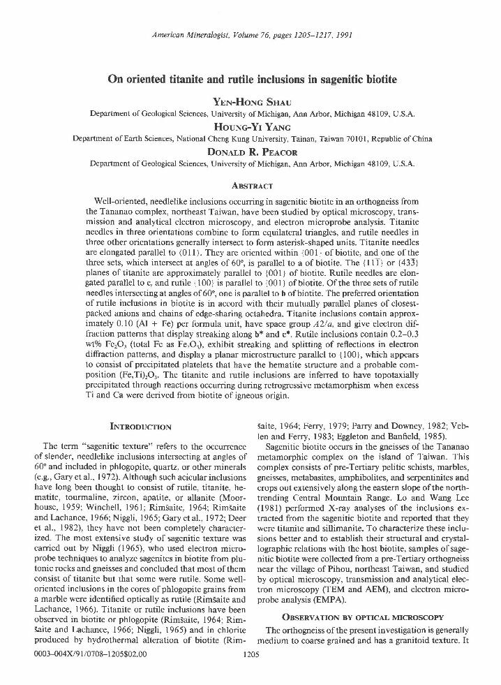
Fig. l. (a) and (b) Photomicrographs of two sagenitic biotite flakes. Titanite (T) needles form equilateral triangles and rutile (R)needles form asterisks in the biotite host. One of the equilateral triangles and one of the asterisks are indicated by white solid lines.The a and b axes of biotite were chosen arbitrarily using one of the three directions of titanite and one of the three directions ofrutile prisms, respectively. Plane-polarized light.
is composed of sodic plagioclase (Anro-or), quartz, biotite,and minor amounts of muscovite, potash feldspar, apa-tite, titanite (intergranular), garnet, chlorite, ilmenite, andtrace amounts of calcite, which generally occurs as smallinclusions (l-20 pm in diameter) lying along the partingplanes or cleavages of plagioclase. The biotite commonlyexhibits well-developed sagenitic texture. Some biotitegrains are overgrown by chlorite or have layers interca-lated with chlorite. No sagenitic inclusions have been ob-served in chlorite. However, a few isolated anhedral ti-tanite inclusions up to l0 pm wide and 40 pm long havebeen observed in chlorite, with their long dimensionssubparallel to {001} ofchlorite. Subhedral titanite grainsup to I mm long also occur at the interyranular sitesamong other major phases.
The sagenitic texture can best be examined in {001}sections of biotite. Very thin {001} sheets can easily bepeeled from a biotite crystal, and the following charac-teristic features of sagenitic texture can be readily ob-served in immenion oil with a polarizing microscope (Fig.l). Needlelike inclusions 0.1-2 p.m (most are <0.5 pm)in width with high relief generally form oriented patternsconsisting of equilateral triangles and asterisk-shapedunits. Triangles are formed by three sets of parallel nee-dles extending in three directions and intersecting at an-gles of 60' (+0.5). An asterisk-shaped pattern is formed
by three needles intersecting at angles of 60" but with acommon origin. The needles forming asterisks are in gen-eral shorter than those forming equilateral triangles.However, the needles radiating from nearby asterisks mayalso intersect to form smaller triangles. When the equi-lateral triangle and asterisk patterns occur together in asingle biotite flake, each of the three needles forming as-terisks is oriented perpendicular to one ofthe three setsof needles forming equilateral triangles. The distributionofequilateral triangles is generally quite even throughoutan entire flake of biotite, but the asterisks are clusteredand distributed unevenly. Inclusions of other types andshapes, such as lath-shaped ilmenite and prismatic apa-tite crystals that are usually much larger than the needle-like inclusions, were also observed in some biotite grains.Edges of ilmenite grains appear to be partially replacedby anhedral titanite.
Equilateral hiangles
Optical studies of single flakes of biotite have revealedthat the abundant needlelike inclusions forming equilat-eral triangles are oriented parallel to {00 I } ofbiotite. Oneset ofsuch needles is parallel to the a axis ofbiotite (Fig.l). As it is difficult to distinguish the directions of a fromb for the monoclinic cell of biotite (because of the smalloptic angle, 2V < 10") when biotite is observed in an
SHAU ET AL.: ORIENTED INCLUSIONS IN BIOTITE
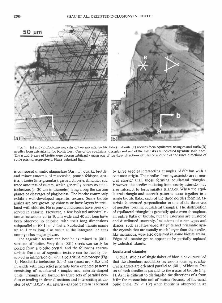
Taele 1. AEM analyses of rutile and titanite inclusions in biotite and biotite from the Tananao orthogneiss, northeast Taiwan
Rutile Titanite (ten analyses) Biotite (eight analyses)
t207
Oxides (wt%) 1 E-RU7 Range Range
sio,Al,o3Tio,FerO.*FeO'MnOMgoCaOKrO
Totalt'
SirrtAlretAlTiFep-F#*MnMgCaK
Total
0.880.00
98.920-200.000.000.000.000.00
100.00
0.010.000-000.99
<0.010.000.000.000.000.001.00
1.320.00
98.400.280.000.000.000.000.00
100.00
29.57-31.631.54-2.75
37.44-39.040.50-1.69
0.000.000.00
25.35-28.560.37-0.53
1.000.00
0.06-0.1 10.92-0.970.01-0.04
0.000.000.00
0.86-1.020.02-0.02
33.96J6.1115.68-18.051.49-3.1 1
0.0020.50-23.73o.174.268.21-9.420.00-0.108.88-10.0s
5.36-5.592.64-2.410.31-0.750.19-0.37
0.002.63J.130.02-0.031.93-2.170.00-0.021.75-2.02
35.301 7 . 1 62.240.00
21.710.219.01
<0.029.37
95.00
5.472.530.600.260.002.810.032.08
<0.011.85
15.63
30.532.04
38.480.960.000.000.00
26.540.45
99.00Normaf ization tactorsr 2 O (rutile), 1 Si (titanite), and 22 O (biotite)
0.o20.00
1.000.000.080.950.020.000.000.000.930.023.00
0.000.98
<0.010.000.000.000.000.001.00
ivote.'Values of 2o (trom counting statistics) tor cations of rutile: Si : 0.004, Ti : 0.01 ; for cations of titanite: Si : 0.02, Al : 0.008, Ti : 0.02, Fe: 0 . 0 0 2 , C a : 0 . 0 2 , K : 0 . 0 0 5 ; f o r c a t i o n s o f b i o t i t e : S i : 0 . 0 9 , > A t : 0 . 0 8 , T i : 0 . 0 2 , F e : 0 . 0 4 , M n : 0 . 0 1 , M g : 0 . 0 8 , K : 0 . 0 4 .
'Total Fe as FerO3 for rutile and titanite, and as FeO for biotite.". Oxide weight percents normalized to total 100.00% for rutile, 99.00% for titanite, and 95.00% for biotite.
ordinary optical microscope, X-ray precession photo-graphs were obtained to determine the orientations of aand b in the same biotite flakes as used in optical studies,however, reflections from the well-oriented inclusionswere not visible in precession photographs, probably be-cause of their very small volume. Optical observations athigh magnification show that most of the needles do notreally intersect but merely pass over or below others atdifferent levels. The needles forming equilateral trianglesexhibit inclined extinction, with an acute angle betweenthe fast ray and the elongated axis (length fast). For nee-dles belonging to any one of the three sets, some becomeextinct simultaneously with 18-25' clockwise rotationfrom the direction of polarization and the others with l8-25" counterclockwise rotation.
Asterisks
The needles forming asterisk-shaped patterns are alsooriented parallel to {001} ofbiotite. Sets ofthree needlesintersect with their ends meeting at a common centralpoint. Clustering and overlapping ofseveral asterisks werecommonly observed (Fig. la). One of the three sets isparallel to the b axis of biotite. Each direction of theneedles forming asterisks is therefore normal to the threedirections, respectively, of the needles forming equilateraltriangles. In contrast to the needles forming equilateraltriangles, those forming asterisks exhibit parallel extinc-tion and are length slow.
OssnnvA,rroN AND ANALysrs By TEM AND AEM
The biotite grains prepared for TEM observation wereoriented in two directions. For electron beam orienta-
tions approximately parallel to c of biotite, very thin {001}sheets were glued onto 3-mm diameter Cu or Al washers(or slot-shaped grids) with epoxy resin. For beam orien-tations parallel to {00 I }, biotite grains having {00 I } near-ly perpendicular to thin section surfaces were attached tothe washers. The 3-mm diameter specimens were ionmilled and then coated with a carbon film for observationwirh JEOL JEM-200CX, Hitachi H700-H, and PhilipsCM- l2 scanning transmission electron microscopes(STEMs). Conventional TEM bright field and dark fieldimages, selected area electron diffraction (SAED) pat-terns, and AEM analyses using the CM-12 STEMequipped with a Kevex energy dispersive spectrometer(EDS, Quantum detector) were obtained. AEM analyseswere obtained at counting rates of200-400 cps with ac-quisition times of 200 s (live time) and from thin edgesof ion-milled specimens where absorption effects werenegligible. The needlelike inclusions of titanite and rutileare usually less than 0.5 pm in width, and although un-resolved by EMPA, they can be precisely analyzed byAEM with a probe -0.01 pm in diameter (Table l). Thehost biotite was also analyzed, by AEM (with a rasteringarea of 0.2 pm x 0.2 pm) for comparison with EMPAresults (Table 2). Standard deviations calculated fromcounting statistics for major elements are similar for bothEMPA and AEM analyses. The thin foil approximationas proposed by Cliff and Lorimer (197 5) was used to cal-culate element proportions using experimentally deter-mined k-factors. As only the relative concentrations ofelements can be determined by these k-factors, weightpercents of the oxides for a mineral were calculated bynormalization, based on anhydrous totals that were as-
I 208 SHAU ET AL.: ORIENTED INCLUSIONS IN BIOTITE
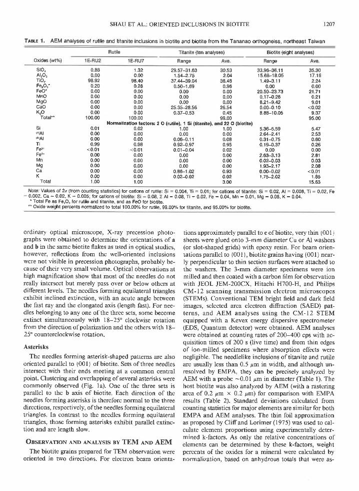
Trele 2. Electron microprobe analyses of biotite, muscovite, chlorite, and titanite from Tananao orthogneiss, northeast Taiwan
Biotite (27 analyses) Muscovite Titanite-Oxides and elements
(wto/o) Range Ave. Mu19
sio,Alro3Tio,Cr,O3FeO(Fe,O3)-'MnOMgoCaOBaONaroKrOFcl
Total
Sir4tAlrelAlTi
FeF(Fe3*)*MnMg16r>CaBaNaK> cationsFcl
35.03J5.561 6 .13-17.141.71J.830.00-o.04
20.80-22.570.20-0.367.65-8.640.00-0.1 00.06-0.320.05-0.1 29.33-9.650.03-0.300.00-0.05
5.479-5.5452.521-2.4550.431-0.6940.208-0.4460.000-0.0052.712-2.9360.026-0.0471.771-2.016
0.000-0.0170.004-0.0200.015-0.036't.851-1.927
0.01 5-0.1 450.000-0.012
5.5052.4950.5280.3390.0012.8620.0381.9045.6720.0050.0100.0231.888
15.5980.0670.005
14.0070.0000.005
0.0070.016
20.0250.0610.006
30.692.26
36.390.000.54o.210.00
28.770.140.03o.o20.570.00
99.62
1.0000.0000.0870.8920.0000.0130.0060.0000.9981.0040.0020.0020.0013.0070.0590.000
30.561.00
38.200.000.430.050.00
28.480.020.000.010.250.02
99.02
1.0000.0000.0390.9400.0000.0110.0010.0000.9910.999
<0.0010.000
<0.0012.9900.0250.001
35.35't6.472.900.01
21.980.298.200.030.170.089.510.140.02
95.15
46.62 24.7532.98 20.680.87 0.040.03 0.01212 28.140.04 0.481.42 13.030.03 0.060.28 0.080.49 0.04
10.40 <0.010.00 0.09o.02 0.02
95.30 87.42
0.0150.128
Normalization factor3: 22 O (B and M), 28 O (C), and 1 Si (T)6.251 5.3461.749 2.6543.464 2.6100.088 0.0070.003 0.0020.238 5.0830.005 0.0880.283 4.1964.081 1 1.9860.004 0.014
1.779 0.002
Nofe. 'Valuesol2o(f romcount ingstat is t ics) forcat ionsofbiot i te:Si :0.077,>Al :0.042,Ti :0.014,Cr:0.0003,Fe:0.092,Mn:0.007,M9:0 .038 , Ca :0 .0008 , Ba :0 .001 , Na :0 .004 , K :0 .045 , F : O .O27 , C l : 0 . 002 .
. | : intergranular, R : replacing ilmenite inclusions in biotite." Total Fe as FeO tor biotite, muscovite, and chlorite, and as FerO3 for titanite.
sumed to be 95.00/o for biotite, 99.00/o for titanite, and100.00/o for rutile (e.g., Shau et al., l99l). Hole-countspectra amount to only 0.50/o of the total counts for biotitespectra (-60000 counts for each analysis) acquired fromthin edges. Nevertheless, such stray radiation contributesup to 300/o of intensities for elements considered minorin titanite and rutile (e.g., K and Fe of titanite). Therefore,analytical data for titanite and rutile were corrected bysubtracting hole-count spectra. For biotite, the contri-bution of the hole-count spectrum was negligible.
Rutile
Major Ti and O and minor Si and Fe were detectedwith AEM analyses of needles forming the asterisks (Ta-ble 1). The electron diffraction patterns from these threesets of needles were indexed using the cell parameters ofrutile (a : 4.59, c :2.96 A;. ttrese data, in combinationwith the optical properties (very high relief, parallel ex-tinction, and length slow), show that those needles consistof rutile.
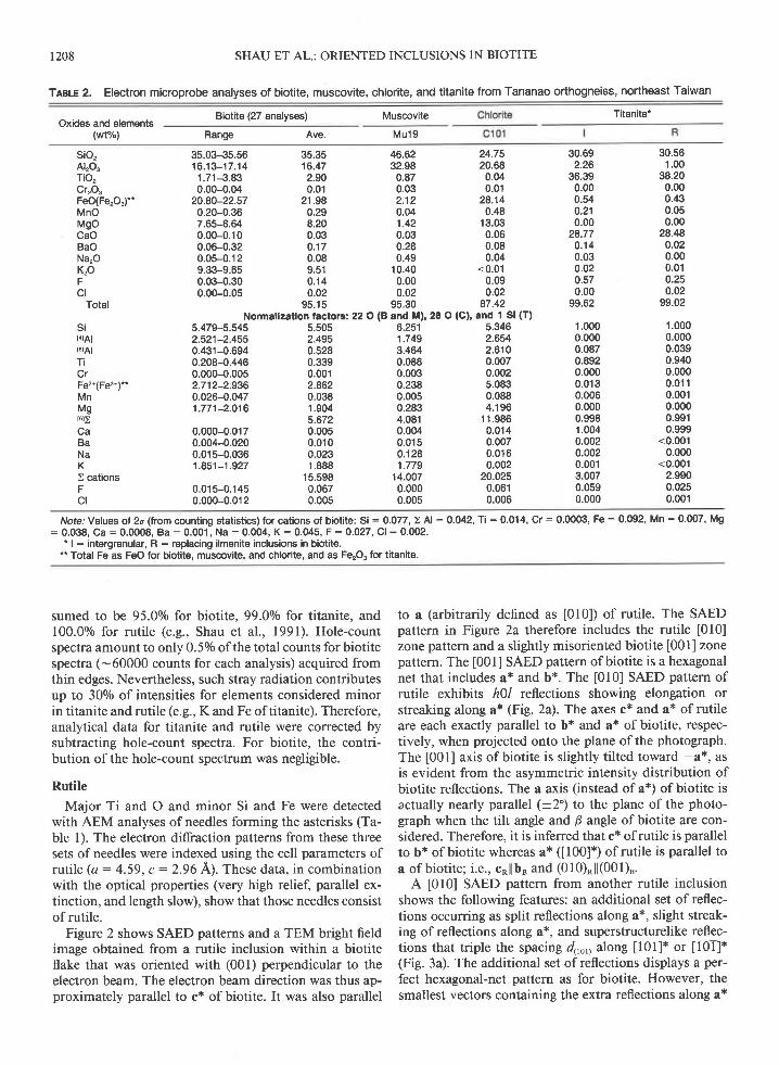
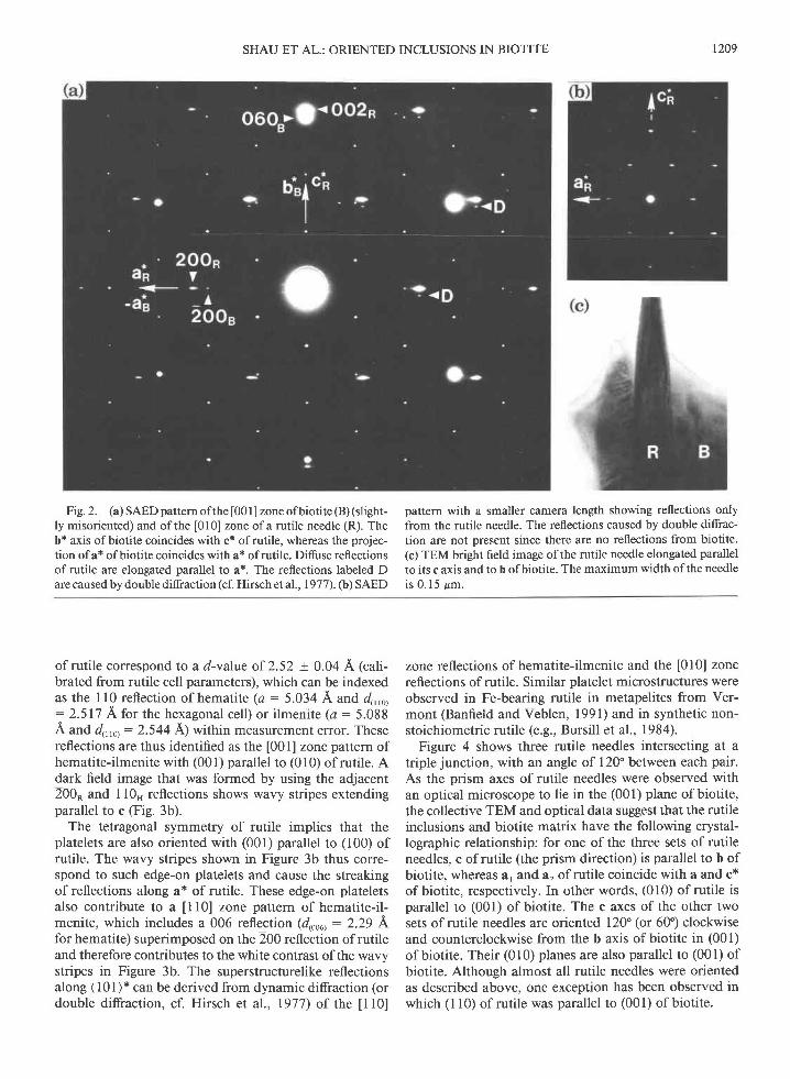
Figure 2 shows SAED patterns and a TEM bright fieldimage obtained from a rutile inclusion within a biotiteflake that was oriented with (001) perpendicular to theelectron beam. The electron beam direction was thus ap-proximately parallel to c* of biotite. It was also parallel
to a (arbitrarily defined as [010]) of rutile. The SAEDpattern in Figure 2a therefore includes the rutile [010]zone pattern and a slightly misoriented biotite [001] zonepattern. The [001] SAED pattern of biotite is a hexagonalnet that includes a* and b*. The [010] SAED pattern ofrutile exhibits h)l reflections showing elongation orstreaking along a* (Fig. 2a). The axes c* and a* of rutileare each exactly parallel to b* and a* of biotite, respec-tively, when projected onto the plane ofthe photograph.The [001] axis of biotite is slightly tilted toward -a*, asis evident from the asymmetric intensity distribution ofbiotite reflections. The a axis (instead ofa*) ofbiotite isactually nearly parallel (+2) to the plane of the photo-graph when the tilt angle and B angle ofbiotite are con-sidered. Therefore, it is inferred that c* of rutile is parallelto b* of biotite whereas a* ([00]*) of rutile is parallel toa of biotite; i.e., c*l lb" and (010)*ll(001)".
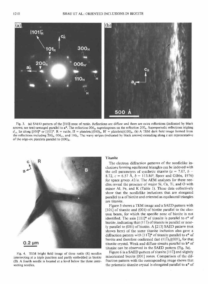
A [010] SAED pattern from another rutile inclusionshows the following features: an additional set of reflec-tions occurring as split reflections along a*, slight streak-ing ofreflections along a*, and superstructurelike reflec-tions that triple the spacing 4,0,y along [01]* or [01]*(Fig. 3a). The additional set ofreflections displays a per-fect hexagonal-net pattern as for biotite. However, thesmallest vectors containing the extra reflections along a*
SHAU ET AL.: ORIENTED INCLUSIONS IN BIOTITE r209
Fig.2. (a) SAED pattern ofthe [001] zone ofbiotite (B) (slight-ly misoriented) and of the [010] zone of a rutile needle (R). Theb* axis ofbiotite coincides with c* ofrutile, whereas the projec-tion ofa* ofbiotite coincides with a* ofrutile. Difuse reflectionsof rutile are elongated parallel to a*. The reflections labeled Dare caused by double diffraction (cf. Hirsch et al.,1977). (b) SAED
pattern with a smaller camera length showing reflections onlyfrom the rutile needle. The reflections caused by double diffrac-tion are not present since there are no reflections from biotite.(c) TEM bright field image ofthe rutile needle elongated parallelto its c axis and to b of biotite. The maximum width of the needleis 0 .15 &m.
of rutile correspond to a d-value of 2.52 + 0.04 A (cali-brated from rutile cell parameters), which can be indexedas the ll0 reflection of hematite (a: 5.034 A and d,,,o,: 2.517 A for the hexagonal cell) or ilmenite (a : 5.088A and d,,,0, :2.544 A1 wittrin measurement error. Thesereflections are thus identified as the [001] zone pattern ofhematite-ilmenite with (001) parallel ro (010) of rutile. Adark field image that was formed by using the adjacent200* and I 10" reflections shows wavy stripes extendingparallel to c (Fig. 3b).
The tetragonal symmetry of rutile implies that theplatelets are also oriented with (001) parallel to (100) ofrutile. The wavy stripes shown in Figure 3b thus corre-spond to such edge-on platelets and cause the streakingof reflections along a* of rutile. These edge-on plateletsalso contribute to a I l0] zone pattern of hematite-il-menite, which includes a 006 reflection (d,oou : 2.29 Lfor hematite) superimposed on the 200 reflection of rutileand therefore contributes to the white contrast ofthe wavystripes in Figure 3b. The superstructurelike reflectionsalong ( l0 I )* can be derived from dynamic diffraction (ordouble diffraction, cf. Hirsch eL al., 1977) of the [10]
zone reflections of hematite-ilmenite and the [010] zonereflections of rutile. Similar platelet microstructures wereobserved in Fe-bearing rutile in metapelites from Ver-mont (Banfield and Veblen, l99l) and in synthetic non-stoichiometric rutile (e.9., Bursill et al., 1984).
Figure 4 shows three rutile needles intersecting at atriple junction, with an angle of 120" between each pair.As the prism axes of rutile needles were observed withan optical microscope to lie in the (001) plane of biotite,the collective TEM and optical data suggest that the rutileinclusions and biotite matrix have the following crystal-lographic relationship: for one of the three sets of rutileneedles, c of rutile (the prism direction) is parallel to b ofbiotite, whereas ar and a2 of rutile coincide with a and c*of biotite, respectively. In other words, (010) of rutile isparallel to (001) of biotite. The c axes of the other twosets of rutile needles are oriented 120" (or 60') clockwiseand counterclockwise from the b axis of biotite in (001)of biotite. Their (010) planes are also parallel to (001) ofbiotite. Although almost all rutile needles were orientedas described above, one exception has been observed inwhich (l l0) of rutile was parallel to (001) of biotite.
t210 SHAU ET AL.: ORIENTED INCLUSIONS IN BIOTITE
Fig. 3. (a) SAED pattern of the [010] zone of rutile. Reflections are difuse and there are extra reflections (indicated by blackarrows; see text) arranged parallel to a*. The reflection 006n, superimposes on the reflection 200". Superperiodic reflections triplingd,o, lie along [01]* or [0T]*. R : rutile, H : plareletsll(010)R, H' : plateletsll(100)R. (b) A TEM dark field image formed fromthe reflections including 200R, 006H,, and I 10". The wavy stripes (indicated by black arrows) extending along c are representativeofthe edge-on platelets parallel to (100)*.
Fig. 4. TEM bright field image of three rutile (R) needlesintersecting at a triple junction and partly embedded in biotite(B). A fourth needle is located at a level below the three inter-secting needles.
Titanite
The electron diffraction patterns of the needlelike in-clusions forming equilateral triangles can be indexed withthe cell parameters of synthetic titanite (a : 7 .07, b :
8 .72 , c :6 .57 A , b :113 .86 ' , Spee r and G ibbs , 1976 )for space gtoup A2/a. The AEM analyses for these nee-dles reveal the presence of major Si, Ca, Ti, and O withminor Al, Fe, and K (Table l). These data collectivelyshow that the needlelike inclusions that are elongatedparallel to a ofbiotite and oriented as equilateral trianglesare titanite.
Figure 5 shows a TEM image and a SAED pattern with
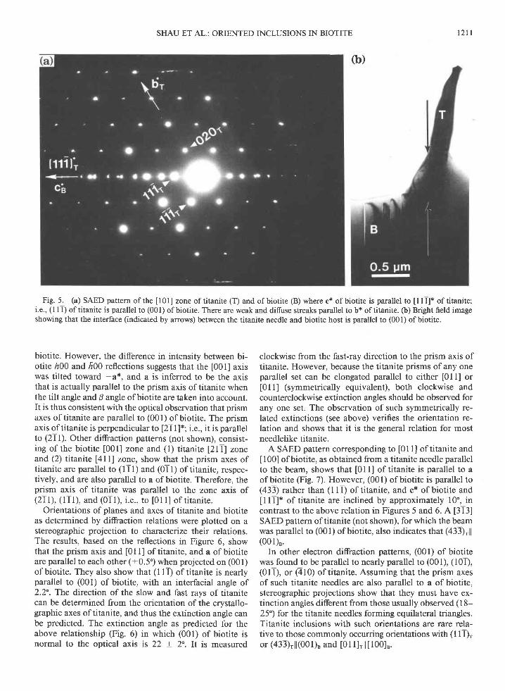
[01] of titanite and (001) of biotite parallel to the elec-tron beam, for which the specific zone of biotite is notidentified. The axis I lT]* of titanite is parallel to c* ofbiotite, indicating that (l I l) oftitanite is parallel or near-ly parallel to (001) of biotite. A [213] SAED pattern (notshown here) of the same titanite inclusion also gave adiffraction pattern with I tT]x oftitanite parallel to c* ofbiotite and therefore confirmed that (l1T)rll(001)" for thattitanite crystal. Weak and diffuse streaks parallel to b* oftitanite can be observed in the SAED pattern (Fig. 5a).
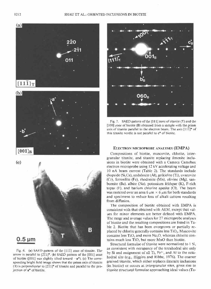
Figure 6 is a SAED pattern of titanite I I l] and slightlymisoriented biotite [001] zones. Comparison of the dif-fraction pattern with the corresponding image shows thatthe prismatic titanite crystal is elongated parallel to a* of
SHAU ET AL.: ORIENTED INCLUSIONS IN BIOTITE L2rr
Fig. 5. (a) SAED pattern of the [l01] zone of titanite (T) and of biotite (B) where c* of biotite is parallel to [111]* of titanite;i.e., (l 1 1) oftitanite is parallel to (001) ofbiotite. There are weak and diffuse streaks parallel to b* oftitanite. (b) Brieht field imageshowing that the interface (indicated by arrows) between the titanite needle and biotite host is parallel to (001) ofbiotite.
biotite. However, the difference in intensity between bi-otite r00 and h00 reflections suggests that the [001] axiswas tilted toward -a*, and a is inferred to be the axisthat is actually parallel to the prism axis oftitanite whenthe tilt angle and B angle ofbiotite are taken into account.It is thus consistent with the optical observation that prismaxes of titanite are parallel to (001) of biotite. The prismaxis oftitanite is perpendicular to [2T I ]*; i.e., it is parallelto (21l). Other diffraction patterns (not shown), consist-ing of the biotite [001] zone and (l) titanite [2lT] zoneand (2) titanite [4ll] zone, show that the prism axes oftitanite are parallel to (1Tl) and (0Tl) of titanire, respec-tively, and are also parallel to a ofbiotite. Therefore, theprism axis of titanite was parallel to the zone axis of(21 l), ( l I l), and (01 l), i .e., to [0] l] of t itanire.
Orientations of planes and axes of titanite and biotiteas determined by diffraction relations were plotted on astereographic projection to characterize their relations.The results, based on the reflections in Figure 6, showthat the prism axis and [0 I I ] of titanite, and a of biotiteare parallel to each other (+0.5') when projected on (001)ofbiotite. They also show that (l lT) oftitanite is nearlyparallel to (001) of biotite, with an interfacial angle of2.2. The direction of the slow and fast rays of titanitecan be determined from the orientation of the crystallo-graphic axes oftitanite, and thus the extinction angle canbe predicted. The extinction angle as predicted for theabove relationship (Fig. 6) in which (001) of biotite isnormal to the optical axis is 22 + 2. It is measured
clockwise from the fast-ray direction to the prism axis oftitanite. However, because the titanite prisms of any oneparallel set can be elongated parallel to either [0ll] or[0Tl] (symmetrically equivalent), both clockwise andcounterclockwise extinction angles should be observed forany one set. The observation of such symmetrically re-lated extinctions (see above) verifies the orientation re-lation and shows that it is the general relation for mostneedlelike titanite.
A SAED pattern corresponding to [0] l] of titanite and[00] of biotite, as obtained from a titanite needle parallelto the beam, shows that [0ll] of titanite is parallel to aof biotite (Fig. 7). However, (001) of biotite is parallel to(433) rather than (l lT) of titanite, and c* of biotite andI l l]* of titanite are inclined by approximately 10", incontrast to the above relation in Figures 5 and 6. A [313]SAED pattern of titanite (not shown), for which the beamwas parallel to (001) of biotite, also indicates that (433)rll(001)".
In other electron diffraction patterns, (001) ofbiotitewas found to be parallel to nearly parallel to (001), (101),(0lT), or (410) of titanite. Assuming that the prism axesof such titanite needles are also parallel to a of biotite,stereographic projections show that they must have ex-tinction angles different from those usually observed (l 8-25') for the titanite needles forming equilateral triangles.Titanite inclusions with such orientations are rare rela-tive to those commonly occurring orientations with (11l)ror (433),l l(001)" and [0] l]r l l l l00lB.
(b)
t 2 1 2 SHAU ET AL.: ORIENTED INCLUSIONS IN BIOTITE
(c)
Fig.6. (a) SAED pattern of the [1ll zone of titanite. Thearrow is parallel to [211]*. (b) SAED pattern ofthe [001] zoneof biotite ([001] was slightly tilted toward -a*). (c) The corre-sponding bright field image shows that the prism axis of titanite(T) is perpendicular to [2 I I ]* of titanite and parallel to the pro-iection of a* of biotite.
Fig. 7 . SAED pattern of the [0 ] I ] zone of titanite (T) and theu00l zone of biotite (B) obtained from a sample with the prismaxis of titanite parallel to the electron beam. The axis I I1]* ofthis titanite needle is not parallel to c* ofbiotite.
Er,ncrnoN MrcRopRoBE ANALvsES (EMPA)
Compositions of biotite, muscovite, chlorite, inter-granular titanite, and titanite replacing ilmenite inclu-sions in biotite were obtained with a Cameca Camebaxelectron microprobe using l2 kV accelerating voltage andl0 nA beam current (Table 2). The standards includediopside (Si,Ca), andalusite (Al), geikielite (Ti), uvarovite(Cr), ferrosilite (Fe), rhodonite (Mn), olivine (Mg), san-bornite (Ba), albite (Na), potassium feldspar (K), F-richtopaz (F), and barium chlorine apatite (Cl). The beamwas rastered overan area6 pm x 6 pm forboth standardsand specimens to reduce loss of alkali cations resultingfrom diffusion.
The composition of biotite obtained with EMPA isconsistent with that obtained with AEM, except that val-ues for minor elements are better defined with EMPA.The range and average values for 27 microprobe analysesof biotite and the resulting compositions are listed in Ta-ble 2. Biotite that has been overgrown or partially re-placed by chlorite generally contains less TiOr. Muscovitecontains less TiO, and more NarO, whereas chlorite con-tains much less TiO, but more MnO than biotite.
Structural formulae of titanite were normalized to I Si,as consistent with occupancy ofthe tetrahedral site onlyby Si and assignment of all Ti, Fe3*, and Al to the octa-hedral site (e.g., Higgins and Ribbe, 1976). The coarsergrained titanite, which either replaces ilmenite inclusions(in biotite) or occurs at intergranular sites, gives rise totitanite structural formulae approaching ideal values (Ta-
[001]s
ble 2). The titanite also contains 0.25-0.79 wto/o of F.However, the titanite replacing ilmenite contains muchless AlrO. and F but slightly more TiOr. When comparedwith the acicular titanite inclusions in biotite, the coarser-grained titanite exhibits lower FerO, and KrO contentsand slightly higher CaO content.
DrscussroN
Mineral chernistry and microstructures ofrutile and titanite
Optical microscopy, AEM microanalysis, and electrondiffraction have shown that the needlelike inclusionsforming the asterisks are rutile and those forming theequilateral triangles in projection on (001) ofbiotite aretitanite. The optical observations and electron diffractiondata show that the rutile and titanite inclusions generally,but not always, have a preferred orientation in biotite.
The rutile inclusions contain minor amounts of Si andFe but no detectable Ta5* or Nb5* that may be coupledwith substitution of Fe3* or Fe2*, as observed by othersfor rutile (cf. analyses compiled in Deer et al., 1962b,Rumble, 1976). As described above, the SAED patternsofrutile exhibit features ofstreaking, reflections from anextra phase, and superstructurelike reflections. Thestreaking of reflections along a* of rutile in SAED pat-terns (Figs. 2 and3) implies the presence of platelet struc-tures parallel to (100) of rutile, as directly seen in darkfield images (Fig. 3b). The reflections from these platelets,though most are superimposed on the reflections of rutile,can be indexed as hematite-ilmenite with (001)rll(100)*.The superstructurelike reflections can also be attributedto dynamic diffraction from these platelets and rutile.Furthermore, the additional set of reflections (Fig. 3a) isconsistent with the [001] zone pattern of hematite-ilmen-ite that is oriented with [T20]Hllc* (i.e., a..n I c.) and(001)H l l (010)R.
Similar streaking and splitting of reflections, super-structurelike reflections, and platelet structures were ob-served by Banfield and Veblen ( I 99 I ) in Fe-bearing rutile(FeO : 0.5-3 wto/o) from chlorite- to staurolite-grademetapelites. They have concluded that coherently inter-grown hematite (or ilmenite) platelets precipitated par-allel to (100) and (010) of rutile and that the streaking ofreflections is due to platelets of irregular spacing, whereasthe superstructurelike reflections are caused by dynamicdifraction. Putnis and Wilson (1978) and Putnis (1978)noted that experimental annealing of natural Fe-bearingrutile (FeO : 0.55 wto/o) at temperatures between 47 5 and595'C resulted in precipitation of two transitional phases(coherent Fe-rich precipitates) and hematite. Bursill et al.( I 984) observed { I 00} platelet defects, often occurring inassociation with crystallographic shear planes, in syn-thetic nonstoichiometric rutile (TiOr_", 0 = x < 0.0035).They srrggested that the platelet defects precipitated as aTirO, phase with the corundum-type structure and thatthe microstructures are strongly determined by coolinghistory. The { 100} platelet defects that they observed have
r213
0.0
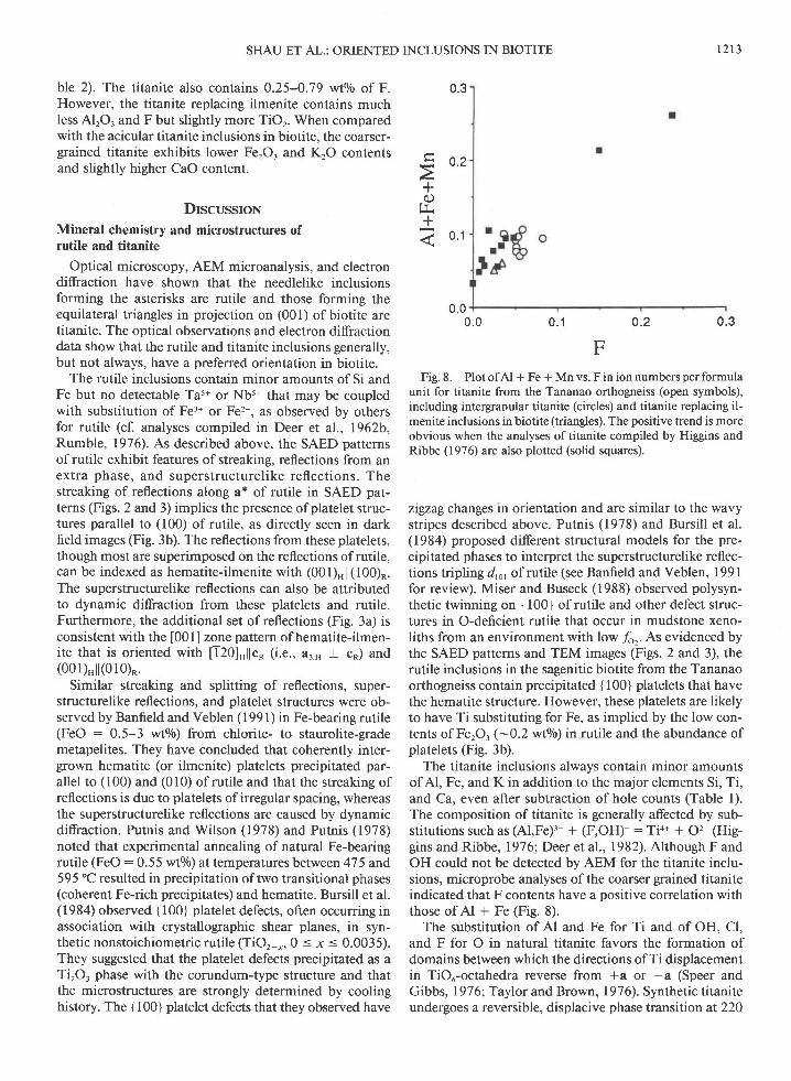
FFig. 8. Plot of Al + Fe + Mn vs. F in ion numbers per formula
unit for titanite from the Tananao orthogneiss (open symbols),including intergranular titanite (circles) and ritanite replacing il-menite inclusions in biotite (triangles). The positive trend is moreobvious when the analyses of titanite compiled by Higgins andRibbe (1976) are also plotted (solid squares).
zigzag changes in orientation and are similar to the wavystripes described above. Putnis (1978) and Bursill et al.(1984) proposed different structural models for the pre-cipitated phases to interpret the superstructurelike reflec-tions tripling d,o, of rutile (see Banfield and Veblen, l99lfor review). Miser and Buseck (1988) observed polysyn-thetic twinning on {100} of rutile and other defect struc-tures in O-deficient rutile that occur in mudstone xeno-liths from an environment with low /o' As evidenced bythe SAED patterns and TEM images (Figs. 2 and 3), therutile inclusions in the sagenitic biotite from the Tananaoorthogneiss contain precipitated { 100} platelets that havethe hematite structure. However, these platelets are likelyto have Ti substituting for Fe, as implied by the low con-tents of FerOr(-0.2 wtolo) in rutile and the abundance ofplatelets (Fig. 3b).
The titanite inclusions always contain minor amountsof Al, Fe, and K in addition to the major elements Si, Ti,and Ca, even after subtraction of hole counts (Table l).The composition of titanite is generally afected by sub-stitutions such as (Al,Fe)3* + (F,OH)- : Ti4+ + 6:- lHig-gins and Ribbe, 197 6; Deer et al., 1982). Although F andOH could not be detected by AEM for the titanite inclu-sions, microprobe analyses of the coarser grained titaniteindicated that F contents have a positive correlation withthose of Al + Fe (Fig. 8).
The substitution of Al and Fe for Ti and of OH. Cl.and F for O in natural titanite favors the formation ofdomains between which the directions of Ti displacementin TiOu-octahedra reverse from +a or -a (Speer andGibbs, 19761-Taylor and Brown, 1976). Synthetic titaniteundergoes a reversible, displacive phase transition at 220
SHAU ET AL.: ORIENTED INCLUSIONS IN BIOTITE
0.3
+f r
+e 0.1
0.30 .20 . 1
tzt4 SHAU ET AL.: ORIENTED INCLUSIONS IN BIOTITE
oo
+.g)
(.)f Y
0.60
0.55
0.500 . 1 0.2 0 .3
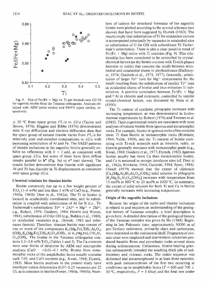
TiFig. 9. Plot of Fe/(Fe + Mg) vs. Ti per formula unit (22 O)
for sagenitic biotite from the Tananao orthogneiss. Analyses ob-tained with AEM (solid circles) and EMPA (open circles), re-spectively.
+ 20 "C from space group P2,/a to A2/a (Taylor andBrown, 1976). Higgins and Ribbe (1976) demonstratedwith X-ray diffraction and electron diffraction data thatthe space group of natural titanite varies from P2,/a forrelatively pure end-member compositions to A2/a wthincreasing substitution of Al and Fe. The SAED patternsoftitanite inclusions in the sagenitic biotite generally ex-hibit no reflections with k + / odd, as consistent withspace group A2/a, but some of them have faint diffusestreaks parallel to b* (Fig. 5a) or c* (not shown). Theresults further demonstrate that titanite with significantAl and Fe has disorder in Ti displacements as consistentwith space group A2/a.
Chemical relations for titanian biotite
Biotite commonly has up to a few weight percent ofTiO, (l-5 wto/o) and less than I wo/o of CaO (e.g., Foster,1960a, 1960b; Deer er al., 1962a). The Ti in biotite islocated in octahedrally coordinated sites, and its substi-tution is coupled with substitution of Al for Si (i.e., Ti-Tschermak's substitution: Tio* + 2,\lt* : Mg2r + 2Si4+'e.g., Robert, 1976; Guidotti, 1984; Hewitt and Wones,1984), substitution of O for OH (e.g., Bohlen et al., 1980),or octahedral vacancies (e.g., Dymek, 1983 and refer-ences therein). Therefore, titanian biotite may consist ofone or more of the components Kr(Mg,Fe)5TiSi4Al4Oro(OH)",Kr(Mg,Fe)rTiSiuAlrOrr(OH)r, or K,(Mg,Fe)oTiSi6Al,Oro(OH)o. The biotite in the Tananao orthogneiss con-tains I .5-3.8 wto/o TiO, (Tables I and 2). The Ca contentswere near limits of detection by AEM and microprobeanalyses (CaO : <0.01-0.1 wto/o). Biotite from meta-morphic rocks of the amphibolite facies usually containssuch TiO, and CaO contents (e.g., Kwak, 1968; Dymek,1983). Most biotite analyses in the present study haveinterlayer-cation defi ciencies (0. 0 7-0. 2 5 v acancies per 22O), as is common in biotite (Foster, 1960a, 1960b). Num-
bers of cations for structural formulae of the sageniticbiotite were plotted according to the several schemes (notshown) that have been suggested by Dymek (1983). Theresults imply that substitution ofTi for octahedral cationsis accompanied principally by vacancies in octahedral sitesor substitution of O for OH with subordinate Ti-Tscher-mak's substitution. There is also a clear positive trend ofFe/(Fe + Mg) ratios with Ti contents (Fig. 9). This rela-tionship has been considered to be controlled by crystal-chemical factors (as the biotite coexists with Ti-rich phasesilmenite or rutile) that concern the misfit between tetra-hedral and octahedral sheets in phyllosilicates (Dallmey-er, 197 4; Guidotti et al., 197 5, 1977). Generally, substi-tution of larger Fe2* ions for Mg2n compensates for themisfit resulting from the substitution of smaller Tio* ionsin octahedral sheets ofbiotite and thus enhances Ti sub-stitution. A positive correlation between Fe/(Fe + Mg)and I4rAl in chlorite and corrensite, controlled by similarcrystal-chemical factors, was discussed by Shau et al.(1990).
The Ti content of synthetic phlogopite increases withincreasing temperature, as was demonstrated in hydro-thermal experiments by Robert (1976) and Tronnes et al.(1985). Their experimental results are consistent with mostanalyses of natural biotite from igneous and metamorphicrocks. For example, biotite in igneous rocks often containsmore Ti than biotite in metamorphic rocks (RimSaite,1964; Velde, 1969), and the Ti content ofbiotite coex-isting with Ti-rich minerals such as ilmenite, rutile, ortitanite generally increases with metamorphic grade (e.g.,Kwak, I 968 ; Guidotti et al ., 197 7 ; Dymek, I 9 8 3). Igneousbiotite usually has more Ca than metamorphic biotite,and Ca is assumed to occupy interlayer sites (cf. Deer etal.,I962a; Rim5aite, 1964;Guidotti, 1984; Speer, 1984).Olesch (1979) showed that the extent of clintonite[Car(MgoAl,)Si,Al6Or0(OH)"] solid solution in phlogopite
[KrMguSiuAlrOr0(OH)o] increases with temperature from13 molo/o at 800 "C to 20 molo/o al 1020 "C. In summary,the extent of solid solution for both Ti and Ca in biotitegenerally increases with increasing temperature.
Origin of the sagenitic inclusions
Because the origin of the rutile and titanite inclusionsis related to and requires an understanding ofthe geolog-ical history of Tananao complex, a brief description isgiven here. A detailed description ofthe geological historyof the Tananao complex was given by Ho (1988). Begin-ning in late Paleozoic time, approximately 50000 m ofpre-Tertiary sediments, primarily clays and carbonates,were deposited on the continental shelf. Fragments of oce-anic crust were emplaced and intermittent volcanism pro-duced basaltic flows and pyroclastic rocks several timesduring sedimentation. Cretaceous, biotite-bearing gran-ites subsequently intruded the resulting thick pile of sed-imentary and volcanic rocks. The entire sequence wasdeformed and metamorphosed in at least three episodes,with peak metamorphism of the first two occurring atconditions up to amphibolite facies (Z: 600 and 700 +50 "C, respectively, P: 5 kbar) and the final one under
n q0 .4
SHAU ET AL.:ORIENTED INCLUSIONS IN BIOTITE L2I5
conditions ofthe greenschist facies (T:350-475 "C andP : 5 kbar) (Chen et al., 1983). The orthogneiss wasderived from the granitic intrusions following the secondamphibolite facies metamorphism. The entire sequenceof metamorphic rocks, consisting of schists, amphibolites,serpentinites, marbles, metabasites, and gneisses, wasknown as the Tananao schists in the past but as the Tana-nao complex in recent years.
Yang and Wang Lee ( 198 I ) found that titanite is a com-mon accessory mineral in the gneisses and other meta-morphic rocks of the Tananao complex and that titanitehas wholly or partially replaced ilmenite in the ortho-gneisses and amphibolites derived from basic igneousrocks. They thus concluded that titanite was stable andshould have crystallized if Ti, Ca, and Si were availableunder the metamorphic conditions for the Tananao com-plex. Rutile was also shown to be stable in the tempera-ture and pressure ranges of the metamorphism (cf. Jam-ieson and Olinger, 1969).
Preferred orientation occurring in two intimately co-existing phases is commonly a result of exsolution or to-potaxial (topotactic) or epitaxial intergrowth. However,exsolution requires that the reactant components are equalto those of the sum of the products; simple exsolution oftitanite and rutile is therefore precluded, as the additionof rutile and titanite components to the biotite leads to anonsensible formula for the hypothetical precursor. Theprocess must therefore involve addition or loss of one ormore components. Because Ti and Ca are common com-ponents of biotite and because rutile and titanite are ran-domly distributed throughout biotite, the source of the Tiand Ca of titanite and rutile was most likely as a solidsolution component of the biotite. These relations collec-tively require dissolution and recrystallization of at leasta portion of the original biotite, with gain or loss of com-ponents.
As discussed above, the solubilities of Ti and Ca inbiotite generally increase with increasing temperature. Ifbiotite of an igneous origin was metamorphosed underthe amphibolite or greenschist facies conditions deter-mined for the Tananao complex, high Ti and Ca com-ponents inherited from higher temperature conditionscould well be in excess of the limits allowed at lowertemperatures. Reactions involving the breakdown of theTi- and Ca-rich biotite to Ti- and Ca-depleted biotite plustitanite, rutile, and other phases might thus take placeduring metamorphism when biotite with high Ti and Cacontents was no longer stable. Studies of hydrothermalexperiments also showed that Ti-rich phlogopite formingat higher temperatures (= 1000 "C) breaks down to assem-blages of Ti-depleted phlogopite, rutile, and sanidine orvapor at lower temperatures (s800 "C) (Forbes and Flow-er,1974; Tronnes et al., 1985). They also showed that theTi content of phlogopite increases with increasing tem-perature and decreasing pressure for a given bulk TiO,content.
Alternatively, titanite and rutile could have formed con-comitantly with the topotaxial breakdown of biotite tochlorite (cf. Veblen and Ferry, 1983; Eggleton and Ban-
field, 1985) during an early retrogressive episode ofpoly-metamorphism. Titanite or rutile formed in this mannerwould likely be observed as well-oriented acicular inclu-sions in chlorite, as reported by Rim5aite (1964), al-though titanite usually occurs as relatively large crystalswithout preferred orientation in chlorite (Dodge, 1973;Ferry,1979 Parry and Downey, 1982; Veblen and Ferry,1983). Baxter and Peacor (unpublished data) observedboth titanite and rutile intergrown with interlayered chlo-rite and biotite in samples of altered tufaceous rocks fromNew Z.ealand, where the chlorite was an alteration prod-uct of Ti-rich detrital biotite. Ferry (1979) suggested areaction that accounts for the formation of chlorite andtitanite from Ti-bearing biotite by hydrothermal alter-ation in granitic rocks. The chlorite thus formed in anearly stage of retrograde metamorphism might havetransformed topotaxially to biotite and retained well-ori-ented titanite and rutile in biotite during a final progres-sive episode of the polymetamorphism, which occurredat the conditions well above the biotite isograd accordingto several prograde reactions (e.g., Ernst, 1963; Brown,r97 t).
However, since a sagenitic biotite with well-orientedinclusions of titanite and rutile may also be altered tochlorite and retain the preexisting inclusions in chlorite,existence of sagenitic chlorite does not necessarily implythat the titanite and rutile inclusions formed during al-teration of biotite to chlorite. In addition, titanite, whichhas been characterized as one ofthe products during al-teration ofbiotite to chlorite, does not generally occur aswell-oriented, needlelike inclusions like those in this study(Dodge, 1973; Ferry, 1919; Parry and Downey, 1982;-Veblen and Ferry, 1983). Therefore, it is more likely thatthe sagenitic biotite formed by a precipitation mechanismwhere titanite, rutile, and Ti-depleted biotite crystallizedtopotaxially during metamorphism through the break-down of a Ti- (and Ca-) rich biotite that had formed underconditions of higher temperatures (e.g., igneous biotite)than those of the metamorphism.
Chlorite observed in the present investigation, which ispresumed to replace biotite or overgrow biotite during thelatest greenschist facies metamorphism, does not containwell-oriented acicular inclusions. It apparently has no re-lationship with the formation of acicular inclusions inbiotite. In addition, the rutile inclusions in biotite containprecipitated hematite-ilmenite platelets that should post-date the formation of rutile (cf. Banfield and Veblen, 199 I ).Precipitation of hematite in rutile took place within anapproximate temperature range of 450-600 "C, accordingto observations of experimentally annealed rutile (Putnis,1978; Putnis and Wilson, 1978), though the precipitationtemperatures are probably lower, based on the observa-tions by Banfield and Veblen (1991) and Bursill et al.(1984). Therefore, precipitation of rutile and titanite inthe sagenitic biotite probably took place during the am-phibolite facies metamorphism or following retrogrademetamorphism at temperatures higher than those of thelatest greenschist facies metamorphism for the Tananaocomplex. Other examples of precipitated inclusions, such
t 2 t 6
as well-oriented magnetite inclusions in pyroxenes (Fleetet al., 1980) and hematite inclusions in sillimanite (Fleetand Arima, 1985), have been reported as occurring inmetamorphic rocks with a complex retrograde history.
Structural control of the well-oriented inclusionsin biotite
Optical and TEM observations showed that one of thethree sets of intersecting rutile needles forming asterisksis parallel to b of biotite. The prism axes of rutile needleare parallel to c, and (100) or (010) of rutile are parallelto (001) of biotite. The rutile structure is composed ofparallel chains ofedge-sharing TiOu octahedra that extendparallel to c and are connected laterally by sharing vertices.The O layer, which is parallel to ( 100) or (0 10) of rutile,is nearly closest-packed, though slightly puckered. Thebiotite structure is also based on closest packing ofanions,with anion layers being parallel to (001) of biotite. (Draw-ings of the rutile and biotite structures can be found inmany standard textbooks of mineralogy and will not begiven here.) Preferred orientation involving ( I 00) or (0 I 0)of rutile and (001) of biotite is therefore understandablein terms of their similar closest-packed geometries. Fur-thermore, the (Mg,Fe,Al,Ti)(O,OH)u octahedra ofthe bru-cite-like octahedral sheet of biotite form chains of octa-hedra by sharing edges, as in rutile. Such chains extendin three directions, each oriented 60" from the others andconforming to the pseudohexagonal symmetry of (001) ofbiotite. During dissolution (of biotite) and crystallization(of rutile), migration of Ti into those octahedra in sub-stitution of Mg, Al, and Fe and replacement of OH by Ocould easily convert the edge-sharing (Mg,Fe,Al,Ti)-(O,OH)6 octahedra into chains of edge-sharing TiOu oc-tahedra to form a pattern ofthree rutile needles with endsintersecting at angles of 120" (Fig. a). Superposition ofanother pattern that consists of three needles extendingin the directions opposite to those of the first patterncreates an asterisk pattern with adjacent needles at anglesof 60' (Figs. I and 4). Growth of rutile needles thus in-volves minimum reconstruction and cation diffusion.
Fleet and Arima (1985) studied oriented hematite in-clusions in sillimanite and indicated that (120) of he-matite is parallel to c of sillimanite, which is the directioncontaining chains of edge-sharing octahedra for bothphases. The orientation relationships of magnetite inclu-s ions in pyroxenes, inc luding { I I I }Ml l { 100}" and( I I 2 ), ll c", are also related to closest-packed O layers andchains of edge-sharing octahedra (Fleet et al., 1980).Platelets of hematite or (Fe,Ti)rO, inclusions in rutile with{001}Hll{100}. and (120)"l lc. have been observed in thepresent and other studies (Putnis and Wilson, 1978; Put-nis, 1978; Bursill el al., 1984; Banfield and Veblen, 1991).Gilkes and Suddhiprakarn (1979) reported a preferredorientation between goethite and biotite with{100}cll {001}u and collbu in deeply weathered graniticrocks, although other orientations between goethite andbiotite have been observed by Banfield and Eggleton(1988). All these preferred crystallographic relations be-
SHAU ET AL.: ORIENTED INCLUSIONS IN BIOTITE
tween rutile and biotite, magnetite and pyroxenes, rutileand hematite, and goethite and biotite have a commonstructural similarity, in that the two coexisting phasesshare nearly closest-packed anion layers and chains ofedge-sharing octahedra within the anion layers and arethus topotaxially controlled (cf. Fleet, 1982; Fleet andArima. 1985).
Although most titanite inclusions exhibit prefened ori-entation in biotite, TEM data have shown that many dif-ferent orientations with respect to (001) of biotite alsooccur. It is estimated that more than 800/o of the titaniteinclusions are elongated parallel to (0 I I ) and to the threedirections forming equilateral triangles, with { l l l } or {43 3 }of titanite approximately parallel to {001 } ofbiotite. How-ever, in contrast to the single kind oforientation occurringalmost exclusively for rutile, other orientations in which
{001}, {l0T}, {0lT}, or {410} of t itanite are parallel to
{001} of biotite were also observed. An orientation with
{ I l0} of titanite parallel to {001} of mixed-layer biotite-chlorite in altered granitic biotite from southeastern Aus-tralia was observed by Eggleton and Banfield (1985). Thevariable orientations of titanite with respect to {001} ofbiotite indicate that titanite does not have well-definedstructural features in common with biotite. Nevertheless,the preferred orientation (0ll)'lla" implies some struc-tural control, but we have been unable to define one.
AcrNowr,nocMENTS
The authors are grateful to Eric J. Essene, Tamsin C. McCormick, andJ. Alexander Speer for critical reviews ofthe manuscript. We thank PouyanShen and Su-Cheng Yu for many helpful discussions. Transmission andanalltical electron microscopic work was carried out at the Institute ofMining, Metallurgy and Material Sciences of the National Cheng KungUniversity and the Institute of Materials Science and Engineering of theNational Sun Yat-Sen University in Taiwan, Republic of China, and inthe Electron Microbeam Analysis laboratory, the University of Michigan,Ann Arbor, Michigan. A titanite standard (B20360) for AEM analyses waskindly provided by the National Museum ofNatural History, SmithsonianInstitution. The present study was supported by the grants NSC-67M-0202- | 2(02\, NSC-7 5-M006-0202-03, and NSC-7 6-M006-0202-02 toH.-Y.Y. from the National Science Council of the government of theRepublic of China, and by NSF grant EAR-8817080 to D.R.P. The CM-l2 STEM was acquired under NSF grant EAR-8708276 and the electronmicroprobe under NSF grant EAR-8212764. Contribution no. 476 fromthe Mineralogical l:boratory, Department of Geological Sciences, Uni-versity of Michigan, Ann Arbor, Michigan 48109, U S.A.
RnrnnoNcns crrED
Banfield, J.F, and Eggleton, R A (1988) Transmission electron micro-scope study ofbiotite weathering. Clays and Clay Minerals, 36,47-60.
Banfield, J F., and Veblen, D.R. (1991) The stmcture and origin of Fe-bearing platelets in metamorphic rutile. American Mineralogist, 76,ll3-127.
Bohlen, S.R., Peacor, D.R., and Essene, E.J. (1980) Crystal chemistry ofa metamorphic biotite and its significance in waterbarometry. AmericanMineralogist, 65, 55-62.
Brown, E.H. (1971) Phase relations of biotite and stilpnomelane in thegreenschist facies. Contributions to Mineralogy and Petrology, 3l,27 5-299.
Bursill, L.A., Blanchin, M.G., and Smith, D.J. (1984) Precipitation phe-
nomena in non-stoichiometric oxides II. {100} platelet defects in re-duced rutiles. Proceedings ofthe Royal Society oflondon, A 391,373-3 9 1 .
Chen, C.H., Chu, H.T., Liou, J.G., and Ernst, W.c. (1983) Explanatorynotes for the metamorphic facies map of Taiwan. Special Publicationof the Central Geological Survey, no. 2, p. l-32. The Ministry of Eco-nomic Affairs, Taipei, Republic of China.
Clitr, G., and Lorimer, G.W. (1975) The quantitative analysis of thinspecimens. Journal of Microscopy, I 03, 203-207.
Dallmeyer, R.D. (1974) The role of crystal strucrure in controlling thepartitioning of Mg and Fe,* between coexisting garnet and biotite. Amer-ican Mineralogist, 59, 201-203.
Deer, W.A., Howie, R.A., and Zussman, J. (1962a) Rock-forming min-erals, vol. 3: Sheet silicates, 270 p. l-ar.gman, London.
- (l 962b) Rock-forming minerals, vol. 5: Non-silicates, 37 1 p. Long-man, London.
- (1982) Rock-forming minerals, vol 1A: Orthosilicates (2nd edi-tion), 919 p. Longman, I-ondon.
Dodge, F.C.W. (1973) Chlorites from granitic rocks ofthe central SierraNevada batholith, California. Mineralogical Magazine, 39, 58-64.
Dymek, R.F. (1983) Titanium, aluminum and interlayer cation substi-tutions in biotite from high-grade gneisses, West Greenland. AmericanMineralogist, 68, 880-899.
Eggleton, R.A., and Banfield, J.F. (1985) The alteration ofgranitic biotiteto chlorite. American Mineralogist, 7 O, 902-9 10.
Ernst, W.G. (1963) Significance of phengitic micas from low-grade schists.American Mineralogist, 48, 1357 -137 3.
Ferry, J.M. (1979) Reaction mechanism, physical conditions, and masstransfer during hydrothermal alteration ofmica and feldspar in graniticrocks from south-central Maine, USA. Contributions to Mineralogy andPetrology, 68, 125-139.
Fleet, M.E. (1982) Orientation of phase and domain boundaries in crys-talline solids. American Mineralogist, 67, 926-936.
Fleet, M.E., and Arima, M. (1985) Oriented hematite inclusions in silli-manite. American Mineralogist, 7 0, 1232- | 237.
Fleet, M.E., Bilcox, G.A., and Barnett, R.L. (1980) Oriented magnetiteinclusions in pyroxenes from the Grenville province. Canadian Min-eralogist, 18,89-99
Forbes, W.C., and Flower, M.F.J. (1974) Phase relations of titan-phlog-opite, KrMgTiAl,SiuOro(OH)o: A refractory phase in the upper mantle?Earth and Planetary Science l€tters, 22,60-66.
Foster, M.D. (1960a) Layer charge relations in the dioctahedral and trioc-tahedral micas. American Mineralogist, 45, 383-398
- (1960b) Interpretation ofthe compositions oftrioctahedral micas.U.S. Geological Survey Professional Paper 354-8, I l-48.
Gary, M., McAfee, R., Jr., and Wolf, C.F (1972) Glossary ofgeology, 805p. American Geological Institute, Washington, DC.
Gilkes, R.J, and Suddhiprakarn, A. (1979) Biotite alteration in deeplyweathered gftlnite. II. The oriented gowth ofsecondary minerals. Claysand Clay Minerals, 27, 361-367 .
Guidotti, C.V. (1984) Micas in metamorphic rocks. In Mineralogical So-ciety of America Reviews in Mineralogy, 13, 357 -467 .
Guidotti, C.V., Cheney, J.T., and Conatore, P.D. (1975) Intenelationshipbetween Mg/Fe ratio and octahedral Al content in biotite. AmericanMineralogist, 60, 849-853.
Guidotti, C.V., Cheney, J.T., and Guggenhein, S. (1977) Disrriburion oftitanium between coexisting muscovite and biotite in pelitic schists fromnorthwestern Maine. American Mineralogist, 62, 438-448.
Hewitt, D.A., and Wones, D.R. (1984) Experimental phase relations ofthe micas. In Mineralogical Society of America Reviews in Mineralogy,t3, 20t-247.
Higgins, J.B., and Ribbe, P.H. (1976) The crystal chemisrry and spacegroups of natural and synthetic titanites. American Mineralogist, 61,878-888.
Hirsch, P.B., Howie, A., Nicholson, R.B., Pashley, D.W., and Whelan,M.J. (1977) Electron microscopy of thin crystals (2nd edition), 563 p.Kreiger, Huntington, New York.
Ho, C.S. (1988) An introduction to the geology ofTaiwan. Explanatorytext of the geological map of Taiwan (2nd edition), 192 p CentralGeological Survey, The Ministry of Economic Affairs, Taipei, RepublicofChina.
Jamieson, J.G., and Olinger, B. (1969) Pressure-temperature studies of
t 2 t 7
anatase, brookite, rutile, and TiO, (II): A discussion. American Min-eralogist, 54, 1477 -1481.
Kwak, T.A.P. (1968) Ti in biotite and muscovite as an indication ofmetamorphic grade in almandine amphibolite facies rocks from Sud-bury, Ontario. Geochimica et Cosmochimica Acta,32, 1222-1229.
Lo, C.H., and Wangl-ee, C.M. (1981) Mineral chemistry in some gneissicbodies, the Hoping-Chipan area, Hualien, eastern Taiwan. Proceedingsof the Geological Society of China, 24, 40-55.
Miser, D.E., and Buseck, P.R. (1988) Defect microstructures in oxygen-deficient rutile from Feo-bearing xenoliths from Dsko, Greenland. Geo-logical Society of America Abstracts vrith Progam, 20, Al0l.
Moorhouse, W.W. (1959). The study of rocks in thin sections, 514 p.Harper and Row, New York.
Niggli, Von C.R. (1965) Uber die Natur sagenitartig angeordneter Nadelnin Biotit. Schweizer Mineralogische und Petrographische Mitteilungen,45, 807-8 I 7.
Olesch, M. (1 979) Ca-bearing phlogopite: Synthesis and solid solubility athigh temperatures and pressures of5 and l0 kilobars. Bulletin de Mi-n6ralogie, 102, l4-2O.
Parry, W.T., and Downey, L.M. (1982) Geochemistry of hydrothermalchlorite replacing igneous biotite. Clays and Clay Minerals, 30, 8 l-90.
Putnis, A. (1978) The mechanism ofexsolution ofhematite from naturaliron-bearing rutiles. Physics and Chemistry of Minerals, 3, I 83- I 97
Putnis, A., and Wilson, M.M. (1978) A study of iron-bearing rutiles inthe paragenesis TiO,-AlrO3-PrO5-SiOr. Mineralogical Magnine, 42, 255-263.
RimSaite, J. (1964) On micas from magmatic and metamorphic rocks.Beitrage zur Mineralogie und Petro$aphie, 10, 152-183.
Rimsaite, J, and Lachance, G.R. (1966) Illustrations ofheterogeneity inphlogopite, feldspar, euxenite and associated minerals. Fourth GeneralMeeting, International Mineralogical Association, Mineralogical Soci-ety of India IMA Volume, 209-229
Robert, J.-L. (1976) Titanium solubility in synthetic phlogopite solid so-lution. Chemical Geology, 17, 213-227 .
Rumble, D. (1976) Oxide minerals in metamorphicrocks. In MineralogicalSociety of America Reviews in Mineralogy, 3, R-l-24.
Shau, Y.-H., Peacor, D.R., and Essene, E.J. (1990) Conensite and mixed-layer chlorite/conensite in metabasalt from northern Taiwan: TEM/AEM, EMPA, XRD, and optical studies. Contributions to Mineralogyand Petrology, 105, 123-142.
Shau, Y.-H , Feather, M.E., Essene, E.J., and Peacor, D.R. (1991) Genesisand solvus relations of submicroscopically intergrown paragonite andphengite in a blueschist from northern California. Contributions toMineralogy and Petrology, 106, 367 -37 8.
Speer, J.A. (1984) Micas in igneous rocks. In Mineralogical Society ofAmerica Reviews in Mineralogy, 13,299-349.
Speer, J.A., and Gibbs, G.V. (1976) The crystal structure of synthetictitanite, CaTiOSiOo, and the domain textures ofnatural titanites. Amer-ican Mineralogist, 61, 238-247.
Taylor, M., and Brown, G.E. (1976) High-temperature structual study ofthe P2t/a: A2/a phase transition in s)'nthetic titanite, CaTiSiOr. Amer-ican Mineralogist, 61, 435-447.
Tronnes, R.G., Edgar, A.D., and Arima, M (1985) A high pressure-hightemperature study ofTiO, solubility in Mg-rich phlogopite: Implicationsto phlogopite chernistry. Geochimica et Cosmochimic.a Actz, 49,2323-2329.
Veblen, D.R., and Ferry, J.M. (1983) A TEM study of the biotite-chloritereaction and comparison with petrologic observation. American Min-eralogist, 68, I 160-1 168
Velde, D. (1969) ks micas des lamprophyres: Kersantites, minettes etlamproites. Bulletin de la Soci6t6 Francaise de Min6ralogie et de Cris-tallographie, 92, 203-223.
Winchell, A.N. (1961) Elementsofoptical mineralogy. Part IL Descriptionof minerals (4th edition), 283 p. Wiley, New York.
Yang, H.Y., and Wang Lee, C.M. (1981) Fe-Ti oxide minerals in someTananao schists and their bearing on metamorphism. Memoir of theGeological Society of China, 4, 537 -550.
MeNuscrurr RECEI!'ED Apnrr 23, 1990Mnm-rscnrrr AccEmED Apntr 17, l99l
SHAU ET AL.: ORIENTED INCLUSIONS IN BIOTITE