Embed Size (px)
Citation preview
C H A P T E R T W O
New experimental techniques for studying root herbivores
RICHARD W. MANKIN1, SCOTT N. JOHNSON2, DIMITRI V. GRINEV3
AND PETER J. GREGORY2
1Center for Medical, Agricultural and Veterinary Entomology, Florida, U.S.A. 2Scottish Crop Research Institute, Dundee, U.K. 3University of Abertay Dundee, Dundee, U.K. 1. Introduction ..................................................................................................................... 31 2. Acoustic detection ........................................................................................................... 32
2.1. Background to acoustic techniques .......................................................................... 32 2.2. Potential applications................................................................................................ 33 2.3. Linking sounds with root herbivore behaviour......................................................... 33
3. X-ray tomography ........................................................................................................... 34 3.1. Origins of the technique ........................................................................................... 34 3.2. Use of X-ray tomography for studying root feeding insects .................................... 35 3.3. Case study: using X-ray microtomography to study wireworm herbivory within
potato tubers ............................................................................................................ 36 3.4. New developments in X-ray tomography for studying root herbivores................... 37
4. Conclusions ..................................................................................................................... 37 References ........................................................................................................................... 38
Chapter 2 - Page 30
C H A P T E R T W O M a n k i n e t a l .
1. Introduction
Many of the chapters in this volume illustrate the importance that root herbivores play in ecosystem processes in both applied and ecological contexts. In most cases, however, relatively less is known about belowground herbivores than their aboveground counterparts (Brown and Gange, 1990; Hunter, 2001). This is largely because root feeding herbivores live in the soil, an opaque, tri-phasic medium, which makes them harder to study and perhaps a less perceptible part of terrestrial ecosystems. Conventional methods for studying root herbivores (reviewed by Dawson & Byers, Chapter One, this volume) have proved successful for unravelling a number of aspects of belowground herbivory, but these techniques frequently still have a ‘black box’ characteristic to them. In this chapter, we focus on recent developments in non-invasive methods for studying root herbivores, both in the field (acoustic detection) and the laboratory (X-ray tomography). We focus on these two non-invasive techniques because they seem to offer the most potential for investigating root herbivory, based on recent studies using a range of root feeders. Other non-invasive methods for studying subterranean herbivores exist (e.g. telemetric techniques reviewed in brief by Reynolds and Riley, 2002) but detailed studies concerning their usage remain scarce, and are therefore not covered in this chapter. We also restrict ourselves to discussing root feeding insects in this chapter because these techniques cannot yet be properly exploited for smaller root herbivores such as nematodes, and are probably inappropriate for larger root herbivores such as rodents. A full glossary of the terms used in the text is given below.
GLOSSARY
Accelerometer A device that generates an electrical signal proportional to the acceleration of an applied vibration.
Acoustic detection Use of sounds or vibrations produced by incidental movement or communication activities of a hidden target organism to estimate the likelihood that the target is present or absent at a sampled site.
Geophone A device that generates an electrical signal proportional to the velocity of ground movement, used to detect earthquakes or seismic vibrations.
Gray (Gy) SI unit of absorbed radiation dose; 1 Gray is the absorption of one joule of radiation energy by one kg of matter.
Hounsfield units Quantitative scale for describing radiodensity. The radiodensity of distilled water at standard pressure and temperature is defined as zero Hounsfield units (HU), while the radiodensity of air under the same conditions is defined as -1000 HU.
Isotropic voxel 3-D pixels with faces that are all square (i.e. it is a cube and not a cuboid).
Piezoelectric probe An assembly that combines a piezoelectric sensor, which generates an electric charge when it is stressed, with a pointed blade or rod to detect weak
Chapter 2 - Page 31
C H A P T E R T W O N e w t e c h n i q u e s f o r s t u d y i n g r o o t h e r b i v o r y
signals produced by target organisms deep in the soil. Usually, the probe is inserted into the soil first, and then the sensor is attached to it.
Quasi-monoenergetic X-ray beam Polyenergetic X-ray beam in which the spectrum has one dominant wavelength peak and energy spread in the beam is minimised by means of filtering and/or particular choice of target in the X-ray gun.
X-ray attenuation Quantified penetration of materials by X-ray beamsX-ray radiography of soils Generation of two dimensional images by passing
X-rays through a thin channel of soil. X-ray tomography of soils A method of generating a three dimensional image
of the inside of a soil column from a series of two dimensional X-ray images taken around a single axis of rotation.
X-ray microtomography of soils As above, but typically with a much higher resolution and using a smaller sample size.
2. Acoustic detection
2.1. Background to acoustic techniques Acoustic technology, with a long history of use for the detection of hidden insect
infestations in food and wood, has considerable potential for addressing important questions involving the physiology, behaviour, and ecology of root feeding insects (see review by Johnson et al., 2007). In the last few years, several different types of sensors, including microphones (Mankin et al., 2000; Zhang et al., 2003a), accelerometers (Mankin et al., 2001), piezoelectric probes (Mankin and Lapointe, 2003; Mankin and Fisher, 2007), and geophones (Mankin and Benshemesh, 2006) have been tested successfully in detection and monitoring applications. Microphones and accelerometers provide the most easily interpretable signals in low background noise, but the signals from piezoelectric sensors are inexpensive to amplify and easily filtered to reduce background noise. Geophones are inexpensive and highly sensitive to low-frequency sounds, although the signals may be difficult to interpret in high background noise.
Because soil strongly attenuates vibrations above 200 Hz, an underground sensor is exposed to lower levels of mid- to high-frequency background noise than in many open-air environments, which can facilitate detection of the low-amplitude, 500-1800-Hz sounds produced by soil insects in agricultural (Mankin et al., 2001), or even urban (Mankin et al., 2002) environments. The same attenuation process, however, restricts detection to distances of only 10-30 cm (Mankin and Lapointe, 2003). When the approximate position of the insect is known, this problem can be reduced for accelerometers and piezoelectric sensors by first inserting a probe of appropriate length close the insect, and then attaching the sensor to the probe. Larvae of the citrus fruit weevil, Diaprepes abbreviatus L. (Coleoptera: Curculionidae), for example, often feed in the root crown of a citrus tree, which can be accessed by inserting a 30 cm probe about 10 cm from the trunk, pointing slightly inwards (Mankin and Lapointe, 2003). To evaluate spatial distributions of soil insects over large areas, regularly spaced probe assemblies can be multiplexed or
Chapter 2 - Page 32
C H A P T E R T W O M a n k i n e t a l .
monitored continuously, or multiple sites can be monitored over brief periods (e.g. Brandhorst-Hubbard et al., 2001; Mankin et al., 2007). Some insects initially become quiescent when disturbed, but resume activity within 3–5 min. Consequently, it is good practice to monitor over at least a 3 minute period after moving from one recording site to another.
2.2. Potential applications One type of root herbivory study, for which acoustic technology may be suited but
has not yet been applied, is analysis of larval movement within the soil. In this case, sensors could be permanently embedded in a one, two or three dimensional grid before the beginning of the experiment and the larval position could be estimated by triangulation or procedures similar to those described in Shuman et al. (1993). These methods would have lower resolution than X-ray tomography (see below) but cover a much greater scale and can be used in the field.
Two concerns that have delayed widespread transfer of acoustic technology from research to general agricultural usage involve the complex problems of how best to discriminate insect-produced signals from background noise, and how to interpret the spectral characteristics and temporal patterns of detected insect sounds. Although there is overwhelming interest in completely automated, instrument-based monitoring methods, most of the progress until now has come from combining subjective, listener-based assessments with computer-based assessments of detected signals. In field tests, the output from acoustic sensors inserted at a series of regularly spaced sites is usually assessed in real time by headphones and saved for confirmatory signal processing analysis on a digital flash-memory or tape recorder. To facilitate objective signal processing, examples of independently verified sounds recorded in the same field are screened by experienced listeners to ensure their quality. These signals are then used to construct spectral profiles (Mankin et al., 2000) against which a computer program compares all of the sounds recorded at each monitoring site. Sounds that adequately match the profiles are judged to be valid insect sounds and sounds that do not match are discarded as background noise. To assess the likelihood that an insect infestation is present at a given site, the computer program sets the values of discrete indicator variables by comparing the rates of valid sounds against threshold sound-rate criteria (Mankin et al., 2007). For example, a low likelihood of infestation might be specified if the rate is less than 2 sounds min-1, and a high likelihood if the rate is greater than 20 sounds min-1. This procedure yields assessments approximating to those of experienced listeners who classify sites into discrete categories (e.g. high, medium, or low likelihood of infestation), based on the rate and quality of detected sounds. In addition to their utility in assessing infestations at monitoring sites, acoustic indicator variables also can be used in geostatistical and clustering analyses (Perry and Dixon, 2002) to spatially quantify soil insect populations (Mankin et al., 2007).
2.3. Linking sounds with root herbivore behaviour Some limited progress has been made in the development of techniques to identify
different types of sounds and relate them to behavioural activities, primarily through analyses of sound durations and temporal patterns. In assessments of sounds produced by white grubs Phyllophaga spp. (Zhang et al., 2003b), short (< 10 ms) and variable-amplitude impulses, including high-amplitude ‘snaps’, were suggestive of feeding. This
Chapter 2 - Page 33
C H A P T E R T W O N e w t e c h n i q u e s f o r s t u d y i n g r o o t h e r b i v o r y
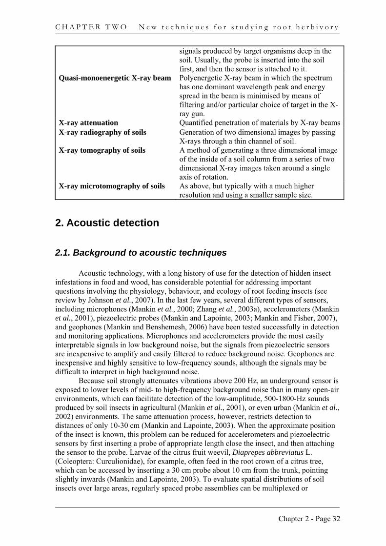
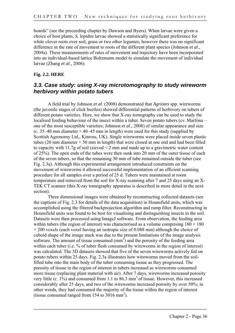
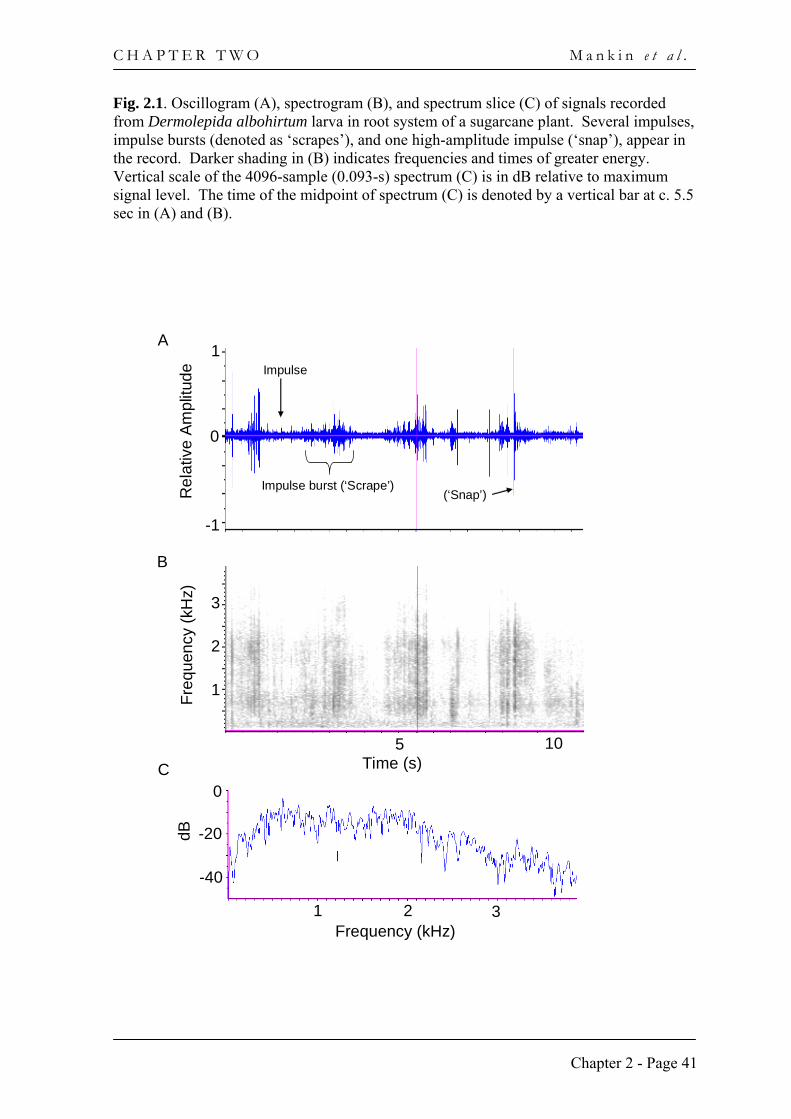
relationship was based on similarities with sounds observed from aboveground insects involved in biting and chewing activities (e.g. Yack et al., 2001). Longer (500–1000 ms), moderate- to weak-intensity ‘rustles’ were suggestive of general rubbing or dragging movements, or digging activity. Other examples of insect larval sounds are shown in Fig. 2.1, recorded from a cane grub, Dermolepida albohirtum Waterhouse (Coleoptera: Scarabaeidae), moving and feeding in the underground stubble in a field of sugarcane (Saccharum officinarum L.). The signals were collected using an accelerometer attached to a 30 cm probe inserted into a sugarcane stool. Afterwards, the signals were filtered at 200 Hz, digitized at 44.1 kHz on a computer, and analyzed as in Mankin et al. (2000) and (Mankin et al., 2001). In the canegrub recording (Fig. 2.1), distinctive impulse bursts were detected that listeners identified as ‘scraping’ sounds. Also detected were sounds similar to the ‘snaps’ in Zhang et al., (2003b). The individual impulses, impulse bursts, and snaps contained energy in a broad band between 0.5 and 3 kHz (Fig. 2.1, B and C). In addition to the sounds above, ant stridulations (Rauth and Vinson, 2006), termite head-banging (Mankin and Benshemesh, 2006), and other patterned sounds have been identified and analyzed, but less obvious or easily interrupted patterns have not yet been studied intensively.
In several respects the identification of insect sound patterns is similar to the problem of identifying beats in music (Gouyon et al., 2006) or ‘voice-print analysis’, which identifies a speaker based on unique acoustic characteristics of their utterances (Bimbot et al., 2004). Analysis of insect sound patterns is likely to become an important focus of future soil insect research, both for its value in interpreting behaviour and for its capability to detect weak insect-produced signals in high levels of background noise (Mankin and Benshemesh, 2006). The increasing availability of sound analysis tools like Raven (Cornell Lab. of Ornithology, Ithaca, NY), general-purpose signal analysis programs like Matlab (MathWorks Inc., Natick, MA), and signal processing tools like Gaussian mixtures and vector quantization (Bimbot et al., 2004) will facilitate such research, driven by the economic importance of root herbivores in agriculture and their wider importance in ecosystem processes. Fig. 2.1. HERE
3. X-ray tomography
3.1. Origins of the technique The use of tomography for studying biological processes in soils is becoming
increasingly common as equipment improves (Asseng et al., 2000). Several energy sources can be used for tomographic imaging of the soil environment, but X-rays have been most frequently used (Young et al., 2001; Gregory et al., 2003). The underlying basis of this technique relies on the differential attenuation of energy by air, water, soil solids and biological material. The differential attenuation of X-rays permits images to be constructed of the soil matrix (including biological material within it) and, when measured at different angles, the spatial characteristics of the soil (and its contents) can be determined by means of computer reconstruction.
X-ray attenuation has been widely used in industrial and medical applications for many years, and provided the impetus for plant and soil scientists to explore the possibility of using these same instruments for studying root growth in the soil. Heeraman et al. (1997), for instance, used an industrial scanner to produce three dimensional images of a
Chapter 2 - Page 34
C H A P T E R T W O M a n k i n e t a l .
bean (Phaseolus vulgaris L.) root system in 50 mm diameter soil columns. Pierret et al. (1999) used a medical scanner to image tree roots in a larger soil column (200 mm in diameter). The main limitation of such instruments was that the resolution was too coarse for obtaining detailed images of the root system, with most industrial and medical scanners having a resolution of c. 1 mm. This limitation was addressed with X-ray scanners that were specifically designed to image roots in the soil. In particular, the instrument described in Jenneson et al. (2003) obtained much higher resolution images with a relatively low dose of radiation. The instrument was based on a third-generation cone-beam scanner and used a filtered silver anode to produce a quasi-monoenergetic X-ray beam. The instrument used an optimum energy level to achieve about three mean-free-path lengths when passing through 25 mm thickness of soil. The design specifications for the instrument allow resolution of c. 100 µm over a scanning period of approximately 40 minutes for a sample 25 mm in diameter and 30 mm high. The radiation dose in the centre of the column is in the region of 0.1 Gy, which is well below the level required to cause significant cell damage. In particular, the scanner has been used to image growing roots of wheat (Triticum aestivum) and rape (Brassica napus) in soil columns (Gregory et al., 2003).
3.2. Use of X-ray tomography for studying root feeding insects In addition to measuring root growth and development, some researchers have
attempted to study root feeding insects in the soil using X-ray radiography – a forerunner of X-ray tomography (Villani and Gould, 1986; Villani and Wright, 1988; Villani and Nyrop, 1991). This proved to be successful for a number of root feeding insects including the Japanese chafer, Popillia japonica Newman (Coleoptera: Scarabaeidae), the European chafer, Rhizotrogus majalis Razoumowsky (Coleoptera: Scarabaeidae) and the corn wireworm, Melanotus communis Gyllenhal (Coleoptera: Elateridae). However, X-ray radiography has not been widely exploited as a technique beyond these studies, probably due to the fact that it mostly provides spatial information in two dimensions. The study by Harrison et al. (1993) was amongst the first to use X-ray tomography (which provides spatial information in three dimensions; 3D) to monitor the behaviour of a root-feeding insect, in this case the fourth instar Pecan weevil Curculio caryae Horn (Coleoptera: Curculionidae). Like the studies that investigated root growth and development with X-ray tomography, these studies were limited by the coarse resolution that was available at the time, meaning that only very large root feeding insects could be detected (> 2 cm long in the case of Harrison et al., 1993). This limitation represented a serious obstacle, since the early stages of most root feeding insects are considerably smaller than 2 cm. However, with the development of devices that could scan soil columns at higher resolution and with minimal energy dosage (e.g. Gregory et al., 2003; Jenneson et al., 2003), it became possible to consider using these techniques for studying much smaller root feeding insects.
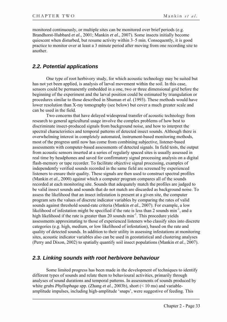
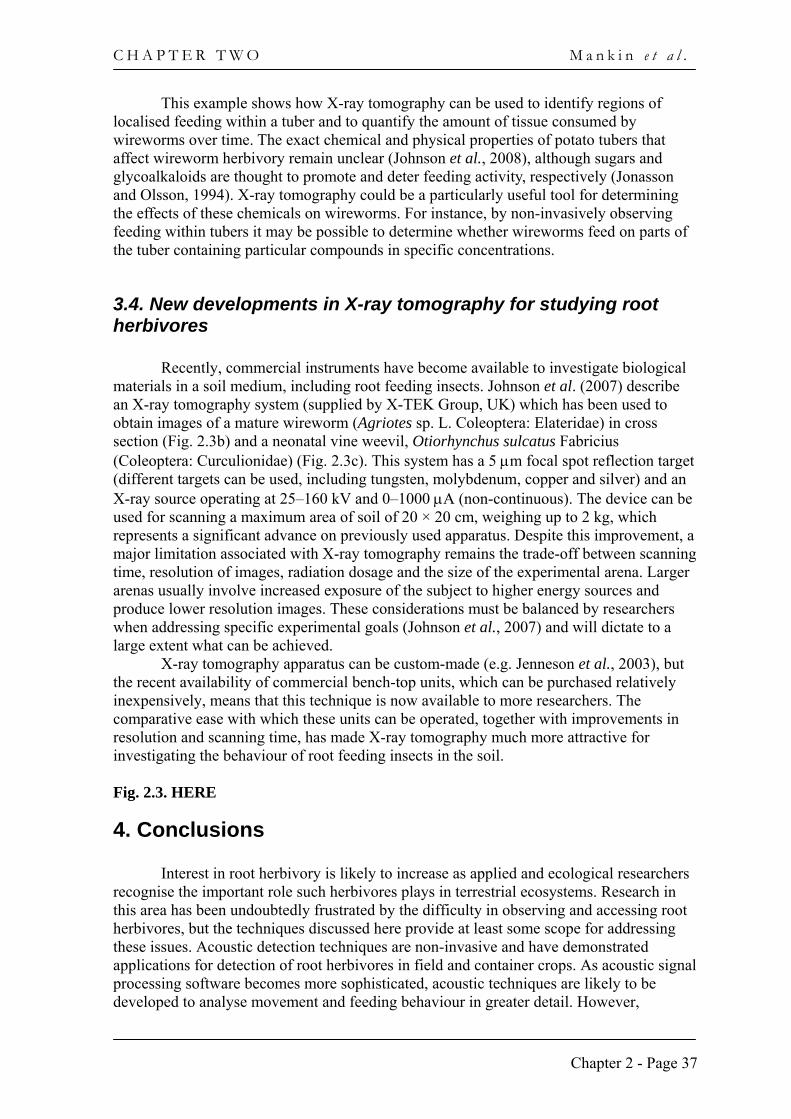
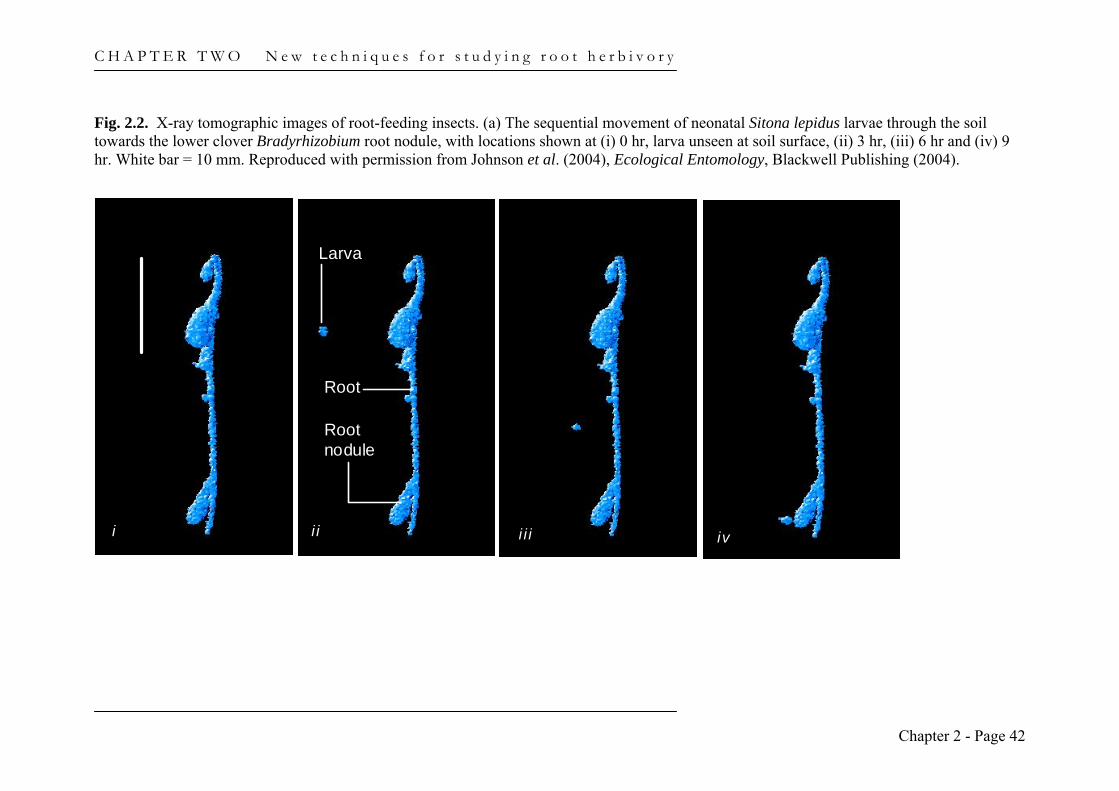
The use of X-ray tomography, and in particular X-ray microtomography, for studying root feeding insects has been employed most recently for studying the soil-dwelling larvae of the clover root weevil (Sitona lepidus), a serious pest of white clover. (Trifolium repens L.). The soil-dwelling stages cause most damage to the plant, particularly when the newly hatched (or neonatal) larvae attack root nodules that house N2-fixing Bradyrhizobium bacteria (Gerard, 2001). In a series of laboratory experiments, Johnson et al. (2004b) demonstrated that S. lepidus larvae burrowed between 9 and 27 mm in nine hours towards nodules on white clover roots at a mean speed of 1.8 mm h-1 (Fig. 2.2). Burrowing patterns were usually convoluted rather than linear with changes in trajectory evident from this study that would be masked in more commonly used “slant
Chapter 2 - Page 35
C H A P T E R T W O N e w t e c h n i q u e s f o r s t u d y i n g r o o t h e r b i v o r y
boards” (see the preceeding chapter by Dawson and Byers). When larvae were given a choice of host plants, S. lepidus larvae showed a statistically significant preference for white clover roots over soil, grass or two other legumes; however there was no significant difference in the rate of movement to roots of the different plant species (Johnson et al., 2004a). These measurements of rates of movement and trajectory have been incorporated into an individual-based lattice Boltzmann model to simulate the movement of individual larvae (Zhang et al., 2006). Fig. 2.2. HERE
3.3. Case study: using X-ray microtomography to study wireworm herbivory within potato tubers
A field trial by Johnson et al. (2008) demonstrated that Agriotes spp. wireworms
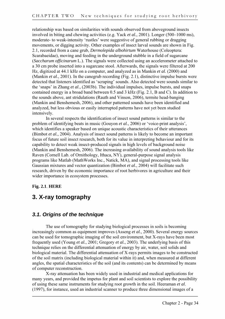
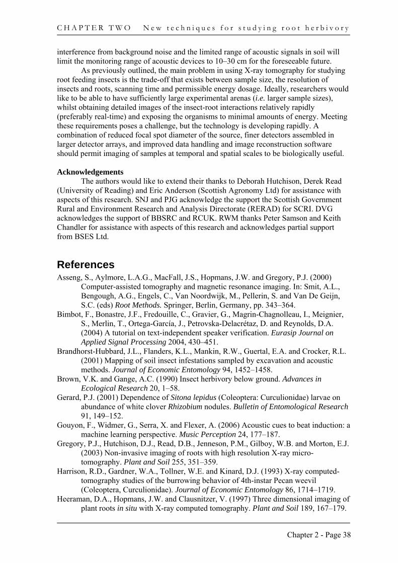
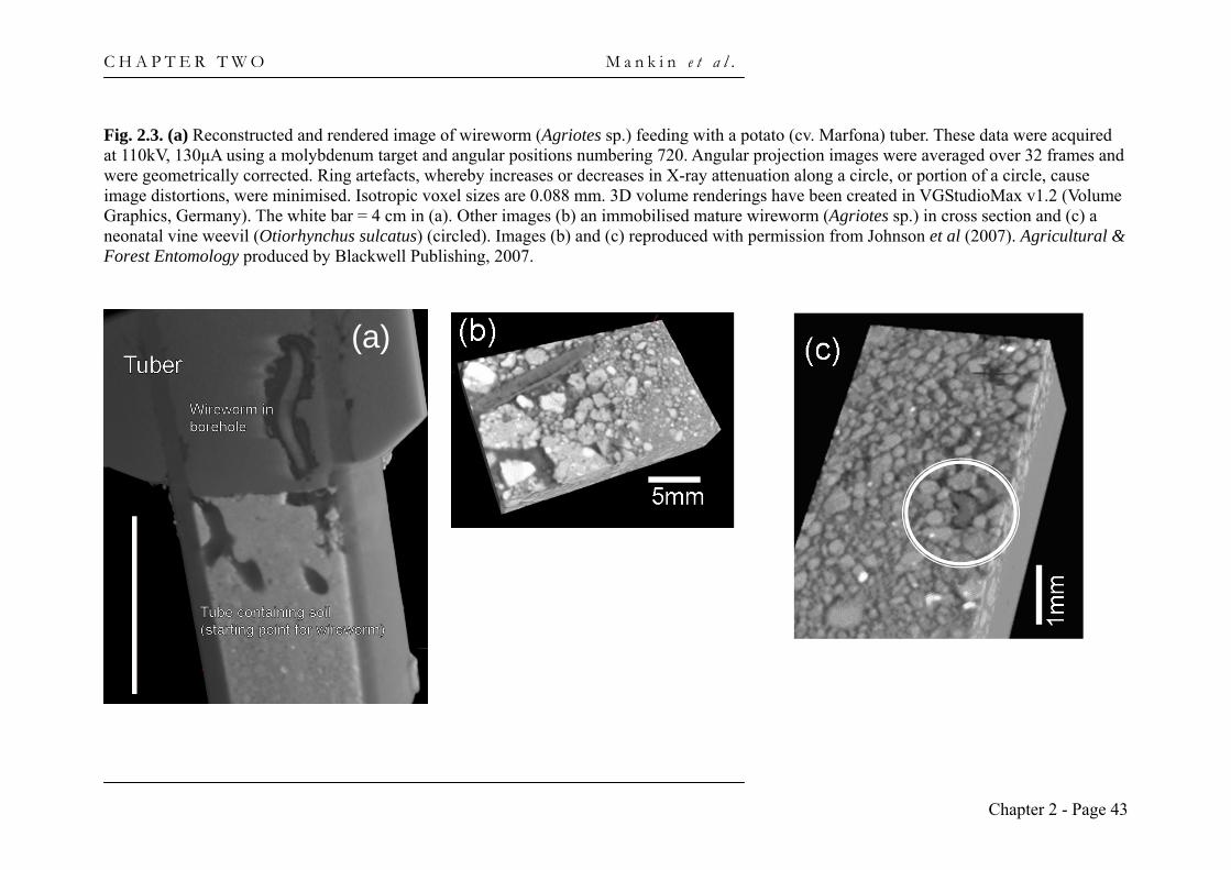
(the juvenile stages of click beetles) showed differential patterns of herbivory on tubers of different potato varieties. Here, we show that X-ray tomography can be used to study the localised feeding behaviour of the insect within a tuber. Seven potato tubers (cv. Marfona – one of the most susceptible varieties; Johnson et al., 2008) of similar appearance and size (c. 35–40 mm diameter × 40–45 mm in length) were used for this study (supplied by Scottish Agronomy Ltd., Kinross, UK). Single wireworms were placed inside seven plastic tubes (20 mm diameter × 50 mm in length) that were closed at one end and had been filled to capacity with 11.7g of soil (sieved < 2 mm and made up to a gravimetric water content of 25%). The open ends of the tubes were then sunk into 20 mm of the outer tissue of each of the seven tubers, so that the remaining 30 mm of tube remained outside the tuber (see Fig. 2.3a). Although this experimental arrangement introduced constraints on the movement of wireworms it allowed successful implementation of an efficient scanning procedure for all samples over a period of 25 d. Tubers were maintained at room temperature and removed from the soil for X-ray scanning after 7 and 25 days using an X-TEK CT scanner (this X-ray tomography apparatus is described in more detail in the next section).
Three dimensional images were obtained by reconstructing collected datasets (see the captions of Fig. 2.3 for details of the data acquisition) in Hounsfield units, which was accomplished using the filtered backprojection algorithm and ramp filter. Reconstructing in Hounsfield units was found to be best for visualising and distinguishing insects in the soil. Datasets were then processed using ImageJ software. From observation, the feeding area within tubers (the region of interest) was characterised as a volume comprising 180 × 180 × 200 voxels (each voxel having an isotropic size of 0.088 mm) although the choice of cuboid shape of the image stack was due to the present limitations of the image analysis software. The amount of tissue consumed (mm3) and the porosity of the feeding area within each tuber (i.e. % of tuber flesh consumed by wireworms in the region of interest) was calculated. The 3D datasets showed that five of the seven wireworms actively fed on potato tubers within 25 days. Fig. 2.3a illustrates how wireworms moved from the soil-filled tube into the main body of the tuber consuming tissue as they progressed. The porosity of tissue in the region of interest in tubers increased as wireworms consumed more tissue (replacing plant material with air). After 7 days, wireworms increased porosity very little (c. 1%) and consumed from 1.1 to 86.3 mm3 of tissue. However, this increased considerably after 25 days, and two of the wireworms increased porosity by over 50%; in other words, they had consumed the majority of the tissue within the region of interest (tissue consumed ranged from 154 to 3016 mm3).
Chapter 2 - Page 36
C H A P T E R T W O M a n k i n e t a l .
This example shows how X-ray tomography can be used to identify regions of localised feeding within a tuber and to quantify the amount of tissue consumed by wireworms over time. The exact chemical and physical properties of potato tubers that affect wireworm herbivory remain unclear (Johnson et al., 2008), although sugars and glycoalkaloids are thought to promote and deter feeding activity, respectively (Jonasson and Olsson, 1994). X-ray tomography could be a particularly useful tool for determining the effects of these chemicals on wireworms. For instance, by non-invasively observing feeding within tubers it may be possible to determine whether wireworms feed on parts of the tuber containing particular compounds in specific concentrations.
3.4. New developments in X-ray tomography for studying root herbivores
Recently, commercial instruments have become available to investigate biological
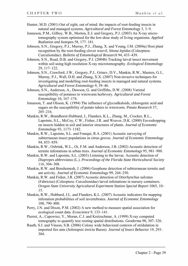
materials in a soil medium, including root feeding insects. Johnson et al. (2007) describe an X-ray tomography system (supplied by X-TEK Group, UK) which has been used to obtain images of a mature wireworm (Agriotes sp. L. Coleoptera: Elateridae) in cross section (Fig. 2.3b) and a neonatal vine weevil, Otiorhynchus sulcatus Fabricius (Coleoptera: Curculionidae) (Fig. 2.3c). This system has a 5 μm focal spot reflection target (different targets can be used, including tungsten, molybdenum, copper and silver) and an X-ray source operating at 25–160 kV and 0–1000 μA (non-continuous). The device can be used for scanning a maximum area of soil of 20 × 20 cm, weighing up to 2 kg, which represents a significant advance on previously used apparatus. Despite this improvement, a major limitation associated with X-ray tomography remains the trade-off between scanning time, resolution of images, radiation dosage and the size of the experimental arena. Larger arenas usually involve increased exposure of the subject to higher energy sources and produce lower resolution images. These considerations must be balanced by researchers when addressing specific experimental goals (Johnson et al., 2007) and will dictate to a large extent what can be achieved.
X-ray tomography apparatus can be custom-made (e.g. Jenneson et al., 2003), but the recent availability of commercial bench-top units, which can be purchased relatively inexpensively, means that this technique is now available to more researchers. The comparative ease with which these units can be operated, together with improvements in resolution and scanning time, has made X-ray tomography much more attractive for investigating the behaviour of root feeding insects in the soil. Fig. 2.3. HERE
4. Conclusions Interest in root herbivory is likely to increase as applied and ecological researchers
recognise the important role such herbivores plays in terrestrial ecosystems. Research in this area has been undoubtedly frustrated by the difficulty in observing and accessing root herbivores, but the techniques discussed here provide at least some scope for addressing these issues. Acoustic detection techniques are non-invasive and have demonstrated applications for detection of root herbivores in field and container crops. As acoustic signal processing software becomes more sophisticated, acoustic techniques are likely to be developed to analyse movement and feeding behaviour in greater detail. However,
Chapter 2 - Page 37
C H A P T E R T W O N e w t e c h n i q u e s f o r s t u d y i n g r o o t h e r b i v o r y
interference from background noise and the limited range of acoustic signals in soil will limit the monitoring range of acoustic devices to 10–30 cm for the foreseeable future.
As previously outlined, the main problem in using X-ray tomography for studying root feeding insects is the trade-off that exists between sample size, the resolution of insects and roots, scanning time and permissible energy dosage. Ideally, researchers would like to be able to have sufficiently large experimental arenas (i.e. larger sample sizes), whilst obtaining detailed images of the insect-root interactions relatively rapidly (preferably real-time) and exposing the organisms to minimal amounts of energy. Meeting these requirements poses a challenge, but the technology is developing rapidly. A combination of reduced focal spot diameter of the source, finer detectors assembled in larger detector arrays, and improved data handling and image reconstruction software should permit imaging of samples at temporal and spatial scales to be biologically useful. Acknowledgements
The authors would like to extend their thanks to Deborah Hutchison, Derek Read (University of Reading) and Eric Anderson (Scottish Agronomy Ltd) for assistance with aspects of this research. SNJ and PJG acknowledge the support the Scottish Government Rural and Environment Research and Analysis Directorate (RERAD) for SCRI. DVG acknowledges the support of BBSRC and RCUK. RWM thanks Peter Samson and Keith Chandler for assistance with aspects of this research and acknowledges partial support from BSES Ltd.
References Asseng, S., Aylmore, L.A.G., MacFall, J.S., Hopmans, J.W. and Gregory, P.J. (2000)
Computer-assisted tomography and magnetic resonance imaging. In: Smit, A.L., Bengough, A.G., Engels, C., Van Noordwijk, M., Pellerin, S. and Van De Geijn, S.C. (eds) Root Methods. Springer, Berlin, Germany, pp. 343–364.
Bimbot, F., Bonastre, J.F., Fredouille, C., Gravier, G., Magrin-Chagnolleau, I., Meignier, S., Merlin, T., Ortega-García, J., Petrovska-Delacrétaz, D. and Reynolds, D.A. (2004) A tutorial on text-independent speaker verification. Eurasip Journal on Applied Signal Processing 2004, 430–451.
Brandhorst-Hubbard, J.L., Flanders, K.L., Mankin, R.W., Guertal, E.A. and Crocker, R.L. (2001) Mapping of soil insect infestations sampled by excavation and acoustic methods. Journal of Economic Entomology 94, 1452–1458.
Brown, V.K. and Gange, A.C. (1990) Insect herbivory below ground. Advances in Ecological Research 20, 1–58.
Gerard, P.J. (2001) Dependence of Sitona lepidus (Coleoptera: Curculionidae) larvae on abundance of white clover Rhizobium nodules. Bulletin of Entomological Research 91, 149–152.
Gouyon, F., Widmer, G., Serra, X. and Flexer, A. (2006) Acoustic cues to beat induction: a machine learning perspective. Music Perception 24, 177–187.
Gregory, P.J., Hutchison, D.J., Read, D.B., Jenneson, P.M., Gilboy, W.B. and Morton, E.J. (2003) Non-invasive imaging of roots with high resolution X-ray micro-tomography. Plant and Soil 255, 351–359.
Harrison, R.D., Gardner, W.A., Tollner, W.E. and Kinard, D.J. (1993) X-ray computed-tomography studies of the burrowing behavior of 4th-instar Pecan weevil (Coleoptera, Curculionidae). Journal of Economic Entomology 86, 1714–1719.
Heeraman, D.A., Hopmans, J.W. and Clausnitzer, V. (1997) Three dimensional imaging of plant roots in situ with X-ray computed tomography. Plant and Soil 189, 167–179.
Chapter 2 - Page 38
C H A P T E R T W O M a n k i n e t a l .
Hunter, M.D. (2001) Out of sight, out of mind: the impacts of root-feeding insects in natural and managed systems. Agricultural and Forest Entomology 3, 3–9.
Jenneson, P.M., Gilboy, W.B., Morton, E.J. and Gregory, P.J. (2003) An X-ray micro-tomography system optimised for the low-dose study of living organisms. Applied Radiation and Isotopes 58, 177–181.
Johnson, S.N., Gregory, P.J., Murray, P.J., Zhang, X. and Young, I.M. (2004a) Host plant recognition by the root-feeding clover weevil, Sitona lepidus (Coleoptera: Curculionidae). Bulletin of Entomological Research 94, 433–439.
Johnson, S.N., Read, D.B. and Gregory, P.J. (2004b) Tracking larval insect movement within soil using high resolution X-ray microtomography. Ecological Entomology 29, 117–122.
Johnson, S.N., Crawford, J.W., Gregory, P.J., Grinev, D.V., Mankin, R.W., Masters, G.J., Murray, P.J., Wall, D.H. and Zhang, X.X. (2007) Non-invasive techniques for investigating and modelling root-feeding insects in managed and natural systems. Agricultural and Forest Entomology 9, 39–46.
Johnson, S.N., Anderson, A., Dawson, G. and Griffiths, D.W. (2008) Varietal susceptibility of potatoes to wireworm herbivory. Agricultural and Forest Entomology 10, 167–174.
Jonasson, T. and Olsson, K. (1994) The influence of glycoalkaloids, chlorogenic acid and sugars on the susceptibility of potato tubers to wireworm. Potato Research 37, 205–216.
Mankin, R.W., Brandhorst-Hubbard, J., Flanders, K.L., Zhang, M., Crocker, R.L., Lapointe, S.L., McCoy, C.W., Fisher, J.R. and Weaver, D.K. (2000) Eavesdropping on insects hidden in soil and interior structures of plants. Journal of Economic Entomology 93, 1173–1182.
Mankin, R.W., Lapointe, S.L. and Franqui, R.A. (2001) Acoustic surveying of subterranean insect populations in citrus groves. Journal of Economic Entomology 94, 853–859.
Mankin, R.W., Osbrink, W.L., Oi, F.M. and Anderson, J.B. (2002) Acoustic detection of termite infestations in urban trees. Journal of Economic Entomology 95, 981–988.
Mankin, R.W. and Lapointe, S.L. (2003) Listening to the larvae. Acoustic detection of Diaprepes abbreviatus (L.). Proceedings of the Florida State Horticultural Society 116, 304–308.
Mankin, R.W. and Benshemesh, J. (2006) Geophone detection of subterranean termite and ant activity. Journal of Economic Entomology 99, 244–250.
Mankin, R.W. and Fisher, J.R. (2007) Acoustic detection of Otiorhynchus sulcatus (Fabricius) (Coleoptera: Curculionidae) larval infestations in nursery containers. Oregon State University Agricultural Experiment Station Special Report 1065, 10–15.
Mankin, R.W., Hubbard, J.L. and Flanders, K.L. (2007) Acoustic indicators for mapping infestation probabilities of soil invertebrates. Journal of Economic Entomology 100, 790–800.
Perry, J.N. and Dixon, P.M. (2002) A new method to measure spatial association for ecological count data. Ecoscience 9, 133–141.
Pierret, A., Capowiez, Y., Moran, C.J. and Kretzschmar, A. (1999) X-ray computed tomography to quantify tree rooting spatial distributions. Geoderma 90, 307–326.
Rauth, S.J. and Vinson, S.B. (2006) Colony wide behavioral contexts of stridulation in imported fire ants (Solenopsis invicta Buren). Journal of Insect Behavior 19, 293–304.
Chapter 2 - Page 39
C H A P T E R T W O N e w t e c h n i q u e s f o r s t u d y i n g r o o t h e r b i v o r y
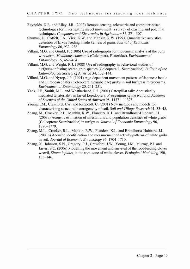
Reynolds, D.R. and Riley, J.R. (2002) Remote-sensing, telemetric and computer-based technologies for investigating insect movement: a survey of existing and potential techniques. Computers and Electronics in Agriculture 35, 271–307.
Shuman, D., Coffelt, J.A., Vick, K.W. and Mankin, R.W. (1993) Quantitative acoustical detection of larvae feeding inside kernels of grain. Journal of Economic Entomology 86, 933–938.
Villani, M.G. and Gould, F. (1986) Use of radiographs for movement analysis of the corn wireworm, Melanotus communis (Coleoptera, Elateridae). Environmental Entomology 15, 462–464.
Villani, M.G. and Wright, R.J. (1988) Use of radiography in behavioral studies of turfgrass-infesting scarab grub species (Coleoptera L. Scarabaeidae). Bulletin of the Entomological Society of America 34, 132–144.
Villani, M.G. and Nyrop, J.P. (1991) Age-dependent movement patterns of Japanese beetle and European chafer (Coleoptera, Scarabeidae) grubs in soil turfgrass microcosms. Environmental Entomology 20, 241–251.
Yack, J.E., Smith, M.L. and Weatherhead, P.J. (2001) Caterpillar talk: Acoustically mediated territoriality in larval Lepidoptera. Proceedings of the National Academy of Sciences of the United States of America 98, 11371–11375.
Young, I.M., Crawford, J.W. and Rappoldt, C. (2001) New methods and models for characterising structural heterogeneity of soil. Soil and Tillage Research 61, 33–45.
Zhang, M., Crocker, R.L., Mankin, R.W., Flanders, K.L. and Brandhorst-Hubbard, J.L. (2003a) Acoustic estimation of infestations and population densities of white grubs (Coleoptera: Scarabaeidae) in turfgrass. Journal of Economic Entomology 96, 1770–1779.
Zhang, M.L., Crocker, R.L., Mankin, R.W., Flanders, K.L. and Brandhorst-Hubbard, J.L. (2003b) Acoustic identification and measurement of activity patterns of white grubs in soil. Journal of Economic Entomology 96, 1704–1710.
Zhang, X., Johnson, S.N., Gregory, P.J., Crawford, J.W., Young, I.M., Murray, P.J. and Jarvis, S.C. (2006) Modelling the movement and survival of the root-feeding clover weevil, Sitona lepidus, in the root-zone of white clover. Ecological Modelling 190, 133–146.
Chapter 2 - Page 40
C H A P T E R T W O M a n k i n e t a l .
Chapter 2 - Page 41
Fig. 2.1. Oscillogram (A), spectrogram (B), and spectrum slice (C) of signals recorded from Dermolepida albohirtum larva in root system of a sugarcane plant. Several impulses, impulse bursts (denoted as ‘scrapes’), and one high-amplitude impulse (‘snap’), appear in the record. Darker shading in (B) indicates frequencies and times of greater energy. Vertical scale of the 4096-sample (0.093-s) spectrum (C) is in dB relative to maximum signal level. The time of the midpoint of spectrum (C) is denoted by a vertical bar at c. 5.5 sec in (A) and (B).
1
2
3
Freq
uenc
y (k
Hz)
Rel
ativ
e Am
plitu
de
1
-1
0
dB
0
-20
-40
1 2 3Frequency (kHz)
5 10Time (s)
(‘Snap’)
A
B
C
Impulse burst (‘Scrape’)
Impulse
C H A P T E R T W O N e w t e c h n i q u e s f o r s t u d y i n g r o o t h e r b i v o r y
Fig. 2.2. X-ray tomographic images of root-feeding insects. (a) The sequential movement of neonatal Sitona lepidus larvae through the soil towards the lower clover Bradyrhizobium root nodule, with locations shown at (i) 0 hr, larva unseen at soil surface, (ii) 3 hr, (iii) 6 hr and (iv) 9 hr. White bar = 10 mm. Reproduced with permission from Johnson et al. (2004), Ecological Entomology, Blackwell Publishing (2004).
i ii iii iv
Larva
Root
Root nodule
Chapter 2 - Page 42
C H A P T E R T W O M a n k i n e t a l .
Fig. 2.3. (a) Reconstructed and rendered image of wireworm (Agriotes sp.) feeding with a potato (cv. Marfona) tuber. These data were acquired at 110kV, 130μA using a molybdenum target and angular positions numbering 720. Angular projection images were averaged over 32 frames and were geometrically corrected. Ring artefacts, whereby increases or decreases in X-ray attenuation along a circle, or portion of a circle, cause image distortions, were minimised. Isotropic voxel sizes are 0.088 mm. 3D volume renderings have been created in VGStudioMax v1.2 (Volume Graphics, Germany). The white bar = 4 cm in (a). Other images (b) an immobilised mature wireworm (Agriotes sp.) in cross section and (c) a neonatal vine weevil (Otiorhynchus sulcatus) (circled). Images (b) and (c) reproduced with permission from Johnson et al (2007). Agricultural & Forest Entomology produced by Blackwell Publishing, 2007.
(a)
Chapter 2 - Page 43