Embed Size (px)
Citation preview
Faculty Scholarship
2019
Neutrophil Extracellular Traps in Breast Cancer and Beyond: Neutrophil Extracellular Traps in Breast Cancer and Beyond:
Current Perspectives on NET Stimuli, Thrombosis and Metastasis, Current Perspectives on NET Stimuli, Thrombosis and Metastasis,
and Clinical Utility for Diagnosis and Treatment and Clinical Utility for Diagnosis and Treatment
Hunter T. Snoderly
Brian A. Boone
Margaret F. Bennewitz
Follow this and additional works at: https://researchrepository.wvu.edu/faculty_publications
Part of the Chemical Engineering Commons
REVIEW Open Access
Neutrophil extracellular traps in breastcancer and beyond: current perspectiveson NET stimuli, thrombosis and metastasis,and clinical utility for diagnosis andtreatmentHunter T. Snoderly1, Brian A. Boone2 and Margaret F. Bennewitz1*
Abstract
The formation of neutrophil extracellular traps (NETs), known as NETosis, was first observed as a novel immuneresponse to bacterial infection, but has since been found to occur abnormally in a variety of other inflammatorydisease states including cancer. Breast cancer is the most commonly diagnosed malignancy in women. In breastcancer, NETosis has been linked to increased disease progression, metastasis, and complications such as venousthromboembolism. NET-targeted therapies have shown success in preclinical cancer models and may provevaluable clinical targets in slowing or halting tumor progression in breast cancer patients. We will briefly outline themechanisms by which NETs may form in the tumor microenvironment and circulation, including the crosstalkbetween neutrophils, tumor cells, endothelial cells, and platelets as well as the role of cancer-associatedextracellular vesicles in modulating neutrophil behavior and NET extrusion. The prognostic implications of cancer-associated NETosis will be explored in addition to development of novel therapeutics aimed at targeting NETinteractions to improve outcomes in patients with breast cancer.
Keywords: Neutrophil extracellular traps, Breast cancer, Metastasis, Venous thromboembolism
BackgroundNeutrophils are the most abundant type of white blood cellsin the circulation and are often considered the frontline de-fenders in innate immunity [1]. These leukocytes were onlyrecently observed to be capable of a novel immune responsein which they expel their DNA and intracellular contents ina web-like structure known as a neutrophil extracellular trap(NET). NETs form when activated neutrophils release DNA,histones, and granular content, exposing antimicrobial andproinflammatory proteins [2]. NETosis occurs as specificproteases are translocated into the neutrophil nucleus,which causes their chromatin to decondense through citrul-lination. These loosely networked strands are then ultim-ately expelled from the cell, rupturing it or leaving the
membrane intact. Subsequent membrane integrity dependson the nature of the stimulus provoking NETosis [3]. NETswere first observed as a response to bacterial infection, ashistones, and released neutrophil granular content have anti-microbial properties and the fibrous NET structure canphysically entrap and kill bacteria [2]. However, NETs havesince been associated with sterile inflammation in a varietyof disease states, including gout, cystic fibrosis, type 1 dia-betes, rheumatoid arthritis, preeclampsia, and others [4–9].NETs have also been associated with tumor cell proliferationand metastasis [10–16], cancer-related thrombosis [17–21],and primary tumor growth [22, 23].In this review, we will focus on the role of NETs primar-
ily in breast cancer. Globally, breast cancer accounted foraround 11.6% of new cancer diagnoses in 2018 and wasestimated to be responsible for more than 6% of all cancerdeaths [24]. Current evidence suggests that NET produc-tion in cancer involves a complex interplay between a
© The Author(s). 2019 Open Access This article is distributed under the terms of the Creative Commons Attribution 4.0International License (http://creativecommons.org/licenses/by/4.0/), which permits unrestricted use, distribution, andreproduction in any medium, provided you give appropriate credit to the original author(s) and the source, provide a link tothe Creative Commons license, and indicate if changes were made. The Creative Commons Public Domain Dedication waiver(http://creativecommons.org/publicdomain/zero/1.0/) applies to the data made available in this article, unless otherwise stated.
* Correspondence: [email protected] of Chemical and Biomedical Engineering, West VirginiaUniversity, 1306 Evansdale Drive, ESB 521, Morgantown, WV 26506, USAFull list of author information is available at the end of the article
Snoderly et al. Breast Cancer Research (2019) 21:145 https://doi.org/10.1186/s13058-019-1237-6
variety of cells and blood components, including platelets,leukocytes, pioneering metastatic tumor cells, and the pri-mary tumor site itself [10, 19, 21, 25–28]. NETs promotethe progression of an inflammatory microenvironment,which develops a positive feedback loop: NETs releasedinto the circulation damage endothelial cells, which pro-motes further inflammation, causing activation of plateletsand other neutrophils which can cause further NET re-lease. Platelet activation caused by NETs can also promoteseveral negative outcomes associated with late-stage meta-static breast cancer, including venous thromboembolism(VTE) [29]. This review will discuss both established andpotential stimuli that promote oncogenic NETosis, bothon a molecular level and in terms of interactions betweenneutrophils, other blood components in cancer-affectedorganisms, and tumor cells themselves. We will also dis-cuss the consequences of NETosis, especially as it relatesto breast cancer progression. Finally, the use of NETs aspotential diagnostic biomarkers and/or clinical therapeutictargets in cancer will be discussed.
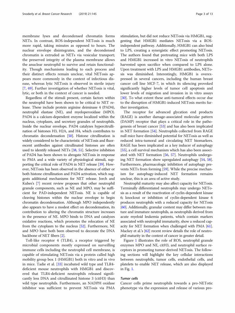
Cellular and molecular stimulants of NETosisPro-NETotic stimuli and neutrophil components required forNETosisSeveral potential pro-NETotic stimuli relevant to cancerprogression are listed in Table 1. The most classical andpotent stimuli provoking NET formation are products of
bacterial infection, such as lipopolysaccharide (LPS), ornon-endogenous inflammatory pathway activators suchas phorbol 12-myristate 13-acetate (PMA) [2]. LPS andPMA promote NETosis through production of reactiveoxygen species (ROS) in which oxygen is transformedinto damaging superoxide radicals and secondary oxi-dants. ROS are key to cancer and inflammatory signalingas well as neutrophil behavior modulation [45, 46]. Theinflammatory state associated with cancer also may pro-voke systemic oxidative stress. The presence of higherlevels of NETosis observed in many cancers may (at leastpartially) be attributed directly to tumor cells, as well asindirectly via ROS generation by other cells and granulesactivated by downstream effects of tumor released fac-tors. It has been shown that PMA provokes NETosisthrough activation of p38 MAPK via NADPH oxidasegeneration of ROS [32]; thus, endogenous stimulantsmay follow similar pathways. Interestingly, p38 activa-tion has also been shown to promote breast cancer cellsurvival and proliferation and has been linked to poorclinical outcomes in humans [47, 48].Although NADPH oxidase inhibition has been shown
to prevent NETosis, not all NETosis appears to be ROS-dependent. In fact, the mechanism of NET release ap-pears to be influenced by the presence or absence ofROS [3, 49]. ROS-dependent NETosis results in neutro-phil cell death, known as lytic NETosis, wherein the cell
Table 1 Key NET stimuli involved in cancer progression. References are annotated to indicate whether NETotic effect has beenshown in human (H) neutrophils, mouse (M) neutrophils, or both (HM)
Stimulus/model: Relevance to cancer progression: Origin:
LPS [2, 30, 31]HM May simulate response to infection; repeatedintranasal dosage in mice activated dormantcancer cells and enhanced metastatic proliferation
Gram-negative bacteria
PMA [2, 32]H N/A Synthetic/pharmaceutical
Platelet-activating factor [19]M Promotes tumor cell proliferation, neovascularization,and immunosuppressive phenotype
Leukocyte, platelet, and endothelial secretionin inflammation
HMGB1 [14, 25, 33]HM Associates with existing NETs; role in platelet andneutrophil activation; synergizes with LPS and thusmay exacerbate response to infection
Leukocyte and platelet secretion in inflammation;expressed in some tumors; released duringcell death
IL-8 [5, 34, 35]H Drives neutrophilia; positive correlation with pooroutcome in women with breast cancer
Expressed in some tumors; released fromactivated endothelial cells
G-CSF [19, 36, 37]M Drives neutrophilia; positive correlation withmetastasis; potentiates extracellular vesicle drivenNETosis
Expressed in some tumors
PAD4 [38–40]HM Catalyzes histone citrullination; inhibition preventsNETosis in most circumstances
Neutrophils; expressed in some tumors
P-selectin [41]M Facilitates neutrophil motility; drivesplatelet-neutrophil aggregation
Endothelial cells; platelets
TF [42–44]H Activates platelets which activate neutrophilsand causes NETosis, potentially through multiplepathways
Secreted during NETosis; expressed in some tumors;contained in tumor EVs
Tumor EVs [21]M May influence neutrophil behavior once taken up;contain inflammatory cytokines and are vital tooncogenic signaling; prothrombotic
Released from tumor cells
Snoderly et al. Breast Cancer Research (2019) 21:145 Page 2 of 13
membrane lyses and decondensed chromatin formsNETs. In contrast, ROS-independent NETosis is muchmore rapid, taking minutes as opposed to hours. Thenuclear envelope disintegrates, and the decondensedchromatin is extruded as NETs via vesicular transport;the preserved integrity of the plasma membrane allowsthe anuclear neutrophil to survive and retain functional-ity. Though mechanisms leading to each process andtheir distinct effects remain unclear, vital NETosis ap-pears more commonly in the context of infectious dis-ease, whereas lytic NETosis is observed in sterile injury[7, 49]. Further investigation of whether NETosis is vital,lytic, or both in the context of cancer is needed.Regardless of the stimuli present, certain factors within
the neutrophil have been shown to be critical to NET re-lease. These include protein arginine deiminase 4 (PAD4),neutrophil elastase (NE), and myeloperoxidase (MPO).PAD4 is a calcium-dependent enzyme localized within thenucleus, cytoplasm, and secretory granules of neutrophils.Inside the nuclear envelope, PAD4 catalyzes hypercitrulli-nation of histones H3, H2A, and H4, which contributes tochromatin decondensation [50]. Histone citrullination iswidely considered to be characteristic of NETosis and fluor-escent antibodies against citrullinated histones are oftenused to identify released NETs [38, 51]. Selective inhibitionof PAD4 has been shown to abrogate NETosis in responseto PMA and a wide variety of physiological stimuli, sup-porting the critical role of PAD4 in NET release [39]. How-ever, NETosis has been observed in the absence of either orboth histone citrullination and PAD4 activation, which sug-gests additional mechanisms for NET release. Jorch andKubes’s [7] recent review proposes that other neutrophilgranule components, such as NE and MPO, may be suffi-cient for PAD-independent NETosis. NE is capable ofcleaving histones within the nuclear envelope to beginchromatin decondensation. Although MPO independentlyalso appears to have a modest effect on decondensation, itscontribution to altering the chromatin structure increasesin the presence of NE. MPO binds to DNA and catalyzesoxidative reactions, which promotes the relocation of NEfrom the cytoplasm to the nucleus [52]. Furthermore, NEand MPO have both been observed to decorate the DNAbackbone of NET fibers [2].Toll-like receptor 4 (TLR4), a receptor triggered by
microbial components mostly expressed on surveillingimmune cells including the neutrophil cell membrane, iscapable of stimulating NETosis via a protein called highmobility group box 1 (HMGB1) both in vitro and in vivoin mice. Tadie et al. [33] incubated wild type and TLR4-deficient mouse neutrophils with HMGB1 and discov-ered that TLR4-deficient neutrophils released signifi-cantly less DNA and citrullinated histone 3 (citH3) thanwild type neutrophils. Furthermore, an NADPH oxidaseinhibitor was sufficient to prevent NETosis via PMA
stimulation, but did not reduce NETosis via HMGB1, sug-gesting that HMGB1 mediates NETosis via a ROS-independent pathway. Additionally, HMGB1 can also bindto LPS, creating a synergistic effect promoting NETosis.The authors found that pretreating mice with both LPSand HMGB1 increased in vitro NETosis of neutrophilsharvested upon sacrifice when compared to LPS alone.Upon treatment with LPS and HMGB1 antibodies, NETo-sis was diminished. Interestingly, HMGB1 is overex-pressed in several cancers, including the human breastcancer cell line MCF-7, in which its silencing provokedsignificantly higher levels of tumor cell apoptosis andlower levels of migration and invasion in in vitro assays[30]. To what extent these anti-tumoral effects occur dueto the disruption of HMGB1-induced NETosis merits fur-ther investigation.The receptor for advanced glycation end products
(RAGE) is another damage-associated molecular pattern(DAMP) receptor that plays a critical role in the patho-genesis of breast cancer [53] and has also been implicatedin NET formation [54]. Neutrophils collected from RAGEnull mice have diminished potential for NETosis as well asreduced intra-tumoral and circulating NET biomarkers.RAGE has been implicated as a key inducer of autophagy[55], a cell survival mechanism which has also been associ-ated with NET formation [56, 57]. Neutrophils undergo-ing NET formation show upregulated autophagy [54, 58].Furthermore, pharmacologic inhibition of autophagy pre-vents NETs from forming [59]. While the precise mechan-ism for autophagy-induced NET formation remainsunclear, this is an area of active study.Neutrophil maturity may also affect capacity for NETosis.
Terminally differentiated neutrophils may undergo NETo-sis as a result of the reactivation of cyclin-dependent kinase6; knockout or inhibition of cyclin-dependent kinase 6produces neutrophils with a reduced capacity for NETosis[60]. Additionally, granular content may differ between ma-ture and immature neutrophils, as neutrophils derived fromacute myeloid leukemia patients, which contain markersassociated with neutrophil immaturity, show a reduced cap-acity for NET formation when challenged with PMA [61].Mackey et al.’s [62] recent review details the role of neutro-phil maturity in the context of cancer in greater detail.Figure 1 illustrates the role of ROS, neutrophil granule
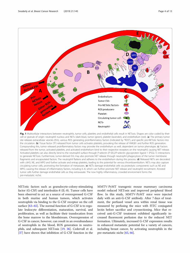
enzymes MPO and NE, citH3, and neutrophil surface re-ceptors in promoting tumor-derived NETosis. The follow-ing sections will highlight the key cellular interactionsbetween neutrophils, tumor cells, endothelial cells, andplatelets to enable NET release, which are also displayedin Fig. 1.
Tumor cellsCancer cells prime neutrophils towards a pro-NEToticphenotype via the expression and release of various pro-
Snoderly et al. Breast Cancer Research (2019) 21:145 Page 3 of 13
NETotic factors such as granulocyte-colony-stimulatingfactor (G-CSF) and interleukin-8 (IL-8). Tumor cells havebeen observed to act as a source of overexpressed G-CSFin both murine and human tumors, which activatesneutrophils via binding to the G-CSF receptor on the cellsurface [63–65]. The normal function of G-CSF is to regu-late leukocyte differentiation, maturation, survival, andproliferation, as well as facilitate their translocation fromthe bone marrow to the bloodstream. Overexpression ofG-CSF in cancer, however, can result in an overabundanceof neutrophils in the blood, ROS generation in neutro-phils, and subsequent NETosis [19, 36]. Cedervall et al.[37] have shown that inhibition of G-CSF function in the
MMTV-PyMT transgenic mouse mammary carcinomamodel reduced NETosis and improved peripheral bloodflow. In this study, MMTV-PyMT mice were injecteddaily with an anti-G-CSF antibody. After 7 days of treat-ment, the perfused vessel area within renal tissue wasmeasured by perfusing the mice with FITC conjugatedlectin before sacrifice and cryosectioning. Mice that re-ceived anti-G-CSF treatment exhibited significantly in-creased fluorescent perfusion due to the reduced NETformation. Ultimately, increased G-CSF expression resultsin enhanced metastatic potential for a variety of cancers,including breast cancer, by activating neutrophils in thepre-metastatic niche [65, 66].
Fig. 1 Multicellular interactions between neutrophils, tumor cells, platelets and endothelial cells result in NETosis. Shapes are color coded by theircell or granule of origin: neutrophil nucleus and NETs (dark blue), tumor (green), platelet (lavender), and endothelium (red). (a) The primary tumorsite releases extracellular vesicles (EVs), various ROS generating proinflammatory factors (indicated by “ROS”), and specific pro-NETotic factors intothe circulation. (b) Tissue factor (TF) released from tumor cells activates platelets, provoking the release of HMGB1 and further ROS generation.Compounding this, tumor released proinflammatory factors may provoke the endothelium as well, dependent on tumor phenotype. (c) Factorsreleased from the tumor, activated platelets, and activated endothelium bind to their respective receptors on the neutrophil, causing NET release.Activated platelets can also directly bind to the neutrophil surface through P-selectin (P-SEL)/P-selectin glycoprotein ligand 1 (PSGL-1) interactionsto generate NETosis. Furthermore, tumor-derived EVs may also promote NET release through neutrophil phagocytosis of the tumor membranefragments and encapsulated factors. The neutrophil flattens and adheres to the endothelium during this process. (d) Released NETs are decoratedwith citH3, NE, and MPO and further activate and entrap platelets, leading to the potential for venous thromboembolism. NETs may also capturecirculating tumor cells, promoting the formation of metastases. (e) NETs damage endothelial cells via proteolytic components such as NE andMPO, causing the release of inflammatory factors, including IL-8, which can further promote NET release and neutrophil recruitment. Arrestedtumor cells further damage endothelial cells as they extravasate. The now highly inflammatory, crowded environment forms thepre-metastatic niche.
Snoderly et al. Breast Cancer Research (2019) 21:145 Page 4 of 13
Neutrophils are chemotactically attracted to tumor cellsthrough secretion of IL-8 (also known as CXCL8). Itshould be noted that human IL-8 does not have a directcounterpart in mice. IL-8 binds to G-protein coupled re-ceptors, CXCR1 and CXCR2, which are expressed by neu-trophils [67]. IL-8 plays an important role in recruitingneutrophils to sites of inflammation; as such, women withbreast cancer have higher serum levels of IL-8 comparedto healthy patients. Additionally, IL-8 levels strongly cor-relate with disease progression [68]. In infectious disease,recruitment towards inflammation may be beneficial, asXu et al. [69] have shown that reduced CXCR1 andCXCR2 expression on neutrophils correlated with nega-tive clinical outcomes in hepatitis B due to insufficientneutrophil recruitment. Other studies have confirmed thatinhibition of IL-8 receptors prevents human neutrophilchemotaxis in vitro [70]. IL-8 production in multiple can-cer types, including breast cancer, has also been associatedwith increased metastatic potential [35]. IL-8 is capable ofstimulating NETosis in human neutrophils in vitro, andthe addition of IL-8 antibodies abolishes this effect [5, 34].In mice, CXCL1 (KC), CXCL2 (MIP-2), and CXCL5 and 6(LIX) serve as functional homologs of IL-8 promotingmurine neutrophil chemotaxis; KC and MIP-2 bind withCXCR2. While the roles of MIP-2 and LIX in NETosis areunclear, KC has been shown to promote NETosis in mur-ine sepsis models [71, 72].Finally, while PAD4 is localized within the nucleus,
cytoplasm, and secretory granules of neutrophils, it hasalso been shown to be expressed in multiple tumor celllines. Chang et al. [40] showed that breast tumors inparticular had the greatest PAD4 expression in a varietyof human malignancies, including lung adenocarcin-omas, colorectal adenocarcinomas, renal cancer cells,and others; additionally, elevated levels of PAD4 weredetected in patient plasma and associated with the pres-ence of other tumor biomarkers. The mechanism con-cerning how PAD4 is exported from tumor cells andwhether extracellular PAD4 can stimulate NETosis hasnot been previously studied.
Endothelial cellsIn addition to being secreted by tumor cells, IL-8 is alsoknown to be produced via endothelial cell (EC) activa-tion [73]. EC activation occurs when the vasculature isexposed to oxidative stress via injury, inflammation,chemotherapy, or ionizing radiation [74]. Activated ECsrelease inflammatory cytokines and growth factors andalso express several adhesion molecules on their surfacesuch as P-selectin, E-selectin, and ICAM-1 to facilitateneutrophil rolling, adhesion, and transmigration to theinflamed site [75]. Gupta et al. [34] investigated the roleof ECs in promoting NETosis and found that activatedECs co-cultured with neutrophils in vitro resulted in
NET formation that is partially mediated by IL-8. Re-leased NETs exposed to the surface of ECs for prolongedtime periods (18 h of neutrophil-EC co-culture) resultedin eventual EC injury and death, which could be inhib-ited through NET dissolution by a DNA-degrading en-zyme, DNase I. NET-induced EC injury and death hasalso been observed in vivo, though this has been demon-strated indirectly. Schreiber et al. [76] found that DNaseI treatment reduced NET formation and protected micefrom blood vessel inflammation, known as vasculitis.Additionally, Knight et al. [77] showed that PAD4 inhib-ition via daily injections of Cl-amidine was effective inreducing NETosis in mice, as well as preventing furthervascular damage and atherosclerosis. Little research hasbeen done to elucidate the link between NETosis andcancer-induced endothelial damage. However, tumorcells themselves can contribute to EC inflammation,which can enhance the potential to induce NETosis byfurther increasing EC damage [34, 78]. The link betweencancer-associated EC activation and NETosis may beworth further investigation; however, since many of thesame stimuli provoke both neutrophil and EC response,establishing causality may be difficult.
PlateletsActivated platelets also stimulate NETosis, which sets upa positive feedback loop, as released NETs are known tostrongly promote a prothrombotic state that further en-hances platelet activation [79]. Much like endothelialcells, platelets must undergo activation prior to stimulat-ing NETosis [25, 31]. Many tumor cell lines includingcertain breast cancers have been shown to overexpressand release tissue factor (TF) [44], which is a well-established platelet activator. TF levels have been shownto correlate with mortality in breast cancer patients [80].However, the use of TF as a biomarker for specificallydefining VTE risk has been demonstrated for some can-cers yet remains inconclusive for others [81]. Neverthe-less, Razak et al. [82] suggest that cancer may activateplatelets through uptake of small tumor-derived extra-cellular vesicles, which often contain TF. Neutrophilsalso contain tissue factor, which is released from NETsto further promote a positive feedback loop by stimulat-ing platelets [42, 43]. Further investigation into themechanisms of TF-mediated increases in mortality inde-pendent of VTE risk would be interesting.Post activation, platelets can stimulate NET release
through direct adhesive interactions with neutrophils[41, 83]; upon activation, platelets rapidly translocate anadhesion molecule known as P-selectin to their surface[84], which can bind to the neutrophil surface receptorP-selectin glycoprotein ligand-1 (PSGL-1) to promoteneutrophil-platelet adhesion [85], neutrophil activation[86], and subsequent NET release. Etulain et al. [41]
Snoderly et al. Breast Cancer Research (2019) 21:145 Page 5 of 13
show thrombin activated platelets elicit NETosis bothin vitro and in vivo in murine neutrophils, and NET for-mation does not occur when either P-selectin or PSGL-1inhibitory antibodies are introduced. NETosis was alsoabolished in P-selectin knockout mice. Interestingly, sol-ubilized P-selectin alone was also observed to stimulateNETosis, but to a lesser extent than activated platelets[41]. This potential NETosis pathway could also be rele-vant in cancer where high levels of soluble P-selectinfound in patient blood plasma have been linked tohigher rates of VTE [87].Both TLR4 and HMGB1 are also expressed by platelets
and have been shown to be another means of platelet-stimulated NETosis relevant to cancer [25, 31]. In septicmice, Clark et al. [31] were the first to show that LPS bindsto TLR4 to enable platelet activation, neutrophil-platelet ag-gregate formation, subsequent neutrophil activation, andNET release. Platelet HMGB1 can cause NETosis throughneutrophil TLR4 activation, or alternatively can bind to theneutrophil RAGE receptor to stimulate NETosis. Maugeriet al. [25] found that when human platelets were activatedwith a variety of factors, including thrombin or collagen,they were able to stimulate NETosis via HMGB1. NETosiswas abolished when RAGE was blocked via antibodies. Theauthors also show that HMGB1 is no longer present inplatelets post activation, indicating that it is released ratherthan translocated to the membrane. It is conceivable thatplatelets may serve as an intermediary between tumor cellsto influence neutrophils and promote NETosis via the re-lease of platelet-activating soluble factors, such as HMGB1.
Extracellular vesiclesThough initially thought to solely be biomarkers, currentliterature suggests that extracellular vesicles (EVs) activelycontribute to angiogenesis, metastasis, and coagulation[21, 88]. The role of EVs in promoting NETosis in thecontext of cancer is only just being explored. Broadly, EVsare formed when a piece of membrane sheds from theparent cell to form membrane-enclosed particles, the con-tents of which depend on the phenotype of the parent cell.Ultimately, any cytoplasmic material in the parent cell canbe present in its EVs; EVs are extremely heterogenous andcan also form from the Golgi or endosomal membrane[89]. Though EVs can be further subcategorized based onsize or origin, the term “extracellular vesicle” refers to anyparticle 50–1500 nm in diameter [90]. EV release often oc-curs as a stress response. Consequently, EVs are morehighly concentrated in cancer patients than in healthy in-dividuals. Elevated EV content in breast cancer patientblood serves as an indicator of more advanced diseasestage and is associated with worse therapeutic success andlower 3-year survival rates [91]. While the cargo, RNA,DNA, and membrane proteins present in EVs from cancerpatients have not yet been fully characterized, cancer-
derived EVs have been associated with high expression ofpro-NETotic and pro-tumoral factors such as interleukinsand G-CSF [92–94]. We will discuss EVs derived fromtumor-burdened organisms and from tumor cell culture.As tumor-derived EVs are just recently being observed tomodulate neutrophil behavior, including NETosis, it is notsurprising that the growth factors and cytokines these EVscarry can further contribute to the inflammatory micro-environment of a nascent pre-metastatic niche.Leal et al.’s recent study [21] shows that EVs derived
from cultured 4T1 mouse breast cancer cells stimulatedNETosis in vitro in neutrophils primed with G-CSF.BALB/c mice with orthotopic mammary 4T1 tumorswere shown to have significantly more EVs present inblood plasma compared to control mice without tumors.The evaluated population contained particles approxi-mately 80–110 nm in diameter. Mice containing 4T1tumors exhibited more rapid coagulation in venous andarterial injury models compared to control mice. Theenhanced prothrombotic state of 4T1 mice could beinhibited through use of DNase I, suggesting a role ofNETs in platelet activation. Notably, healthy mice injectedwith G-CSF and culture-derived 4T1 EVs experiencedmore rapid coagulation induced via photochemical vascu-lar injury than did healthy mice given G-CSF only. NETswere observed (though not quantified) within thesethrombi, suggesting that EVs could lead to NET releaseand subsequent coagulation in vivo. However, the use ofexclusively tumor-derived EVs is limiting, as it does notaccount for the release and content of EVs derived fromother blood cells in tumor-burdened organisms. EVs re-leased from other cells such as platelets, endothelial cells,and macrophages may also be tumor mediated, since EVsfacilitate intracellular communication between tumorsand other cells [92]. Despite this, to our knowledge, Lealet al.’s study has been the only published work to examinethe direct stimulatory effect of tumor-derived EVs onNETosis.Similarly, the specific mechanisms of interaction between
neutrophils and EVs leading to NETosis are largely un-known. However, Headley et al. [95] utilized fluorescenceintravital microscopy of lungs in live mice to show that B16melanoma cells, injected intravascularly via the tail vein, at-tached to the pulmonary endothelium and subsequently re-leased large membrane bound particles of around 5 μm.Fascinatingly, the authors observed that neutrophils andother immune cells had phagocytosed fragments of thesetumor-derived microparticles in vivo. As such, it is not un-reasonable to conclude that ingested tumor material mayhave a stimulatory effect on immune cells. These implica-tions are supported by evidence showing that neutrophilsuptake tumor-derived DNA delivered via EVs, which maycontain pro-NETotic cargo. In fact, Chennakrishnaiah et al.[96] recently showed that white blood cells contained the
Snoderly et al. Breast Cancer Research (2019) 21:145 Page 6 of 13
highest concentration of human epidermal growth factorreceptor 2 (HER2) oncogenic DNA in SCID mice bearingBT474 breast tumor xenografts (a HER2-positive humanbreast carcinoma) compared to other blood components,including plasma, suggesting that neutrophils may be espe-cially prone to stimulation from tumor-derived EVs. A par-allel experiment examining the oncogenic DNA content ofa different human breast cancer oncogene, HRAS, withinthe white blood cells of RAS-3 burdened SCID miceshowed that neutrophils were the major contributor to thisuptake and that neutrophil depletion resulted in far higherplasma oncogenic DNA concentration. Finally, RAS-3-derived exosomes were shown to trigger a significant in-crease in endogenous expression of IL-8 in vitro in humanneutrophil-like cells, or HL60. These findings provide inter-esting insights into the NETosis stimulation exhibited bytumor-derived EVs. NETosis may be both directly inducedvia stimulants expressed by the tumor cell and containedwithin EVs, and EVs may induce neutrophils to producetheir own NETosis stimulants. However, our understandingof the role of EVs in causing NETosis remains limited.Though proteomic analysis has been performed on a var-iety of tumor-derived EV populations, the content ofknown NETotic agents has not been examined. Addition-ally, whether neutrophils internalize EVs predominantlythrough phagocytosis or receptor mediated endocytosis isalso unknown.
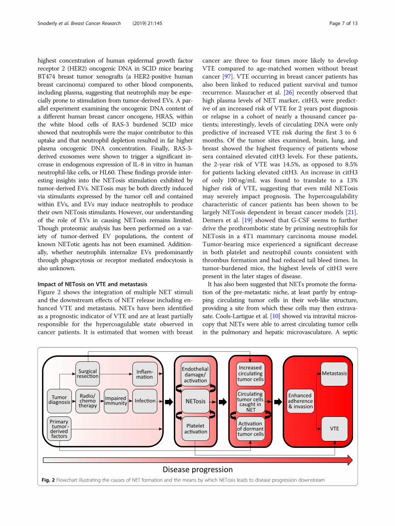
Impact of NETosis on VTE and metastasisFigure 2 shows the integration of multiple NET stimuliand the downstream effects of NET release including en-hanced VTE and metastasis. NETs have been identifiedas a prognostic indicator of VTE and are at least partiallyresponsible for the hypercoagulable state observed incancer patients. It is estimated that women with breast
cancer are three to four times more likely to developVTE compared to age-matched women without breastcancer [97]. VTE occurring in breast cancer patients hasalso been linked to reduced patient survival and tumorrecurrence. Mauracher et al. [26] recently observed thathigh plasma levels of NET marker, citH3, were predict-ive of an increased risk of VTE for 2 years post diagnosisor relapse in a cohort of nearly a thousand cancer pa-tients; interestingly, levels of circulating DNA were onlypredictive of increased VTE risk during the first 3 to 6months. Of the tumor sites examined, brain, lung, andbreast showed the highest frequency of patients whosesera contained elevated citH3 levels. For these patients,the 2-year risk of VTE was 14.5%, as opposed to 8.5%for patients lacking elevated citH3. An increase in citH3of only 100 ng/mL was found to translate to a 13%higher risk of VTE, suggesting that even mild NETosismay severely impact prognosis. The hypercoagulabilitycharacteristic of cancer patients has been shown to belargely NETosis dependent in breast cancer models [21].Demers et al. [19] showed that G-CSF seems to furtherdrive the prothrombotic state by priming neutrophils forNETosis in a 4T1 mammary carcinoma mouse model.Tumor-bearing mice experienced a significant decreasein both platelet and neutrophil counts consistent withthrombus formation and had reduced tail bleed times. Intumor-burdened mice, the highest levels of citH3 werepresent in the later stages of disease.It has also been suggested that NETs promote the forma-
tion of the pre-metastatic niche, at least partly by entrap-ping circulating tumor cells in their web-like structure,providing a site from which these cells may then extrava-sate. Cools-Lartigue et al. [10] showed via intravital micros-copy that NETs were able to arrest circulating tumor cellsin the pulmonary and hepatic microvasculature. A septic
Fig. 2 Flowchart illustrating the causes of NET formation and the means by which NETosis leads to disease progression downstream
Snoderly et al. Breast Cancer Research (2019) 21:145 Page 7 of 13

state was induced in C57BL/6 J mice prior to intrasple-nic injection of H59 Lewis lung carcinoma cells. Micro-metastases were observed within 48 h, with both NEinhibitor and DNase I treatment abolishing this effect;non-septic mice showed few micrometastases, suggest-ing that pro-NETotic stimuli are required to enhanceNET-mediated metastasis. Park et al.’s [27] recent studyshows that 4T1 breast cancer cells injected into the tailvein of LysM-EGFP mice were found within lungs andcaused NET formation; immunofluorescence staining oflung tissue sections showed via DNA and NE fluorescencethat tumor cells were sufficient to provoke increased ratesof lytic NETosis for up to 4 days post injection. DNase I-coated nanoparticle treatment reduced tumor invasionin vitro and the number and size of lung metastasesin vivo.Interestingly, surgical resection aimed at decreasing
tumor burden can actually promote metastasis throughneutrophil activation and subsequent NET formation. In-creased NETosis in patients undergoing liver resectiondue to metastatic colorectal cancer correlates to markedlylower cancer-free survival [14]. This effect of increasedmetastasis following surgical stress was reproduced inmouse models and was abolished by DNase I treatment orinhibiting PAD4 to dissociate NETs or prevent their re-lease, respectively. Neutrophil HMGB1 release occurredconcurrently with pro-NETotic stimulation, and HMGB1was also associated with NETs. This represents a meansby which NETs may directly activate platelets and otherneutrophils, eventually forming a thrombus. Circulatingtumor cells (the presence of which may be increased bysurgical disruption of the primary tumor) could then becaptured due to partial vessel blockage and the coagulat-ing microenvironment around the NET. Simultaneously,the capacity of NETs to damage endothelial cells likely en-ables arrested tumor cells to adhere to the activated endo-thelium, eventually extravasating and establishing a newmetastatic site. Interestingly, NETs have also been shownto activate dormant single breast tumor cells in mouselungs, which can then lead to metastasis development.Cancer cell activation from dormancy is thought to occurvia the remodeling of extracellular matrix due to NET-associated NE and is further facilitated by G-CSF [13].It is reasonable to conclude that tumor-driven NETo-
sis alone, even without surgical stress or major infection,can also serve to drive metastasis. Pro-NETotic factorsare known to be overexpressed by many tumor lines,and multiple murine breast cancer models have beenshown to promote NETosis. However, much remainsunclear about the specific mechanism in which cancerpromotes metastasis through NET formation. It is cur-rently unknown whether NETs predominantly contrib-ute to metastatic establishment via endothelial damageor direct sequestration of tumor cells. Additionally, little
is known about the timeline of NET generation. It ispossible that the primary tumor site must reach enoughdevelopment to elicit NETosis, which then promotes theestablishment of metastases. Alternatively, pioneeringtumor cells may secrete pro-NETotic factors which thenprovoke a NET-induced inflammatory state from sur-rounding neutrophils, favoring tumor cell invasion andfurther sequestration of circulating tumor cells.
NETs as biomarkers and clinical targetsThe ability to detect NETs would likely be of significantprognostic use in differentiating patients at higher risk ofmetastatic progression or VTE, thereby enabling cliniciansto better personalize treatment regimens. To develop aclinical screening tool for NETs, a standardized definitionof “normal” levels of NETosis would need to be estab-lished and has not yet been presented in the literature.The simplest means of in vivo NET detection involvesmeasurement of NET-associated products in the bloodsuch as circulating cell-free DNA, citH3, NE, and MPO.For example, free circulating DNA has been quantified inboth colorectal and breast cancer patient serum samplesvia a simple nucleic acid staining assay [98, 99]. However,even though circulating DNA is known to correlate withbreast tumor size and malignancy [100], it lacks specificityin measuring NETosis. An increased amount of DNA incancer patient serum can also be attributable to otherfactors such as apoptotic and necrotic cells. Measuringcirculating MPO/DNA conjugates is more specific forNET formation than evaluation of cell-free DNA alone[101]. Citrullinated histone H3 (citH3) is formed as a re-sult of PAD4-mediated citrullination during NET forma-tion and represents the most specific biomarker forcirculating NETs [26]. In addition, citH3 may be of prog-nostic significance, as Thålin et al. [102] observed thathigh plasma content of citH3 was a significant indicator ofshort-term mortality in late-stage cancer patients, evenwhen compared to severely ill patients without cancer.Additionally, IL-8 levels were found to correlate withlevels of citH3. Since higher levels of IL-8 would result inincreased neutrophil recruitment, it would be reasonableto conclude that this higher density of neutrophils wouldsubsequently lead to increased NETosis. Despite this,other markers associated with NETs including NE andMPO were not found to differ significantly between se-verely ill patients with and without malignancy; however,these neutrophil-derived enzymes can be independentlyreleased during neutrophil degranulation in the absence ofNET formation, and therefore may not be reliable NET-specific biomarkers. Indeed, citH3 seems to be the mostconsistent indicator of NETosis. While levels of othermarkers may provide useful insight into neutrophil behav-ior, citH3 is highly specific to NETosis and thus would bevaluable in understanding variances between other NET-
Snoderly et al. Breast Cancer Research (2019) 21:145 Page 8 of 13
associated biomarkers. CitH3 levels are also predictive ofVTE risk in newly diagnosed patients, further supportingits diagnostic utility [26].The development of clinical therapies specifically target-
ing NETs in cancer is in its infancy. Inhibition of NETosishas been achieved through several means, though thesevary in their potential for clinical therapies. For instance,DNase I treatment degrades NETs and results in a loss ofthe web-like structure and a reduction in the capacity topromote metastasis in several studies [10, 14, 21, 34]. Inaddition, DNase I has been shown to decrease tumor vol-ume in rats when injected intramuscularly or intraperitone-ally in conjunction with other proteases (papain, trypsin,and chymotrypsin) [103]; however, it is not known whetherthese effects are due primarily to NET inhibition. Currently,DNase I is used clinically in the treatment of cystic fibrosis,as it decreases the NETosis-mediated buildup of mucousviscosity, resulting in improved lung function [6]. However,in this context, DNase I is delivered via nebulizer, whichwould likely be ineffectual in most cancer treatments,though it would be fascinating to observe whether nebu-lized DNase I would have a preventative effect on lung me-tastasis. Additionally, DNase I injection may have off-targeteffects, including compromising the immunoprotectivefunction of NETs.Inhibition of components integral to NETosis, such as
NE or PAD4, would likely have similar off-target effectsdue to their involvement in other key pathways, potentiallydisrupting normal neutrophil function. Small moleculeinhibitors of PAD4 for NET inhibition are under activeinvestigation and include Cl-amidine and F-amidine, irre-versible inhibitors that inactivate calcium-bound PAD4[104]. However, these lack specificity and interact withother PAD-family enzymes. Recently, Lewis et al. [105]synthesized two reversible inhibitors which overcome thishurdle, GSK199 and GSK484, both of which exhibit highspecificity for PAD4 and inhibit NETosis in both mouseand human neutrophils. GSK484 was recently shown toprevent tumor-associated renal dysfunction in mice, whichwas determined to be NET-mediated; the inhibitory effectsof GSK484 were as effective as DNase I [106]. Additionally,a recent study by Yazdani et al. [107] indicates that PAD4-knockout mice challenged with subcutaneous tumor injec-tion of colorectal and hepatocellular carcinoma tumor cellsexperienced slower tumor growth and smaller metastasessimilar to mice treated with daily DNase I injection.NETs were not observable in excised tumor tissue inPAD4-knockout mice. Finally, the authors showed thatNETosis at the primary tumor site may contribute totumor cell survival through enhanced mitochondrialbiogenesis. This data further supports the need to de-velop NET-targeting treatments, as these would be ofgreat therapeutic benefit in both the context of the pri-mary tumor site and the pre-metastatic niche.
Efforts targeting cell adhesive molecules, such as P-selectin, could also prove problematic. Though successfulresults of a stage II clinical trial for the use of the P-selectin inhibitor crizanlizumab in sickle cell anemia toprevent vaso-occlusion were recently published [108], itwould be reasonable to conclude that such a therapy mayinterfere with leukocyte function. Though P-selectin andPSGL-1 antibodies have been shown to inhibit NETosis inmice [41], the disruption of leukocyte adhesion moleculebinding capacity could decrease neutrophil recruitment inresponse to infection in cancer patients already sufferingfrom an immunocompromised state. Off-target effectscould potentially be mitigated via the development of new,more specific delivery vehicles, such as functionalized, tar-geted nanoparticles.Alternatively, the adaptation of FDA-approved drugs
could facilitate the development of effective anti-NETtreatments. For instance, the inhibitory effect of aspirin onNETs has yielded some promising results in animalmodels. Lapponi et al. [109] showed that aspirin preventedNET-induced injury of the lung endothelium by inhibitingplatelet activation and subsequent NET formation in mice.The inhibitory effect of aspirin on NF-κB, an inflamma-tory transcriptional regulator that plays a role in somepathways promoting NETosis, was also demonstrated.The authors found that aspirin treatment effectively inhib-ited NETs in human neutrophils in vitro and resulted inhigher bacteria counts in infection-burdened mice in vivo,suggesting a loss of normal NET functionality. There isevidence to support the use of aspirin in clinical treat-ment. In one meta-analysis, patients using aspirin dailyhad significantly reduced mortality and risk of distant me-tastases for adenocarcinomas. Interestingly, this effect didnot appear to be dose dependent [110]. Aspirin has alsobeen shown to be effective in reducing metastasis in pa-tients suffering from breast cancer specifically [111].Another FDA-approved drug, hydroxychloroquine, origin-
ally used to treat malaria, has been shown to inhibit NETosis[17, 54, 112]. While the mechanism behind NET inhibitionby hydroxychloroquine is unclear, it may be related to au-tophagy inhibition [113]. However, a phase II clinical studyon patients with advanced pancreatic cancer produced littleclinical effect. The authors do suggest, however, that combin-ation therapy may prove more effective [114]. Furthermore,use of hydroxychloroquine as a neoadjuvant treatment inearlier stage disease holds significant promise [115]. Remark-ably, and perhaps not coincidentally, hydroxychloroquinealso inhibits leukocyte phagocytosis [116]. Thus, it may bepossible that hydroxychloroquine could inhibit neutrophiluptake of tumor-derived EVs, thus reducing NETosis. How-ever, the precise mechanism by which this uptake occurs isunknown, as are the mechanisms behind tumor-derived EVstimulated NETosis. Due to the associated complications ofNETs including increased VTE risk and metastasis, which
Snoderly et al. Breast Cancer Research (2019) 21:145 Page 9 of 13
are both negatively associated with breast cancer patient out-come, it is crucial for future research efforts to focus on fur-ther investigation of new specific targets to prevent NETformation.
ConclusionEvidence is mounting that NETs play a significant detri-mental role in the inflammatory state of cancer. We havepresented several classical NETotic stimuli, as well as stim-uli that have been implicitly or explicitly demonstrated toinduce NETosis specifically within the context of cancer,though the mechanisms by which such stimuli occur arenot yet entirely defined. We have also discussed the nega-tive outcomes NETs promote and have highlighted poten-tial NET-specific targets to investigate and utilize todevelop therapies for clinical translation. The next vital stepwill be untangling the web of crosstalk between neutrophils,tumor cells, endothelial cells, platelets, and extracellularvesicles, and eventually the influence of other componentsof the innate and adaptive immune systems on cancer pro-gression. Better understanding of these processes will en-able the development of precise NET-targeted therapiesand diagnostic tools, potentially allowing the identificationof tumors with the potential for metastasis, earlier diagno-sis, and more personalized and effective treatments forbreast cancer patients.
AbbreviationsCitH3: Citrullinated histone 3; DAMP: Damage-associated molecular pattern;EC: Endothelial cell; EV: Extracellular vesicle; G-CSF: Granulocyte-colony-stimulating factor; HER2: Human epidermal growth factor 2; HMGB1: Highmobility group box 1; IL-8: Interleukin-8; LPS: Lipopolysaccharide;MPO: Myeloperoxidase; NE: Neutrophil elastase; NET: Neutrophil extracellulartrap; PAD4: Protein arginine deiminase 4; PMA: Phorbol 12-myristate 13-acetate; P-SEL: P-selectin; PSGL-1: P-selectin glycoprotein ligand-1;RAGE: Receptor for advanced glycation end products; ROS: Reactive oxygenspecies; TF: Tissue factor; TLR4: Toll-like receptor 4; VTE: Venousthromboembolism
AcknowledgementsThe authors would like to thank Dr. Tim Eubank for his helpful suggestionson review content.
Authors’ contributionsHTS performed the literature search, wrote the manuscript, and created thefigures. BAB provided additional content, citations, and insight relevant toNETs throughout the article. MFB wrote the manuscript and edited thefigures. All authors read and approved the final manuscript.
FundingNot applicable.
Availability of data and materialsNot applicable.
Ethics approval and consent to participateNot applicable.
Consent for publicationNot applicable.
Competing interestsThe authors declare that they have no competing interests.
Author details1Department of Chemical and Biomedical Engineering, West VirginiaUniversity, 1306 Evansdale Drive, ESB 521, Morgantown, WV 26506, USA.2Department of Surgery, West Virginia University, Morgantown, WV 26506,USA.
Received: 28 August 2019 Accepted: 4 December 2019
References1. Kobayashi SD, Voyich JM, Burlak C, DeLeo FR. Neutrophils in the innate
immune response. Archivum Immunologiae ET Therapiae Experimentalis-English Edition. 2005;53(6):505.
2. Brinkmann V, Reichard U, Goosmann C, Fauler B, Uhlemann Y, Weiss DS,et al. Neutrophil extracellular traps kill bacteria. Science. 2004;303(5663):1532–5.
3. Yipp BG, Kubes P. NETosis: how vital is it? Blood J Am Soc Hematol. 2013;122(16):2784–94.
4. Chowdhury CS, Giaglis S, Walker UA, Buser A, Hahn S, Hasler P. Enhancedneutrophil extracellular trap generation in rheumatoid arthritis: analysis ofunderlying signal transduction pathways and potential diagnostic utility.Arthritis Res Ther. 2014;16(3):R122.
5. Gupta AK, Hasler P, Holzgreve W, Gebhardt S, Hahn S. Induction ofneutrophil extracellular DNA lattices by placental microparticles and IL-8and their presence in preeclampsia. Hum Immunol. 2005;66(11):1146–54.
6. Gray R, McCullagh B, McCray P. NETs and CF lung disease: current statusand future prospects. Antibiotics. 2015;4(1):62–75.
7. Jorch SK, Kubes P. An emerging role for neutrophil extracellular traps innoninfectious disease. Nat Med. 2017;23(3):279.
8. Mitroulis I, Kambas K, Chrysanthopoulou A, Skendros P, Apostolidou E,Kourtzelis I, et al. Neutrophil extracellular trap formation is associated withIL-1β and autophagy-related signaling in gout. PLoS One. 2011;6(12):e29318.
9. Wang Y, Xiao Y, Zhong L, Ye D, Zhang J, Tu Y, et al. Increased neutrophilelastase and proteinase 3 and augmented NETosis are closely associatedwith β-cell autoimmunity in patients with type 1 diabetes. Diabetes. 2014;63(12):4239–48.
10. Cools-Lartigue J, Spicer J, McDonald B, Gowing S, Chow S, Giannias B, et al.Neutrophil extracellular traps sequester circulating tumor cells and promotemetastasis. J Clin Invest. 2013;123(8):3446–58.
11. Spicer JD, McDonald B, Cools-Lartigue JJ, Chow SC, Giannias B, Kubes P,et al. Neutrophils promote liver metastasis via mac-1-mediated interactionswith circulating tumor cells. Cancer Res. 2012;72(16):3919–27.
12. Pieterse E, Rother N, Garsen M, Hofstra JM, Satchell SC, Hoffmann M, et al.Neutrophil extracellular traps drive endothelial-to-Mesenchymal transition.Arterioscler Thromb Vasc Biol. 2017;37(7):1371–9.
13. Albrengues J, Shields MA, Ng D, Park CG, Ambrico A, Poindexter ME, et al.Neutrophil extracellular traps produced during inflammation awakendormant cancer cells in mice. Science. 2018;361(6409):eaao4227.
14. Tohme S, Yazdani HO, Al-Khafaji AB, Chidi AP, Loughran P, Mowen K, et al.Neutrophil extracellular traps promote the development and progression ofliver metastases after surgical stress. Cancer Res. 2016;76(6):1367–80.
15. Al-Haidari AA, Algethami N, Lepsenyi M, Rahman M, Syk I, Thorlacius H.Neutrophil extracellular traps promote peritoneal metastasis of colon cancercells. Oncotarget. 2019;10(12):1238–49.
16. Kanamaru R, Ohzawa H, Miyato H, Matsumoto S, Haruta H, Kurashina K,et al. Low density neutrophils (LDN) in postoperative abdominal cavityassist the peritoneal recurrence through the production of neutrophilextracellular traps (NETs). Sci Rep. 2018;8(1):632.
17. Boone BA, Murthy P, Miller-Ocuin J, Doerfler WR, Ellis JT, Liang X, et al.Chloroquine reduces hypercoagulability in pancreatic cancer throughinhibition of neutrophil extracellular traps. BMC Cancer. 2018;18(1):678.
18. Fuchs TA, Brill A, Wagner DD. Neutrophil extracellular trap (NET) impact ondeep vein thrombosis. Arterioscler Thromb Vasc Biol. 2012;32(8):1777–83.
19. Demers M, Krause DS, Schatzberg D, Martinod K, Voorhees JR, Fuchs TA,et al. Cancers predispose neutrophils to release extracellular DNA traps thatcontribute to cancer-associated thrombosis. Proc Natl Acad Sci U S A. 2012;109(32):13076–81.
20. Hisada Y, Grover SP, Maqsood A, Houston R, Ay C, Noubouossie DF, et al.Neutrophils and neutrophil extracellular traps enhance venous thrombosisin mice bearing human pancreatic tumors. Haematologica. 2019. [Epub
Snoderly et al. Breast Cancer Research (2019) 21:145 Page 10 of 13
ahead of print]. http://www.haematologica.org/content/early/2019/04/28/haematol.2019.217083.long.
21. Leal AC, Mizurini DM, Gomes T, Rochael NC, Saraiva EM, Dias MS, et al.Tumor-derived exosomes induce the formation of neutrophil extracellulartraps: implications for the establishment of cancer-associated thrombosis.Sci Rep. 2017;7(1):6438.
22. Arelaki S, Arampatzioglou A, Kambas K, Papagoras C, Miltiades P, AngelidouI, et al. Gradient infiltration of neutrophil extracellular traps in colon cancerand evidence for their involvement in tumour growth. PLoS One. 2016;11(5):e0154484.
23. Demers M, Wong SL, Martinod K, Gallant M, Cabral JE, Wang Y, et al.Priming of neutrophils toward NETosis promotes tumor growth.Oncoimmunology. 2016;5(5):e1134073.
24. Bray F, Ferlay J, Soerjomataram I, Siegel RL, Torre LA, Jemal A. Global cancerstatistics 2018: GLOBOCAN estimates of incidence and mortality worldwidefor 36 cancers in 185 countries. CA Cancer J Clin. 2018;68(6):394–424.
25. Maugeri N, Campana L, Gavina M, Covino C, De Metrio M, Panciroli C, et al.Activated platelets present high mobility group box 1 to neutrophils,inducing autophagy and promoting the extrusion of neutrophil extracellulartraps. J Thromb Haemost. 2014;12(12):2074–88.
26. Mauracher LM, Posch F, Martinod K, Grilz E, Däullary T, Hell L, et al.Citrullinated histone H3, a biomarker of neutrophil extracellular trapformation, predicts the risk of venous thromboembolism in cancer patients.J Thromb Haemost. 2018;16(3):508–18.
27. Park J, Wysocki RW, Amoozgar Z, Maiorino L, Fein MR, Jorns J, et al. Cancercells induce metastasis-supporting neutrophil extracellular DNA traps. SciTransl Med. 2016;8(361):361ra138.
28. Saffarzadeh M, Juenemann C, Queisser MA, Lochnit G, Barreto G,Galuska SP, et al. Neutrophil extracellular traps directly induce epithelialand endothelial cell death: a predominant role of histones. PLoS One.2012;7(2):e32366.
29. Semeraro F, Ammollo CT, Morrissey JH, Dale GL, Friese P, Esmon NL, et al.Extracellular histones promote thrombin generation through platelet-dependent mechanisms: involvement of platelet TLR2 and TLR4. Blood.2011;118(7):1952–61.
30. Ni P, Zhang Y, Liu Y, Lin X, Su X, Lu H, et al. HMGB1 silence could promoteMCF-7 cell apoptosis and inhibit invasion and metastasis. Int J Clin ExpPathol. 2015;8(12):15940.
31. Clark SR, Ma AC, Tavener SA, McDonald B, Goodarzi Z, Kelly MM, et al.Platelet TLR4 activates neutrophil extracellular traps to ensnare bacteria inseptic blood. Nat Med. 2007;13(4):463.
32. Keshari RS, Verma A, Barthwal MK, Dikshit M. Reactive oxygen species-induced activation of ERK and p38 MAPK mediates PMA-induced NETsrelease from human neutrophils. J Cell Biochem. 2013;114(3):532–40.
33. Tadie J-M, Bae H-B, Jiang S, Park DW, Bell CP, Yang H, et al. HMGB1promotes neutrophil extracellular trap formation through interactions withtoll-like receptor 4. Am J Phys Lung Cell Mol Phys. 2013;304(5):L342–L9.
34. Gupta AK, Joshi MB, Philippova M, Erne P, Hasler P, Hahn S, et al. Activatedendothelial cells induce neutrophil extracellular traps and are susceptible toNETosis-mediated cell death. FEBS Lett. 2010;584(14):3193–7.
35. De Larco JE, Wuertz BR, Furcht LT. The potential role of neutrophils inpromoting the metastatic phenotype of tumors releasing interleukin-8. ClinCancer Res. 2004;10(15):4895–900.
36. Avalos BR, Gasson JC, Hedvat C, Quan S, Baldwin G, Weisbart R, et al.Human granulocyte colony-stimulating factor: biologic activities andreceptor characterization on hematopoietic cells and small cell lung cancercell lines. Blood. 1990;75(4):851–7.
37. Cedervall J, Zhang Y, Huang H, Zhang L, Femel J, Dimberg A, et al.Neutrophil extracellular traps accumulate in peripheral blood vessels andcompromise organ function in tumor-bearing animals. Cancer Res. 2015;75(13):2653–62.
38. Li P, Li M, Lindberg MR, Kennett MJ, Xiong N, Wang Y. PAD4 is essential forantibacterial innate immunity mediated by neutrophil extracellular traps. JExp Med. 2010;207(9):1853–62.
39. Tatsiy O, McDonald PP. Physiological stimuli induce PAD4-dependent, ROS-independent NETosis, with early and late events controlled by discretesignaling pathways. Front Immunol. 2018;9:2036. https://www.ncbi.nlm.nih.gov/pubmed/30279690.
40. Chang X, Han J, Pang L, Zhao Y, Yang Y, Shen Z. Increased PADI4expression in blood and tissues of patients with malignant tumors. BMCCancer. 2009;9(1):40.
41. Etulain J, Martinod K, Wong SL, Cifuni SM, Schattner M, Wagner DD. P-selectin promotes neutrophil extracellular trap formation in mice. Blood.2015;126(2):242–6.
42. Kambas K, Chrysanthopoulou A, Vassilopoulos D, Apostolidou E, Skendros P,Girod A, et al. Tissue factor expression in neutrophil extracellular traps andneutrophil derived microparticles in antineutrophil cytoplasmic antibodyassociated vasculitis may promote thromboinflammation and thethrombophilic state associated with the disease. Ann Rheum Dis. 2014;73(10):1854–63.
43. Stakos DA, Kambas K, Konstantinidis T, Mitroulis I, Apostolidou E, Arelaki S,et al. Expression of functional tissue factor by neutrophil extracellular trapsin culprit artery of acute myocardial infarction. Eur Heart J. 2015;36(22):1405–14.
44. Ruf W, Yokota N, Schaffner F. Tissue factor in cancer progression andangiogenesis. Thromb Res. 2010;125:S36–S8.
45. Kirchner T, Möller S, Klinger M, Solbach W, Laskay T, Behnen M. The impactof various reactive oxygen species on the formation of neutrophilextracellular traps. Mediat Inflamm. 2012;2012.
46. Warnatsch A, Tsourouktsoglou T-D, Branzk N, Wang Q, Reincke S, Herbst S,et al. Reactive oxygen species localization programs inflammation to clearmicrobes of different size. Immunity. 2017;46(3):421–32.
47. Davidson B, Konstantinovsky S, Kleinberg L, Nguyen MT, Bassarova A,Kvalheim G, et al. The mitogen-activated protein kinases (MAPK) p38 andJNK are markers of tumor progression in breast carcinoma. Gynecol Oncol.2006;102(3):453–61.
48. Zhao M, Howard EW, Parris AB, Guo Z, Zhao Q, Yang X. Alcohol promotesmigration and invasion of triple-negative breast cancer cells throughactivation of p38 MAPK and JNK. Mol Carcinog. 2017;56(3):849–62.
49. Rochael NC, Guimarães-Costa AB, Nascimento MT, DeSouza-Vieira TS,Oliveira MP, e Souza LFG, et al. Classical ROS-dependent and early/rapidROS-independent release of neutrophil extracellular traps triggered byLeishmania parasites. Scientific Reports 2015;5:18302.
50. Wang Y, Li M, Stadler S, Correll S, Li P, Wang D, et al. Histonehypercitrullination mediates chromatin decondensation and neutrophilextracellular trap formation. J Cell Biol. 2009;184(2):205–13.
51. Rohrbach A, Slade D, Thompson P, Mowen K. Activation of PAD4 in NETformation. Front Immunol. 2012;3:360.
52. Papayannopoulos V, Metzler KD, Hakkim A, Zychlinsky A. Neutrophil elastaseand myeloperoxidase regulate the formation of neutrophil extracellulartraps. J Cell Biol. 2010;191(3):677–91.
53. Kwak T, Drews-Elger K, Ergonul A, Miller PC, Braley A, Hwang GH, et al.Targeting of RAGE-ligand signaling impairs breast cancer cell invasion andmetastasis. Oncogene. 2017;36(11):1559–72.
54. Boone BA, Orlichenko L, Schapiro NE, Loughran P, Gianfrate GC, Ellis JT,et al. The receptor for advanced glycation end products (RAGE) enhancesautophagy and neutrophil extracellular traps in pancreatic cancer. CancerGene Ther. 2015;22(6):326–34.
55. Kang R, Tang D, Lotze MT, Zeh HJ, 3rd. RAGE regulates autophagy andapoptosis following oxidative injury. Autophagy. 2011;7(4):442–4.
56. Pham DL, Ban GY, Kim SH, Shin YS, Ye YM, Chwae YJ, et al. Neutrophilautophagy and extracellular DNA traps contribute to airway inflammation insevere asthma. Clin Exp Allergy. 2017;47(1):57–70.
57. Sha LL, Wang H, Wang C, Peng HY, Chen M, Zhao MH. Autophagy isinduced by anti-neutrophil cytoplasmic Abs and promotes neutrophilextracellular traps formation. Innate Immun. 2016;22(8):658–65.
58. Park SY, Shrestha S, Youn YJ, Kim JK, Kim SY, Kim HJ, et al. Autophagyprimes neutrophils for neutrophil extracellular trap formation during Sepsis.Am J Respir Crit Care Med. 2017;196(5):577–89.
59. Remijsen Q, Vanden Berghe T, Wirawan E, Asselbergh B, Parthoens E, DeRycke R, et al. Neutrophil extracellular trap cell death requires bothautophagy and superoxide generation. Cell Res. 2011;21(2):290–304.
60. Amulic B, Knackstedt SL, Abed UA, Deigendesch N, Harbort CJ, Caffrey BE,et al. Cell-cycle proteins control production of neutrophil extracellular traps.Developmental Cell. 2017;43(4):449–62 e5.
61. Lukášová E, Kořistek Z, Klabusay M, Ondřej V, Grigoryev S, Bačíková A, et al.Granulocyte maturation determines ability to release chromatin NETs andloss of DNA damage response; these properties are absent in immatureAML granulocytes. Biochimica et Biophysica Acta (BBA)-molecular. Cell Res.2013;1833(3):767–79.
62. Mackey JBG, Coffelt SB, Carlin LM. Neutrophil Maturity in Cancer. Frontiers inImmunology. 2019;10(1912).
Snoderly et al. Breast Cancer Research (2019) 21:145 Page 11 of 13
63. Powell DR, Huttenlocher A. Neutrophils in the tumor microenvironment.Trends Immunol. 2016;37(1):41–52.
64. Hunter KW Jr. Murine mammary carcinoma 4T1 induces a leukemoidreaction with splenomegaly: association with tumor-derived growth factors.Exp Mol Pathol. 2007;82(1):12–24.
65. Kowanetz M, Wu X, Lee J, Tan M, Hagenbeek T, Qu X, et al.Granulocyte-colony stimulating factor promotes lung metastasis throughmobilization of Ly6G+ Ly6C+ granulocytes. Proc Natl Acad Sci. 2010;107(50):21248–55.
66. Sugimoto C, Fujieda S, Sunaga H, Noda I, Tanaka N, Kimura Y, et al. Granulocytecolony-stimulating factor (G-CSF)-mediated signaling regulates type IVcollagenase activity in head and neck cancer cells. Int J Cancer. 2001;93(1):42–6.
67. de Oliveira S, Reyes-Aldasoro CC, Candel S, Renshaw SA, Mulero V, Calado Â.Cxcl8 (IL-8) mediates neutrophil recruitment and behavior in the zebrafishinflammatory response. J Immunol. 2013;190(8):4349–59.
68. Kozłowski L, Zakrzewska I, Tokajuk P, Wojtukiewicz M. Concentration ofinterleukin-6 (IL-6), interleukin-8 (IL-8) and interleukin-10 (IL-10) in bloodserum of breast cancer patients. Roczniki Akademii Medycznej wBialymstoku (1995). 2003;48:82–4.
69. Xu R, Bao C, Huang H, Lin F, Yuan Y, Wang S, et al. Low expression ofCXCR1/2 on neutrophils predicts poor survival in patients with hepatitis Bvirus-related acute-on-chronic liver failure. Sci Rep. 2016;6:38714.
70. Alfaro C, Teijeira A, Oñate C, Pérez G, Sanmamed MF, Andueza MP, et al.Tumor-produced interleukin-8 attracts human myeloid-derived suppressorcells and elicits extrusion of neutrophil extracellular traps (NETs). Clin CancerRes. 2016;22(15):3924–36.
71. Jin L, Batra S, Douda DN, Palaniyar N, Jeyaseelan S. CXCL1 contributes tohost defense in polymicrobial sepsis via modulating T cell and neutrophilfunctions. J Immunol. 2014;193(7):3549–58.
72. Jin L, Batra S, Jeyaseelan S. Diminished neutrophil extracellular trap (NET)formation is a novel innate immune deficiency induced by acute ethanolexposure in polymicrobial sepsis, which can be rescued by CXCL1. PLoSPathog. 2017;13(9):e1006637.
73. Jeannin P, Delneste Y, Gosset P, Molet S, Lassalle P, Hamid Q, et al.Histamine induces interleukin-8 secretion by endothelial cells. Blood. 1994;84(7):2229–33.
74. Haddad TC, Greeno EW. Chemotherapy-induced thrombosis. Thromb Res.2006;118(5):555–68.
75. Corre I, Paris F, Huot J. The p38 pathway, a major pleiotropic cascade thattransduces stress and metastatic signals in endothelial cells. Oncotarget.2017;8(33):55684.
76. Schreiber A, Rousselle A, Becker JU, von Mässenhausen A, Linkermann A,Kettritz R. Necroptosis controls NET generation and mediates complementactivation, endothelial damage, and autoimmune vasculitis. Proc Natl AcadSci. 2017;114(45):E9618–E25.
77. Knight JS, Luo W, O’Dell AA, Yalavarthi S, Zhao W, Subramanian V, et al.Peptidylarginine deiminase inhibition reduces vascular damage andmodulates innate immune responses in murine models of atherosclerosis.Circ Res. 2014;114(6):947–56.
78. Goerge T, Barg A, Schnaeker E-M, Poppelmann B, Shpacovitch V, RattenhollA, et al. Tumor-derived matrix metalloproteinase-1 targets endothelialproteinase-activated receptor 1 promoting endothelial cell activation.Cancer Res. 2006;66(15):7766–74.
79. Andrews RK, Arthur JF, Gardiner EE. Neutrophil extracellular traps (NETs) andthe role of platelets in infection. Thromb Haemost. 2014;112(10):659–65.
80. Ueno T, Toi M, Koike M, Nakamura S, Tominaga T. Tissue factor expressionin breast cancer tissues: its correlation with prognosis and plasmaconcentration. Br J Cancer. 2000;83(2):164.
81. Pabinger I, Thaler J, Ay C. Biomarkers for prediction of venousthromboembolism in cancer. Blood. 2013;122(12):2011–8.
82. Abdol Razak N, Elaskalani O, Metharom P. Pancreatic cancer-inducedneutrophil extracellular traps: a potential contributor to cancer-associatedthrombosis. Int J Mol Sci. 2017;18(3):487.
83. Ma A, Kubes P. Platelets, neutrophils, and neutrophil extracellular traps(NETs) in sepsis. J Thromb Haemost. 2008;6(3):415–20.
84. Li J, Kim K, Hahm E, Molokie R, Hay N, Gordeuk VR, et al. Neutrophil AKT2regulates heterotypic cell-cell interactions during vascular inflammation. JClin Invest. 2014;124(4):1483–96.
85. Sreeramkumar V, Adrover JM, Ballesteros I, Cuartero MI, Rossaint J, Bilbao I,et al. Neutrophils scan for activated platelets to initiate inflammation.Science. 2014;346(6214):1234–8.
86. Nagata K, Tsuji T, Todoroki N, Katagiri Y, Tanoue K, Yamazaki H, et al.Activated platelets induce superoxide anion release by monocytes andneutrophils through P-selectin (CD62). J Immunol. 1993;151(6):3267–73.
87. Ay C, Simanek R, Vormittag R, Dunkler D, Alguel G, Koder S, et al. Highplasma levels of soluble P-selectin are predictive of venousthromboembolism in cancer patients: results from the Vienna Cancer andthrombosis study (CATS). Blood. 2008;112(7):2703–8.
88. Zomer A, Maynard C, Verweij FJ, Kamermans A, Schafer R, Beerling E, et al.In vivo imaging reveals extracellular vesicle-mediated phenocopying ofmetastatic behavior. Cell. 2015;161(5):1046–57.
89. Akers JC, Gonda D, Kim R, Carter BS, Chen CC. Biogenesis of extracellularvesicles (EV): exosomes, microvesicles, retrovirus-like vesicles, and apoptoticbodies. J Neuro-Oncol. 2013;113(1):1–11.
90. Xu R, Greening DW, Zhu H-J, Takahashi N, Simpson RJ. Extracellular vesicleisolation and characterization: toward clinical application. J Clin Invest. 2016;126(4):1152–62.
91. König L, Kasimir-Bauer S, Bittner A-K, Hoffmann O, Wagner B, SantosManvailer LF, et al. Elevated levels of extracellular vesicles are associatedwith therapy failure and disease progression in breast cancer patientsundergoing neoadjuvant chemotherapy. Oncoimmunology. 2018;7(1):e1376153.
92. Becker A, Thakur BK, Weiss JM, Kim HS, Peinado H, Lyden D. Extracellularvesicles in cancer: cell-to-cell mediators of metastasis. Cancer Cell. 2016;30(6):836–48.
93. Chen I-H, Xue L, Hsu C-C, Paez JSP, Pan L, Andaluz H, et al. Phosphoproteinsin extracellular vesicles as candidate markers for breast cancer. Proc NatlAcad Sci. 2017;114(12):3175–80.
94. Logozzi M, De Milito A, Lugini L, Borghi M, Calabro L, Spada M, et al. Highlevels of exosomes expressing CD63 and caveolin-1 in plasma of melanomapatients. PLoS One. 2009;4(4):e5219.
95. Headley MB, Bins A, Nip A, Roberts EW, Looney MR, Gerard A, et al.Visualization of immediate immune responses to pioneer metastatic cells inthe lung. Nature. 2016;531(7595):513.
96. Chennakrishnaiah S, Meehan B, D'Asti E, Montermini L, Lee TH, Karatzas N,et al. Leukocytes as a reservoir of circulating oncogenic DNA and regulatorytargets of tumor-derived extracellular vesicles. J Thromb Haemost. 2018;16(9):1800–13.
97. Walker AJ, West J, Card TR, Crooks C, Kirwan CC, Grainge MJ. Whenare breast cancer patients at highest risk of venousthromboembolism? A cohort study using English health care data.Blood. 2016;127(7):849–57.
98. Agassi R, Czeiger D, Shaked G, Avriel A, Sheynin J, Lavrenkov K, et al.Measurement of circulating cell-free DNA levels by a simple fluorescent testin patients with breast cancer. Am J Clin Pathol. 2015;143(1):18–24.
99. Czeiger D, Shaked G, Eini H, Vered I, Belochitski O, Avriel A, et al.Measurement of circulating cell-free DNA levels by a new simplefluorescent test in patients with primary colorectal cancer. Am J Clin Pathol.2011;135(2):264–70.
100. Kohler C, Radpour R, Barekati Z, Asadollahi R, Bitzer J, Wight E, et al. Levelsof plasma circulating cell free nuclear and mitochondrial DNA as potentialbiomarkers for breast tumors. Mol Cancer. 2009;8(1):105.
101. Yoo DG, Floyd M, Winn M, Moskowitz SM, Rada B. NET formation inducedby Pseudomonas aeruginosa cystic fibrosis isolates measured as release ofmyeloperoxidase-DNA and neutrophil elastase-DNA complexes. ImmunolLett. 2014;160(2):186–94.
102. Thålin C, Lundström S, Seignez C, Daleskog M, Lundström A, Henriksson P,et al. Citrullinated histone H3 as a novel prognostic blood marker inpatients with advanced cancer. PLoS One. 2018;13(1):e0191231.
103. Trejo-Becerril C, Pérez-Cardenas E, Gutiérrez-Díaz B, De La Cruz-Sigüenza D,Taja-Chayeb L, González-Ballesteros M, et al. Antitumor effects of systemicDNAse I and proteases in an in vivo model. Integr Cancer Ther. 2016;15(4):NP35–43.
104. Jones J, Causey C, Knuckley B, Slack-Noyes JL, Thompson PR. Proteinarginine deiminase 4 (PAD4): current understanding and future therapeuticpotential. Curr Opin Drug Discov Devel. 2009;12(5):616.
105. Lewis HD, Liddle J, Coote JE, Atkinson SJ, Barker MD, Bax BD, et al. Inhibitionof PAD4 activity is sufficient to disrupt mouse and human NET formation.Nat Chem Biol. 2015;11(3):189.
106. Cedervall J, Dragomir A, Saupe F, Zhang Y, Arnlov J, Larsson E, et al.Pharmacological targeting of peptidylarginine deiminase 4 prevents cancer-associated kidney injury in mice. Oncoimmunol. 2017;6(8):e1320009.
Snoderly et al. Breast Cancer Research (2019) 21:145 Page 12 of 13
107. Yazdani HO, Roy E, Comerci AJ, van der Windt DJ, Zhang H, Huang H, et al.Neutrophil extracellular traps drive mitochondrial homeostasis in tumors toaugment growth. Cancer Research. 2019:canres. 0800.2019.
108. Ataga KI, Kutlar A, Kanter J, Liles D, Cancado R, Friedrisch J, et al.Crizanlizumab for the prevention of pain crises in sickle cell disease. N EnglJ Med. 2017;376(5):429–39.
109. Lapponi MJ, Carestia A, Landoni VI, Rivadeneyra L, Etulain J, Negrotto S,et al. Regulation of neutrophil extracellular trap formation by anti-inflammatory drugs. J Pharmacol Exp Ther. 2013;345(3):430–7.
110. Rothwell PM, Wilson M, Price JF, Belch JF, Meade TW, Mehta Z. Effect ofdaily aspirin on risk of cancer metastasis: a study of incident cancers duringrandomised controlled trials. Lancet. 2012;379(9826):1591–601.
111. Holmes MD, Chen WY, Li L, Hertzmark E, Spiegelman D, Hankinson SE.Aspirin intake and survival after breast cancer. J Clin Oncol. 2010;28(9):1467.
112. Murthy P, Singhi AD, Ross MA, Loughran P, Paragomi P, Papachristou GI,et al. Enhanced neutrophil extracellular trap formation in acute pancreatitiscontributes to disease severity and is reduced by chloroquine. FrontImmunol. 2019;10:28.
113. Cook KL, Warri A, Soto-Pantoja DR, Clarke PA, Cruz MI, Zwart A, et al.Hydroxychloroquine inhibits autophagy to potentiate antiestrogenresponsiveness in ER+ breast cancer. Clin Cancer Res. 2014;20(12):3222–32.
114. Wolpin BM, Rubinson DA, Wang X, Chan JA, Cleary JM, Enzinger PC, et al.Phase II and pharmacodynamic study of autophagy inhibition usinghydroxychloroquine in patients with metastatic pancreatic adenocarcinoma.Oncologist. 2014;19(6):637–8.
115. Boone BA, Bahary N, Zureikat AH, Moser AJ, Normolle DP, Wu WC, et al.Safety and biologic response of pre-operative autophagy inhibition incombination with gemcitabine in patients with pancreatic adenocarcinoma.Ann Surg Oncol. 2015;22(13):4402–10.
116. Labro M, Babin-Chevaye C. Effects of amodiaquine, chloroquine, andmefloquine on human polymorphonuclear neutrophil function in vitro.Antimicrob Agents Chemother. 1988;32(8):1124–30.
Publisher’s NoteSpringer Nature remains neutral with regard to jurisdictional claims inpublished maps and institutional affiliations.
Snoderly et al. Breast Cancer Research (2019) 21:145 Page 13 of 13