Embed Size (px)
Citation preview
Vol.:(0123456789)1 3
Archivum Immunologiae et Therapiae Experimentalis (2018) 66:273–281 https://doi.org/10.1007/s00005-018-0505-y
REVIEW
The Phenomenon of Neutrophil Extracellular Traps in Vascular Diseases
Dorota Dąbrowska1 · Ewa Jabłońska1 · Marzena Garley1 · Jolanta Sawicka‑Powierza2 · Karolina Nowak1
Received: 8 April 2017 / Accepted: 2 January 2018 / Published online: 5 February 2018 © The Author(s) 2018. This article is an open access publication
AbstractVascular diseases constitute a global health issue due to the increasing number of cases of patients with these diseases. The pathogenesis of the majority of these diseases, including atherosclerosis and thrombosis, is complex and not yet fully understood. One of the major causes for their occurrence can be immune disorders resulting in the development of a chronic inflammation within the vessels. In recent years, studies have placed emphasis on the role of neutrophils in the development of these diseases, i.e., the discovery of neutrophil extracellular traps (NETs) demonstrated that the structures released by the cells may contribute to the enhancement of inflammatory reactions and cell damage. This article summarizes current knowledge on the role of NETs during atherosclerosis, thrombosis and small-vessel vasculitis, especially in antineutrophil cytoplasmic antibody (ANCA)-associated small-vessel vasculitis (AAV).
Keywords Neutrophils · NETs · Atherosclerosis · Thrombosis · Small-vessel vasculitis · AAV
Introduction
Vascular diseases constitute a serious clinical, social and economic issue in the modern world. For many years, they have remained the major cause of mortality in developed countries. In recent years, studies have demonstrated that both innate and adaptive mechanisms of immune response are the basis for the majority of vascular diseases, includ-ing atherosclerosis and thrombosis (Chamorro and Hallen-beck 2006). Recently, interest in neutrophils has increased, as, according to reports, they play a significant role in the development and progression of cardiovascular diseases (Ionita et al. 2010; von Brühl et al. 2012). One of the major reasons for the great number of experiments conducted on these immunocompetent cells was the recent discovery of one of their functions—the formation and release of neu-trophil extracellular traps (NETs). The literature reports that the excessive production of the NETs, stemming from the disturbed balance between the production and destruction of
NETs, may contribute to the damage of vascular endothe-lium and to uncontrolled formation of thrombus in their lumina. The available literature data show that NETs may be involved in the development of atherosclerosis and thrombo-sis and also in small-vessel vasculitis (SVV) (Borissoff et al. 2013; Fuchs et al. 2010; Kessenbrock et al. 2009; Warnatsch et al. 2015).
Neutrophil Extracellular Traps
Neutrophils represent the most abundant (50–75% of the total number of the peripheral blood leukocytes) population of white blood system cells. Neutrophils are one of the first effector cells recruited at the inflammation site, where they constitute the first line of defense in the combat against a wide range of microorganisms. Moreover, neutrophils can affect the recruitment and activation of other immunocom-petent cells, as well as participate in regenerative processes of tissues. For many years, the antipathogenic activity of neutrophils was linked solely to the elimination of pathogens through phagocytosis, including degranulation of antibac-terial proteins and generation of reactive oxygen species (ROS) (Borregaard 2010; Mantovani et al. 2011).
A breakthrough in the study on these cells came from the results of the study of Brinkmann et al. (2004) published in Science. The authors of this pioneering article recently
* Dorota Dąbrowska [email protected]
1 Department of Immunology, Medical University of Bialystok, J. Waszyngtona 15A, 15-269 Bialystok, Poland
2 Department of Family Medicine, Medical University of Bialystok, Mieszka I 4B, 15-054, Bialystok, Poland
274 Archivum Immunologiae et Therapiae Experimentalis (2018) 66:273–281
1 3
detected an additional property of neutrophils consisting of the generation of NETs (Brinkmann et al. 2004). Thanks to this discovery, scientists began to realize that the earlier information on neutrophils was insufficient and a new analy-sis of their functions should be performed. Brinkmann et al. (2004) indicated that the NET structure consists of clusters of thin fibers and globular domains able to aggregate and form larger strands of traps. These scientists also revealed that NETs may be subjected to degradation by DNase but not proteases, which confirmed that the deoxyribonucleic acid (DNA) is one of the main NET elements (Brinkmann et al. 2004). Apart from DNA, NETs, contain numerous proteins originating from the primary neutrophil granules: myelop-eroxidase (MPO), neutrophil elastase (NE), proteinase 3 (PR3); secondary neutrophil granules: lactoferrin, pentraxin 3 and tertiary neutrophil granules: gelatinase (Jaillon et al. 2007; Urban et al. 2009; Wartha et al. 2007). In the globu-lar domains of NETs, histones: H1, H2A, H2B, H3, H4 are the dominant group of proteins and they constitute approx. 70% of all proteins related to NETs (Brinkmann et al. 2004; Dwivedi et al. 2014).
Formation and release of NETs demonstrates a unique form of cell death, known as NETosis, which cannot be compared to other known death programs, including apop-tosis and necrosis (Fuchs et al. 2007). The proper course of NETosis requires the activity of the NADPH oxidase complex, histone citrullination, being the posttranslational modification of proteins, catalyzed by peptidylarginine deiminases (PADs), in particular by the PAD4 isoform and autophagy phenomena (Fuchs et al. 2007; Neeli et al. 2008; Remijsen et al. 2011).
Numerous studies have demonstrated that NETs play a crucial role in the body’s immune reaction to an infection. A release of these structures to the extracellular space enables the entrapment and elimination of many pathogens, includ-ing bacteria: Salmonella typhimurium, Shigella flexneri (Brinkmann et al. 2004), fungi: Candida albicans (Urban et al. 2006), Aspergillus fumigatus (McCormick et al. 2010), protozoans: Toxoplasma gondii (Abi Abdallah et al. 2012), Leishmania amazonensis (Guimarães-Costa et al. 2009) and viruses: human immunodeficiency virus 1 (HIV-1) (Saitoh et al. 2012), influenza A virus H1N1 (Narasaraju et al. 2011). In their review article, Kaplan and Radic (2012) described that by forming a physical barrier, NETs facilitate the degradation of bacterial and viral factors of virulence, and thereby prevent the spread of microorganisms.
But, despite these significant advantages resulting from the formation of NETs, numerous scientific studies report on the pathological role of these structures. As has been observed, the process of the generation and elimination of NETs should be strictly regulated. An excessive number of these structures formed in an inappropriate place and time may cause numerous undesirable and unfavorable changes
in an organism (Manda et al. 2014). In the light of current knowledge, formation of NETs, carrying the nuclear mate-rial in the form of DNA and enzymes, such as MPO or NE, constitutes a potential developmental factor for autoimmuni-zation and cardiovascular disorders.
NETs and Atherosclerosis
Atherosclerosis, a civilization disease, has become one of the most common health problems in recent years. The development of atherosclerosis is caused by damaged endothelium, a chronic response of the vessel walls with an inflammatory character, resulting in adhesion of leukocytes and blood platelets and an increase in the permeability of vessels for lipid compounds, primarily low density fraction. Because of the accumulation of the immune system cells and lipids, atherosclerotic plaques are developed, surrounded by smooth muscle cells (Hansson 2005). The atherosclerotic process is influenced by a series of states, such as obesity, hypertension, diabetes, dyslipidemia, which increase the risk of a rapid progression of atherosclerotic changes leading to the occurrence of significant disorders in the function of vital organs (Scott 2002; Singh et al. 2002). The available literature data show that atherosclerosis should be treated not only as a disease related to lipid disorders, but also as a chronic inflammatory disease, for instance due to the cells of the immune system found in atherosclerotic plaques, includ-ing T lymphocytes, macrophages, granulocytes, which, by releasing inflammatory mediators (cytokines, growth fac-tors), influence the development of these atherosclerotic plaques (Falk 2006; Jawień 2008; Singh et al. 2002; Weber et al. 2008).
Initially overshadowed, neutrophils gained more impor-tance when it turned out that they can occur in different regions of an atherosclerotic plaque, including in the fibrous cap, in the shoulder, and in areas toward the media (also known as the base of the plaque). As described, neutrophils play a significant role in both atherosclerosis pathogenesis as well as in the destabilization of atherosclerotic plaque (Ionita et al. 2010). Thanks to their capacity to form numerous fac-tors, including ROS and cytokines, neutrophils participate in the promotion of systemic inflammatory reactions and influence the local concentration of different immunocom-petent cells modulating the permeability of endothelial cells (Baetta and Corsini 2010). In their review article, Chistia-kov et al. (2015) described that for a chronic inflammation accompanying atherosclerosis, the activity of neutrophils may be directed at their own cells and also contribute to gradual vessel damage.
An additional stimulus for further study on the role of neutrophils during the atherosclerotic process was the dis-covery of NETs. Megens et al. (2012) were among the first to detect NET formation in a mouse atherosclerosis model
275Archivum Immunologiae et Therapiae Experimentalis (2018) 66:273–281
1 3
as well as in patients who were subjected to the procedure of endarterectomy, i.e., removal of atherosclerotic plaques. The study conducted by Knight et al. (2014) further proved that neutrophils isolated from mice with atherosclerosis are more susceptible to NET formation. Moreover, the authors of this publication demonstrated that inhibition of PAD4 by Cl-amidine results not only in the reduction of NET formation, but also protects against the development of atherosclerosis and arterial thrombosis, which suggests their significant role in the pathogenesis of these diseases (Knight et al. 2014).
Borissoff et al. (2013), who aimed to determine the rela-tionship between NET release and coronary atherosclero-sis and the presence of prothrombotic state, revealed that elevated levels of NET markers—double-stranded DNA, nucleosomes and MPO-DNA complexes were significantly correlated with the occurrence of serious cardiovascular events. According to these scientists, these biomarkers may prove useful in the forecasting of coronary disease (Boris-soff et al. 2013). Interesting observations were published by Warnatsch et al. (2015) who, using a mouse model of
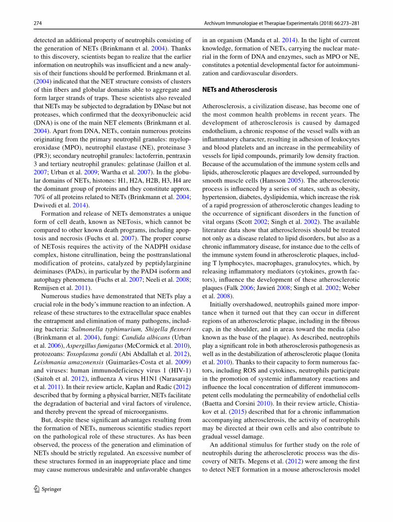
atherosclerosis, demonstrated that cholesterol crystals influ-ence the induction of NETosis. They observed that NETs constituted large amorphic structures within the atheroscle-rotic changes in mice which had previously been introduced with a high-lipid diet to induce hypercholesterolemia. More-over, based on the results of their study, these scientists sug-gested that NETs may also contribute to the development of atherosclerosis through modulation of cytokine formation. Evidence for this assumption was the finding of elevated levels of interleukin (IL)-1α, IL-1β and IL-6 in the blood plasma of mice with excessive generation of NETs (War-natsch et al. 2015) (Fig. 1).
Literature data demonstrate that NETs constitute a con-nection between innate and adaptive immune response and may influence the acuteness of atherosclerosis, i.e., through activation of T lymphocytes and antigen-presenting cells. This is demonstrated in the study conducted by Tillack et al. (2012), which revealed that NETs have the ability to directly prime T lymphocytes by decreasing their acti-vation threshold and thereby contributing to an enhanced
Fig. 1 NET formation in atherosclerosis. a NETs may influence acti-vation of plasmacytoid dendritic cells (pDC), releasing large amounts of type I interferon facilitating the development of atherosclerotic changes (Döring et al. 2012); b Cholesterol crystal induce NETosis (Warnatsch et al. 2015); c NETs stimulate macrophages to release
cytokines, activating Th17 lymphocytes, which enhance the recruit-ment of immunity cells in atherosclerotic plaques (Warnatsch et al. 2015); d NET markers circulating in the bloodstream (nucleosomes, double-stranded DNA (dsDNA) and MPO-DNA) (Borissoff et al. 2013)
276 Archivum Immunologiae et Therapiae Experimentalis (2018) 66:273–281
1 3
adaptive immune response. Moreover, the results of the study obtained by Warnatsch et al. (2015) showed that NETs may stimulate macrophages to release cytokines, activat-ing Th17 lymphocytes, which enhance the recruitment of immune cells in atherosclerotic plaques and which are thus capable of influencing the development of inflammation within vessels. According to other experiments, NETs may also influence activation of plasmacytoid dendritic cells identified in murine and human atherosclerotic plaques, releasing large amounts of type I interferon facilitating the development of atherosclerotic changes (Döring et al. 2012).
Oxidative stress, defined as the balance disorder between the formation of ROS and the oxidative capacity of an organ-ism, constitutes the integral part of the pathogenesis of ath-erosclerosis (Madamanchi and Runge 2013). Recent results of the study conducted by Wang et al. (2017) indicated a significant relationship between mitochondrial oxidative stress (mitoOS) and generation of NETs in aged mice with atherosclerosis. Authors of this publication observed that inhibition of mitoOS in aged mice leads to reduction of NET formation, which could not be observed in younger speci-mens. These results uncover a new possibility for prevention of atherosclerosis development during aging.
NETs and Thrombosis
Thrombosis is one of the most serious vascular diseases and it consists of the formation of a thrombus in the lumen of blood vessels. Following the hypothesis formulated in 1856 by Rudolf Virchow, the main stimuli contributing to the development of thrombosis are: decreased blood flow, damage to the vessel walls and hypercoagulation (Bagot and Arya 2008). Numerous factors increasing the risk of this disease are known, including: obesity, long-lasting immobi-lization, tumors, infections, injuries and surgical procedures. Susceptibility to thrombosis may also stem from congenital or acquired coagulation disorders (Cushman 2007).
Many literature reports indicate the existence of a rela-tionship between blood coagulation and mechanisms of the innate immune response of an organism. Activated throm-bocytes exhibit the capacity to bind a large number of leu-kocytes, including neutrophils, and to generate leukocyte—platelet aggregates (Zarbock et al. 2007). Furthermore, neutrophils may play a significant role in the initial stage of fibrin formation and clot formation resulting in their inter-action with endothelium cells damaged by LFA-1/ICAM-1 adhesion molecules (Darbousset et al. 2012). Researchers have also noticed that generation of NETs may influence the thrombogenicity of these cells during sepsis and deep vein thrombosis (DVT) (Kambas et al. 2012; von Brühl et al. 2012).
Numerous publications provide evidence that NETs possess prothrombotic properties and may function as a
scaffolding for thrombocytes and erythrocytes morphotic elements of the blood, which including fibrin participate in the formation of a clot (Brill et al. 2012; Fuchs et al. 2010). An important step on the way to an understanding of the role of NETs in thrombosis was the study of Fuchs et al. (2010), who observed that a thrombus originating from a baboon experimental model of DVT contains components of NETs, including extracellular DNA, histones H3 and a complex DNA/H2A/H2B. The authors of the publication also revealed that, under in vivo conditions, NETs occur in co-location with the fibers of fibrin. From their observations, they concluded that the potential strict cooperation of NETs and fibrin may influence the organization and stability of a clot (Fuchs et al. 2010). These reports have been confirmed by Varjú et al. (2015) who demonstrated that histones and DNA increase the diameter of fibrin fibers, which consti-tute the scaffolding for blood clots under in vitro conditions. Furthermore, these scientists observed that DNA influences a decrease in the activity of plasminogen, while histones contribute to the inhibition of thrombin effect through antithrombin.
Furthermore, the results of the study conducted by von Brühl et al. (2012) on a mice DVT model demonstrated that NETs formed and released by neutrophils may influence the development of DVT through binding and activation of blood coagulation factor XII. As reported in the scientific literature, during arterial thrombosis, procoagulant activity may also be enhanced as a result of an indispensable ele-ment of NETs neutrophil elastase, which influences inactiva-tion of the tissue factor pathway inhibitor (TFPI) (Massberg et al. 2010). But, this assumption was not confirmed for the experimental DVT model. Martinod et al. (2016) revealed that a deficiency of NE does not contribute to inhibition of the NETs generation, which may indicate that the NE-dependent TFPI inactivation is not of significance in the DVT pathogenesis (Fig. 2).
Fuchs et al. (2010), in their experimental study, observed a completely new mechanism of antithrombotic action of heparin. This well-known anticoagulant may influence destabilization of NETs by releasing histone proteins from a nucleoprotein complex, thus resulting in a restriction of the formation of these structures in DVT under in vivo con-ditions (Fuchs et al. 2010). The study by Martinod et al. (2013) indicated that suppression of thrombus formation is also possible in PAD4-knockout mice (which is indispensa-ble for NETs generation).
Of special interest seems to be the study conducted by Brill et al. (2012), which enabled the discovery of the pos-sibility to prevent thrombus formation with the contribu-tion of DNase I, which, as known, disintegrates NETs, and thus can prevent the initiation of blood coagulation cascade resulting in thrombosis. The observations of von Brühl et al. (2012) published several months later also demonstrated that
277Archivum Immunologiae et Therapiae Experimentalis (2018) 66:273–281
1 3
DNase I influences the restriction of NET generation and inhibits the development of DVT in mice. The recent study of Jiménez-Alcázar et al. (2015) demonstrated that a low level of DNase I leads to an inefficient removal of excessive amounts of NETs, contributing to the promotion of throm-bosis in patients with thrombotic microangiopathy.
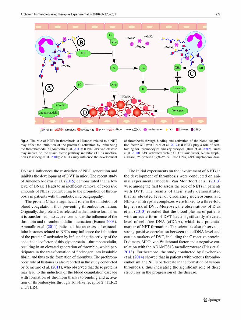
The protein C has a significant role in the inhibition of blood coagulation, thus preventing thrombus formation. Originally, the protein C is released in the inactive form, then it is transformed into active form under the influence of the thrombin and thrombomodulin interaction (Esmon 2003). Ammollo et al. (2011) indicated that an excess of extracel-lular histones related to NETs may influence the inhibition of the protein C activation by influencing the activity of the endothelial cofactor of this glycoprotein—thrombomodulin, resulting in an elevated generation of thrombin, which par-ticipates in the transformation of fibrinogen into insoluble fibrin, and thus to the formation of thrombus. The prothrom-botic role of histones is also reported in the study conducted by Semeraro et al. (2011), who observed that these proteins may lead to the induction of the blood coagulation cascade with formation of thrombin thanks to binding and activa-tion of thrombocytes through Toll-like receptor 2 (TLR2) and TLR4.
The initial experiments on the involvement of NETs in the development of thrombosis were conducted on ani-mal experimental models. Van Montfoort et al. (2013) were among the first to assess the role of NETs in patients with DVT. The results of their study demonstrated that an elevated level of circulating nucleosomes and NE–α1-antitrypsin complexes were linked to a three-fold higher risk of DVT. Moreover, the observations of Diaz et al. (2013) revealed that the blood plasma of patients with an acute form of DVT has a significantly elevated level of cell-free DNA (cfDNA), which is a potential marker of NET formation. The scientists also observed a strong positive correlation between the cfDNA level and certain markers of DVT, including the C reactive protein, D-dimers, MPO, von Willebrand factor and a negative cor-relation with the ADAMTS13 metalloprotease (Diaz et al. 2013). Furthermore, the study conducted by Savchenko et al. (2014) showed that in patients with venous thrombo-embolism, the NETs participate in the formation of venous thrombosis, thus indicating the significant role of these structures in the progression of the disease.
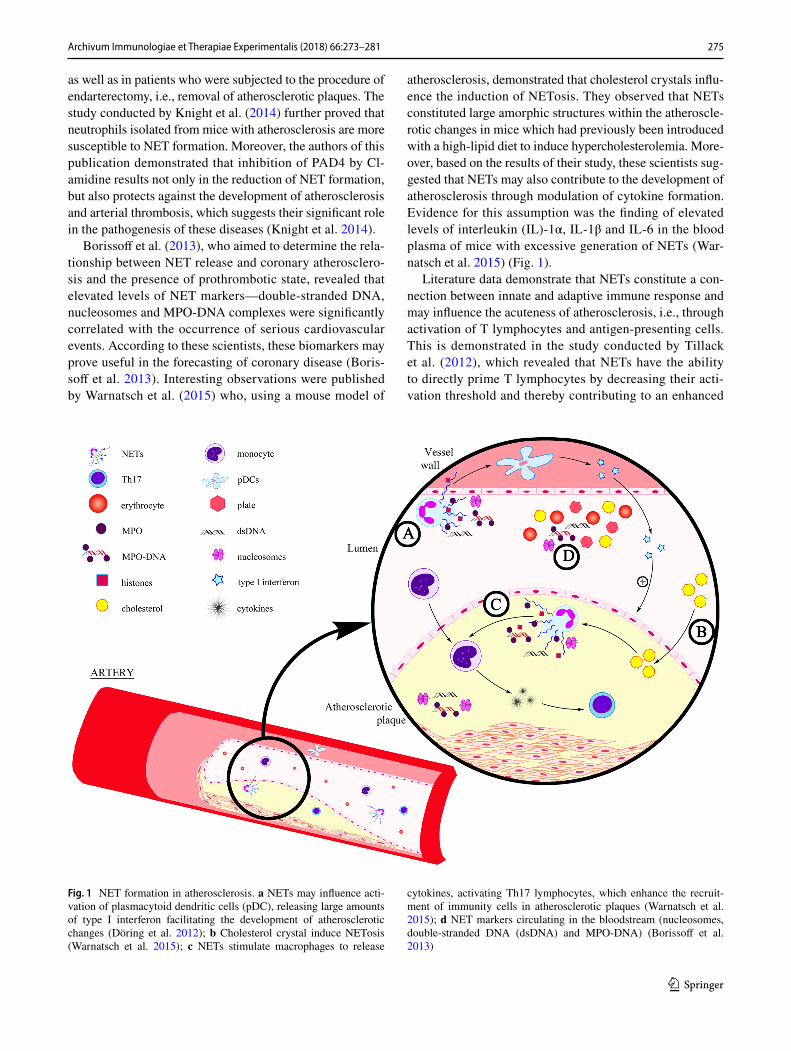
Fig. 2 The role of NETs in thrombosis. a Histones related to a NET may affect the inhibition of the protein C activation by influencing the thrombomodulin (Ammollo et al. 2011); b NET-derived elastase may impact on the tissue factor pathway inhibitor (TFPI) inactiva-tion (Massberg et al. 2010); c NETs may influence the development
of thrombosis through binding and activation of the blood coagula-tion factor XII (von Brühl et al. 2012); d NETs play a role of scaf-folding for thrombocytes and erythrocytes (Brill et al. 2012; Fuchs et al. 2010). APC activated protein C, TF tissue factor, NE neutrophil elastase, PC protein C, cfDNA cell-free DNA, MPO myeloperoxidase
278 Archivum Immunologiae et Therapiae Experimentalis (2018) 66:273–281
1 3
NETs and Small‑Vessel Vasculitis
Small-vessel vasculitis is a systemic autoimmune disease. In its course, an inflammation and necrosis occur in the walls of small vessels including arterioles, venules and capillaries. As reported in the literature, one cannot exclude the possibil-ity that the process also includes mid-sized arteries and veins (Jennette and Falk 1997).
According to the classification established in 2012 at the International Chapel Hill Consensus Conference Nomencla-ture of Vasculitides (Jennette 2013), SVV are divided into:
• antineutrophil cytoplasmic antibody (ANCA)-associ-ated vasculitides (AAV), which are further divided into: microscopic polyangiitis (MPA), granulomatosis with polyangiitis (GPA) and eosinophilic GPA (EGPA);
• immune-complex associated vasculitides, including: anti-glomerular basement membrane disease, cryoglo-bulinemic vasculitis, IgA vasculitis (Henoch-Schönlein) and hypocomplementemic urticarial vasculitis (anti-C1q vasculitis) (Jennette 2013).
The AAV pathogenesis is complex and it is influenced by both genetic predispositions as well as environmental fac-tors (environmental pollution, drugs, infections), leading to induction and excessive activation of lymphocytes and autoantibodies, contributing to tissue damage and predis-posal to disease recrudescence (Cartin-Ceba et al. 2012). Numerous experimental studies and clinical observations have confirmed the pathogenetic role of ANCA, mostly mye-loperoxidase (MPO-ANCA) and proteinase 3 (PR3-ANCA) (Falk et al. 1990; Schreiber et al. 2004). Literature data indi-cates that PR3-ANCA is most common in GPA patients, whereas MPO-ANCA occurs more frequently in patients with MPA (Jennette and Falk 1998; Rowaiye et al. 2015).
Following the discovery of NETs, the potential relation-ship was observed between formation of these structures and ANCA-associated vasculitis progression. This is confirmed by the presence of the autoantigens (MPO and PR3) in the NETs. Kessenbrock et al. (2009) noticed that anti-PR3-anti-bodies stimulate neutrophils to form and release NETs in the course of AAV. Moreover, these scientists demonstrated that in the majority of patients with AAV who have complica-tions in the form of glomerulonephritis, NETs are accumu-lated within the structural elements of the organ, suggesting that the NETs may contribute to the progression of AAV (Kessenbrock et al. 2009). Several months later, a descrip-tion was published in the scientific literature of a case of a patient with SVV in which in skin biopsy material using immunofluorescence, anti-MPO antibodies and NETs were detected (Abreu-Velez et al. 2009).
Propylthiouracil (PTU) is a drug with a proven impact on the development of AAV, which has been found to contribute
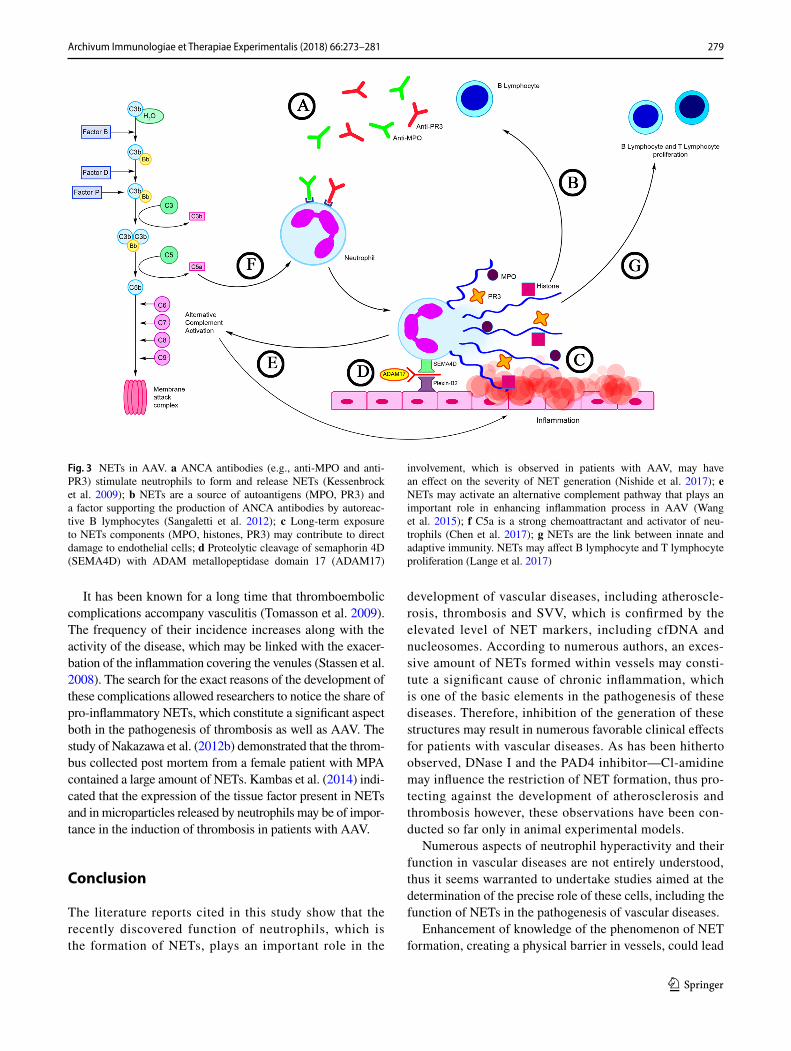
to the induction of ANCA. Due to its thyrostatic action, PTU is administered to treat hyperthyroidism (Cao and Lin 2013). Recent reports have revealed that PTU can affect the NET generation. Interesting observations were obtained by Naka-zawa et al. (2012a) who indicated that PTU may influence a change of NET conformation released by human neutrophils induced by PMA both in vitro and in vivo. These scientists observed that a change of the spatial layout of the NETs leads to their impaired degradation with the contribution of DNase I. An explanation of the occurrence of this phe-nomenon is the fact that PTU metabolites impede identifica-tion of the cutting sites of the endonuclease through binding NETs to the DNA. According to these researchers, this may constitute the critical aspect in the pathogenesis of AAV (Nakazawa et al. 2012a). A continuation of the study con-ducted by Nakazawa et al. (2014) supports earlier reports by Kessenbrock et al. (2009), concerning the strong ability of ANCA for induction of NETosis in patients with AAV. Fur-thermore, these authors discovered that NETs degradation in patients with AAV is significantly inhibited by the low level of DNase I activity and presence of anti-NET antibodies (Nakazawa et al. 2014). In contrast Sangaletti et al. (2012) demonstrated that NETs are highly immunogenic and that they may lead to the induction and support of autoimmuni-zation. They observed that myeloid dendritic cells (mDCs) may capture some NET components, i.e., MPO and PR3, and then present them as antigens. Furthermore, the authors revealed that in skin samples obtained from patients with MPA, mDCs were in direct contact with NETs (Sangaletti et al. 2012) (Fig. 3).
The results of Söderberg et al. (2015) showed that in the circulation of patients with AAV an elevated level of NET remnants is found, which has a positive correlation with the activity of the disease and is negatively correlated to the number of ANCA antibodies in the remission period of the disease. The authors also observed that the neutro-phils of patients with AAV exhibit a stronger tendency for spontaneous cell death through NETosis (Söderberg et al. 2015). On the other hand, Lange et al. (2017) have revealed in their latest publication that in the serum of patients with GPA, cfDNA concentration is significantly increased, which may indicate significant participation of these structures in the development of this disease. They also observed that NETs may influence the B lymphocyte maturation and T lymphocyte (CD4+) and B lymphocyte (CD19+) prolifera-tion (Lange et al. 2017).
Of special interest appears the study conducted by Nator-ska et al. (2017), who were among the first to describe that neutrophils of patients with EGPA are capable of NETs gen-eration. Authors of this publication further reported on the participation of eosinophils in the induction of NETosis, as they observed, the number of these cells correlated with the number of NETs in all examined EGPA patients.
279Archivum Immunologiae et Therapiae Experimentalis (2018) 66:273–281
1 3
It has been known for a long time that thromboembolic complications accompany vasculitis (Tomasson et al. 2009). The frequency of their incidence increases along with the activity of the disease, which may be linked with the exacer-bation of the inflammation covering the venules (Stassen et al. 2008). The search for the exact reasons of the development of these complications allowed researchers to notice the share of pro-inflammatory NETs, which constitute a significant aspect both in the pathogenesis of thrombosis as well as AAV. The study of Nakazawa et al. (2012b) demonstrated that the throm-bus collected post mortem from a female patient with MPA contained a large amount of NETs. Kambas et al. (2014) indi-cated that the expression of the tissue factor present in NETs and in microparticles released by neutrophils may be of impor-tance in the induction of thrombosis in patients with AAV.
Conclusion
The literature reports cited in this study show that the recently discovered function of neutrophils, which is the formation of NETs, plays an important role in the
development of vascular diseases, including atheroscle-rosis, thrombosis and SVV, which is confirmed by the elevated level of NET markers, including cfDNA and nucleosomes. According to numerous authors, an exces-sive amount of NETs formed within vessels may consti-tute a significant cause of chronic inflammation, which is one of the basic elements in the pathogenesis of these diseases. Therefore, inhibition of the generation of these structures may result in numerous favorable clinical effects for patients with vascular diseases. As has been hitherto observed, DNase I and the PAD4 inhibitor—Cl-amidine may influence the restriction of NET formation, thus pro-tecting against the development of atherosclerosis and thrombosis however, these observations have been con-ducted so far only in animal experimental models.
Numerous aspects of neutrophil hyperactivity and their function in vascular diseases are not entirely understood, thus it seems warranted to undertake studies aimed at the determination of the precise role of these cells, including the function of NETs in the pathogenesis of vascular diseases.
Enhancement of knowledge of the phenomenon of NET formation, creating a physical barrier in vessels, could lead
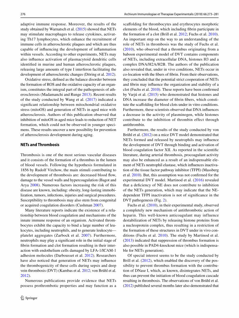
Fig. 3 NETs in AAV. a ANCA antibodies (e.g., anti-MPO and anti-PR3) stimulate neutrophils to form and release NETs (Kessenbrock et al. 2009); b NETs are a source of autoantigens (MPO, PR3) and a factor supporting the production of ANCA antibodies by autoreac-tive B lymphocytes (Sangaletti et al. 2012); c Long-term exposure to NETs components (MPO, histones, PR3) may contribute to direct damage to endothelial cells; d Proteolytic cleavage of semaphorin 4D (SEMA4D) with ADAM metallopeptidase domain 17 (ADAM17)
involvement, which is observed in patients with AAV, may have an effect on the severity of NET generation (Nishide et al. 2017); e NETs may activate an alternative complement pathway that plays an important role in enhancing inflammation process in AAV (Wang et al. 2015); f C5a is a strong chemoattractant and activator of neu-trophils (Chen et al. 2017); g NETs are the link between innate and adaptive immunity. NETs may affect B lymphocyte and T lymphocyte proliferation (Lange et al. 2017)
280 Archivum Immunologiae et Therapiae Experimentalis (2018) 66:273–281
1 3
to the development of new treatment strategies and improve-ment of the quality of life of patients suffering from these diseases in the future.
Compliance with ethical standards
Conflict of interest The authors declare no potential conflicts of inter-est.
Open Access This article is distributed under the terms of the Crea-tive Commons Attribution 4.0 International License (http://creat iveco mmons .org/licen ses/by/4.0/), which permits unrestricted use, distribu-tion, and reproduction in any medium, provided you give appropriate credit to the original author(s) and the source, provide a link to the Creative Commons license, and indicate if changes were made.
References
Abi Abdallah DS, Lin C, Ball CJ et al (2012) Toxoplasma gondii trig-gers release of human and mouse neutrophil extracellular traps. Infect Immun 80:768–777
Abreu-Velez AM, Smith JG Jr, Howard MS (2009) Presence of neutro-phil extracellular traps and antineutrophil cytoplasmic antibodies associated with vasculitides. N Am J Med Sci 1:309–313
Ammollo CT, Semeraro F, Xu J et al (2011) Extracellular histones increase plasma thrombin generation by impairing thrombo-modulin-dependent protein C activation. J Thromb Haemost 9:1795–1803
Baetta R, Corsini A (2010) Role of polymorphonuclear neutrophils in atherosclerosis: current state and future perspectives. Athero-sclerosis 210:1–13
Bagot CN, Arya R (2008) Virchow and his triad: a question of attribu-tion. Br J Haematol 143:180–190
Borissoff JI, Joosen IA, Versteylen MO et al (2013) Elevated levels of circulating DNA and chromatin are independently associated with severe coronary atherosclerosis and a prothrombotic state. Arterioscler Thromb Vasc Biol 33:2032–2040
Borregaard N (2010) Neutrophils, from marrow to microbes. Immunity 33:657–670
Brill A, Fuchs TA, Savchenko AS et al (2012) Neutrophil extracellular traps promote deep vein thrombosis in mice. J Thromb Haemost 10:136–144
Brinkmann V, Reichard U, Goosmann CH et al (2004) Neutrophil extracellular traps kill bacteria. Science 303:1532–1535
Cao X, Lin W (2013) Clinical study of renal impairment in patients with propylthiouracil-induced small-vessel vasculitis and patients with primary ANCA-associated small-vessel vasculitis. Exp Ther Med 5:1619–1622
Cartin-Ceba R, Peikert T, Specks U (2012) Pathogenesis of ANCA-associated vasculitis. Curr Rheumatol Rep 14:481–493
Chamorro A, Hallenbeck J (2006) The harms and benefits of inflam-matory and immune responses in vascular disease. Stroke 37:291–293
Chen M, Jayne DRW, Zhao MH (2017) Complement in ANCA-asso-ciated vasculitis: mechanisms and implications for management. Nat Rev Nephrol 13:359–367
Chistiakov DA, Bobryshev YV, Orekhov AN (2015) Neutrophil’s weapons in atherosclerosis. Exp Mol Pathol 99:663–671
Cushman M (2007) Epidemiology and risk factors for venous throm-bosis. Semin Hematol 44:62–69
Darbousset R, Thomas GM, Mezouar S et al (2012) Tissue factor-positive neutrophils bind to injured endothelial wall and initiate thrombus formation. Blood 120:2133–2143
Diaz JA, Fuchs TA, Jackson TO et al (2013) Plasma DNA is elevated in patients with deep vein thrombosis. J Vasc Surg Venous Lymphat Disord 1(4):341–348
Döring Y, Manthey HD, Drechsler M et al (2012) Auto-antigenic protein-DNA complexes stimulate plasmacytoid dendritic cells to promote atherosclerosis. Circulation 125:1673–1683
Dwivedi N, Neeli I, Schall N et al (2014) Deimination of linker his-tones links neutrophil extracellular trap release with autoantibod-ies in systemic autoimmunity. FASEB J 28:2840–2851
Esmon CT (2003) The protein C pathway. Chest 124(3 Suppl):26S–32SFalk E (2006) Pathogenesis of atherosclerosis. J Am Coll Cardiol 47(8
Suppl):C7–C12Falk RJ, Terrell R, Charles LA et al (1990) Anti-neutrophil cytoplas-
mic autoantibodies induce neutrophils to degranulate and produce oxygen radicals. Proc Natl Acad Sci USA 87:4115–4119
Fuchs T, Abed U, Goosmann C et al (2007) Novel cell death program leads to neutrophil extracellular traps. J Cell Biol 176:231–241
Fuchs TA, Brill A, Duerschmied D et al (2010) Extracellular DNA traps promote thrombosis. Proc Natl Acad Sci USA 107:15880–15885
Guimarães-Costa AB, Nascimento MT, Froment GS et al (2009) Leish-mania amazonensis promastigotes induce and are killed by neutro-phil extracellular traps. Proc Natl Acad Sci USA 106:6748–6753
Hansson GK (2005) Inflammation, atherosclerosis, and coronary artery disease. N Engl J Med 352:1685–1695
Ionita MG, van den Borne P, Catanzariti LM et al (2010) High neutro-phil numbers in human carotid atherosclerotic plaques are asso-ciated with characteristics of rupture-prone lesions. Arterioscler Thromb Vasc Biol 30:1842–1848
Jaillon S, Peri G, Delneste Y et al (2007) The humoral pattern recogni-tion receptor PTX3 is stored in neutrophil granules and localizes in extracellular traps. J Exp Med 204:793–804
Jawień J (2008) New insights into immunological aspects of athero-sclerosis. Pol Arch Med Wewn 118:127–131
Jennette JC (2013) Overview of the 2012 revised International Chapel Hill Consensus Conference nomenclature of vasculitides. Clin Exp Nephrol 17:603–606
Jennette JC, Falk RJ (1997) Small-vessel vasculitis. N Engl J Med 337:1512–1523
Jennette JC, Falk RJ (1998) Pathogenesis of the vascular and glomeru-lar damage in ANCA-positive vasculitis. Nephrol Dial Transplant 13(Suppl 1):16–20
Jiménez-Alcázar M, Napirei M, Panda R et al (2015) Impaired DNase1-mediated degradation of neutrophil extracellular traps is associ-ated with acute thrombotic microangiopathies. J Thromb Haemost 13:732–742
Kambas K, Mitroulis I, Apostolidou E et al (2012) Autophagy mediates the delivery of thrombogenic tissue factor to neutrophil extracel-lular traps in human sepsis. PLoS One 7:e45427
Kambas K, Chrysanthopoulou A, Vassilopoulos D et al (2014) Tissue factor expression in neutrophil extracellular traps and neutrophil derived microparticles in antineutrophil cytoplasmic antibody associated vasculitis may promote thromboinflammation and the thrombophilic state associated with the disease. Ann Rheum Dis 73:1854–1863
Kaplan MJ, Radic M (2012) Neutrophil extracellular traps: double—edged swords of innate immunity. J Immunol 189:2689–2695
Kessenbrock K, Krumbholz M, Schonermarck U et al (2009) Netting neu-trophils in autoimmune small-vessel vasculitis. Nat Med 15:623–625
Knight JS, Luo W, O’Dell AA et al (2014) Peptidylarginine deimi-nase inhibition reduces vascular damage and modulates innate immune responses in murine models of atherosclerosis. Circ Res 114:947–956
281Archivum Immunologiae et Therapiae Experimentalis (2018) 66:273–281
1 3
Lange C, Csernok E, Moosig F et al (2017) Immune stimulatory effects of neutrophil extracellular traps in granulomatosis with polyangiitis. Clin Exp Rheumatol Suppl 103:33–39
Madamanchi NR, Runge MS (2013) Redox signaling in cardiovascular health and disease. Free Radic Biol Med 61:473–501
Manda A, Pruchniak MP, Araźna M et al (2014) Neutrophil extracellular traps in physiology and pathology. Cent Eur J Immunol 39:116–121
Mantovani A, Cassatella MA, Costantini C et al (2011) Neutrophils in the activation and regulation of innate and adaptive immunity. Nat Rev Immunol 11:519–531
Martinod K, Demers M, Fuchs TA et al (2013) Neutrophil histone modi-fication by peptidylarginine deiminase 4 is critical for deep vein thrombosis in mice. Proc Natl Acad Sci USA 110:8674–8679
Martinod K, Witsch T, Farley K et al (2016) Neutrophil elastase-deficient mice form neutrophil extracellular traps in an experimental model of deep vein thrombosis. J Thromb Haemost 14:551–558
Massberg S, Grahl L, von Bruehl ML et al (2010) Reciprocal coupling of coagulation and innate immunity via neutrophil serine proteases. Nat Med 16:887–896
McCormick A, Heesemann L, Wagener J et al (2010) NETs formed by human neutrophils inhibit growth of the pathogenic mold Aspergil-lus fumigatus. Microbes Infect 12:928–936
Megens RT, Vijayan S, Lievens D et al (2012) Presence of luminal neutrophil extracellular traps in atherosclerosis. Thromb Haemost 107:597–598
Nakazawa D, Tomaru U, Suzuki A et al (2012a) Abnormal conformation and impaired degradation of propylthiouracil-induced neutrophil extracellular traps: implications of disordered neutrophil extracellu-lar traps in a rat model of myeloperoxidase antineutrophil cytoplas-mic antibody-associated vasculitis. Arthritis Rheum 64:3779–3787
Nakazawa D, Tomaru U, Yamamoto C et al (2012b) Abundant neutrophil extracellular traps in thrombus of patient with microscopic poly-angiitis. Front Immunol 3:333
Nakazawa D, Shida H, Tomaru U et al (2014) Enhanced formation and disordered regulation of NETs in myeloperoxidase-ANCA–associ-ated microscopic polyangiitis. J Am Soc Nephrol 25:990–997
Narasaraju T, Yang E, Samy RP et al (2011) Excessive neutrophils and neutrophil extracellular traps contribute to acute lung injury of influ-enza pneumonitis. Am J Pathol 179:199–210
Natorska J, Ząbczyk M, Siudut J et al (2017) Neutrophil extracellular traps formation in patients with eosinophilic granulomatosis with polyangiitis: association with eosinophilic inflammation. Clin Exp Rheumatol 103(35 Suppl):27–32
Neeli I, Khan SN, Radic M (2008) Histone deimination as a response to inflammatory stimuli in neutrophils. J Immunol 180:1895–1902
Nishide M, Nojima S, Ito D et al (2017) Semaphorin 4D inhibits neu-trophil activation and is involved in the pathogenesis of neutrophil-mediated autoimmune vasculitis. Ann Rheum Dis 76:1440–1448
Remijsen Q, Berghe TW, Wirawan E et al (2011) Neutrophil extracellular trap cell death requires both autophagy and superoxide generation. Cell Res 21:290–304
Rowaiye OO, Kusztal M, Klinger M (2015) The kidneys and ANCA-associated vasculitis: from pathogenesis to diagnosis. Clin Kidney J 8:343–350
Saitoh T, Komano J, Saitoh Y et al (2012) Neutrophil extracellular traps mediate a host defense response to human immunodeficiency virus-1. Cell Host Microbe 12:109–116
Sangaletti S, Tripodo C, Chiodoni C et al (2012) Neutrophil extracellular traps mediate transfer of cytoplasmic neutrophil antigens to myeloid dendritic cells toward ANCA induction and associated autoimmun-ity. Blood 120:3007–3018
Savchenko AS, Martinod K, Seidman MA et al (2014) Neutrophil extra-cellular traps form predominantly during the organizing stage of human venous thromboembolism development. J Thromb Haemost 12:860–870
Schreiber A, Luft FC, Kettritz R (2004) Membrane proteinase 3 expression and ANCA-induced neutrophil activation. Kidney Int 65:2172–2183
Scott J (2002) The pathogenesis of atherosclerosis and new opportunities for treatment and prevention. J Neural Transm Suppl 63:1–17
Semeraro F, Ammollo CT, Morrissey JH et al (2011) Extracellular his-tones promote thrombin generation through platelet-dependent mechanisms: involvement of platelet TLR2 and TLR4. Blood 118:1952–1961
Singh RB, Mengi SA, Xu YJ et al (2002) Pathogenesis of atherosclerosis: A multifactorial process. Exp Clin Cardiol 7:40–53
Söderberg D, Kurz T, Motamedi A et al (2015) Increased levels of neu-trophil extracellular trap remnants in the circulation of patients with small vessel vasculitis, but an inverse correlation to anti-neu-trophil cytoplasmic antibodies during remission. Rheumatology 54:2085–2094
Stassen PM, Derks RP, Kallenberg CG et al (2008) Venous thromboem-bolism in ANCA-associated vasculitis-incidence and risk factors. Rheumatology 47:530–534
Tillack K, Breiden P, Martin R et al (2012) T lymphocyte priming by neutrophil extracellular traps links innate and adaptive immune responses. J Immunol 188:3150–3159
Tomasson G, Monach PA, Merkel PA (2009) Thromboembolic disease in vasculitis. Curr Opin Rheumatol 21:41–46
Urban CF, Reichard U, Brinkmann V et al (2006) Neutrophil extracellular traps capture and kill Candida albicans yeast and hyphal forms. Cell Microbiol 8:668–676
Urban CF, Ermert D, Schmid M et al (2009) Neutrophil extracellular traps contain calprotectin, a cytosolic protein complex involved in host defense against Candida albicans. PLoS Pathog 5:e1000639
van Montfoort ML, Stephan F, Lauw MN et al (2013) Circulating nucle-osomes and neutrophil activation as risk factors for deep vein throm-bosis. Arterioscler Thromb Vasc Biol 33:147–151
Varjú I, Longstaff C, Szabó L et al (2015) DNA, histones and neutrophil extracellular traps exert anti-fibrinolytic effects in a plasma environ-ment. Thromb Haemost 113:1289–1298
von Brühl ML, Stark K, Steinhart A et al (2012) Monocytes neutrophils and platelets cooperate to initiate and propagate venous thrombosis in mice in vivo. J Exp Med 209:819–835
Wang H, Wang C, Zhao MH et al (2015) Neutrophil extracellular traps can activate alternative complement pathways. Clin Exp Immunol 181:518–527
Wang Y, Wang W, Wang N et al (2017) Mitochondrial oxidative stress promotes atherosclerosis and neutrophil extracellular traps in aged mice. Arterioscler Thromb Vasc Biol 37:e99–e107
Warnatsch A, Ioannou M, Wang Q et al (2015) Neutrophil extracellular traps license macrophages for cytokine production in atherosclero-sis. Science 349:316–320
Wartha F, Beiter K, Normark S et al (2007) Neutrophil extracellular traps: casting the NET over pathogenesis. Curr Opin Microbiol 10:52–56
Weber C, Zernecke A, Libby P (2008) The multifaceted contributions of leukocyte subsets to atherosclerosis: lessons from mouse models. Nat Rev Immunol 8:802–815
Zarbock A, Polanowska-Grabowska RK, Ley K (2007) Platelet-neu-trophil-interactions: linking hemostasis and inflammation. Blood Rev 21:99–111