Embed Size (px)
Citation preview
1
Neither Maternal nor Zygotic med-1/med-2 Genes
Play a Major Role
in
Specifying
the
C. elegans Endoderm
by
Vasile V. Captan,
Barbara Goszczynski
and
James D. McGhee1
Department of Biochemistry and Molecular Biology,
Genes and Development Research Group,
University of Calgary,
Calgary, Alberta, CANADA
T2N 4N1
Running Title: C. elegans Endoderm in the Absence of med Genes
Key Words: C. elegans / development / endoderm specification / med-1 / med-2
1 Corresponding Author: James D. McGhee,
Department of Biochemistry and Molecular Biology,
Genetics: Published Articles Ahead of Print, published on December 6, 2006 as 10.1534/genetics.106.066662
2
University of Calgary,
Health Sciences Centre, Room 2205,
3330 Hospital Drive N.W.,
Calgary, Alberta, CANADA T2N 4N1
phone: 403 – 220 – 4476
FAX: 403 – 270 – 0737
email: [email protected]
3
ABSTRACT
The med-1 and med-2 genes encode small highly-similar proteins related to GATA-type
transcription factors and have been proposed to be necessary for specification of both C.
elegans mesoderm and endoderm. However, we have previously shown that neither
maternal nor zygotic expression of the med-1/2 genes are necessary to specify the C.
elegans endoderm: the majority (>80%) of embryos zygotically lacking both the med-1
gene (removed by a gene specific deletion) and the med-2 gene (removed by a
chromosome deficiency) nonetheless still express standard markers of endoderm
specification (e.g. gut granules); standard med-1/2 RNAi to remove possible med-1/2
maternal transcripts showed no effect. In the present paper, we re-investigate med-2(-);
med-1(-) embryos using a med-2 specific null allele instead of the chromosomal
deficiences used previously: again, we show that the large majority (~84%) of med-2(-);
med-1(-) embryos still express gut granules, in precise agreement with our previous results
obtained using the chromosomal deficiency sDf127 to remove the med-2 gene. A recent
report that the med-1/2 genes show an endoderm-specifying maternal effect was based on
the assumption that multiple transgenic copies of the med-1(+) gene (used to balance the
strain) cause cosuppression, removing putative med-1/2 transcripts from the maternal
germline. Here, we re-investigate this proposal by direct med-1/2 RNAi using the standard
protocol known to be effective in ablating maternal transcripts but, as we found previously,
we could detect no evidence for a maternal med-1/2 effect. We do however show that
expression of gut-granules in med-1/2-deficient embryos is exquisitely sensitive to RNAi
against the vacuolar ATPase-encoding unc-32 gene; multiple copies of unc-32 sequences
were present on the same multicopy med-1(+)-containing transgenic balancer used in
support of the maternal med-1/2 effect. We thus suggest that the experimental evidence
for a maternal med-1/2 effect should be reinterpreted and may instead reflect
cosuppression caused by multiple unc-32 sequences, not by multiple med sequences.
Indeed, the unc-32-mediated cosuppression model provides a simple explanation for

4
several further experimental observations not easily explained by med-based
cosuppression. Embryos lacking the med-1/2 genes are severely deranged and it is clear
that the med-1/2 genes are of great importance for correct embryonic development.
Nonetheless, it is also clear that >80% of med-2(-); med-1(-) embryos still express markers
of endoderm specification (gut granules) and we suggest that recent evidence for a med-
1/2 maternal effect should be re-examined.
5
The med-1 and med-2 genes encode a redundant pair of highly-similar GATA-
factor-related zinc finger proteins that together are crucial for the early development of
the C. elegans embryo (MADURO et al. 2001). The original model for med-1/2 action
(MADURO et al. 2001) was that both genes were strictly zygotic, acting downstream of
the maternally-transcribed but zygotically-translated transcription factor SKN-1 and
essential for specifying E and MS fate (where the E and MS blastomeres give rise to all
of the worm endoderm and much of the worm mesoderm, respectively). In the present
paper, we are concerned only with the question whether med-1/2 are indeed necessary
to specify the C. elegans endoderm.
The evidence that med-1/2 are essential for endoderm specification was based
on two different protocols for administering med-1/2 RNAi (MADURO et al. 2001). In the
first protocol, embryos were collected 7-9 hours following injection of concentrated
dsRNA into the maternal gonad; the rationale for such an early and limited observation
period was to more effectively target the transient expression of the med-1/2 genes in
the early embryo (MADURO et al. 2001; COROIAN et al. 2006). In the more standard
protocol, the RNAi effect is found to be maximally effective from one to three days
following injection (FIRE et al. 1998; ZIPPERLEN et al. 2001; GOSZCZYNSKI and MCGHEE
2005; AHRINGER 2006)); however, these more usual methods of administering RNAi
show no effect with the med-1/2 genes (KAMATH et al. 2003; GOSZCZYNSKI and MCGHEE
2005; SONNICHSEN et al. 2005). A second method of administering med-1/2 RNAi
(MADURO et al. 2001) was by means of a transgenic heat-inducible promoter driving
expression of double-stranded med RNA (TAVERNARAKIS et al. 2000). Together, these
two protocols were reported to produce a small number of embryos that arrested with a
distinctive phenotype (~2-fold elongation with absence of posterior pharynx); ~50% of
these arrested embryos failed to show the standard marker for endoderm specification,
the presence of birefringent gut granules (MADURO et al. 2001; COROIAN et al. 2006).
6
As reported in detail elsewhere (GOSZCZYNSKI and MCGHEE 2005), we had only
limited success producing med-type arrested embryos by either of these two protocols.
With the first protocol, we were able to produce <1 gut-granule negative embryo for
every two injected hermaphrodites, only marginally higher than produced by injection of
non-specific control RNA (see also COROIAN et al. 2006). In our hands, the second
protocol produces either sterile mothers or embryos that arrest prior to the stage where
they would ordinarily express gut granules. We thus proceeded with a more definitive
test of the requirements of the med-1/2 genes for C. elegans endoderm specification,
constructing worm strains segregating embryos that genetically lacked both med-1 and
med-2 zygotic function. In these strains, the med-1 gene was removed by the gene-
specific deletion ok804; the med-2 gene was removed by either of two chromosomal
deficiencies, sDf127 or nDf16, both of which remove several hundred genes including
med-2. Our results were unexpected but clear: we found that only 16-17% or 0-3% of
the doubly homozygous med-2(null); med-1(null) embryos did not express gut granules
(depending on whether sDf127 or nDf16 was used to remove med-2). Both estimates
were far lower than the >70% gut-granule negative embryos predicted by the original
model of MADURO et al. (2001). We thus concluded that the zygotic expression of the
med-1 and med-2 genes could not play a major necessary role in specifying endoderm.
Even though med transcripts cannot be detected in the maternal germline (REINKE et al.
2004), we nonetheless looked for (but did not find) any evidence for a possible maternal
effect of the med-1/2 genes, using the standard protocol of dsRNA injection into
sensitized med-1/2-deficient hermaphrodites. Using the same protocol even with non-
sensitized strains, we routinely produce 100% arrested embryos when targeting the
maternal skn-1 gene (GOSZCZYNSKI and MCGHEE 2005) and 100% arrested larvae when
targeting the relatively early zygotic gene elt-2 (FUKUSHIGE et al. 2005).
There remained two unresolved issues. First, which is the correct estimate for
the % gut granule negative med-2(-); med-1(-) embryos? If the 16-17% estimated with

7
the sDf127-containing strain is correct (and hence the 0-3% estimated with the nDf16-
containing strain is incorrect), can an "endoderm suppressor" be identified among the
genes removed by nDf16 but not by sDf127? The second issue was that, in spite of
our defense of chromosomal deficiencies (GOSZCZYNSKI and MCGHEE 2005), use of
gene specific knockouts for both med-1 and med-2 would strengthen our conclusion
that loss of the med-1/2 genes causes only a weakly impenetrant loss of endoderm.
The recent availability of a Mos transposon insertion (GRANGER et al. 2004;
WILLIAMS et al. 2005) into the med-2 gene (allele cxTi9744) provides a means to re-
assess the importance of the med-1/2 genes for specifying endoderm without relying on
chromosomal deficiencies. Thus, we constructed the two balanced strains described in
Table 1: JM142 (med-2(cxTi9744); + med-1(ok804)/lin-2 +) lacks both copies of med-
2(+) but has one copy of med-1(+), and JM143 (+ med-2(cxTi9744)/sma-3 +; med-
1(ok804)) lacks both copies of med-1(+) but has one copy of med-2(+). Both med
alleles are likely to be null (see footnote to Table 1) and are hereafter referred to simply
as med-1(-) or med-2(-). One-quarter of the embryos produced by either strain should
be med-2(-); med-1(-) and should arrest. This expectation is accurately met with strain
JM142 but 42.2 % of the embryos produced by JM143 arrest prior to hatching,
suggesting a degree of med-2 haploinsufficiency (verified semi-quantitatively by PCR on
arrested embryos). In other words, an embryo with only a single copy of any med gene
has a higher probability of surviving if that single copy is med-1 (~100%) rather than
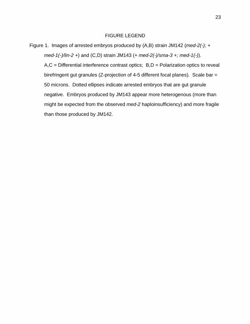
med-2 (~66%). The morphology of the arrested embryos is similar for both strains
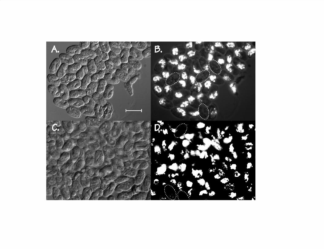
(Figure 1A and 1C): many of the embryos are arrested at two-fold stage with a
morphology like that originally reported by MADURO et al. (2001); others have arrested
earlier and can be grossly vacuolated.
Figure 1B and D show that only a minor fraction of the arrested embryos
produced by either strain do not express gut granules, the standard assay for specified
endoderm. After correcting for haploinsufficiency (Table 1), the proportion of gut
8
granule negative homozygous med-2(-); med-1(-) embryos is estimated at 14.2% and
17.3% for the two different strains, in excellent agreement with the 16.0 to 17.3% gut
granule negative med-1/2-deficient embryos previously measured segregating from the
sDf127-containing strain JM134 (GOSZCZYNSKI and MCGHEE 2005). Thus, our
combined results remain incompatible with the original model of MADURO et al. (2001),
which proposed that the med-1/2 genes "function downstream of SKN-1 in the EMS
lineage and are essential to specify E (and MS) fates in any context".
Our final estimate of ~16% gut granule negative med-2(-); med-1(-) embryos
differs significantly from the 0-3% gut granule negative med-2(-); med-1(-) embryos
segregating from the nDf16-containing strain JM136 (GOSZCZYNSKI and MCGHEE 2005).
As noted above, this discrepancy raises the possibility that a potential "endoderm
suppressor" resides among the ~413 genes (excluding microRNAs) deleted by nDf16
but not by sDf127. We thus fed JM134 (dpy-17 sDf127 unc-32 III; sDp3 (III,f); med-1(-
)) animals on individual E.coli strains containing the 290 of these 413 genes that are
present in the Ahringer RNAi library (KAMATH et al. 2003). However, we were not able
to identify any clone that could lower the proportion of gut granule negative embryos
from the ~16% seen with sDf127 to the <3% seen with nDf16.
As noted above, we had previously performed med RNAi into the sensitized
strain JM134 (one copy of med-2(+) and no copies of med-1(+)) using the standard
protocol that efficiently ablates maternal transcripts (GOSZCZYNSKI and MCGHEE 2005).
We found no significant increase in the proportion of gut granule negative embryos
following injection of dsRNA targeting both med-1 and med-2, compared to injection of
GFP dsRNA, and concluded that the med-1/2 genes do not show a maternal effect.
However, MADURO et al. (2006) have recently made the opposite claim, namely that the
med-1/2 genes show a previously unreported maternal rescue of endoderm
specification (although apparently not maternal rescue of mesoderm specification).
MADURO et al. (2006) did not address our RNAi results (GOSZCZYNSKI and MCGHEE

9
2005) that contradict their proposal. However, because our previous results could
possibly have been influenced by the use of chromosomal deficiencies and could
always be criticized as being "negative" (i.e. no effect was observed, even though both
positive and negative controls behaved as expected), we repeated our experiments
using a strain carrying the two gene-specific med-1/2 nulls. med-1 dsRNA (which
should target both med-1 and med-2 transcripts) was injected into JM143 (med-1(-); +
med-2(-)/sma-3 +) mothers, GFP dsRNA was injected as a control, and arrested
embryos were scored (blind) for expression of gut granules. The results are collected in
Table 2 and our conclusions remain unchanged: we could find no significant evidence
that the med-1/2 genes show a maternal effect.
We thus wish to consider possible explanations for the differences between our
results and those of MADURO et al. (2006). In spite of the large difference in
interpretations and implications, the numerical differences between our observations are
rather modest. We routinely observe that <20% of med-2(-); med-1(-) embryos do not
express gut granules (GOSZCZYNSKI and MCGHEE (2005); Table 1 above) and for all but
two of their strains, MADURO et al. (2006) record a similar number. (Their numbers are
usually slightly higher than ours, in the range of 20-25% gut granule negative; we
ascribe no significance to this slight difference and suggest that it may be due to our
gentler observation technique; see Footnote to Table 1). We also note that when
MADURO et al. (2006) assay embryos segregated from our strain JM134, they reproduce
our results. Only two strains of MADURO et al. (2006), MS162 and MS247, produce
med-2(-); med-1(-) embryos that are in the range of 40-50% gut granule negative. This
~20-30% difference in the proportion of gut granule negative embryos is the basis for
their conclusion that the med-1/2 genes show a maternal effect. We will propose an
alternative explanation that is both simpler and experimentally supported.
The two strains that can be most closely compared between the two studies are
our strain JM134 (dpy-17(e164) sDf127(s2428) unc-32(e189) III; sDp3(III,f); med-

10
1(ok804) X), producing <20% gut granule negative embryos, and their strain MS162
(dpy-17(e164) sDf127(s2428) unc-32(e189) III; irDp1(III,f); med-1(ok804) X),
producing 40-50% gut granule negative embryos. Doubly homozygous arrested
embryos produced by either strain should have exactly the same genotype: dpy-
17(e164) sDf127(s2428) unc-32(e189) III; med-1(ok804) X. The difference lies in the
manner in which the two strains are balanced. Our strain JM134 is balanced by the
well-characterized free duplication sDp3 (III,f) (ROSENBLUTH et al. 1985; HEDGECOCK and
HERMAN 1995), which contains a single copy of the wildtype med-2 gene in its normal
chromosomal context. Strain MS162 used by MADURO et al. (2006) is balanced by
irDp1, a derivative of sDp3 containing a spontaneously integrated transgenic array
composed of multiple copies of unc-119::YFP, med-1(+) and a plasmid containing the
wildtype unc-32 gene (PUJOL et al. 2001). Thus, the key observation is that med-2(-);
med-1(-) embryos segregating from irDp1-containing MS162 mothers are 40-50% gut
granule negative whereas genotypically identical arrested embryos segregating from
sDp3-containing JM134 mothers are <20% gut granule negative. MADURO et al. (2006)
interpret this difference to mean that putative maternal med transcripts are being
removed by the RNAi related phenomenon called cosuppression (DERNBURG et al.
2000; ROBERT et al. 2005) caused by the multiple transgenic copies of med-1(+)
integrated into irDp1. However, they did not test their model by standard methods of
med RNAi. In contrast, as we have noted above, we have performed med RNAi into
sensitized strains carrying only a single copy of a med gene under conditions that
should ablate maternal transcripts but the proportion of gut granule negative embryos is
not significantly increased (GOSZCZYNSKI and MCGHEE (2005) and Table 2). Thus, the
interpretation of MADURO et al. (2006) requires that the putative maternal med
transcripts are somehow susceptible to cosuppression and susceptible to RNAi only for
a narrow window of several hours following injection (MADURO et al. 2001) but are not
11
susceptible to conventional (and usually much more powerful) RNAi effects that
routinely persist for days.
We will now propose a simple alternative explanation for the difference between
our results and those of MADURO et al. (2006). We suggest that cosuppression caused
by the multiple transgenic copies of the unc-32 gene (not the med-1 gene) present on
the irDp1 balancer is the reason that strain MS162 produces the higher levels (40-50%)
of gut granule negative med-2(-); med-1(-) embryos. unc-32 is a complex locus with
multiple transcripts expressed widely in the worm, including in the maternal germline
and throughout the early embryo (PUJOL et al. 2001); the null allele of unc-32 is
associated with a strict maternal effect lethality and the arrested embryos often show
vacuolated intestines (PUJOL et al. 2001); (unc-32(e189) present in both JM134 and
MS162 is a weak allele primarily affecting the nervous system). unc-32 encodes a
subunit of a vacuolar ATPase involved in acidifying intracellular organelles (PUJOL et al.
2001), and since gut granules are lysosome derivatives (CLOKEY and JACOBSON 1986;
HERMANN et al. 2005), it seemed possible that unc-32-mediated cosuppression in the
maternal germline could weaken the subsequent formation of embryonic gut granules.
To test this possibility, we synthesized dsRNA corresponding to a portion of the unc-32
gene (completely included in the transgenic unc-32 sequences present on irDp1),
injected it into strain JM143 (+ med-2(-)/sma-3 +; med-1(-)) hermaphrodites (with
wildtype hermaphrodites as controls) and then assayed gut granule formation in the
subsequently produced embryos. The results are collected in Table 2 and strongly
support our hypothesis. The three key observations are: (i) unc-32 RNAi is effective
and causes essentially complete embryonic arrest in both strains, as expected from the
genetic experiments of PUJOL et al. (2001); (ii) gut granule birefringence in arrested
wildtype control embryos is markedly weakened such that ~25% of the embryos are
scored as gut granule negative, and; (iii) gut granules in embryos produced by the med-
deficient strain JM143 (+ med-2(-)/sma-3 +; med-1(-)) are far more sensitive to unc-32
12
RNAi than in control embryos: >80% of arrested JM143 unc-32 RNAi embryos are
scored as gut granule negative. We do not know whether the unc-32 RNAi effect is
directly on the formation of the gut granule marker (e.g. a block in lysosome maturation
because of aberrant acidification) or is indirect because it causes early embryonic
arrest; in either case, the end result would be the same, namely that the affected
embryos would be scored as gut granule negative. Thus, because of the strong effect
caused by unc-32 RNAi and because of the absence of any effect caused by med RNAi
(GOSZCZYNSKI and MCGHEE (2005) and Table 2), we suggest that even mild
cosuppression caused by the multiple transgenic unc-32 sequences present on irDp1 is
a more likely explanation for the results of MADURO et al. (2006) than is cosuppression
caused by med sequences.
One obvious experiment to distinguish between the two cosuppression-based
explanations is to balance med-2(-); med-1(-) with a multicopy transgenic array
containing med-1(+) sequences but not containing unc-32 sequences. MADURO et al.
(2006) have already performed this experiment. Their strain MS290 (med-2(cxTi9744);
med-1(ok804); Ex[med-1(+); unc-119::CFP]) contains transgenic med-1(+) sequences
but does not contain transgenic unc-32 sequences. Strain MS290 clearly segregates
low levels (~17%) of gut granule negative arrested embryos, agreeing precisely with our
prediction. MADURO et al. (2006) find this result "unexpected" and suggest that there
must be intrinsic differences in the cosuppression abilities of individual transgenic
arrays. However, their explanation contradicts the findings of DERNBURG et al. (2000),
who showed that cosuppression by individual transgenic arrays, if it does occur, is
highly reliable and reproducible.
The results of a second experiment performed by MADURO et al. (2006) also
agree with our hypothesis but not with theirs. MADURO et al. (2006) identified eight
MS290 hermaphrodites that had lost the Ex[med-1(+); unc-119::CFP] balancing array
from the maternal germline, as well as a single hermaphrodite from a different strain
13
that had lost the balancing sDp3 duplication; in other words, none of these nine
mothers should have any maternal wildtype med-1/2 genes in their germline. Again,
contrary to the expectation of MADURO et al. (2006), these germline mosaic mothers
produce arrested embryos that are only 27% gut granule negative, accepted by the
authors as clearly in the low category. According to our model, this result is exactly
what would be predicted, simply because the med genes show no maternal effect. In
contrast, MADURO et al. (2006) introduce an unusual ad hoc hypothesis, namely that the
putative med transcripts in the maternal germline are not actually produced in the
germline but rather are imported from the anterior intestine (although these cells were
not previously reported to express a med-1::GFP transgene (MADURO et al. 2001)). In
defense of their hypothesis, MADURO et al. (2006) point out that such a somatic
transport model could resolve a major discrepancy in their results, namely why an in situ
hybridization signal ascribed to med transcripts in the maternal germline is ablated by
SKN-1 RNAi when, as the authors point out, there is no evidence that SKN-1 functions
in the maternal germline.
In summary, the results of the present paper completely confirm our previous
conclusions (GOSZCZYNSKI and MCGHEE 2005): the large majority (>80%) of embryos
that lack both copies of med-1 and med-2 nonetheless still express markers of
endoderm specification. Thus, the original model of MADURO et al. (2001) in which med-
1/2 were the sole (or even the major) downstream effectors of SKN-1 in specifying
endoderm must be ruled out. Our present results also contradict the revised model of
MADURO et al. (2006) in which it is claimed that the med genes show a maternal effect.
However, their claim is based on the assumption that a particular balancing array is
causing cosuppression involving multiple transgenic copies of the med-1 gene. We
suggest that their results are more likely to be caused by cosuppression associated with
multiple transgenic copies of the unc-32 gene present on the same balancing array;
indeed, we show that expression of gut granules by med deficient embryos appears
14
exquisitely sensitive to unc-32 RNAi. This suggestion provides a much simpler
explanation for several other experiments performed by MADURO et al. (2006) that
otherwise require the introduction of ad hoc hypotheses.
If loss of both med-1/2 genes causes only a weakly penetrant loss of endoderm
and if no maternal med-1/2 effect exists, what is the major regulatory pathway
specifying endoderm? As originally suggested by ZHU et al. (1997) and as we had
pointed out previously (GOSZCZYNSKI and MCGHEE 2005), all evidence points to the
SKN-1 transcription factor having the major direct role in specifying endoderm. In
particular, MADURO et al. (2005) have shown that the activity of the end-1 promoter
(thought to be critically involved in endoderm specification) is severely decreased
("++++" to "+") when an upstream region containing multiple SKN-1 sites is deleted. In
contrast, the short proximal end-1 promoter region containing MED-1/2 sites drives the
low ("+") residual level of end-1 activity.
We certainly do not question the major role that the med-1/2 genes must play in
C. elegans development; the severely deranged morphology of med-2(-); med-1(-)
embryos seen in Figure 1 is convincing evidence of their importance. Nonetheless, the
fact remains that >80 % of these arrested med-2(-); med-1(-) embryos still express
endoderm markers and there is no convincing evidence for any med-1/2 maternal
effect.
15
ACKNOWLEDGEMENTS
We thank Dr. Laurent Segalat (Université Lyon Claude Bernard) for providing the
med-2 (cxT19744) Mos insertion allele, the C. elegans Genetics Center (funded by the
National Center for Research Resources) for providing several strains and Dr. Paul
Mains (University of Calgary) for critical reading of the manuscript. This work was
supported by an operating grant from the Canadian Institutes of Health Research
(CIHR) to J.D.M. V.V.C. received partial support from a CIHR Institute of Genetics
Training Grant. J.D.M. is a Medical Scientist of the Alberta Heritage Foundation for
Medical Research and a Canada Research Chair.

16
LITERATURE CITED AHRINGER, J., 2006 Reverse Genetics. ed. The C. elegans Research Community,
WormBook, doi/10.1895/wormbook.1.47.1, http://www.wormbook.org
CLOKEY, G. V., and L. A. JACOBSON, 1986 The autofluorescent "lipofuscin granules" in
the intestinal cells of Caenorhabditis elegans are secondary lysosomes. Mech
Ageing Dev 35: 79-94.
COROIAN, C., G. BROITMAN-MADURO and M. F. MADURO, 2006 Med-type GATA factors
and the evolution of mesendoderm specification in nematodes. Dev Biol 289:
444-455.
DERNBURG, A. F., J. ZALEVSKY, M. P. COLAIACOVO and A. M. VILLENEUVE, 2000
Transgene-mediated cosuppression in the C. elegans germ line. Genes Dev 14:
1578-1583.
FIRE, A., S. XU, M. K. MONTGOMERY, S. A. KOSTAS, S. E. DRIVER et al., 1998 Potent and
specific genetic interference by double-stranded RNA in Caenorhabditis elegans.
Nature 391: 806-811.
FUKUSHIGE, T., B. GOSZCZYNSKI, J. YAN and J. D. MCGHEE, 2005 Transcriptional control
and patterning of the pho-1 gene, an essential acid phosphatase expressed in
the C. elegans intestine. Developmental Biology 279: 446-461.
GOSZCZYNSKI, B., and J. D. MCGHEE, 2005 Reevaluation of the Role of the med-1 and
med-2 Genes in Specifying the Caenorhabditis elegans Endoderm. Genetics
171: 545-555.
GRANGER, L., E. MARTIN and L. SEGALAT, 2004 Mos as a tool for genome-wide
insertional mutagenesis in Caenorhabditis elegans: results of a pilot study.
Nucleic Acids Res 32: e117.
HEDGECOCK, E. M., and R. K. HERMAN, 1995 The ncl-1 gene and genetic mosaics of
Caenorhabditis elegans. Genetics 141: 989-1006.
HERMANN, G. J., L. K. SCHROEDER, C. A. HIEB, A. M. KERSHNER, B. M. RABBITTS et al.,
2005 Genetic analysis of lysosomal trafficking in Caenorhabditis elegans. Mol
Biol Cell. 16: 3273-3288.

17
KAMATH, R. S., A. G. FRASER, Y. DONG, G. POULIN, R. DURBIN et al., 2003 Systematic
functional analysis of the Caenorhabditis elegans genome using RNAi. Nature
421: 231-237.
MADURO, M. F., G. BROITMAN-MADURO, I. MENGARELLI and J. H. ROTHMAN, 2006
Maternal deployment of the embryonic SKN-1-->MED-1,2 cell specification
pathway in C. elegans. Developmental Biology doi:10.1016/j.ydbio.2006.08.029
MADURO, M. F., J. J. KASMIR, J. ZHU and J. H. ROTHMAN, 2005 The Wnt effector POP-1
and the PAL-1/Caudal homeoprotein collaborate with SKN-1 to activate C.
elegans endoderm development. Dev Biol 285: 510-523.
MADURO, M. F., M. D. MENEGHINI, B. BOWERMAN, G. BROITMAN-MADURO and J. H.
ROTHMAN, 2001 Restriction of mesendoderm to a single blastomere by the
combined action of SKN-1 and a GSK-3beta homolog is mediated by MED-1 and
-2 in C. elegans. Mol Cell 7: 475-485.
PUJOL, N., C. BONNEROT, J. J. EWBANK, Y. KOHARA and D. THIERRY-MIEG, 2001 The
Caenorhabditis elegans unc-32 gene encodes alternative forms of a vacuolar
ATPase a subunit. J Biol Chem 276: 11913-11921.
REINKE, V., I. S. GIL, S. WARD and K. KAZMER, 2004 Genome-wide germline-enriched
and sex-biased expression profiles in Caenorhabditis elegans. Development 131:
311-323.
ROBERT, V. J., T. SIJEN, J. VAN WOLFSWINKEL and R. H. PLASTERK, 2005 Chromatin and
RNAi factors protect the C. elegans germline against repetitive sequences.
Genes Dev 19: 782-787.
ROSENBLUTH, R. E., C. CUDDEFORD and D. L. BAILLIE, 1985 Mutagenesis in
Caenorhabditis elegans. II. A spectrum of mutational events induced with 1500 r
of gamma-radiation. Genetics 109: 493-511.
SONNICHSEN, B., L. B. KOSKI, A. WALSH, P. MARSCHALL, B. NEUMANN et al., 2005 Full-
genome RNAi profiling of early embryogenesis in Caenorhabditis elegans.
Nature 434: 462-469.
TAVERNARAKIS, N., S. L. WANG, M. DOROVKOV, A. RYAZANOV and M. DRISCOLL, 2000
Heritable and inducible genetic interference by double-stranded RNA encoded by
transgenes. Nat Genet 24: 180-183.

18
WILLIAMS, D. C., T. BOULIN, A. F. RUAUD, E. M. JORGENSEN and J. L. BESSEREAU, 2005
Characterization of Mos1 Mediated Mutagenesis in C. elegans: A Method for the
Rapid Identification of Mutated Genes. Genetics 169: 1779-1785.
ZHU, J., R. J. HILL, P. J. HEID, M. FUKUYAMA, A. SUGIMOTO et al., 1997 end-1 encodes an
apparent GATA factor that specifies the endoderm precursor in Caenorhabditis
elegans embryos. Genes Dev 11: 2883-2896.
ZIPPERLEN, P., A. G. FRASER, R. S. KAMATH, M. MARTINEZ-CAMPOS and J. AHRINGER,
2001 Roles for 147 embryonic lethal genes on C.elegans chromosome I
identified by RNA interference and video microscopy. EMBO J 20: 3984-3992.

19
Table I: The Majority (>80%) Of med-2(-); med-1(-) Embryos Express The Endoderm Marker Gut Granules
Strain Genotypea % Arrested Embryosb Observed % Gut Granule Negative Arrested Embryosc
% Gut Granule Negative med-2(-); med-1(-)
Embryosd
JM142 med-2(-); + med-1(-)/lin-2 + 25.2 % (111) 14.1 % (893) 14.2 %
JM143 + med-2(-)/sma-3 +; med-1(-) 42.2 % (2338) 10.2 % (1652) 17.3 %
Average % Gut Granule Negative med-2(-); med-1(-) Embryo 15.8 %
Footnotes to Table I
a med-2(-) refers to Mos insertion allele med-2(cxTi9744), previously designated cxP9744, which was outcrossed
five times; we confirmed the site and sequence of the insertion event; med-2(cxTi9744) is predicted to introduce a stop
codon upstream of the MED-2 DNA binding domain and is likely to be a null. The med-1(-) allele refers to deletion allele
ok804, which removes the med-1 coding sequence and has now been outcrossed a total of five times. The markers used
for balancing are lin-2(e1309) and sma-3(e491), located on cosmids adjacent to med-1 and med-2, respectively.
b The Lin-2 phenotype can be slightly impenetrant so all the counts on % arrest were performed on embryos
produced by mothers whose genotype had been confirmed by PCR. The two strains med-2(-); lin-2 and sma-3; med-1(-)

20
produce 1.3% and 3.4 % arrested embryos, respectively (data not shown). Total number of scored embryos is shown in
parentheses.
c Birefringent gut granules were assayed as previously described (GOSZCZYNSKI and MCGHEE 2005), by allowing
balanced heterozygous mothers to lay eggs for several hours on a thin layer of seeded NGM agar poured on a
microscope slide; after removal of mothers, embryos were incubated at 20oC overnight; gut granules were scored by
inspecting unhatched embryos with polarized light. In this manner, embryos are minimally manipulated and the possibility
of selection of particular classes of embryos is avoided. Total number of arrested embryos scored is shown in
parentheses.
d Corrected by assuming that arrested embryos in excess of the expected 25% are all gut granule positive.
21
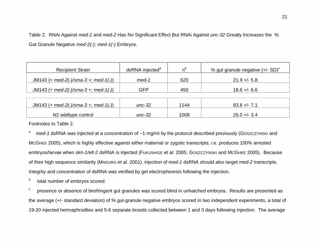
Table 2. RNAi Against med-1 and med-2 Has No Significant Effect But RNAi Against unc-32 Greatly Increases the %
Gut Granule Negative med-2(-); med-1(-) Embryos.
Recipient Strain dsRNA injecteda nb % gut granule negative (+/- SD)c
JM143 (+ med-2(-)/sma-3 +; med-1(-)) med-1 620 21.9 +/- 5.8
JM143 (+ med-2(-)/sma-3 +; med-1(-)) GFP 450 18.6 +/- 6.6
JM143 (+ med-2(-)/sma-3 +; med-1(-)) unc-32 1144 83.6 +/- 7.1
N2 wildtype control unc-32 1008 26.0 +/- 3.4
Footnotes to Table 2.
a med-1 dsRNA was injected at a concentration of ~1 mg/ml by the protocol described previously (GOSZCZYNSKI and
MCGHEE 2005), which is highly effective against either maternal or zygotic transcripts, i.e. produces 100% arrested
embryos/larvae when skn-1/elt-2 dsRNA is injected (FUKUSHIGE et al. 2005; GOSZCZYNSKI and MCGHEE 2005). Because
of their high sequence similarity (MADURO et al. 2001), injection of med-1 dsRNA should also target med-2 transcripts.
Integrity and concentration of dsRNA was verified by gel electrophoresis following the injection.
b total number of embryos scored.
c presence or absence of birefringent gut granules was scored blind in unhatched embryos. Results are presented as
the average (+/- standard deviation) of % gut-granule negative embryos scored in two independent experiments, a total of
19-20 injected hermaphrodites and 5-6 separate broods collected between 1 and 3 days following injection. The average

22
values have not been corrected for possible haploinsufficiency of med-2. The 83.6 +/- 7.1 % gut granule negative
embryos produced by injection of unc-32 dsRNA into strain JM143 is close to the 81.5 % expected if unc-32 RNAi
completely inhibits gut granule formation in med-2(-); med-1(-) homozygous embryos and in med-2(-)/med-2(+); med-1(-)
heterozygous embryos but in only 26% of med-2(+); med-1(-) embryos (as in the wildtype controls).
23
FIGURE LEGEND
Figure 1. Images of arrested embryos produced by (A,B) strain JM142 (med-2(-); +
med-1(-)/lin-2 +) and (C,D) strain JM143 (+ med-2(-)/sma-3 +; med-1(-)).
A,C = Differential interference contrast optics; B,D = Polarization optics to reveal
birefringent gut granules (Z-projection of 4-5 different focal planes). Scale bar =
50 microns. Dotted ellipses indicate arrested embryos that are gut granule
negative. Embryos produced by JM143 appear more heterogenous (more than
might be expected from the observed med-2 haploinsufficiency) and more fragile
than those produced by JM142.