Embed Size (px)
Citation preview
1
Naturally Occurring Carboxypeptidase A6 Mutations: Effect on Enzyme Function and Association with Epilepsy Matthew R. Sapio1, Annick Salzmann2, Monique Vessaz3, Arielle Crespel4, Peter J. Lyons5, Alain Malafosse2,3, Lloyd D. Fricker1,6 1) Department of Neuroscience, Albert Einstein College of Medicine, Bronx, NY 10461 2) Department of Psychiatry, University of Geneva, Switzerland 3) Department of Genetic Medicine and Laboratory, University Hospitals of Geneva, Switzerland 4) Department of Neurology, Montpellier University Hospital, France 5) Department of Biology, Andrews University, Berrien Springs, MI 49104 6) Department of Molecular Pharmacology, Albert Einstein College of Medicine, Bronx, NY 10461 Corresponding Author: Lloyd Fricker, Ph.D., Department of Molecular Pharmacology, Albert Einstein College of Medicine, 1300 Morris Park Avenue, Bronx, NY 10461 Phone: 718-430-4225; Fax: 718-430-8922; E-Mail: [email protected] Running Title: Carboxypeptidase A6 mutations Keywords: Carboxypeptidase, protease, neuropeptide, febrile seizure, temporal lobe epilepsy Background: Two mutations in the
carboxypeptidase A6 (CPA6) gene were previously found in epilepsy patients.
Result: Many additional CPA6 mutations were found. Some inactivated CPA6 and were more frequently found in epilepsy patients than controls.
Conclusion: Several CPA6 mutations greatly reduce enzyme activity, but the most frequently found mutations do not.
Significance: Mutations in CPA6 are associated with rare cases of epilepsy.
SUMMARY Carboxypeptidase A6 (CPA6) is a member of the A/B subfamily of M14 metallo-carboxypeptidases that is expressed in brain and many other tissues during development. Recently, two mutations in human CPA6 were associated with febrile seizures and/or temporal lobe epilepsy. In the present study, we screened for additional CPA6 mutations in patients with febrile seizures and focal epilepsy, which encompasses the temporal lobe epilepsy subtype. Mutations found from this analysis, as well as CPA6 mutations reported in databases of single nucleotide polymorphisms, were
further screened by analysis of the modeled proCPA6 protein structure and the functional role of the mutated amino acid. The point mutations predicted to affect activity and/or protein folding were tested by expression of the mutant in HEK293 cells and analysis of the resulting CPA6 protein. Common polymorphisms in CPA6 were also included in this analysis. Several mutations resulted in reduced enzyme activity or CPA6 protein levels in the extracellular matrix. The mutants with reduced extracellular CPA6 protein levels showed normal levels of 50 kDa proCPA6 in the cell, and this could be converted into 37 kDa CPA6 by trypsin, suggesting that protein folding was not greatly affected by the mutations. Interestingly, three of the mutations that reduced CPA6 enzyme activity were found in patients with epilepsy. Taken together, these results provide further evidence for the involvement of CPA6 mutations in human epilepsy, and reveal additional rare mutations that inactivate CPA6 and could therefore also be associated with epileptic phenotypes.
http://www.jbc.org/cgi/doi/10.1074/jbc.M112.414094The latest version is at JBC Papers in Press. Published on October 26, 2012 as Manuscript M112.414094
Copyright 2012 by The American Society for Biochemistry and Molecular Biology, Inc.
by guest on May 26, 2018
http://ww
w.jbc.org/
Dow
nloaded from
2
INTRODUCTION Epilepsy is one of the most common
neurological disorders, affecting 1-2% of the population (1) with focal epilepsy (FE) accounting for around 60% of this prevalence (2). As proposed by the International League Against Epilepsy, FE includes temporal, frontal, parietal or occipital lobe epilepsy, depending on the localization of the seizure discharge (3). Temporal lobe epilepsy (TLE) is the most frequent form of FE in adults, with hippocampal sclerosis (HS) being the most recurrent pathogenic feature of this illness (4). Moreover, 25% of patients with TLE and HS often show febrile seizures (FS) during childhood (5). Generally, FS are defined as seizures occurring during fever episodes without any central nervous system infection (3). Moreover, FS are the most common convulsive event during early childhood (5), being seen in 5% of the general population (6). As suggested by segregation and linkage studies, FS (7) and TLE (8) could be considered as complex disorders with genetic predisposition. However, few genes have been reproducibly associated with FS (9) and TLE (10). This may be due to the clinical heterogeneity of the patients included in almost all studies. FS and TLE may also be caused by multiple rare and/or de novo mutations. This latter possibility has been recently supported by the identification of two mutations in the carboxypeptidase A6 (CPA6) gene in a family with FS and TLE as well as in unrelated TLE patients (11) .
CPA6 is an extracellular peptidase in the A/B subfamily of the M14 family of carboxypeptidases (12). CPA6 is translated with an N-terminal signal peptide (residues 1-30), which routes it to the secretory pathway. Like most other members of its subfamily, CPA6 is produced as a ~50 kDa inactive proenzyme, proCPA6 (residues 31-437), that is activated when cleaved by a furin-like enzyme into the 37 kDa form (residues 130-437)(13,14). The propeptide found in most members of the A/B carboxypeptidase subfamily acts as an intramolecular chaperone, inhibiting enzymatic activity (15,16). Upon secretion, CPA6 binds to the extracellular matrix (ECM) where it is enzymatically active and cleaves bulky aliphatic and aromatic residues from the C-termini of peptides and proteins (17). CPA6 mRNA is most highly expressed in the olfactory bulb and
epididymus of the adult mouse, and is more broadly expressed in the developing forebrain and cerebellum, as well as in developing skin, bone and osteoblasts (18). CPA6 mRNA has been identified in the human central nervous system, notably the hippocampus (11).
One of the previously identified CPA6 mutations results in a substitution at alanine 270 (Ala270Val) and appears to be linked to a familial autosomal recessive form of FS and TLE while the other, a missense mutation at glycine 267 (Gly267Arg), is associated to unrelated TLE patients in the heterozygous state (11). Biochemical analysis revealed that the Ala270Val mutation retains full carboxypeptidase activity but is expressed at about 40% of wild type (WT) level in the ECM. The Gly267Arg mutation was not detectable in the ECM. Although CPA6 has been shown to perform biologically significant cleavages on several peptides in vitro (13,17), the function of CPA6 as well as the mechanism by which these mutations may lead to epilepsy remains unknown. Because both mutations reduce the level of CPA6 protein in the ECM, it remains unclear whether carboxypeptidase activity or the expression level of the protein is responsible for the observed effects of these mutations. Given what is known about the Gly267Arg and Ala270Val mutations, we hypothesized that loss of function of CPA6 could lead to seizures and epilepsy. Since other mutations in the CPA6 gene should be capable of reducing or eliminating the function of the protein, we searched for and found additional CPA6 mutations in FE and FS populations. Modeling of these mutations, as well as other mutations reported in databases of single nucleotide polymorphisms (SNPs) suggested that many mutations in the CPA6 gene could alter structural integrity, carboxypeptidase activity or both, and thus could potentially be related to the disease. We therefore analyzed these mutant proteins using a variety of methods to better understand their relationship to seizures and epilepsy. EXPERIMENTAL PROCEDURES Subjects The present study was approved by the ethics committees from the departments of neurology and pediatrics from the University Hospitals of Geneva. The FE group consisted of patients who
by guest on May 26, 2018
http://ww
w.jbc.org/
Dow
nloaded from
3
suffered from non-lesional FE and lesional FE such as vascular malformation, cortical dysplasia, and nervous system tumor (Table 1). These unrelated Caucasian patients showed the following distribution of epilepsy syndromes already described in a previous paper (11): 138 patients with TLE (70.8%), 28 patients with undetermined focal epilepsy (15.4%), 12 patients with frontotemporal epilepsy (6.2%), 4 patients with parietal epilepsy (2.1%), 2 patients with temporoparietal junction epilepsy (1.0%), 2 patients with parietooccipital epilepsy (1.0%), 2 patients with temporolateral epilepsy (1.0%), 2 patients with temporooccipital epilepsy (1.0%), 1 patient with parietotemporal epilepsy (0.5%), 1 patient with multifocal epilepsy (0.5%) and 1 patient with Rasmussen syndrome (0.5%). These patients suffered from a severe form of epilepsy with poor control of their seizures. Diagnosis was based on patient history, clinical examination, interictal and ictal electroencephalography (EEG) analysis carried out with monitoring video-EEG, and magnetic resonance imaging evaluation. Unrelated Caucasian patients with FS were admitted to the pediatric emergency rooms at the University Hospitals of Geneva, Lausanne and Neuchâtel (Switzerland) (Table 1). At that time, these patients were recruited following the Consensus statement (19). Simple FS were defined when the seizures were brief with a single seizure occurring during a febrile illness. If duration exceeded 15 min or if multiple seizures occurred during a single febrile illness or within the first 24 hour period, they were defined as complex FS. A healthy Caucasian control group (West European origin for at least two generations) was recruited from blood donors at the University Hospital of Geneva (Table 1). Only unrelated blood donors without a personal and/or familial history of epilepsy and seizures were included.
Mutation and single nucleotide poylmorphism screening All patients and controls or their legal representatives gave their informed consent, and then venous blood was collected in EDTA tubes. DNA was extracted from peripheral blood leukocytes by using the Nucleon BACC 2 kit (Bioscience Amersham, GE Healthcare, Glatbrugg, CH). CPA6 exons were explored by high resolution melt (HRM) assay, which is a
rapid, highly reproducible and resolutive process. Previous experiments have shown a sensitivity and specificity between 97% and 99% for PCR products up to 1,000 bp (20)(21). We used this method followed by direct sequencing to confirm mutations and SNPs, as described (11). PCR and HRM conditions assay and primers sequences are available on request. For HRM data analysis, we normalized melting curves for each sample by calculating the ‘line of best fit’ between two normalization regions before and after the major fluorescence decrease representing the melting of the PCR product using the software provided by the Rotor-GeneTM 6000 (Corbett Research, Australia). This algorithm allows the direct comparison of samples that have different starting fluorescence levels with control samples, which were previously sequenced to determine genotype (22). Statistical analysis First, we checked Hardy-Weinberg equilibrium by using a 2 goodness-of-fit test. Then differences in allele frequencies between cases and controls was determined using UNPHASED software (version 3.0.10)(23). The statistical power to detect association was estimated using the Genetic Power Calculator (http://pngu.mgh.harvard.edu/~purcell /gpc/). For every calculation, we used a genotype relative risk for heterozygote of 2, for homozygote of 3 and an additive model at α level = 0.05. Therefore, we found that for rs17343819, FE patients had 94% power to detect a risk allele G with 13% frequency in controls, and a disease prevalence of 0.5% (24)(1). The power was 84% for FS patients with a disease prevalence of 2% (6). For rs17853192, FE patients had 82% power to detect no significant association with a risk allele G with 7% frequency in controls. The power was 68% for FS patients. Protein analysis and modeling CPA6 protein sequences were accessed from The National Center for Biotechnology Information. Sequence alignment was performed using ClustalW and analyzed using GeneDoc to quantify homology between human CPA6 and orthologs in other vertebrates. Modeling of CPA6 protein structure has been described previously (17). Mutations were modeled using Swiss PDB Viewer and PyMol. Images were created using PyMol.
by guest on May 26, 2018
http://ww
w.jbc.org/
Dow
nloaded from
4
Site-Directed Mutagenesis Point mutations were introduced into the pcDNA3.1(-)hCPA6-HAH6 plasmid (13) using PfuUltra II Hotstart DNA Polymerase (Agilent, Santa Clara, CA) or Pfu Turbo (Stratagene, La Jolla, CA) using the QuickChange Mutagenesis protocol (Stratagene). The resulting cDNA constructs code for full-length human preproCPA6 protein with the hemagglutinin (HA) and hexahistidine (H6) epitopes at the C-terminus (13). All mutations were confirmed by DNA sequence analysis. Cell Culture and Analysis of CPA6 mutant proteins HEK293T cells were cultured in Dulbecco’s modified Eagle’s medium supplemented with 10% fetal bovine serum and penicillin / streptomycin at 37°C and 5% CO2. Cells were transfected with human CPA6 cDNA using TransFectin (Bio-Rad, Hercules, CA) according to manufacturer’s instructions. Amount of plasmid used in transfection was adjusted so that expression levels of proCPA6 in cell lysates were equal when examined by Western Blot (data not shown). At 48 hours post-transfection, cells were harvested in phosphate buffered saline (PBS), centrifuged, and the pellet sonicated in PBS solution containing 0.1% NP-40. Aliquots were analyzed on a denaturing polyacrylamide gel. After harvesting cells from the culture dish, the ECM remaining on the dish was washed several times with PBS to remove any remaining cells. ECM samples were analyzed for CPA6 activity by incubating for 2 hours at 37°C with 0.5 mM 3-(2-furyl) acryloyl-Phe-Phe (FA-Phe-Phe; Bachem, Bubendorf, Switzerland) in Tris buffer (pH 7.8) containing 150 mM NaCl. Reactions were stopped by transferring the soluble substrate to polypropylene tubes, separating it from the enzyme which remained bound to the ECM. Solutions were transferred to cuvettes, and carboxypeptidase activity was detected by a decrease in absorbance at 336nm as described previously (13). After incubation with FA-Phe-Phe, the same ECM samples were collected by the addition of SDS-PAGE sample buffer, fractionated on SDS-PAGE, and transferred to nitrocellulose. Western blotting was performed according to standard protocol with mouse anti-HA (clone HA-7, Sigma, St. Louis,
MO, 1:5000 dilution) primary antibody and either IRdye800-congugated anti-mouse secondary antibody (Rockland, Gilbertsville, PA, 1:3000 dilution) or horseradish peroxidase-conjugated anti-mouse secondary antibody (Cell Signaling, Danvers, MA 1:2000). Images were obtained and quantified using the Li-COR Odyssey System (Lincoln, NE) or with enhanced chemiluminescence detection reagent.
In experiments where levels of CPA6 protein expression were compared between media and ECM fractions, cells were cultured in reduced volumes of media beginning at 24 hours post-transfection to ensure detection of CPA6 protein in the media fractions. As a positive control, 400 µg/ml heparin was added at 24 hours post-transfection to solubilize CPA6, in order to show that CPA6 could be detected in the media when present. At 48 hours post-transfection, media samples were collected, cells were harvested in PBS, combined with SDS-PAGE sample buffer, and analyzed by Western blotting, as above.
In experiments testing the thermal stability of CPA6, ECM samples were washed to remove cells and cell fragments, as described above. Samples were incubated in PBS at the indicated temperature for 1 hour. After heating, PBS was removed and samples were assayed for carboxypeptidase activity as described above. In experiments to determine if oxidation reduces the enzymatic activity of CPA6, a similar method was employed. In place of heating for 1 hour, ECM samples were incubated with 2% hydrogen peroxide (w/w) for 30 minutes at 22°C on a rotating platform (25). Samples were then washed several times with PBS to remove remaining hydrogen peroxide before assaying for carboxypeptidase activity.
In experiments where trypsin was used to digest proCPA6, cells were lysed as described above and incubated with trypsin for 1 hour at 37°C at a broad range of concentrations. A 1:1 concentration of trypsin represents a stock solution of 0.2 µg/µl trypsin in trypsin resuspension buffer (Promega, Fitchburg, WI), from which other concentrations were prepared by serial dilution.
RESULTS Mutations and SNPs screening To screen for additional mutations in CPA6, exonic HRM analysis was performed. A
by guest on May 26, 2018
http://ww
w.jbc.org/
Dow
nloaded from
5
previously unpublished missense mutation c.843A>G was found in a heterozygote FE patient (ET 174). This mutation changes a histidine at position 196 into an arginine (His196Arg) and was not found in 242 Caucasian controls. The parents of the individual harboring this mutation were not available. One FS patient (CF-29-0) was found with a putative missense heterozygous mutation (Table 2). This non-synonymous mutation replaces a proline at position 16 with a threonine (Pro16Thr). The patient’s unaffected father (CF-29-2) was heterozygous, whereas his affected mother (CF-29-1) did not harbor the mutation (Table 2). The mutation was not found in 242 Caucasian controls. The screening of CPA6 exons also revealed a published genetic variant, rs35993949 (c.875C>G) found in the heterozygous condition in one FE patient (Table 2, subject ET 158). The SNP rs35993949 affects a glutamine residue, which is replaced by a glutamic acid residue (Gln207Glu). The putative pathogenic G-allele was not found in 242 Caucasian controls. In a previous study we reported a heterozygous missense mutation (Gly267Arg) for patient ET 158 (11). Further analysis shows that patient ET 158 is compound heterozygous for the two pathogenic non-synonymous mutations c.875C>G (Gln207Glu) and c.799G>A (Gly267Arg) on exons 6 and 8, respectively. As parents of ET 158 were not available, we could not determine whether these two mutations are de novo. Three common allelic variations were examined. First, we checked Hardy-Weinberg equilibrium in order to test if both allele and genotype frequencies in our patient and control populations remained constant through generations (26). The rs17853192 (C>G Ser173Cys) and rs17343819 (A>G Asn249Ser) genotypic distributions were at Hardy-Weinberg equilibrium among FE and FS patients and controls (Table 3). The rs10957393 (T>C Phe45Leu) genotypic distributions were at Hardy-Weinberg equilibrium among FS patients (p = 0.39) but at the limit in FE patients (p = 0.03) and in controls (p = 0.02). The SNP rs17343819 showed a significant difference between FE patients and controls (Table 3). The G-allele displayed a higher frequency in FE patients (21.0%) than in unaffected cases (13.7%). This difference reached the statistical significance with a p-value of 0.0041. Moreover,
the AA-genotype appears to be protective against FE because controls show a higher frequency (74.0%) than FE patients (61.0%) (p = 0.013). No significant genotypic and allelic differences were found between FS patients and controls. For rs17853192 and rs10957393, no associations were found for FE and FS patients as compared to controls. Modeling of CPA6 mutations within predicted protein structure In addition to the mutations described above in FE and FS patients, a large number of additional mutations in the CPA6 gene were reported in the National Center for Biotechnology Information SNP database (http://www.ncbi.nlm.nih.gov/ projects/SNP/) and in the 1000 Genomes Project (http://browser.1000genomes.org/). We first examined all SNP mutations and selected those that were exonic and which resulted in a missense mutation (supplemental Table S1). These mutations were examined for conservation from human to zebrafish (supplemental Fig. S1) and across members of the carboxypeptidase A subfamily (supplemental Fig. S2). SNPs were selected based on a variety of factors including frequency of the allele, conservation, severity of the change in amino acid, location of the residue, and predictions from PolyPhen and SIFT, which analyze conservation of mutated residues across species, and the predicted severity of amino acid substitutions (supplemental Table S1). Several nonsense mutations were found in the SNP database that produced truncations of the protein due to frameshifts and/or introduction of stop codons. All of these were predicted to result in inactive CPA6 protein, based on the requirement for an active site Glu residue known to be involved with catalytic function (13). Missense mutations in CPA6 were visualized within the structural model of the CPA6 protein, as previously described (17)(supplemental Fig. S3). These mutations were organized into several categories based on biochemical properties, the frequency at which they occur in humans, and the level of conservation of the amino acid that they alter. The Phe45Leu, Ser173Cys and Asn249Ser mutations, which occur frequently in humans, were predicted to be harmless or minimally damaging to the protein. These mutations represent modest changes in that these amino acids
by guest on May 26, 2018
http://ww
w.jbc.org/
Dow
nloaded from
6
are not highly conserved, and the substituted amino acid was not predicted from our analysis to alter the conformation or function of the protein (supplemental Table S1). The His196Arg, Gln207Glu, Arg311Glu and Ala424Val mutations were predicted to greatly diminish CPA6 activity. The His196Arg mutation occurs at a residue that is crucial for the coordination of the zinc ion in the catalytic region of the protein and is necessary for the function of all metallocarboxypeptidases in the A/B subfamily (supplemental Fig. S2) (27). The Val306Ile, Pro342Ala and Leu413Phe mutations were predicted to be possibly damaging (supplemental Table S1). These substitutions occur at conserved amino acids, but do not cause substitutions which represent severe changes in properties of the residue. Another mutation, Pro16Thr, occurs within the signal peptide, and was analyzed using SignalP (28), however this substitution was not predicted to result in a change in signal peptide cleavage (supplemental Fig. S4). Mutations which were selected for further analysis were visualized within the CPA6 protein structure (Fig. 1). Mutations were subjected to additional analysis to predict whether any would alter mRNA splicing using Human Splicing Finder (29). One SNP, rs17343819 (Asn249Ser) occurs within an exon splice enhancer motif in exon 7, a predicted recognition site for 9G8. This SNP results in a modest decrease in the value assigned by Human Splicing Finder for this motif (supplemental Table 2). Biochemical analysis of CPA6 mutants
Twelve of the naturally-occurring mutations were selected for direct testing of the biochemical properties. Two of these, Gly267Arg and Ala270Val were mutations we had previously tested for enzyme activity; these were further characterized in the present study (11). Although not identified in humans, the active site mutation Glu398Gln which has no detectable carboxypeptidase activity (13) was included as a negative control.
Mutants were expressed in HEK293 cells, and ECM samples were assayed for carboxypeptidase activity (Fig. 2). The Phe45Leu mutation was the only mutation which showed no statistically significant difference in activity from WT. The Asn249Ser mutation retained 89% of WT activity. Surprisingly, the Ser173Cys mutation
retained only approximately half of WT activity. The His196Arg and Ala424Val mutations had no detectable activity, while the Gln207Glu mutant had 11% of WT activity. We estimate that we would be able to detect activity comparable to 5% of WT activity by our assay, indicating that these mutations remove >95% of carboxypeptidase activity. The Gly267Arg and Glu398Gln mutations also resulted in a similar near total reduction in carboxypeptidase activity, as previously reported (11). The remaining mutations resulted in an intermediate level of retained carboxypeptidase activity. When we examined expression levels of CPA6 protein in the ECM we found that many of the mutants that showed a reduction in carboxypeptidase activity were expressed at reduced levels in the ECM. For the Ala270Val and Val306Ile mutants, reduced expression likely accounts for the observed reduction in activity. While it cannot be ruled out, this is unlikely for the Asn249Ser mutant, which shows a measurable reduction in activity but not in expression levels in the ECM. Notably, some mutations such as the Ala424Val had no detectable carboxypeptidase activity, but an intermediate level of protein expression in the ECM. Conversely, the Ser173Cys and Val306Ile mutations showed a vast reduction in ECM expression, and an intermediate reduction in carboxypeptidase activity. No CPA6 protein was detectable in the media for WT or any of the mutant proteins (Fig. 2B), and when heparin was added to displace protein from the ECM into the media, mutant protein expression in the media was detected in approximately the same ratio as it was in the ECM (Fig. 2B-E).
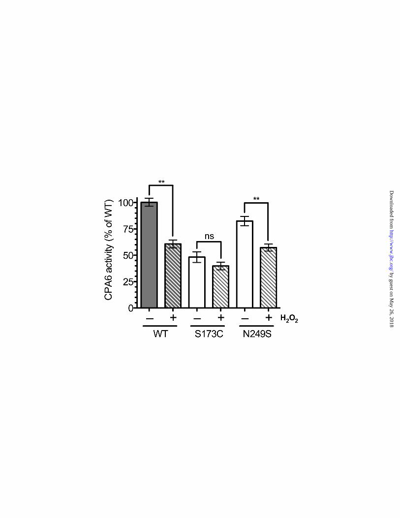
From the comparison of activity versus protein levels, it appeared that the Ser173Cys mutant had more activity than expected based on the amount of CPA6 protein, relative to WT CPA6 (Fig. 2A, C). A potential explanation is that the WT CPA6 in the ECM was not fully active, while a greater proportion of the Ser173Cys mutant had full activity. One mechanism by which WT CPA6 activity could be reduced without a corresponding decrease in protein would be through oxidization of the methionine residue present in a key position within the substrate-binding pocket. In all species examined, CPA6 contains a methionine in the position that confers specificity towards hydrophobic amino acids (supplemental Fig. S1,
by guest on May 26, 2018
http://ww
w.jbc.org/
Dow
nloaded from
7
S2); in other related enzymes (carboxypeptidases A1, A2, A3, A4, and A5) the comparable residue is a leucine, isoleucine, or valine, and never a methionine. Methionine can become oxidized into the sulfoxide and sulfone, and this change is predicted to eliminate the ability of CPA6 to bind peptides with hydrophobic C-termini. Therefore, we tested whether oxidation reduced CPA6 activity. Treatment of WT CPA6 with hydrogen peroxide lowered the amount of enzyme activity by about 60% (Fig. 3). Ser173Cys, which has an outward-facing cysteine residue, did not show a significant reduction in activity after exposure to the oxidizing chemical. As a control, the Asn249Ser mutation was also examined. This mutant showed a similar reduction in activity as WT CPA6 upon exposure to hydrogen peroxide. To test the possibility that CPA6 could have altered enzymatic activity when the methionine was converted into the sulfoxide or the sulfone, the hydrogen peroxide-treated CPA6 was tested with residues representing bulky hydrophobic residues, small hydrophobic residues, and basic residues. However no change in substrate specificity was observed between treated and untreated CPA6 (supplemental Fig. S5).
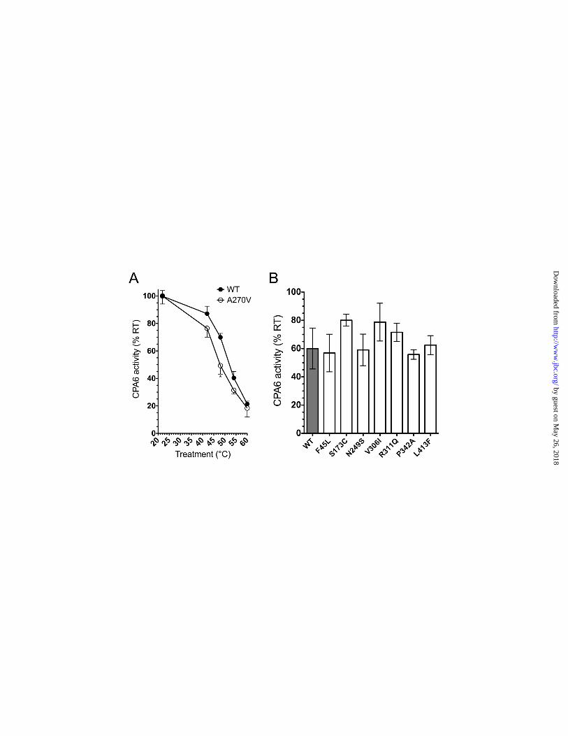
The Ala270Val mutation was analyzed for heat stability because of its connection to febrile seizures, and to examine possible reasons for its reduced expression in the ECM. A change in heat stability indicates a difference in folding, which could serve as an explanation for the pathological properties of the Ala270Val mutation. This assay has been used to characterize mutants of other carboxypeptidases (30). A modest reduction of carboxypeptidase activity was detected at 48°C, and the Ala270Val mutant showed a greater reduction than the WT form (Fig. 4A). At temperatures above 48°C, both WT and Ala270Val enzyme activity were greatly reduced, and were not significantly different between WT and Ala270Val. Other mutants with detectable activity were tested following a 48°C heat treatment, however none showed a significant difference when compared to WT CPA6 (Fig. 4B), indicating that the Ala270Val may be unique in this property among the mutants tested.
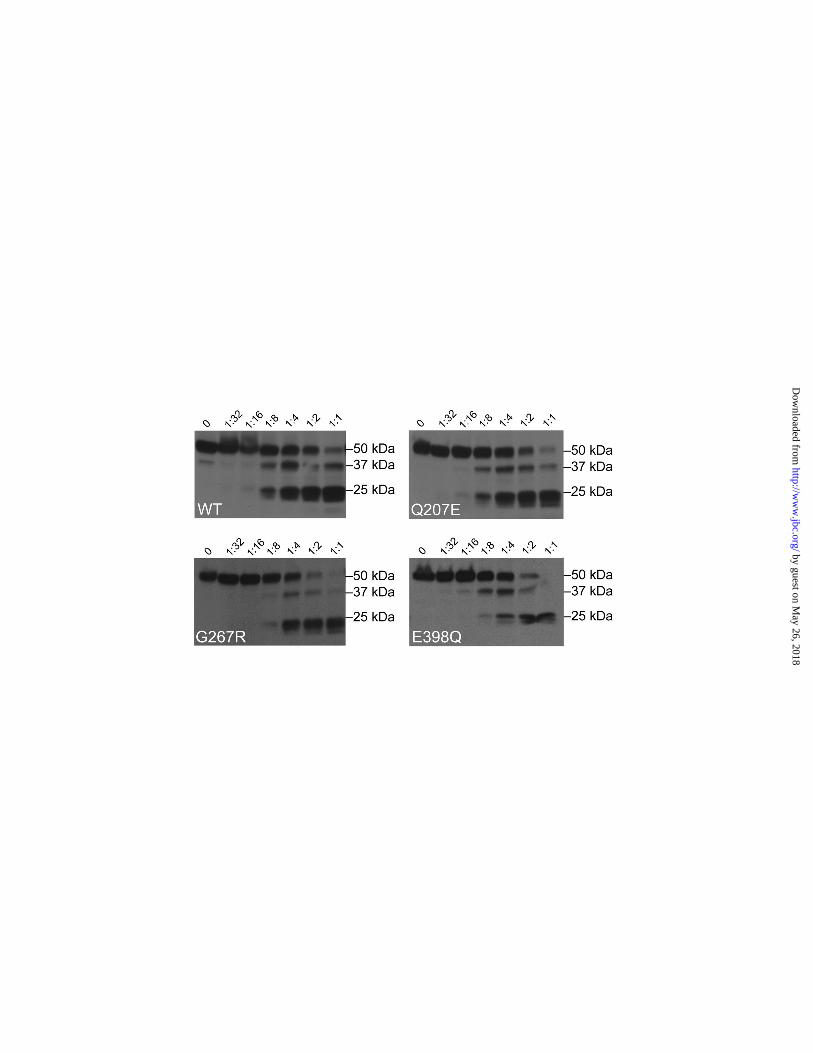
Because proteins that are misfolded are often degraded before secretion, we tested if CPA6 mutants that have no detectable expression in the ECM are more sensitive to trypsin digestion,
which could indicate a change in folding pattern and domain structure. All of the mutant proteins tested showed the same bands representing the 50 kDa proCPA6, the ~37 kDa active CPA6, and a ~25 kDa degradation product which likely corresponds to a cleavage at a region rich in basic amino acids (residues 178 – 187)(Fig. 5, supplemental Fig. S6). Grossly misfolded proteins often show smears, representing trypsin’s equal accessibility to many basic residues, while correctly folded proteins will be preferentially cleaved by trypsin at basic residues which are accessible to the enzyme. The similar sizes of the trypsin digestion products indicates that the mutant proteins form stable structures that resemble the WT form of CPA6. To test the possibility of a dominant negative action for CPA6 mutants found in the heterozygous condition, co-expression of WT and mutant CPA6 was performed. For this, WT CPA6 was tagged with the FLAG epitope tag and mutants were tagged with the His6 tag. Combinations of the plasmid encoding WT CPA6 with plasmids encoding each of the mutants were transfected into HEK293T and the ECM assayed for enzyme activity (supplemental Fig. S7). The combination of WT and mutant either led to greater activity than WT alone (for those mutants with activity) or no difference with WT alone (for those mutants with no activity). None of the combinations showed activity lower than that of WT alone, indicating that the mutants were not able to act in a dominant negative fashion. DISCUSSION Previously, two CPA6 mutations were identified that affected enzymatic activity and protein levels; one mutation was linked to familial FS and TLE, and the other mutation was associated with sporadic TLE (11). A major finding of the present study is that a number of additional mutations within CPA6 similarly affect the protein and are found in patients with epilepsy. Because both of the previously identified mutations were located in the same substrate binding loop of the CPA6 protein, it was unclear if the mutations acted in a related and specific way, or if other mutations affecting different parts of the protein could also be associated with epilepsy. The new mutations identified in the present study extend the previous results and suggest that loss-of-function mutations
by guest on May 26, 2018
http://ww
w.jbc.org/
Dow
nloaded from
8
in CPA6 are related to epilepsy and seizures. Our finding that the Ala270Val mutation and hydrogen peroxide treatment, each manipulations that affect the region of the protein involved in substrate binding, had no effect on the substrate specificity of the enzyme suggests that it is unlikely that other mutations which occur far from the substrate binding loop affect specificity of CPA6. However, because all of the other mutants were examined using only one substrate, it remains a possibility that some mutations in CPA6 alter the selectivity of the enzyme. Both gain-of-function (31,32) and loss-of-function (33-35) mutations of other genes have been found to cause epilepsy. We expect that both types of mutations in CPA6 could lead to epilepsy, however in vitro experiments present difficulties in discriminating between the two, as it is not always clear how these mutations will affect the human brain in vivo (36). Generally, common variations in the CPA6 gene were not found to markedly reduce the activity or expression of the enzyme, and were not found to be associated with epilepsy. However, our finding that the SNP rs17343819 (A>G Asn249Ser) shows a small but statistically significant decrease in enzyme activity and is more common in FE patients than in controls is consistent with the common disease–common variant hypothesis. Previous studies that identified susceptibility markers for complex epilepsy predicted considerable underlying polygenic heterogeneity (37). It is possible that the modest reduction in carboxypeptidase activity of the Asn249Ser variant contributes to seizures. The cause of the reduction in enzyme activity of this mutant is not known, and although ECM levels of mutant CPA6 protein were comparable to WT CPA6 protein, we cannot rule out a small decrease in expression of the Asn249Ser mutant. Alternatively, this mutation may cause changes in protein function or expression that were not detected in the present study. For example, Asn249 is the last residue coded by exon 7, and the adenine to guanine substitution could affect splicing because it occurs within an exon splice enhancer, offering a potential alternative explantion for the consequences of this mutation. While the reduction in the score assigned by Human Splicing Finder is modest (supplemental Table S2) this is consistent with a subtle effect of this SNP. Such
results have already been found in some epileptic contexts. The well-studied SNP rs3812718 (IV5N+5 G>A), which modifies the proportion of two SCN1A alternative transcripts is a susceptibility common variant for epilepsy. This SNP has been associated with FS and FE with history of FS (38,39). Moreover, rare non-synonymous mutations in SCN1A have been linked to severe myoclonic epilepsy in infancy (40). Collectively, these data demonstrate that common variants and rare mutations of the same gene are implicated in related epileptic syndromes. This is consistent with the findings of the present study, as rare missense mutations and one common marker, rs17343819 (Asn249Ser), are detected in FE patients. Several rare mutations were identified in patients with epilepsy that greatly reduce CPA6 activity and expression. The missense mutation (Gly267Arg) was previously found in three TLE patients (11). In the present study, we found that one of these three patients also showed another missense mutation, Gln207Glu. We could not confirm if these pathogenic variants were de novo because the parents of the individual harboring these mutations were not available. We were also unable to determine whether or not the two mutant alleles were located on the same parental chromosome. Because of this uncertainty, this patient is considered to be compound heterozygous for Gly267Arg and Gln207Glu (11). Because each of these mutations alone causes a >95% reduction in CPA6 carboxypeptidase activity and protein levels in the ECM, it is likely that a compound heterozygote for these mutations would show a complete loss of functional protein. Analogous events have been described in patients with missense mutations in POLG, the gene encoding mitochondrial DNA polymerase gamma (41). Patients with these mutations display ataxia syndrome, which is a progressive neurological disorder characterized by FE as a primary feature (41). Some compound heterozygotes have been reported among these patients, who harbor both Ala467Thr and Trp748Ser mutations in POLG (42,43). One patient, heterozygote for Trp748Ser only, also showed a similar neurological phenotype with epilepsy (42). Except for the Asn249Ser polymorphism discussed above, all other CPA6 polymorphisms which occur frequently in the population did not
by guest on May 26, 2018
http://ww
w.jbc.org/
Dow
nloaded from
9
show any association with epilepsy. Our finding that the SNP rs10957393 (T>C Phe45Leu) is not significantly associated with FE and FS groups is consistent with the observation that the Phe45Leu mutation showed normal levels of enzyme activity when expressed in cells. The Ser173Cys mutation is another relatively common variant in the general population. Although the Ser173Cys mutation significantly reduced CPA6 protein levels in the ECM, it had no apparent association with epilepsy. Notably, the Ser173Cys mutation has never been reported in the homozygous state, and it is possible that like the Ala270Val mutation, which also causes a partial reduction in ECM expression of CPA6, the Ser173Cys mutation may be found to be associated with epilepsy only when both alleles are mutated (11).
While most of the mutants tested in the present study showed a reduction in enzyme activity relative to the amount of protein present in the ECM, the Ser173Cys enzyme appears to be more active than WT CPA6 when enzyme activity is adjusted for protein level. It is possible that CPA6 exists in a variety of states, some more active than others. We hypothesized that CPA6 may be regulated by oxidation, and that some percentage of CPA6 in the ECM may be oxidized at a given time. This prediction comes from the observation that CPA6 has a methionine residue at the position which confers substrate specificity; this methionine is conserved throughout evolution for all species where a clear CPA6 orthologue has been identified. Methionine is not present in the comparable position in any other member of the M14 metallocarboxypeptidase family. Because methionine is less hydrophobic when oxidized to either the sulfoxide or sulfone, this oxidation was predicted to reduce the ability of CPA6 to cleave peptides with hydrophobic C-termini. This prediction was confirmed; CPA6 activity is diminished when treated with hydrogen peroxide, a standard technique to oxidize methionines in proteins. In contrast, the Ser173Cys mutant is resistant to hydrogen peroxide and does not lose enzyme activity, possibly because the free Cys residue is oxidized preferentially, protecting the critical methionine from oxidation. If the WT CPA6 in the ECM represents a mixture of protein with reduced and oxidized methionine residues while the Ser173Cys is primarily the reduced
form, this could account for the observation that the mutant has more activity than expected due to the low level of protein.
Several of the mutations in CPA6 were predicted to affect protein folding based on the steric hinderance imposed by the mutation, or the loss of a salt-bridge. Consistent with this prediction, we found a number of the mutations had no enzymatic activity and were not secreted from cells. Misfolded proteins are often degraded before being routed to the trans Golgi complex for secretion, potentially providing an explanation for the lack of secretion of some CPA6 mutants. However, all of the mutants presumably have the same general domain structures as they are all cleaved at the same basic regions by tryspin. Furthermore, except for the Ala270Val mutation, none of the mutants showed greater thermo-instability, further supporting similar folding patterns to WT CPA6. Although the Ala270Val mutant showed a significant decrease after being heated to 48°C, indicating it may be less stable than the WT variant, this temperature is not physiological even during extreme fever. Similar results were found for a naturally occuring mutation of carboxypeptidase E, with greater sensitivity to elevated temperatures presumably reflecting decreased stability of the mutant (30). The mechanism by which CPA6 affects predisposition to seizures is not known. CPA6 is an extracellular protease that cleaves C-terminal amino acids with a preference for large aromatic and aliphatic residues (17). When tested in in vitro assays, CPA6 cleaves neurotensin, Met-enkephalin, and Leu-enkephalin into inactive products, but converts inactive angiotensin I into angiotensin II, the biologically active form. Many other peptides are cleaved by CPA6; the effect on biological activity is not currently known (13). The working hypothesis is that CPA6 plays a role in the activation of peptides that have neuroprotective effects or in the inactivation of peptides that cause neuronal stimulation, and that the reduction of CPA6 activity leads to an imbalance of excitation and inhibition. Alternatively, it is possible that substrates or products of CPA6 play a role in development such that the reduction of CPA6 activity leads to a nervous system that is more prone to seizures. Previously, CPA6 deficiency was found in one patient with Duane Syndrome, an eye movement
by guest on May 26, 2018
http://ww
w.jbc.org/
Dow
nloaded from
10
disorder caused by the failure of cranial nerve VI to innervate the lateral rectus muscle (44). Interestingly, CPA6 is expressed strongly in the region posterior to the developing eye. However, reduction of cpa6 levels in zebrafish embryos had no effect on eye tracking or cranial nerve pathfinding (45). Furthermore, there are no reports
of eye movement disorders in the FS or FE patients found to have point mutations within CPA6. Thus, the link between CPA6 and Duane Syndrome is based on a single patient, in contrast to the link between CPA6 and epilepsy, which has now been extended to several mutations.
FOOTNOTES * Funding was provided by National Institutes of Health grant DA-004494 (to L.D.F.) 1Abbreviations used: CPA6, carboxypeptidase A6; ECM, extracellular matrix; EEG, electroencephalography; ESE, exon splice enhancer; FA, 3-(2-furyl) acryloyl-Phe-Phe; FE, focal epilepsy; FS, febrile seizures; HRM, high resolution melt; HS, hippocampal sclerosis; PBS, phosphate buffered saline; SNP, single nucleotide polymorphisms; TLE, Temporal lobe epilepsy; WT, wild type REFERENCES
1. Hauser, W. A., Annegers, J. F., and Rocca, W. A. (1996) Descriptive epidemiology of epilepsy: contributions of population-based studies from Rochester, Minnesota. Mayo Clin Proc 71, 576-586
2. Sander, J. W., Hart, Y. M., Johnson, A. L., and Shorvon, S. D. (1990) National General Practice Study of Epilepsy: newly diagnosed epileptic seizures in a general population. Lancet 336, 1267-1271
3. (1989) Proposal for revised classification of epilepsies and epileptic syndromes. Commission on Classification and Terminology of the International League Against Epilepsy. Epilepsia 30, 389-399
4. Falconer, M. A., Serafetinides, E. A., and Corsellis, J. A. (1964) Etiology and Pathogenesis of Temporal Lobe Epilepsy. Arch Neurol 10, 233-248
5. Baulac, S., Gourfinkel-An, I., Nabbout, R., Huberfeld, G., Serratosa, J., Leguern, E., and Baulac, M. (2004) Fever, genes, and epilepsy. Lancet Neurol 3, 421-430
6. Hauser, W. A. (1994) The prevalence and incidence of convulsive disorders in children. Epilepsia 35 Suppl 2, S1-6
7. Rich, S. S., Annegers, J. F., Hauser, W. A., and Anderson, V. E. (1987) Complex segregation analysis of febrile convulsions. Am J Hum Genet 41, 249-257
8. Ottman, R. (1989) Genetics of the partial epilepsies: a review. Epilepsia 30, 107-111 9. Nakayama, J. (2009) Progress in searching for the febrile seizure susceptibility genes. Brain Dev
31, 359-365 10. Salzmann, A., Malafosse, A. (2012) Genetics of Temporal Lobe Epilepsy: A Review. Epilepsy
Res Treat, 863702 11. Salzmann, A., Guipponi, M., Lyons, P. J., Fricker, L. D., Sapio, M., Lambercy, C., Buresi, C.,
Ouled Amar Bencheikh, B., Lahjouji, F., Ouazzani, R., Crespel, A., Chaigne, D., and Malafosse, A. (2012) Carboxypeptidase A6 gene (CPA6) mutations in a recessive familial form of febrile seizures and temporal lobe epilepsy and in sporadic temporal lobe epilepsy. Hum Mutat 33, 124-135
12. Wei, S., Segura, S., Vendrell, J., Aviles, F. X., Lanoue, E., Day, R., Feng, Y., and Fricker, L. D. (2002) Identification and characterization of three members of the human metallocarboxypeptidase gene family. J Biol Chem 277, 14954-14964
13. Lyons, P. J., Callaway, M. B., and Fricker, L. D. (2008) Characterization of carboxypeptidase A6, an extracellular matrix peptidase. J Biol Chem 283, 7054-7063
by guest on May 26, 2018
http://ww
w.jbc.org/
Dow
nloaded from
11
14. Vendrell, J., Querol, E., and Aviles, F. X. (2000) Metallocarboxypeptidases and their protein inhibitors. Structure, function and biomedical properties. Biochim Biophys Acta 1477, 284-298
15. Phillips, M. A., and Rutter, W. J. (1996) Role of the prodomain in folding and secretion of rat pancreatic carboxypeptidase A1. Biochemistry 35, 6771-6776
16. Gomis-Ruth, F. X. (2008) Structure and mechanism of metallocarboxypeptidases. Crit Rev Biochem Mol Biol 43, 319-345
17. Lyons, P. J., and Fricker, L. D. (2010) Substrate specificity of human carboxypeptidase A6. J Biol Chem 285, 38234-38242
18. Fontenele-Neto, J. D., Kalinina, E., Feng, Y., and Fricker, L. D. (2005) Identification and distribution of mouse carboxypeptidase A-6. Brain Res Mol Brain Res 137, 132-142
19. (1980) Consensus statement. Febrile seizures: long-term management of children with fever-associated seizures. Pediatrics 66, 1009-1012
20. Montgomery, J., Wittwer, C. T., Palais, R., and Zhou, L. (2007) Simultaneous mutation scanning and genotyping by high-resolution DNA melting analysis. Nat Protoc 2, 59-66
21. Taylor, C. F. (2009) Mutation scanning using high-resolution melting. Biochem Soc Trans 37, 433-437
22. Wojdacz, T. K., Dobrovic, A., and Hansen, L. L. (2008) Methylation-sensitive high-resolution melting. Nat Protoc 3, 1903-1908
23. Dudbridge, F., and Koeleman, B. P. (2003) Rank truncated product of P-values, with application to genomewide association scans. Genet Epidemiol 25, 360-366
24. Wallace, H., Shorvon, S., and Tallis, R. (1998) Age-specific incidence and prevalence rates of treated epilepsy in an unselected population of 2,052,922 and age-specific fertility rates of women with epilepsy. Lancet 352, 1970-1973
25. Kim, Y. H., Berry, A. H., Spencer, D. S., and Stites, W. E. (2001) Comparing the effect on protein stability of methionine oxidation versus mutagenesis: steps toward engineering oxidative resistance in proteins. Protein Eng 14, 343-347
26. Stern, C. (1943) The Hardy-Weinberg Law. Science 97, 137-138 27. Gomis-Ruth, F. X., Botelho, T. O., and Bode, W. (2012) A standard orientation for
metallopeptidases. Biochim Biophys Acta 1824, 157-163 28. Petersen, T. N., Brunak, S., von Heijne, G., and Nielsen, H. (2011) SignalP 4.0: discriminating
signal peptides from transmembrane regions. Nat Methods 8, 785-786 29. Desmet, F. O., Hamroun, D., Lalande, M., Collod-Beroud, G., Claustres, M., and Beroud, C.
(2009) Human Splicing Finder: an online bioinformatics tool to predict splicing signals. Nucleic Acids Res 37, e67
30. Chen, H., Jawahar, S., Qian, Y., Duong, Q., Chan, G., Parker, A., Meyer, J. M., Moore, K. J., Chayen, S., Gross, D. J., Glaser, B., Permutt, M. A., and Fricker, L. D. (2001) Missense polymorphism in the human carboxypeptidase E gene alters enzymatic activity. Hum Mutat 18, 120-131
31. Dibbens, L. M., Reid, C. A., Hodgson, B., Thomas, E. A., Phillips, A. M., Gazina, E., Cromer, B. A., Clarke, A. L., Baram, T. Z., Scheffer, I. E., Berkovic, S. F., and Petrou, S. (2010) Augmented currents of an HCN2 variant in patients with febrile seizure syndromes. Ann Neurol 67, 542-546
32. Volkers, L., Kahlig, K. M., Verbeek, N. E., Das, J. H., van Kempen, M. J., Stroink, H., Augustijn, P., van Nieuwenhuizen, O., Lindhout, D., George, A. L., Jr., Koeleman, B. P., and Rook, M. B. (2011) Nav 1.1 dysfunction in genetic epilepsy with febrile seizures-plus or Dravet syndrome. Eur J Neurosci 34, 1268-1275
33. Escayg, A., and Goldin, A. L. (2010) Sodium channel SCN1A and epilepsy: mutations and mechanisms. Epilepsia 51, 1650-1658
34. Kang, J. Q., Shen, W., and Macdonald, R. L. (2009) The GABRG2 mutation, Q351X, associated with generalized epilepsy with febrile seizures plus, has both loss of function and dominant-negative suppression. J Neurosci 29, 2845-2856
by guest on May 26, 2018
http://ww
w.jbc.org/
Dow
nloaded from
12
35. Liao, W. P., Shi, Y. W., Long, Y. S., Zeng, Y., Li, T., Yu, M. J., Su, T., Deng, P., Lei, Z. G., Xu, S. J., Deng, W. Y., Liu, X. R., Sun, W. W., Yi, Y. H., Xu, Z. C., and Duan, S. (2010) Partial epilepsy with antecedent febrile seizures and seizure aggravation by antiepileptic drugs: associated with loss of function of Na(v) 1.1. Epilepsia 51, 1669-1678
36. Moulard, B., Picard, F., le Hellard, S., Agulhon, C., Weiland, S., Favre, I., Bertrand, S., Malafosse, A., and Bertrand, D. (2001) Ion channel variation causes epilepsies. Brain Res Brain Res Rev 36, 275-284
37. Mulley, J. C., Scheffer, I. E., Harkin, L. A., Berkovic, S. F., and Dibbens, L. M. (2005) Susceptibility genes for complex epilepsy. Hum Mol Genet 14 Spec No. 2, R243-249
38. Schlachter, K., Gruber-Sedlmayr, U., Stogmann, E., Lausecker, M., Hotzy, C., Balzar, J., Schuh, E., Baumgartner, C., Mueller, J. C., Illig, T., Wichmann, H. E., Lichtner, P., Meitinger, T., Strom, T. M., Zimprich, A., and Zimprich, F. (2009) A splice site variant in the sodium channel gene SCN1A confers risk of febrile seizures. Neurology 72, 974-978
39. Le Gal, F., Salzmann, A., Crespel, A., and Malafosse, A. (2011) Replication of association between a SCN1A splice variant and febrile seizures. Epilepsia 52, e135-138
40. Lossin, C. (2009) A catalog of SCN1A variants. Brain Dev 31, 114-130 41. Winterthun, S., Ferrari, G., He, L., Taylor, R. W., Zeviani, M., Turnbull, D. M., Engelsen, B. A.,
Moen, G., and Bindoff, L. A. (2005) Autosomal recessive mitochondrial ataxic syndrome due to mitochondrial polymerase gamma mutations. Neurology 64, 1204-1208
42. Tzoulis, C., Engelsen, B. A., Telstad, W., Aasly, J., Zeviani, M., Winterthun, S., Ferrari, G., Aarseth, J. H., and Bindoff, L. A. (2006) The spectrum of clinical disease caused by the A467T and W748S POLG mutations: a study of 26 cases. Brain 129, 1685-1692
43. Roshal, D., Glosser, D., and Zangaladze, A. (2011) Parieto-occipital lobe epilepsy caused by a POLG1 compound heterozygous A467T/W748S genotype. Epilepsy Behav 21, 206-210
44. Pizzuti, A., Calabrese, G., Bozzali, M., Telvi, L., Morizio, E., Guida, V., Gatta, V., Stuppia, L., Ion, A., Palka, G., and Dallapiccola, B. (2002) A peptidase gene in chromosome 8q is disrupted by a balanced translocation in a duane syndrome patient. Invest Ophthalmol Vis Sci 43, 3609-3612
45. Lyons, P. J., Ma, L. H., Baker, R., and Fricker, L. D. (2010) Carboxypeptidase A6 in zebrafish development and implications for VIth cranial nerve pathfinding. PLoS One 5, e12967
by guest on May 26, 2018
http://ww
w.jbc.org/
Dow
nloaded from
13
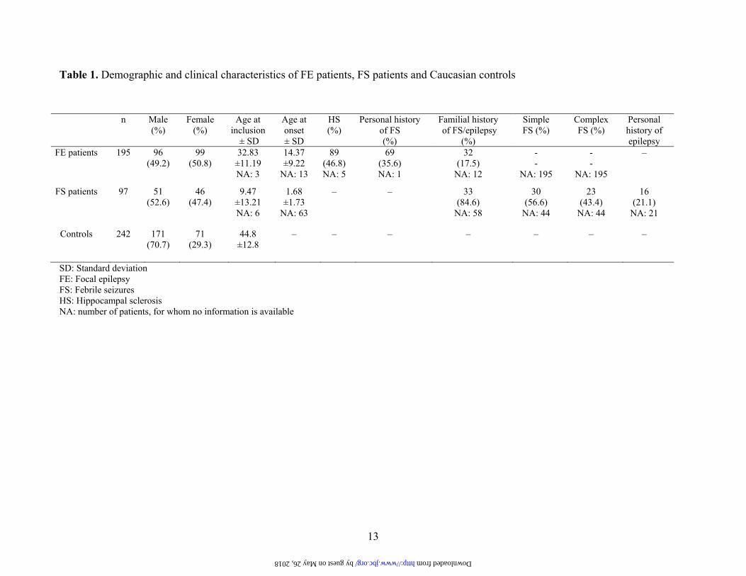
Table 1. Demographic and clinical characteristics of FE patients, FS patients and Caucasian controls
n Male (%)
Female (%)
Age at inclusion
± SD
Age at onset ± SD
HS (%)
Personal history of FS (%)
Familial history of FS/epilepsy
(%)
Simple FS (%)
Complex FS (%)
Personal history of epilepsy
FE patients
195 96 (49.2)
99 (50.8)
32.83 ±11.19 NA: 3
14.37 ±9.22
NA: 13
89 (46.8) NA: 5
69 (35.6) NA: 1
32 (17.5)
NA: 12
- -
NA: 195
- -
NA: 195
–
FS patients
97 51 (52.6)
46 (47.4)
9.47 ±13.21 NA: 6
1.68 ±1.73
NA: 63
– – 33 (84.6)
NA: 58
30 (56.6)
NA: 44
23 (43.4)
NA: 44
16 (21.1)
NA: 21
Controls
242 171 (70.7)
71 (29.3)
44.8 ±12.8
– – – – – – –
SD: Standard deviation FE: Focal epilepsy FS: Febrile seizures HS: Hippocampal sclerosis NA: number of patients, for whom no information is available
by guest on May 26, 2018 http://www.jbc.org/ Downloaded from
14
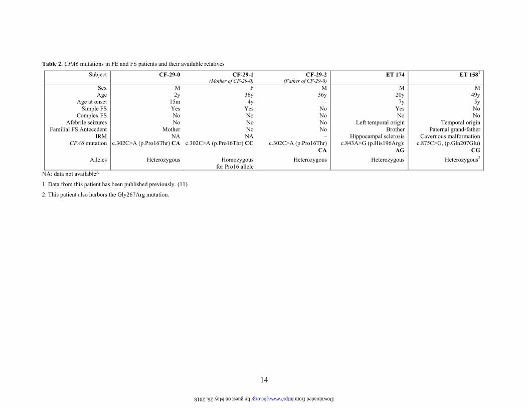
Table 2. CPA6 mutations in FE and FS patients and their available relatives
Subject CF-29-0 CF-29-1 (Mother of CF-29-0)
CF-29-2 (Father of CF-29-0)
ET 174 ET 1581
Sex M F M M M Age 2y 36y 36y 20y 49y
Age at onset 15m 4y – 7y 5y Simple FS Yes Yes No Yes No
Complex FS No No No No No Afebrile seizures No No No Left temporal origin Temporal origin
Familial FS Antecedent Mother No No Brother Paternal grand-father IRM NA NA – Hippocampal sclerosis Cavernous malformation
CPA6 mutation c.302C>A (p.Pro16Thr) CA c.302C>A (p.Pro16Thr) CC c.302C>A (p.Pro16Thr) CA
c.843A>G (p.His196Arg): AG
c.875C>G, (p.Gln207Glu) CG
Alleles Heterozygous Homozygous for Pro16 allele
Heterozygous Heterozygous Heterozygous2
NA: data not available^
1. Data from this patient has been published previously. (11)
2. This patient also harbors the Gly267Arg mutation.
by guest on May 26, 2018 http://www.jbc.org/ Downloaded from
15
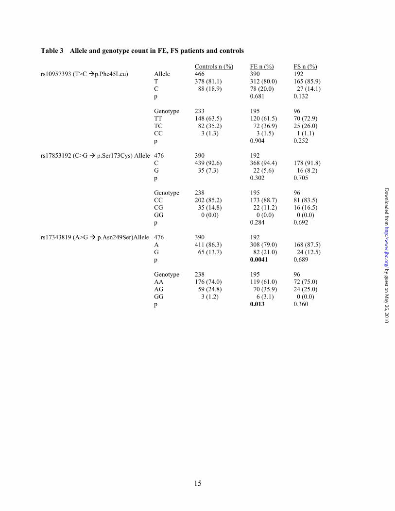
Table 3 Allele and genotype count in FE, FS patients and controls
Controls n (%) FE n (%) FS n (%) rs10957393 (T>C p.Phe45Leu) Allele 466 390 192 T 378 (81.1) 312 (80.0) 165 (85.9) C 88 (18.9) 78 (20.0) 27 (14.1) p 0.681 0.132 Genotype 233 195 96 TT 148 (63.5) 120 (61.5) 70 (72.9) TC 82 (35.2) 72 (36.9) 25 (26.0) CC 3 (1.3) 3 (1.5) 1 (1.1) p 0.904 0.252 rs17853192 (C>G p.Ser173Cys) Allele 476 390 192 C 439 (92.6) 368 (94.4) 178 (91.8) G 35 (7.3) 22 (5.6) 16 (8.2) p 0.302 0.705 Genotype 238 195 96 CC 202 (85.2) 173 (88.7) 81 (83.5) CG 35 (14.8) 22 (11.2) 16 (16.5) GG 0 (0.0) 0 (0.0) 0 (0.0) p 0.284 0.692 rs17343819 (A>G p.Asn249Ser)Allele 476 390 192 A 411 (86.3) 308 (79.0) 168 (87.5) G 65 (13.7) 82 (21.0) 24 (12.5) p 0.0041 0.689 Genotype 238 195 96 AA 176 (74.0) 119 (61.0) 72 (75.0) AG 59 (24.8) 70 (35.9) 24 (25.0) GG 3 (1.2) 6 (3.1) 0 (0.0) p 0.013 0.360
by guest on May 26, 2018
http://ww
w.jbc.org/
Dow
nloaded from
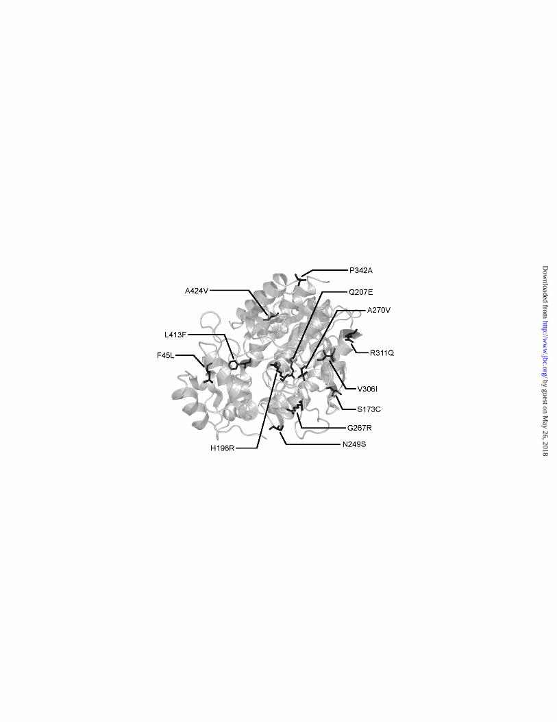
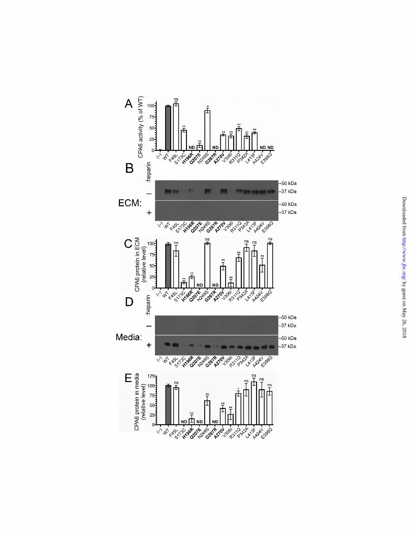
FIGURE LEGENDS Figure 1. Mutations identified in the CPA6 protein. The present study examines the biochemical properties of twelve mutations in the CPA6 protein (sticks). These mutations occur throughout the protein, and the residues they alter likely have diverse effects on structure and function. The zinc ion (sphere) is shown to indicate the location of the active site. Figure 2. Activity and expression level of CPA6 mutants. WT CPA6 and CPA6 mutants were expressed in HEK293T cells and tested for activity and extracellular expression levels. Mock transfected cells (–) and cells transfected with an active site mutant, E398Q, were included as negative controls. All CPA6 constructs contained an HA tag on the C-terminus for protein detection. A: The ECM-bound CPA6 was assayed for enzyme activity using the chromogenic carboxypeptidase substrate FA-Phe-Phe. B, C: The ECM samples were resolved by SDS-PAGE, and Western blots were performed using an antibody against the HA epitope to determine the relative level of CPA6 protein bound to the ECM. A number of mutants showed a reduction in the amount of protein bound to the ECM (quantified in panel C). D, E: Media were analyzed on Western blots. In the absence of heparin, CPA6 protein was undetectable in media. With the addition of 400 µg/ml heparin, which displaces CPA6 from the ECM, CPA6 was detectable in the media in approximately the same ratio as in the ECM (quantified in panel E). Error bars indicate standard error of the mean (n=3-25). Rare mutations found in patients with epilepsy are indicated in bold. ND, not detected; ns, not statistically significant. Figure 3. Effects of hydrogen peroxide treatment on CPA6 activity. HEK293T cells were transfected with WT, Ser173Cys or Asn249Ser CPA6 constructs. ECM-bound CPA6 was incubated with or without 2% hydrogen peroxide for 30 minutes at 22°C, and assayed for activity using the chromogenic carboxypeptidase substrate FA-Phe-Phe. After treatment with hydrogen peroxide, WT activity was reduced by approximately 40% (n = 4, p < 0.001) while Ser173Cys showed only a modest reduction that was not statistically significant (n = 5, p > 0.2). The Asn249Ser mutation was reduced by approximately 30% (n = 5, p < 0.01). Figure 4. Heat stability of CPA6 mutants. HEK293T cells were transfected with WT and mutant CPA6. ECM-bound CPA6 was incubated at the temperature indicated for 1 hour, and then cooled to 37ºC and assayed for CPA6 activity using the chromogenic carboxypeptidase substrate, FA-Phe-Phe. A: The Ala270Val CPA6 mutant showed slightly decreased activity when incubated at 48°C (WT, n = 12; Ala270Val, n = 6; p < 0.05). Other temperatures showed a tendency towards reduced activity but this was not significant. B: Other CPA6 mutants with activity were tested by incubating at 48°C for 1 hour, and then cooling to 37ºC for enzyme assay. No statistically significant differences were observed between these mutants and WT CPA6. Error bars represent standard error of the mean (n=5). Figure 5. Conversion of proCPA6 mutants into active CPA6. HEK293T cells were transfected with WT or mutant CPA6. Cells were harvested in PBS, lysed in PBS with 0.1% NP40 and sonicated. Samples were then centrifuged and supernatants were divided into aliquots and incubated for 1 hour at 37°C with varying concentrations of trypsin, as indicated. A 1:1 concentration of trypsin represents a stock solution of 0.2 µg/µl trypsin, from which other concentrations were prepared by serial dilution. Both WT CPA6 and mutants which displayed no detectable CPA6 expression in the ECM show distinct bands corresponding to 50 kDa, 37 kDa and 25 kDa, representing proCPA6, CPA6, and a cleavage product, respectively. The Glu398Gln mutation was included as a control.
by guest on May 26, 2018
http://ww
w.jbc.org/
Dow
nloaded from
by guest on May 26, 2018
http://ww
w.jbc.org/
Dow
nloaded from
by guest on May 26, 2018
http://ww
w.jbc.org/
Dow
nloaded from
by guest on May 26, 2018
http://ww
w.jbc.org/
Dow
nloaded from
by guest on May 26, 2018
http://ww
w.jbc.org/
Dow
nloaded from
by guest on May 26, 2018
http://ww
w.jbc.org/
Dow
nloaded from

Alain Malafosse and Lloyd D. FrickerMatthew R. Sapio, Annick Salzmann, Monique Vessaz, Arielle Crespel, Peter J. Lyons,
and Association with EpilepsyNaturally Occurring Carboxypeptidase A6 Mutations: Effect on Enzyme Function
published online October 26, 2012J. Biol. Chem.
10.1074/jbc.M112.414094Access the most updated version of this article at doi:
Alerts:
When a correction for this article is posted•
When this article is cited•
to choose from all of JBC's e-mail alertsClick here
Supplemental material:
http://www.jbc.org/content/suppl/2012/10/26/M112.414094.DC1
by guest on May 26, 2018
http://ww
w.jbc.org/
Dow
nloaded from