Embed Size (px)
Citation preview
Bond UniversityResearch Repository
Carboxypeptidase X-1 (CPX-1) is a secreted collagen-binding glycoprotein
Kim, Yu Hee; O'Neill, Hayley M; Whitehead, Jonathan P
Published in:Biochemical and Biophysical Research Communications
DOI:10.1016/j.bbrc.2015.11.053
Licence:CC BY-NC-ND
Link to output in Bond University research repository.
Recommended citation(APA):Kim, Y. H., O'Neill, H. M., & Whitehead, J. P. (2015). Carboxypeptidase X-1 (CPX-1) is a secreted collagen-binding glycoprotein. Biochemical and Biophysical Research Communications, 468(4), 894-899.https://doi.org/10.1016/j.bbrc.2015.11.053
General rightsCopyright and moral rights for the publications made accessible in the public portal are retained by the authors and/or other copyright ownersand it is a condition of accessing publications that users recognise and abide by the legal requirements associated with these rights.
For more information, or if you believe that this document breaches copyright, please contact the Bond University research repositorycoordinator.
Download date: 07 Oct 2021
Accepted Manuscript
Carboxypeptidase X-1 (CPX-1) is a secreted collagen-binding glycoprotein
Yu-Hee Kim, Hayley M. O’Neill, Jonathan P. Whitehead
PII: S0006-291X(15)30922-0
DOI: 10.1016/j.bbrc.2015.11.053
Reference: YBBRC 34910
To appear in: Biochemical and Biophysical Research Communications
Received Date: 6 November 2015
Accepted Date: 11 November 2015
Please cite this article as: Y.-H. Kim, H.M. O’Neill, J.P. Whitehead, Carboxypeptidase X-1 (CPX-1) is asecreted collagen-binding glycoprotein, Biochemical and Biophysical Research Communications (2015),doi: 10.1016/j.bbrc.2015.11.053.
This is a PDF file of an unedited manuscript that has been accepted for publication. As a service toour customers we are providing this early version of the manuscript. The manuscript will undergocopyediting, typesetting, and review of the resulting proof before it is published in its final form. Pleasenote that during the production process errors may be discovered which could affect the content, and alllegal disclaimers that apply to the journal pertain.
MANUSCRIP
T
ACCEPTED
ACCEPTED MANUSCRIPT
1
Carboxypeptidase X-1 (CPX-1) is a secreted collagen-binding glycoprotein
Yu-Hee Kim1,2
, Hayley M. O’Neill1,3
, Jonathan P. Whitehead1,4
1Mater Research Institute-University of Queensland, Translational Research Institute, Brisbane,
Queensland, Australia
2Current address, Department of Microbiology, Ewha Womans University School of Medicine,
Yangcheon-Gu, Seoul, Korea
3Current address, Bond University, Institute of Health and Sport, Robina, Queensland, Australia
4Corresponding author:
Level 4, Translational Research Institute, 37 Kent Road, Brisbane, QLD 4102, Australia
Email: [email protected]
MANUSCRIP
T
ACCEPTED
ACCEPTED MANUSCRIPT
2
Abstract
Carboxypeptidase X-1 (CPX-1) is an atypical member of the carboxypeptidase (CP) family of
proteins involved in a variety of physiological and pathological processes. However, unlike most
other family members CPX-1 lacks catalytic activity making its biological function unclear. CPX-1
contains a 160 amino acid discoidin domain (DSD) that serves as a binding domain in other
proteins prompting us to investigate a putative functional role for this domain in CPX-1.
Sequence alignment confirmed the overarching homology between the DSD of CPX-1 and other
DSDs whilst more detailed analysis revealed conservation of the residues known to form the
collagen-binding trench within the DSD of the discoidin domain receptors (DDRs) 1 and 2.
Biochemical characterisation of transiently expressed human CPX-1 revealed that CPX-1 was
secreted in an N-glycosylation-dependent manner as treatment with the N-glycosylation
inhibitor tunicamycin inhibited secretion concomitant with a reduction in CPX-1 mobility on
Western blot. Using a collagen pull-down assay we found that secreted CPX-1 bound collagen
and this appeared independent of N-glycosylation as treatment with PNGaseF did not affect
binding. Further analysis under non-reducing and reducing (+DTT) conditions revealed that CPX-
1 was secreted in both monomeric and dimeric forms and only the former bound collagen.
Finally, mutation of a key residue situated within the putative collagen-binding trench within
the DSD of CPX-1 (R192A) significantly reduced secretion and collagen-binding by 40% and 60%,
respectively. Collectively these results demonstrate that CPX-1 is a secreted collagen-binding
glycoprotein and provide a foundation for future studies investigating the function of CPX-1.
Keywords
Carboxypeptidase, Discoidin Domain, Collagen
MANUSCRIP
T
ACCEPTED
ACCEPTED MANUSCRIPT
3
Introduction
The Carboxypeptidase (CP) superfamily of enzymes typically performs a broad range of
functions by removing C-terminal amino acids from proteins and peptides. As such, they play
key roles in processes including the biosynthesis of neuropeptides and peptide hormones as
well as in the degradation of proteins within the digestive tract. The M14 family of
metallocarboxypeptidases is the largest family of CPs and most, but not all, are active as
peptidases [1]. The carboxypeptidase E (CPE) subfamily consists of eight members, of which five
are enzymatically active peptidases [2]. The three members that lack catalytic activity, CPX-1,
CPX-2 and aortic carboxypeptidase-like protein/adipocyte enhancer binding protein 1
(ACLP/AEBP1) form a discrete subset [2]. Their CP domains show the highest degree of
homology within the family with each lacking one or more residues critical for enzymatic activity
and/or substrate binding [2,3,4]. Furthermore, each contains an N-terminal signal peptide
followed by a discoidin domain (DSD), a domain not present in the other family members [2].
DSDs, which are also called factor 5/8 type C domains [5], are 150-160 amino acid domains
present in a large number of proteins with a wide range of functions [6]. They typically serve as
binding domains, binding to a diverse array of molecules including growth factors,
phospholipids and lipids, galactose and collagen, and are implicated in a range of physiological
and pathophysiological processes [6,7,8].
CPX-1 was first identified and characterised almost 20 years ago when it was shown to be a
secreted protein subject to N-glycosylation [3]. Empirical evidence suggests it lacks catalytic
activity [3] and this finding is consistent with sequence alignments which reveal substitution of
two residues critical for substrate binding (R117 and Y248 in CPB are replaced by Val and His
residues, V408 and H558, in CPX-1) [2,3]. Thus, the molecular role and biological function of
CPX-1 are unclear. One study has described a role for CPX-1 in osteoclastogenesis [9]. A
proteomics based approach revealed that CPX-1 levels were transiently increased upon RANKL-
stimulated induction of differentiation of pre-osteoclasts with a subsequent decrease in CPX-1
levels as the cells progressed to mature osteoclasts [9]. Additional investigations demonstrated
that constitutive overexpression of CPX-1 inhibited the formation of multinucleated osteoclasts,
but not the generation of mononuclear pre-osteoclasts, prompting the suggestion that tightly-
MANUSCRIP
T
ACCEPTED
ACCEPTED MANUSCRIPT
4
coordinated expression of CPX-1 may be required for the progression of pre-osteoclasts to
osteoclasts [9]. Whilst establishing a putative role for CPX-1 in the efficient differentiation of
osteoclasts these investigations provided no insight into the underlying molecular mechanisms.
At the molecular level CPX-1 is a modular protein containing a classic signal peptide, a DSD and
a catalytically inactive CP domain [3] (see Fig 1A). Given the absence of enzymatic activity and
the recognised functional and biological importance of the DSDs in a range of other proteins
[6,7,8] we reasoned that increased understanding of the putative role of the DSD in CPX-1
would help direct future studies investigating the biological function of CPX-1. To this end, we
have employed a complementary range of computational, cellular and biochemical approaches
that show that CPX-1 is a secreted, collagen-binding glycoprotein.
Materials and methods
Reagents and antibodies
Unless otherwise stated, general reagents were obtained from Sigma-Aldrich (Castle Hill, NSW,
Australia) and tissue culture reagents were from Invitrogen (Mount Waverley, Victoria,
Australia). CPX-1 antibody was from R&D Systems (Minneapolis, MN, USA). β-tubulin antibody
was from Sigma-Aldrich. Secondary antibodies were from Rockland Immunochemicals
(Limerick, PA, USA).
In silico approaches
NetNGlyc 1.0 was used to predict putative N-Glycosylation sites
(http://www.cbs.dtu.dk/services/NetNGlyc/)[10,11]. CLUSTAL multiple sequence alignment
was used to perform sequence alignments (www.drive5.com/muscle/)[12].
CPX-1 overexpression
Overexpression of human CPX-1 was performed using a CPX-1 expression vector from
GeneCopoeia (Rockville, MD, USA). Transfection into Chinese hamster ovary (CHO) cells or
Human embryonic kidney cells (HEK) cells was performed using Lipofectamine and PLUS
MANUSCRIP
T
ACCEPTED
ACCEPTED MANUSCRIPT
5
Reagents according to the manufacturer’s instructions (Invitrogen). Endogenous CPX-1 was
undetectable by Western blot in both cell lines (not shown).
Site directed mutagenesis
Site directed mutagenesis was performed to generate CPX-1R192A (R192 to Ala) using the
QuikChange mutagenesis kit according to the manufacturer’s instructions (Invitrogen).
Sequence confirmation of CPX-1R192A was performed using Sanger sequencing.
In vitro pull-down assay
Binding conditions were optimised in preliminary studies to reduce non-specific binding to IgG-
agarose beads (which were used throughout as a negative control). Prior to use collagen-
agarose beads, featuring collagen III, (Sigma) were washed once with PBS and twice with pull-
down buffer (150mM NaCl, 0.1% Triton X-100) and resuspended to give a 1:1 slurry of
bead:buffer by volume. CPX-1 in conditioned, serum free medium was prepared for use by
concentrating (1 in 10-20) using Amicon Ultra Centrifugal Filters (30kDa) according to the
manufacturer’s instructions (Millipore, Victoria, Australia). This (5-10µl) was then incubated
with collagen-agarose beads (30-50 µl) in pull-down buffer (total volume of 500µl) overnight at
4°C or for 3h at 37°C with constant mixing. Samples were centrifuged at 3,000 rpm for 30s and
supernatant (unbound fraction) was transferred to a new tube and pellet (bound fraction) was
washed 4 times with pull-down buffer. Fractions were analysed by Western blot.
Western blotting
Western blotting was carried out using SDS-PAGE as described [13].
Statistical analyses
Data are presented as mean ± SEM. Significance was determined using students t-test with
statistical significance defined as p < 0.05.
3. Results
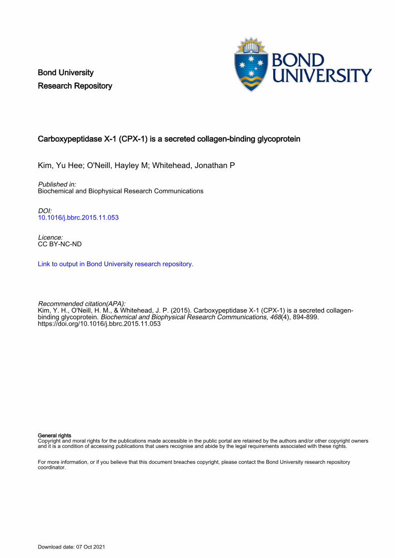
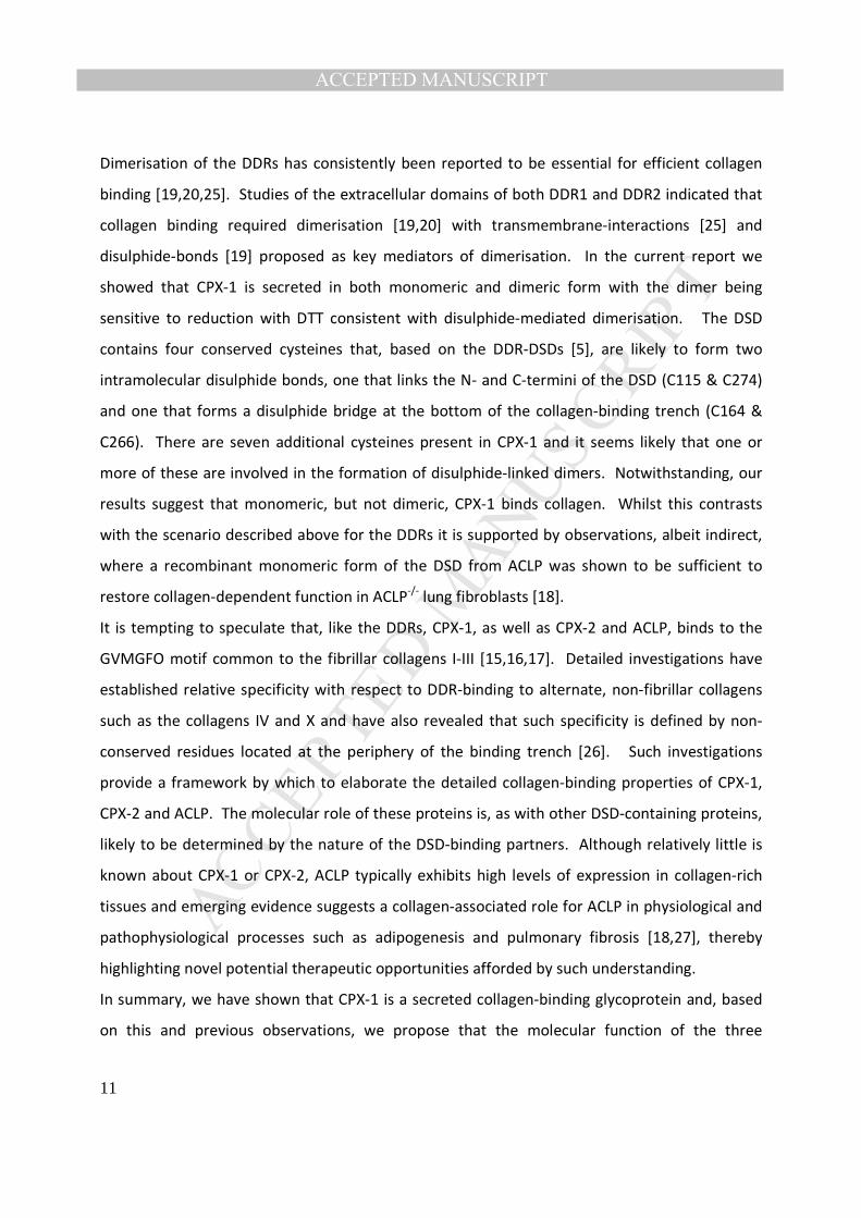
CPX-1 is secreted in an N-glycosylation dependent manner
MANUSCRIP
T
ACCEPTED
ACCEPTED MANUSCRIPT
6
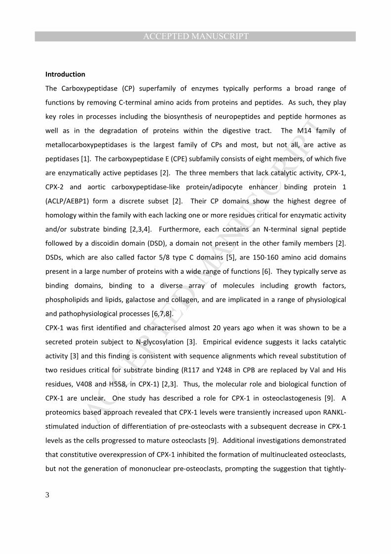
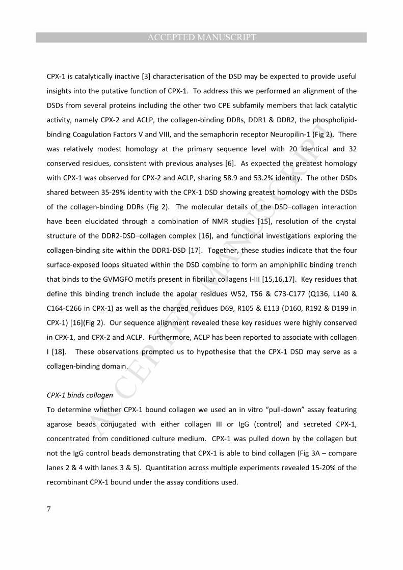
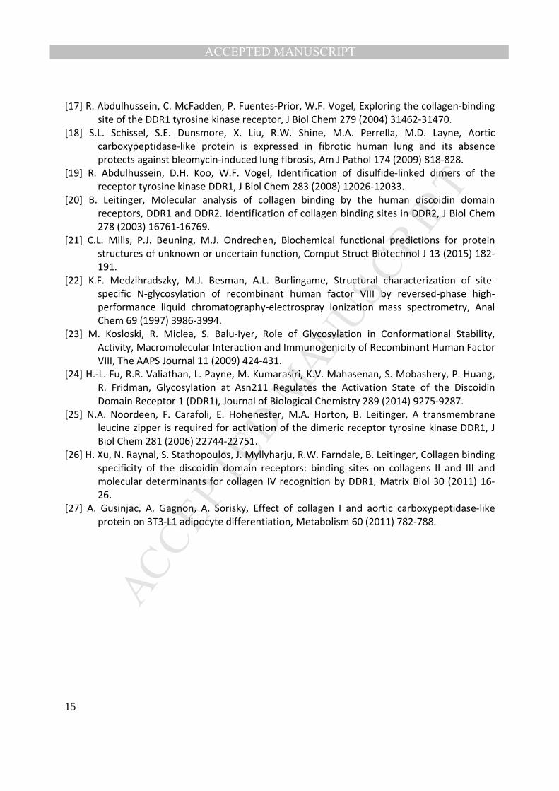
CPX-1 has been reported to be secreted in an N-glycosylated form [3]. In silico analysis revealed
4 putative N-glycosylation sites in human CPX-1 including one situated within the DSD (Fig 1A).
We reasoned that elucidation of the characteristics of N-glycosylation would increase our
understanding of CPX-1 at the molecular level. As N-glycosylation has been shown to be
essential for efficient secretion of some but not all N-glycosylated proteins [14] we first
determined whether N-glycosylation was required for efficient secretion of CPX-1. Human CPX-
1 was transiently expressed in CHO cells and intracellular and secreted CPX-1 was analysed by
Western blot. The predicted molecular weight of CPX-1 (based on amino acid sequence) is
80kDa. Intracellular CPX-1 migrated at 80 and 100kDa whilst secreted CPX-1, present in
conditioned medium, migrated predominantly at 120kDa, respectively (Fig 1B). These
observations are consistent with CPX-1 undergoing a series of post-translational modifications
(PTMs) during transit through the secretory pathway.
We used tunicamycin, an inhibitor of the enzyme N-acetylglucosamine phosphotransferase
which catalyses the first step of N-glycosylation, to investigate a putative role for N-
glycosylation in the secretion of CPX-1. Incubation of CPX-1 expressing cells with tunicamycin
for 3h or 24h selectively reduced the intensity of the 100kDa band, with only the 80kDa form of
intracellular CPX-1 detected after 24h (Fig 1C). Moreover, 24h incubation with tunicamycin
completely abolished secretion of CPX-1 (Fig 1D). We used PNGaseF, which cleaves N-linked
oligosaccharides, to complement and extend these observations. Treatment of cell lysates with
PNGaseF resulted in conversion of the intracellular 100kDa band to 80kDa (Fig 1E).
Interestingly, treatment of conditioned media with PNGaseF resulted in conversion of the
120kDa band to a band that migrated at around 100kDa but not 80kDa (Fig 1F). Collectively
these results demonstrate that N-glycosylation of CPX-1 is essential for efficient secretion and
also raise the possibility that secreted CPX-1 may be subject to additional, PNGaseF-
independent PTM.
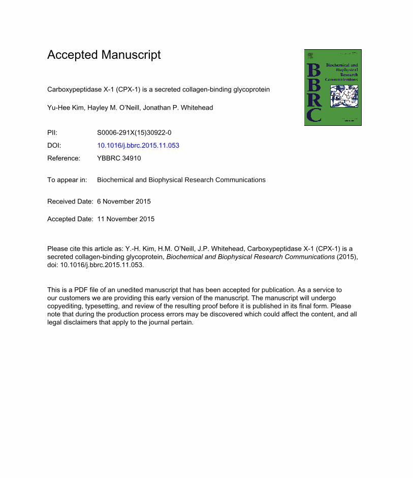
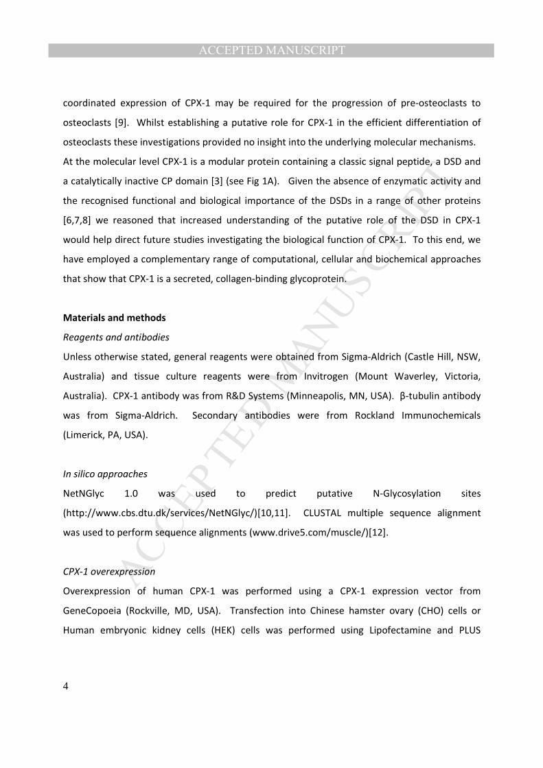
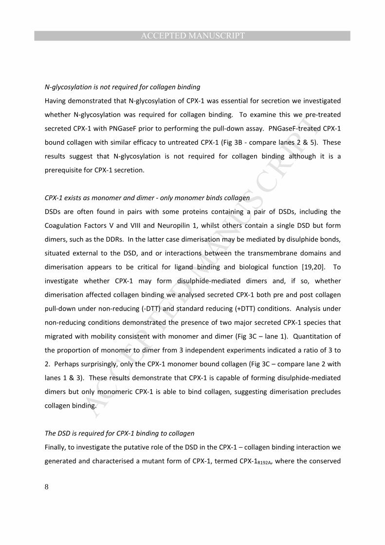
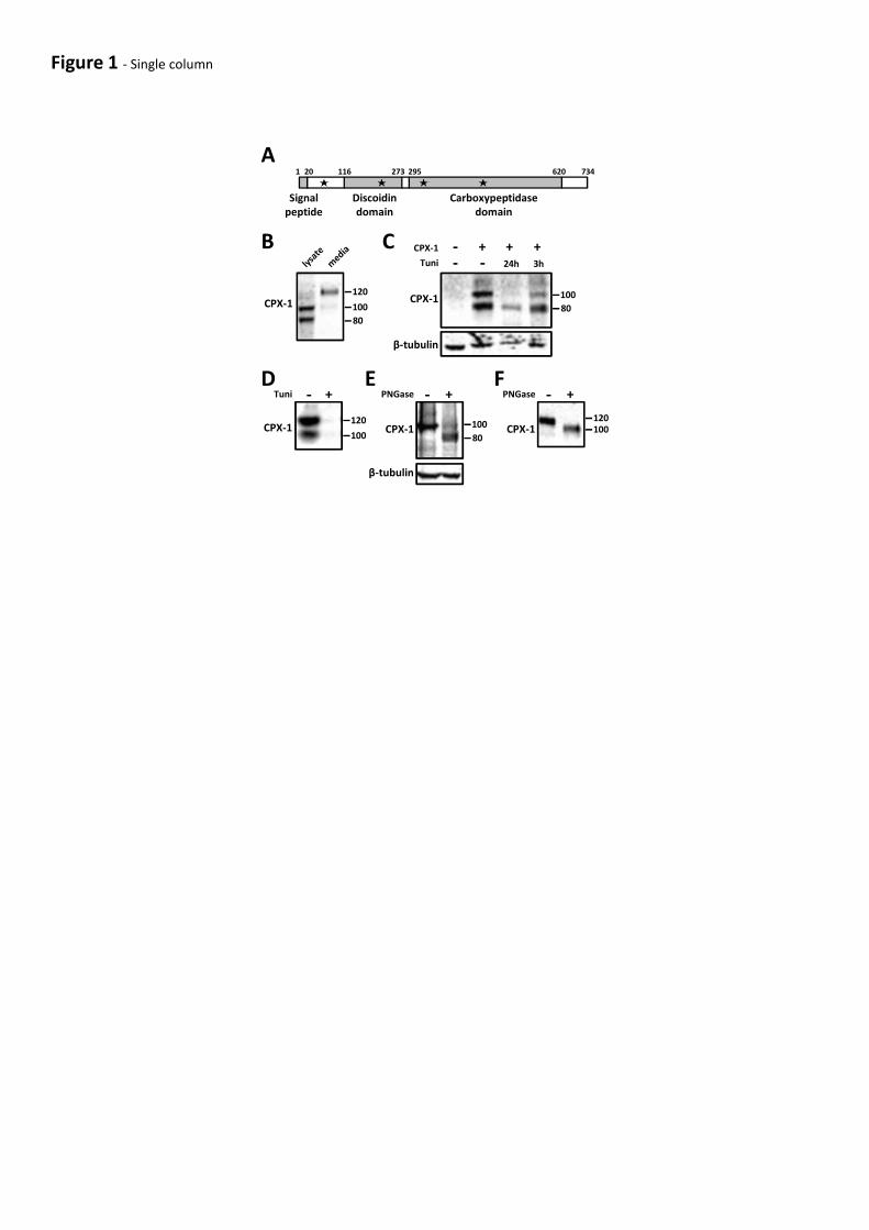
In silico analysis suggests the DSD of CPX-1 may bind collagen
In other proteins the DSD typically serves as a binding domain, where its biological role is
determined predominantly by the nature of the binding partner [6]. Given the CP domain of
MANUSCRIP
T
ACCEPTED
ACCEPTED MANUSCRIPT
7
CPX-1 is catalytically inactive [3] characterisation of the DSD may be expected to provide useful
insights into the putative function of CPX-1. To address this we performed an alignment of the
DSDs from several proteins including the other two CPE subfamily members that lack catalytic
activity, namely CPX-2 and ACLP, the collagen-binding DDRs, DDR1 & DDR2, the phospholipid-
binding Coagulation Factors V and VIII, and the semaphorin receptor Neuropilin-1 (Fig 2). There
was relatively modest homology at the primary sequence level with 20 identical and 32
conserved residues, consistent with previous analyses [6]. As expected the greatest homology
with CPX-1 was observed for CPX-2 and ACLP, sharing 58.9 and 53.2% identity. The other DSDs
shared between 35-29% identity with the CPX-1 DSD showing greatest homology with the DSDs
of the collagen-binding DDRs (Fig 2). The molecular details of the DSD–collagen interaction
have been elucidated through a combination of NMR studies [15], resolution of the crystal
structure of the DDR2-DSD–collagen complex [16], and functional investigations exploring the
collagen-binding site within the DDR1-DSD [17]. Together, these studies indicate that the four
surface-exposed loops situated within the DSD combine to form an amphiphilic binding trench
that binds to the GVMGFO motifs present in fibrillar collagens I-III [15,16,17]. Key residues that
define this binding trench include the apolar residues W52, T56 & C73-C177 (Q136, L140 &
C164-C266 in CPX-1) as well as the charged residues D69, R105 & E113 (D160, R192 & D199 in
CPX-1) [16](Fig 2). Our sequence alignment revealed these key residues were highly conserved
in CPX-1, and CPX-2 and ACLP. Furthermore, ACLP has been reported to associate with collagen
I [18]. These observations prompted us to hypothesise that the CPX-1 DSD may serve as a
collagen-binding domain.
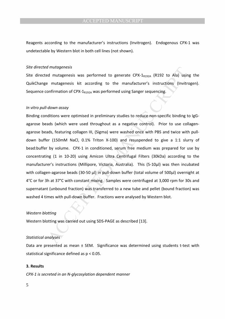
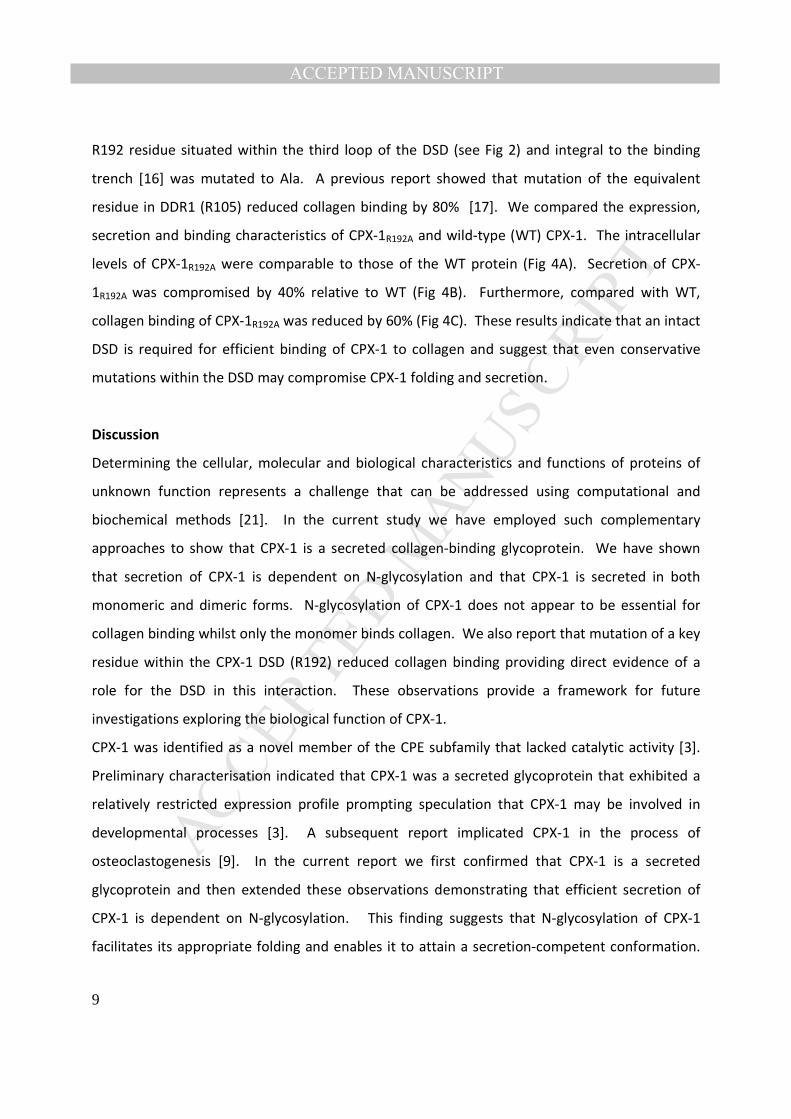
CPX-1 binds collagen
To determine whether CPX-1 bound collagen we used an in vitro “pull-down” assay featuring
agarose beads conjugated with either collagen III or IgG (control) and secreted CPX-1,
concentrated from conditioned culture medium. CPX-1 was pulled down by the collagen but
not the IgG control beads demonstrating that CPX-1 is able to bind collagen (Fig 3A – compare
lanes 2 & 4 with lanes 3 & 5). Quantitation across multiple experiments revealed 15-20% of the
recombinant CPX-1 bound under the assay conditions used.
MANUSCRIP
T
ACCEPTED
ACCEPTED MANUSCRIPT
8
N-glycosylation is not required for collagen binding
Having demonstrated that N-glycosylation of CPX-1 was essential for secretion we investigated
whether N-glycosylation was required for collagen binding. To examine this we pre-treated
secreted CPX-1 with PNGaseF prior to performing the pull-down assay. PNGaseF-treated CPX-1
bound collagen with similar efficacy to untreated CPX-1 (Fig 3B - compare lanes 2 & 5). These
results suggest that N-glycosylation is not required for collagen binding although it is a
prerequisite for CPX-1 secretion.
CPX-1 exists as monomer and dimer - only monomer binds collagen
DSDs are often found in pairs with some proteins containing a pair of DSDs, including the
Coagulation Factors V and VIII and Neuropilin 1, whilst others contain a single DSD but form
dimers, such as the DDRs. In the latter case dimerisation may be mediated by disulphide bonds,
situated external to the DSD, and or interactions between the transmembrane domains and
dimerisation appears to be critical for ligand binding and biological function [19,20]. To
investigate whether CPX-1 may form disulphide-mediated dimers and, if so, whether
dimerisation affected collagen binding we analysed secreted CPX-1 both pre and post collagen
pull-down under non-reducing (-DTT) and standard reducing (+DTT) conditions. Analysis under
non-reducing conditions demonstrated the presence of two major secreted CPX-1 species that
migrated with mobility consistent with monomer and dimer (Fig 3C – lane 1). Quantitation of
the proportion of monomer to dimer from 3 independent experiments indicated a ratio of 3 to
2. Perhaps surprisingly, only the CPX-1 monomer bound collagen (Fig 3C – compare lane 2 with
lanes 1 & 3). These results demonstrate that CPX-1 is capable of forming disulphide-mediated
dimers but only monomeric CPX-1 is able to bind collagen, suggesting dimerisation precludes
collagen binding.
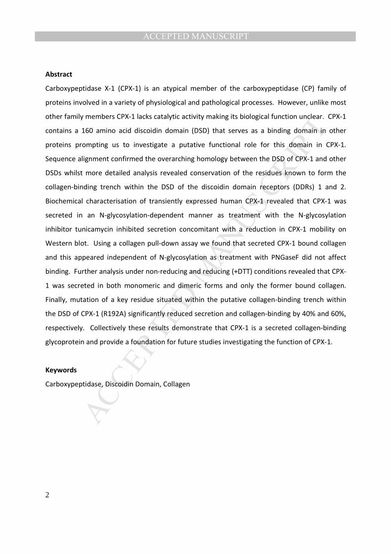
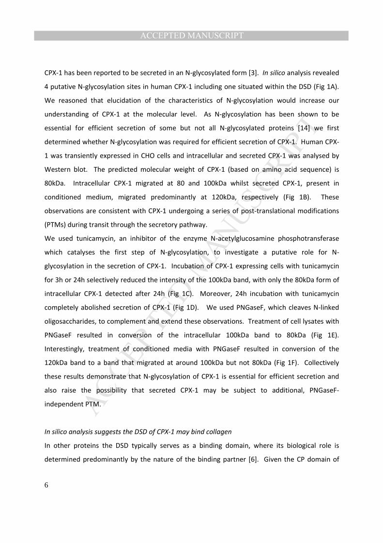
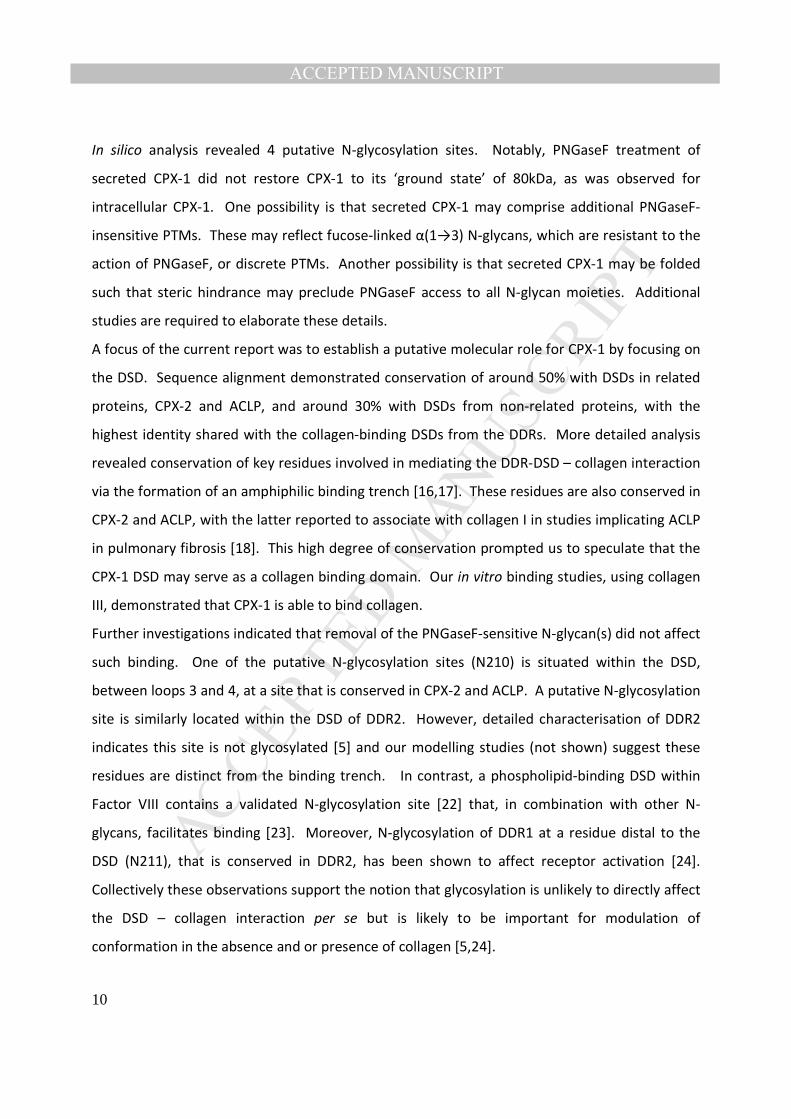
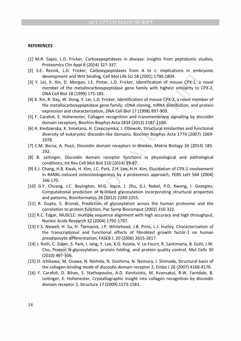
The DSD is required for CPX-1 binding to collagen
Finally, to investigate the putative role of the DSD in the CPX-1 – collagen binding interaction we
generated and characterised a mutant form of CPX-1, termed CPX-1R192A, where the conserved
MANUSCRIP
T
ACCEPTED
ACCEPTED MANUSCRIPT
9
R192 residue situated within the third loop of the DSD (see Fig 2) and integral to the binding
trench [16] was mutated to Ala. A previous report showed that mutation of the equivalent
residue in DDR1 (R105) reduced collagen binding by 80% [17]. We compared the expression,
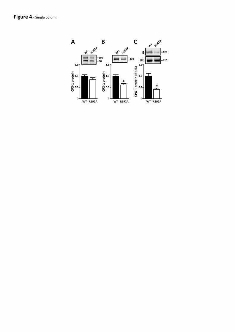
secretion and binding characteristics of CPX-1R192A and wild-type (WT) CPX-1. The intracellular
levels of CPX-1R192A were comparable to those of the WT protein (Fig 4A). Secretion of CPX-
1R192A was compromised by 40% relative to WT (Fig 4B). Furthermore, compared with WT,
collagen binding of CPX-1R192A was reduced by 60% (Fig 4C). These results indicate that an intact
DSD is required for efficient binding of CPX-1 to collagen and suggest that even conservative
mutations within the DSD may compromise CPX-1 folding and secretion.
Discussion
Determining the cellular, molecular and biological characteristics and functions of proteins of
unknown function represents a challenge that can be addressed using computational and
biochemical methods [21]. In the current study we have employed such complementary
approaches to show that CPX-1 is a secreted collagen-binding glycoprotein. We have shown
that secretion of CPX-1 is dependent on N-glycosylation and that CPX-1 is secreted in both
monomeric and dimeric forms. N-glycosylation of CPX-1 does not appear to be essential for
collagen binding whilst only the monomer binds collagen. We also report that mutation of a key
residue within the CPX-1 DSD (R192) reduced collagen binding providing direct evidence of a
role for the DSD in this interaction. These observations provide a framework for future
investigations exploring the biological function of CPX-1.
CPX-1 was identified as a novel member of the CPE subfamily that lacked catalytic activity [3].
Preliminary characterisation indicated that CPX-1 was a secreted glycoprotein that exhibited a
relatively restricted expression profile prompting speculation that CPX-1 may be involved in
developmental processes [3]. A subsequent report implicated CPX-1 in the process of
osteoclastogenesis [9]. In the current report we first confirmed that CPX-1 is a secreted
glycoprotein and then extended these observations demonstrating that efficient secretion of
CPX-1 is dependent on N-glycosylation. This finding suggests that N-glycosylation of CPX-1
facilitates its appropriate folding and enables it to attain a secretion-competent conformation.
MANUSCRIP
T
ACCEPTED
ACCEPTED MANUSCRIPT
10
In silico analysis revealed 4 putative N-glycosylation sites. Notably, PNGaseF treatment of
secreted CPX-1 did not restore CPX-1 to its ‘ground state’ of 80kDa, as was observed for
intracellular CPX-1. One possibility is that secreted CPX-1 may comprise additional PNGaseF-
insensitive PTMs. These may reflect fucose-linked α(1→3) N-glycans, which are resistant to the
action of PNGaseF, or discrete PTMs. Another possibility is that secreted CPX-1 may be folded
such that steric hindrance may preclude PNGaseF access to all N-glycan moieties. Additional
studies are required to elaborate these details.
A focus of the current report was to establish a putative molecular role for CPX-1 by focusing on
the DSD. Sequence alignment demonstrated conservation of around 50% with DSDs in related
proteins, CPX-2 and ACLP, and around 30% with DSDs from non-related proteins, with the
highest identity shared with the collagen-binding DSDs from the DDRs. More detailed analysis
revealed conservation of key residues involved in mediating the DDR-DSD – collagen interaction
via the formation of an amphiphilic binding trench [16,17]. These residues are also conserved in
CPX-2 and ACLP, with the latter reported to associate with collagen I in studies implicating ACLP
in pulmonary fibrosis [18]. This high degree of conservation prompted us to speculate that the
CPX-1 DSD may serve as a collagen binding domain. Our in vitro binding studies, using collagen
III, demonstrated that CPX-1 is able to bind collagen.
Further investigations indicated that removal of the PNGaseF-sensitive N-glycan(s) did not affect
such binding. One of the putative N-glycosylation sites (N210) is situated within the DSD,
between loops 3 and 4, at a site that is conserved in CPX-2 and ACLP. A putative N-glycosylation
site is similarly located within the DSD of DDR2. However, detailed characterisation of DDR2
indicates this site is not glycosylated [5] and our modelling studies (not shown) suggest these
residues are distinct from the binding trench. In contrast, a phospholipid-binding DSD within
Factor VIII contains a validated N-glycosylation site [22] that, in combination with other N-
glycans, facilitates binding [23]. Moreover, N-glycosylation of DDR1 at a residue distal to the
DSD (N211), that is conserved in DDR2, has been shown to affect receptor activation [24].
Collectively these observations support the notion that glycosylation is unlikely to directly affect
the DSD – collagen interaction per se but is likely to be important for modulation of
conformation in the absence and or presence of collagen [5,24].
MANUSCRIP
T
ACCEPTED
ACCEPTED MANUSCRIPT
11
Dimerisation of the DDRs has consistently been reported to be essential for efficient collagen
binding [19,20,25]. Studies of the extracellular domains of both DDR1 and DDR2 indicated that
collagen binding required dimerisation [19,20] with transmembrane-interactions [25] and
disulphide-bonds [19] proposed as key mediators of dimerisation. In the current report we
showed that CPX-1 is secreted in both monomeric and dimeric form with the dimer being
sensitive to reduction with DTT consistent with disulphide-mediated dimerisation. The DSD
contains four conserved cysteines that, based on the DDR-DSDs [5], are likely to form two
intramolecular disulphide bonds, one that links the N- and C-termini of the DSD (C115 & C274)
and one that forms a disulphide bridge at the bottom of the collagen-binding trench (C164 &
C266). There are seven additional cysteines present in CPX-1 and it seems likely that one or
more of these are involved in the formation of disulphide-linked dimers. Notwithstanding, our
results suggest that monomeric, but not dimeric, CPX-1 binds collagen. Whilst this contrasts
with the scenario described above for the DDRs it is supported by observations, albeit indirect,
where a recombinant monomeric form of the DSD from ACLP was shown to be sufficient to
restore collagen-dependent function in ACLP-/-
lung fibroblasts [18].
It is tempting to speculate that, like the DDRs, CPX-1, as well as CPX-2 and ACLP, binds to the
GVMGFO motif common to the fibrillar collagens I-III [15,16,17]. Detailed investigations have
established relative specificity with respect to DDR-binding to alternate, non-fibrillar collagens
such as the collagens IV and X and have also revealed that such specificity is defined by non-
conserved residues located at the periphery of the binding trench [26]. Such investigations
provide a framework by which to elaborate the detailed collagen-binding properties of CPX-1,
CPX-2 and ACLP. The molecular role of these proteins is, as with other DSD-containing proteins,
likely to be determined by the nature of the DSD-binding partners. Although relatively little is
known about CPX-1 or CPX-2, ACLP typically exhibits high levels of expression in collagen-rich
tissues and emerging evidence suggests a collagen-associated role for ACLP in physiological and
pathophysiological processes such as adipogenesis and pulmonary fibrosis [18,27], thereby
highlighting novel potential therapeutic opportunities afforded by such understanding.
In summary, we have shown that CPX-1 is a secreted collagen-binding glycoprotein and, based
on this and previous observations, we propose that the molecular function of the three
MANUSCRIP
T
ACCEPTED
ACCEPTED MANUSCRIPT
12
secreted, catalytically-inactive CPE subfamily of proteins, namely CPX-1, CPX-2 and ACLP, is
likely to be elaborated, at least in part, by defining the molecular details of the respective DSD-
collagen interaction.
Acknowledgements
Work was funded by grants from the Australian National Health & Medical Research Council
(JPW – 511104, 1047625; HMON - 1074074) and a University of Queensland-International
Postgraduate Research Scholarship (YHK). The authors thank Johanna Barclay and John Hooper
for discussions and critical reading of the manuscript.
Figure Legends
Figure 1 – N-glycosylation of CPX-1 is required for secretion. (A) Schematic showing
organisation of CPX-1 and location of putative N-glycosylation sites (stars) at residues 57, 210,
318 and 472. (B) CPX-1 was transiently expressed in CHO cells and intracellular (lysate) and
secreted (media) CPX-1 was analysed by Western blot. (C & D) CPX-1 expressing CHO cells were
treated with tunicamycin (Tuni - 10 μg/ml) for 24h or 3h and intracellular and secreted CPX-1
was analysed by Western blot. (E & F) Intracellular and secreted CPX-1 were treated with
PNGaseF (2.5 units for 2h) and analysed by Western blot. Blots are representative of 3
independent experiments.
Figure 2 – In silico analysis of the DSD within CPX-1. Multiple sequence alignment of the DSD
in human CPX-1, CPX-2, ACLP, DDR1, DDR2, Factor V, Neuropilin I and Factor VIII in decreasing
order of homology. Loop regions (1-4) are boxed in grey. Conserved residues that form the
collagen-binding trench are boxed in yellow. Putative N-glycosylation sites are underlined in
green. The degree of conservation across all 8 DSDs is denoted as (*) identical, (:) highly
conserved and (.) conserved; Additional conservation across CPX-1/2, ACLP & DDR1/2 is
denoted as (*, : and .) respectively. Numbers in parentheses depict % identity with CPX-1.
Figure 3 – CPX-1 binds collagen. (A) Secreted CPX-1 was incubated with collagen-agarose beads
(col), or IgG-agarose beads (IgG) as control, and bound (B) and unbound (UB) material was
MANUSCRIP
T
ACCEPTED
ACCEPTED MANUSCRIPT
13
analysed by Western blot. ST – starting material. (B) As in (A) except samples were pre-treated
with PNGaseF as indicated prior to incubation with the collagen-agarose beads. (C) As in (A)
except following incubation with the collagen-agarose beads both bound and unbound samples
were treated -/+ DTT prior to Western blot. Blots are representative of 3 independent
experiments.
Figure 4 – Mutation of the CPX-1 DSD compromises collagen binding. (A & B) Wild-type CPX-1
(WT) and CPX-1R192A (R192A) were transiently expressed in HEK cells and intracellular (cell
lysates - A) and secreted (conditioned media - B) CPX-1 was analysed by Western blot. (C)
Secreted WT and mutant CPX-1 were incubated with collagen agarose beads and bound (B) and
unbound (UB) material was analysed by Western blot. Graphs show quantitation from 6
independent experiments. *p<0.05 WT cf R192A.
MANUSCRIP
T
ACCEPTED
ACCEPTED MANUSCRIPT
14
REFERENCES
[1] M.R. Sapio, L.D. Fricker, Carboxypeptidases in disease: insights from peptidomic studies,
Proteomics Clin Appl 8 (2014) 327-337.
[2] S.E. Reznik, L.D. Fricker, Carboxypeptidases from A to z: implications in embryonic
development and Wnt binding, Cell Mol Life Sci 58 (2001) 1790-1804.
[3] Y. Lei, X. Xin, D. Morgan, J.E. Pintar, L.D. Fricker, Identification of mouse CPX-1, a novel
member of the metallocarboxypeptidase gene family with highest similarity to CPX-2,
DNA Cell Biol 18 (1999) 175-185.
[4] X. Xin, R. Day, W. Dong, Y. Lei, L.D. Fricker, Identification of mouse CPX-2, a novel member of
the metallocarboxypeptidase gene family: cDNA cloning, mRNA distribution, and protein
expression and characterization, DNA Cell Biol 17 (1998) 897-909.
[5] F. Carafoli, E. Hohenester, Collagen recognition and transmembrane signalling by discoidin
domain receptors, Biochim Biophys Acta 1834 (2013) 2187-2194.
[6] A. Kiedzierska, K. Smietana, H. Czepczynska, J. Otlewski, Structural similarities and functional
diversity of eukaryotic discoidin-like domains, Biochim Biophys Acta 1774 (2007) 1069-
1078.
[7] C.M. Borza, A. Pozzi, Discoidin domain receptors in disease, Matrix Biology 34 (2014) 185-
192.
[8] B. Leitinger, Discoidin domain receptor functions in physiological and pathological
conditions, Int Rev Cell Mol Biol 310 (2014) 39-87.
[9] E.J. Chang, H.B. Kwak, H. Kim, J.C. Park, Z.H. Lee, H.H. Kim, Elucidation of CPX-1 involvement
in RANKL-induced osteoclastogenesis by a proteomics approach, FEBS Lett 564 (2004)
166-170.
[10] G.Y. Chuang, J.C. Boyington, M.G. Joyce, J. Zhu, G.J. Nabel, P.D. Kwong, I. Georgiev,
Computational prediction of N-linked glycosylation incorporating structural properties
and patterns, Bioinformatics 28 (2012) 2249-2255.
[11] R. Gupta, S. Brunak, Prediction of glycosylation across the human proteome and the
correlation to protein function, Pac Symp Biocomput (2002) 310-322.
[12] R.C. Edgar, MUSCLE: multiple sequence alignment with high accuracy and high throughput,
Nucleic Acids Research 32 (2004) 1792-1797.
[13] F.S. Newell, H. Su, H. Tornqvist, J.P. Whitehead, J.B. Prins, L.J. Hutley, Characterization of
the transcriptional and functional effects of fibroblast growth factor-1 on human
preadipocyte differentiation, FASEB J. 20 (2006) 2615-2617.
[14] J. Roth, C. Zuber, S. Park, I. Jang, Y. Lee, K.G. Kysela, V. Le Fourn, R. Santimaria, B. Guhl, J.W.
Cho, Protein N-glycosylation, protein folding, and protein quality control, Mol Cells 30
(2010) 497-506.
[15] O. Ichikawa, M. Osawa, N. Nishida, N. Goshima, N. Nomura, I. Shimada, Structural basis of
the collagen-binding mode of discoidin domain receptor 2, Embo J 26 (2007) 4168-4176.
[16] F. Carafoli, D. Bihan, S. Stathopoulos, A.D. Konitsiotis, M. Kvansakul, R.W. Farndale, B.
Leitinger, E. Hohenester, Crystallographic insight into collagen recognition by discoidin
domain receptor 2, Structure 17 (2009) 1573-1581.
MANUSCRIP
T
ACCEPTED
ACCEPTED MANUSCRIPT
15
[17] R. Abdulhussein, C. McFadden, P. Fuentes-Prior, W.F. Vogel, Exploring the collagen-binding
site of the DDR1 tyrosine kinase receptor, J Biol Chem 279 (2004) 31462-31470.
[18] S.L. Schissel, S.E. Dunsmore, X. Liu, R.W. Shine, M.A. Perrella, M.D. Layne, Aortic
carboxypeptidase-like protein is expressed in fibrotic human lung and its absence
protects against bleomycin-induced lung fibrosis, Am J Pathol 174 (2009) 818-828.
[19] R. Abdulhussein, D.H. Koo, W.F. Vogel, Identification of disulfide-linked dimers of the
receptor tyrosine kinase DDR1, J Biol Chem 283 (2008) 12026-12033.
[20] B. Leitinger, Molecular analysis of collagen binding by the human discoidin domain
receptors, DDR1 and DDR2. Identification of collagen binding sites in DDR2, J Biol Chem
278 (2003) 16761-16769.
[21] C.L. Mills, P.J. Beuning, M.J. Ondrechen, Biochemical functional predictions for protein
structures of unknown or uncertain function, Comput Struct Biotechnol J 13 (2015) 182-
191.
[22] K.F. Medzihradszky, M.J. Besman, A.L. Burlingame, Structural characterization of site-
specific N-glycosylation of recombinant human factor VIII by reversed-phase high-
performance liquid chromatography-electrospray ionization mass spectrometry, Anal
Chem 69 (1997) 3986-3994.
[23] M. Kosloski, R. Miclea, S. Balu-Iyer, Role of Glycosylation in Conformational Stability,
Activity, Macromolecular Interaction and Immunogenicity of Recombinant Human Factor
VIII, The AAPS Journal 11 (2009) 424-431.
[24] H.-L. Fu, R.R. Valiathan, L. Payne, M. Kumarasiri, K.V. Mahasenan, S. Mobashery, P. Huang,
R. Fridman, Glycosylation at Asn211 Regulates the Activation State of the Discoidin
Domain Receptor 1 (DDR1), Journal of Biological Chemistry 289 (2014) 9275-9287.
[25] N.A. Noordeen, F. Carafoli, E. Hohenester, M.A. Horton, B. Leitinger, A transmembrane
leucine zipper is required for activation of the dimeric receptor tyrosine kinase DDR1, J
Biol Chem 281 (2006) 22744-22751.
[26] H. Xu, N. Raynal, S. Stathopoulos, J. Myllyharju, R.W. Farndale, B. Leitinger, Collagen binding
specificity of the discoidin domain receptors: binding sites on collagens II and III and
molecular determinants for collagen IV recognition by DDR1, Matrix Biol 30 (2011) 16-
26.
[27] A. Gusinjac, A. Gagnon, A. Sorisky, Effect of collagen I and aortic carboxypeptidase-like
protein on 3T3-L1 adipocyte differentiation, Metabolism 60 (2011) 782-788.
MANUSCRIP
T
ACCEPTED
ACCEPTED MANUSCRIPTFigure 1 - Single column
A
B C
Carboxypeptidase
domain
Discoidin
domain
Signal
peptide
1 20 116 273 295 620 734
80
100
120
CPX-1
-
80
100CPX-1
β-tubulin
CPX-1
Tuni -+ + +
- 24h 3h
FD ETuni -
CPX-1100
120CPX-1
β-tubulin
80
100
PNGase - +
100
120
CPX-1
PNGase - ++
MANUSCRIP
T
ACCEPTED
ACCEPTED MANUSCRIPTFigure 2 - Double column
115 Loop 1 Loop 2 CPX-1 CP-PLGLESLRVSDSRLEASSSQSFGLGPHRGRLNIQSGLEDGDLYDGAWCAE----EQD CPX-2 CP-PLGLETLKITDFQLHASTVKRYGLGAHRGRLNIQAGINENDFYDGAWCAG----RND ACLP CP-PIGMESHRIEDNQIRASSMLRHGLGAQRGRLNMQTGATEDDYYDGAWCAE----DDA DDR2 CRYPLGMSGGQIPDEDITASSQWSESTAAKYGRLDSEEG-------DGAWCPEIPVEPDD DDR1 CRYALGMQDRTIPDSDISASSSWSDSTAARHSRLESSDG-------DGAWCPAGSVFPKE Factor V(1) CRMPMGLSTGIISDSQIKASEFLGY-WEPRLARLNNGGS-------YNAWSVEKLAAEFA Neuropilin 1(1) CMEALGMESGEIHSDQITASSQYSTNWSAERSRLNYP---------ENGWTPG----EDS Factor VIII(1) CQTPLGMASGHIRDFQITASGQYGQ-WAPKLARLHYSGS-------INAWS------TKE * .:*: : * : **: . ... .**: . * *****. .
170 Loop 3 CPX-1 ADPWFQVDAGHPTRFSGVITQGRNSVWRY-D-WVTSYKVQFSNDSRTWWGSRNHSSGMDA CPX-2 LQQWIEVDARRLTRFTGVITQGRNSLWLS-D-WVTSYKVMVSNDSHTWVTVKNGSGDM-- ACLP RTQWIEVDTRRTTRFTGVITQGRDSSIHD-D-FVTTFFVGFSNDSQTWVMYTNGYEEM-- DDR2 LKEFLQIDLHTLHFITLVGTQGRHAGGHGIE-FAPMYKINYSRDGTRWISWRNRHGKQ-- DDR1 -EEYLQVDLQRLHLVALVGTQGRHAGGLGKE-FSRSYRLRYSRDGRRWMGWKDRWGQE-- Factor V(1) SKPWIQVDMQKEVIITGIQTQGAKHYLKSC--YTTEFYVAYSSNQINWQIFKGNSTRNVM Neuropilin 1(1) YREWIQVDLGLLRFVTAVGTQGAISKETKKKYYVKTYKIDVSSNGEDWITIKEGNKPV-- Factor VIII(1) PFSWIKVDLLAPMIIHGIKTQGARQKFSSL--YISQFIIMYSLDGKKWQTYRGNSTGTLM ::::* .: : **** : : : : *.:. * .
230 Loop 4 274 (% identity) CPX-1 VFPANSDPETPVLNLLPEPQVARFIRLLPQTWLQGGAPCLRAEILAC (100.0) CPX-2 IFEGNSEKEIPVLNELPVPMVARYIRINPQSWFDNGSICMRMEILGC (58.9) ACLP TFHGNVDKDTPVLSELPEPVVARFIRIYPLTW--NGSLCMRLEVLGC (53.2) DDR2 VLDGNSNPYDIFLKDLEPPIVARFVRFIPVTD-HSMNVCMRVELYGC (34.7) DDR1 VISGNEDPEGVVLKDLGPPMVARLVHFYPRAD-RVMSVCLRVELYGC (34.2) Factor V(1) YFNGNSDASTIKENQFDPPIVARYIRISPTRA--YNRPTLRLELQGC (32.7) Neuropilin 1(1) LFQGNTNPTDVVVAVFPKPLITRFVRIKPATW--ETGISMRFEVYGC (30.6) Factor VIII(1) VFFGNVDSSGIKHNIFNPPIIARYIRLHPTHY--SIRSTLRMELMGC (29.1) : .* : .*. : * ::* :.: * : *:* *: .*
MANUSCRIP
T
ACCEPTED
ACCEPTED MANUSCRIPTFigure 3 - Single column
A
B
C
120
Col bead - +IgG bead
Material ST B B B B UB UB
- - - -+ +
++ +- - -
Lane 1 2 3 4 5 6 7
100120
PNGase
Material ST B UB ST B UB
- - - ++ +
Lane 1 2 3 4 5 6
120
240
DTT
Material ST B UB ST B UB
- - - ++ +
Lane 1 2 3 4 5 6
MANUSCRIP
T
ACCEPTED
ACCEPTED MANUSCRIPTFigure 4 - Single column
A
1.5
1.0
0.5
0
WT R192A
CP
X-1
pro
tein
80
100
B
1.5
1.0
0.5
0
WT R192AC
PX
-1 p
rote
in
120
*
C
1.0
1.5
120
*
B 120
UB
0.5
0
WT R192A
CP
X-1
pro
tein
(B
:UB
)
MANUSCRIP
T
ACCEPTED
ACCEPTED MANUSCRIPT
Highlights
• The discoidin domain-containing protein CPX-1 is secreted o in an N-glycosylation dependent manner o in monomeric and dimeric forms
• CPX-1 binds collagen o in an N-glycosylation independent manner o in monomeric form only
• Mutation of the discoidin domain compromises collagen binding
![CPX terminal - Festo Electronics CPX field bus node Type CPX-FB32 Fieldbus protocol EtherNet/IP CPX terminal Manual 541 305 en 1111a [761 331] Contents and general instructions Festo](https://img.pdfslide.us/doc/110x75/5abd6a997f8b9aa15e8b8c21/cpx-terminal-festo-electronics-cpx-field-bus-node-type-cpx-fb32-fieldbus-protocol.jpg)
![Terminal CPX Bus node CPX-FB14 - Festo USA · Description CANopen network protocol 526410 en 1411d [8041138] Terminal CPX Bus node CPX-FB14](https://img.pdfslide.us/doc/110x75/5b4c9fef7f8b9ad1338b9f4c/terminal-cpx-bus-node-cpx-fb14-festo-usa-description-canopen-network-protocol.jpg)


![CPX terminal · 2020-03-11 · Electronics manual CPX−Front−End Controller Type CPX−FEC CPX terminal Manual 538 475 en 0404NH [677 480]](https://img.pdfslide.us/doc/110x75/5f0a30077e708231d42a6f3f/cpx-terminal-2020-03-11-electronics-manual-cpxafrontaend-controller-type-cpxafec.jpg)


![Terminal CPX Nœudde bus CPX-(M)-FB33/34/35 · Description Protocole réseau PROFINET IO 548762 fr 1407c [8032735] Terminal CPX Nœudde bus CPX-(M)-FB33/34/35](https://img.pdfslide.us/doc/110x75/5f1579b44f77305bca353355/terminal-cpx-nudde-bus-cpx-m-fb333435-description-protocole-rseau-profinet.jpg)
![Terminale CPX - festo.com · Manual elettronica NodoFieldbus CPX Tipo CPX-FB14 Protocollo busdi campoCANopen Terminale CPX Beschreibung 526 413 it 0206NH [653635]](https://img.pdfslide.us/doc/110x75/5e0d4aa4b6306836287c1f72/terminale-cpx-festocom-manual-elettronica-nodofieldbus-cpx-tipo-cpx-fb14-protocollo.jpg)


![CPX Terminal...Manual electronics CPX control block Type CPX−SF34 Type CPX−SF35 PCWORX integrated Network protocol PROFINET IO CPX Terminal Manual 570 541 en 1007NH [748 167] Contents](https://img.pdfslide.us/doc/110x75/5f0378aa7e708231d4094b43/cpx-terminal-manual-electronics-cpx-control-block-type-cpxasf34-type-cpxasf35.jpg)