Embed Size (px)
Citation preview

NANO EXPRESS Open Access
Sn2+ Doping: A Strategy for Tuning ofFe3O4 Nanoparticles Magnetization DippingTemperature/Amplitude, Irreversibility, andCurie PointUmaima S. H. Al-Kindi1, Salim H. Al-Harthi1*, Hisham M. Widatallah1, Mohamed E. Elzain1, Myo T. Z. Myint1 andHtet H. Kyaw2
Abstract
Doped magnetite (SnxFe3-2/3xO4) nanoparticles (NPs) (12–50 nm) with different amount of Sn2+ ions (x) weresynthesized using co-precipitation method. Sn2+ doping reduces the anticipated oxidation of Fe3O4 NPs tomaghemite (γ-Fe2O3), making them attractive in several magnetic applications. Detailed characterizations duringheating–cooling cycles revealed the possibility of tuning the unusual observed magnetization dipping temperature/amplitude, irreversibility, and Curie point of these NPs. We attribute this dip to the chemical reduction of γ-Fe2O3 atthe NPs surfaces. Along with an increase in the dipping temperature, we found that doping with Sn2+ reduces thedipping amplitude, until it approximately disappears when x = 0.150. Based on the core-shell structure of these NPs,a phenomenological expression that combines both modified Bloch law (M = M0[1 − γ(T/TC)]β) and a modifiedCurie–Weiss law (M = − α[1/(T − TC)
δ]) is developed in order to explain the observed M-T behavior at differentapplied external magnetic fields and for different Sn2+ concentrations. By applying high enough magnetic field, thevalue of the parameters γ and δ ≈ 1 which are the same in modified Bloch and Curie–Weiss laws. They do notchange with the magnetic field and depend only on the material structure and size. The power β for highmagnetic field was 2.6 which is as expected for this size of nanoparticles with the core dominated magnetization.However, the β value fluctuates between 3 and 10 for small magnetic fields indicating an extra magneticcontribution from the shell structure presented by Curie–Weiss term. The parameter (α) has a very small value andit turns to negative values for high magnetic fields.
Keywords: Curie, Dipping, Irreversible, Maghemite, Magnetite, Modified Bloch law
IntroductionMetal oxide nanoparticles are attractive from both tech-nical and theoretical perspective. Among them, iron oxidenanoparticles are very popular due to their massive appli-cations in the fields of ferrofluids, pigments, informationstorage disks, and medical applications as magneticallyguided drug delivery, cell separation, and cancer diagnoses
[1–9]. Magnetite (Fe3O4) nanoparticles are particularlywell suited for medical applications, due to their biologicalcompatibility and the large saturation magnetization (Ms)of 92 emu/g at 300 K for the bulk [10, 11]. However, thethermal instability of these nanoparticles can be a draw-back for these applications since nanoparticles with size of~ 8–22 nm can be easily oxidized to maghemite (γ-Fe2O3)even at ambient conditions of temperature and pressure—although the bulk can be accomplished at ~ 220 °C [12].Maghemite is a ferrimagnetic material like magnetite withthe same spinel structure but with lower Ms of 78 emu/g
© The Author(s). 2020 Open Access This article is licensed under a Creative Commons Attribution 4.0 International License,which permits use, sharing, adaptation, distribution and reproduction in any medium or format, as long as you giveappropriate credit to the original author(s) and the source, provide a link to the Creative Commons licence, and indicate ifchanges were made. The images or other third party material in this article are included in the article's Creative Commonslicence, unless indicated otherwise in a credit line to the material. If material is not included in the article's Creative Commonslicence and your intended use is not permitted by statutory regulation or exceeds the permitted use, you will need to obtainpermission directly from the copyright holder. To view a copy of this licence, visit http://creativecommons.org/licenses/by/4.0/.
* Correspondence: [email protected] of Physics, College of Science, Sultan Qaboos University, P.O.Box 36, Al-Khoudh, Muscat 123, Sultanate of OmanFull list of author information is available at the end of the article
Al-Kindi et al. Nanoscale Research Letters (2020) 15:192 https://doi.org/10.1186/s11671-020-03423-9

at 300 K [10]. By heating up to about 850 K (Curie point),Fe3O4 can be structurally changed to the antiferromag-netic corundum-like structure hematite with zero Ms [13].These transformations are controlled by particle size,temperature, and pressure. Scarce studies are made forFe3O4 particles at high temperature because of thermal in-stability. Recently more attention has been given to the ef-fects of organic capping—such as an oleate-capped Fe3O4
nanoparticles—on the magnetization of the nanoparticles(NPs) [14]. It was found that, in heating–cooling cycles,Fe3O4 NPs exhibited irreversible M behavior with two pe-culiar effects, namely, dips and loops in theirM (T) curves.The dipping and the irreversible magnetization were at-tributed to the induced reduction of Fe3+ to Fe2+ and sin-tering upon the decomposition of the capping ligandsrespectively. Our intention in this study is to thoroughlyunderstand the cause of these peculiar effects, their na-ture, stability, effects on magnetization, and surface reduc-tion of Fe3+ to Fe2+ and their relationship with sinteringprocess of the NPs at elevated temperatures. Motivated bythe fact that Fe3O4 NPs can be easily oxidized to form γ-Fe2O3 shell (i.e., thin layer thereafter called a shell) on thesurface which acts as a capping layer and exploiting theknowledge that doping Fe3O4 with certain ions like Sn4+
and Ti4+ shows a decrease in the Fe3+ to Fe2+ reductionprocess [15, 16], we therefore explore the possibility oftuning those peculiar effects (i.e., dipping and loops) intemperature dependent magnetization curves by Sn2+
doping of Fe3O4 NPs.In order to study the effect of Sn2+ doping on the sta-
bility of magnetite nanoparticles, magnetization dipping,and irreversibility at high temperatures, SnxFe3-2/3xO4
nanoparticles (12–50 nm) with (x = 0.000, 0.045, 0.090,and 0.150), were prepared and characterized using sev-eral complementary techniques. The magnetization wasmeasured using a vibrating sample magnetometer(VSM) while repeatedly heating the sample up to 900 K(5 K/min) and cooling back to room temperature (300K). An irreversible dip in magnetization was noticed at aspecific temperature and with certain amplitude duringthe first heating–cooling cycle. Evidences of the changein dipping temperature, and amplitude, irreversibility, di-vergence in magnetization (i.e., magnetization values aredifferent at specific temperature in heating and coolingcycles) and Curie point with x were observed and ex-plained. Contrary to the explanation that the observedirreversibility in heating–cooling regime can only be ex-pected for the ligand-free Fe3O4 NPs, we show that di-vergence can be controlled by the external magneticfield applied to the Fe3O4 NPs during the magnetic mea-surements and disappears at higher applied field. Fur-thermore, we show that the M-T of the pristine and theSn2+-doped Fe3O4 NPs after the first heating–coolingcycle can be predicted by a new approach that combines
both a modified Bloch and Curie–Weiss laws for differ-ent Sn2+ concentrations and different applied externalmagnetic fields.
Methods/ExperimentalMaterialsAqueous ammonia (Mw = 17.03, 30%) and absoluteethanol were purchased from Merck, ferric chloridehexahydrate (Mw = 270.3, ≥ 99%) and ferrous chloridetetrahydrate (Mw = 198.8, ≥ 99%) were procured fromSigma-Aldrich, and stannous chloride (Mw = 189.60, ≥98%) was obtained from Fluka. All the chemicals wereused without further purification.
MethodsNanoparticles of Sn2+ doped Fe3O4 with the nominalcomposition SnxFe3-2/3xO4 (x = 0.000, 0.045, 0.090, and0.150), where Sn2+ substitutes Fe3+, were prepared usingco-precipitation under reflux at 80 °C for 4 h. Aqueousammonia was added to stoichiometric solutions of ferricchloride hexahydrate, ferrous chloride tetrahydrate, andstannous chloride at 50 °C until a pH ≈ 10.4 wasattained. The precipitates were then removed by filtra-tion, washed with distilled water followed by ethanol,and very carefully dried at room temperature avoidinghigh temperature that would result in the formation ofSn-doped maghemite as was demonstrated by Berryet al. [16].The surface of pristine Fe3O4 nanoparticles was
covered with 2 nm layer of gold (99.99% gold target,Scotech) using e-beam evaporation (deposition rate ~0.47 Å/s) attached to the Nanosys 550 nanoparticle de-position system from Mantis Deposition Ltd. in order toexamine the surface effect.
CharacterizationsA VSM attached to a quantum design physical propertymeasurement system (Dynacool PPMS) was used formagnetic measurements at temperature ranging from 2to 900 K with magnetic fields up to 9 (Tesla). Curiepoint was taken by the extrapolation of M curve to thex-axis during the first heating regime following theprocedure used in reference [17]. The morphology ofthe samples was characterized using JOEL digital high-resolution (JEN-2100F) transmission electron micro-scope (HRTEM) and (X’Pert PRO) diffractometer for X-ray powder diffraction (XRD) patterns using a standardCu-Kα radiation. The MAUD software was used to per-form simple XRD Rietveld refinements [18]. Elementalmapping (EDX) was carried out using field emissionscanning electron microscope (JOEL, JSM 7600F). X-rayphotoemission spectra (XPS) were acquired using Omi-cron Nanotechnology multiprobe photoelectron instru-ment equipped with a hemispherical electron analyzer
Al-Kindi et al. Nanoscale Research Letters (2020) 15:192 Page 2 of 14

where Al Kα radiation (1486.6 eV) was used at 10−9
mbar. Intrinsic carbon peak at 284.6 eV was employedfor calibration. The Casa XPS software was used for XPSdata analysis [19]. Fourier transform infrared (FTIR)spectrum was obtained from PerkinElmer (SpectraOne)using transmission mode with KBr pellets in the rangeof 400–4000 cm−1.
Results and DiscussionThe Main Features of M-T Curves During the First HeatingCycleFigure 1a–d shows the change of magnetization (M) as afunction of temperature of the samples; pristine Fe3O4-and tin–doped SnxFe3-2/3xO4 nanoparticles with differentamount of x. The samples were heated from 300 up to900 K (Fig. 1 point A to B) and cooled back (point B toC) for the first heating–cooling cycle while applying anexternal magnetic field of 200 Oe. The heating–coolingcycle’s measurements as depicted from curves D to Ewere repeated under same magnetic field until stablemagnetization data was reached. The pristine Fe3O4
nanoparticles (Fig. 1a) undergo heating–cooling cycle forfive times. For clarity, we only present three cycles sinceafter that there were no more changes in magnetizationduring heating–cooling process. The doped samples(Fig. 1b–d) were heated and cooled for three times onlysince there was no obvious change in M after the secondcycle (two cycles are presented in the figures). Fourobvious features were noticed where temperature isranging from 300 to 900 K. First, there is a dip inmagnetization of about 10 emu/g that occurred in thepristine sample (x = 0.000) between T1 (564 K) and T2
(655 K), while going from point A to B in the firstheating–cooling cycle. This dip also occurred in thedoped samples but with increased dipping temperatures(T1, T2) as x increases (Fig. 2a). This increase may beattributed to the increase in particle size due to Sn-doping as confirmed by HRTEM measurements shownin Fig. S1. To ensure that Sn2+ ions spread uniformlythroughout the structure, an elemental mapping for thepure and SnxFe3-2x/3O4 doped sample with x = 0.150(Figs. S2 and S3).Similar dipping was also reported as mentioned above
in oleate-capped magnetite nanoparticles with 20 nm insize which is attributed to the thermal decomposition ofthe capping ligands. Along with the decomposition, a re-duction of Fe3+ to Fe2+ after heating was also observedusing Raman and Mössbauer spectroscopy [14].Interestingly, the dipping feature was not detected in
the un-capped Fe3O4 sample reported by Kolen’ko et al.[14]. Although, there were no capping ligands used inour sample’s preparation, the surface of nanoparticleswas influenced by oxidation either to maghemite (γ-Fe2O3) or Sn2+-related oxides, both of which could actas a capping layer. Consequently, the dipping of M inthe first heating–cooling cycles indicates that there wasa thermal decomposition of the oxidized layer on thesurface of these nanoparticles (i.e., a reduction of Fe3+
and Sn2+, Sn 4+ ions).This decomposition will take placeat lower temperature for smaller particles due to theirlarger specific surface area. This explanation is sup-ported by a previously reported reduction of amorphousγ-Fe2O3 nanoparticles in an evacuated environment at523 K [20]. The second observed feature is related to the
Fig. 1 Change of magnetization (M) with temperature of pristine and SnxFe3-2/3xO4 nanoparticles of Sn2+ (x) amount a 0.000 (pristine Fe3O4), b
0.045, c 0.090, and d 0.150 respectively, for different heating–cooling cycles [for a and b, black indicates 1st; red, 2nd; blue, 3rd and for c and d,only 2-cycles are indicated] (magnetic field H = 200 Oe) (solid line, heating; dotted line, cooling)
Al-Kindi et al. Nanoscale Research Letters (2020) 15:192 Page 3 of 14

M dipping amplitude (labeled as ΔM in Fig. 1a). ΔM de-creases as the amount of Sn2+ increases (Fig. 2a) due todecrease in the amount of γ-Fe2O3 caused by dopingprocess [11, 16].The third feature is that the heating–cooling curves
are irreversible (i.e., M curves during heating are differ-ent from cooling). This is related to the blockingfeatures since after heating there is an increase in theparticle size confirmed by TEM images (Fig. 3). The in-crease in particle size will increase the magnetocrystal-line anisotropic energy (EA) of a single domain particleaccording to Wolfarth model as shown below.
EA ¼ KV sin2θ ð1Þ
where K is the magnetocrystalline anisotropy constant,V is the volume of the nanoparticle, and θ is the anglebetween the magnetization direction and the easy axis ofmagnetization of the nanoparticles [21, 22]. Hence, morethermal energy is needed to overcome the magnetic ani-sotrpic energy and randomize the magnetic spins. Therandomly oriented spins as a result of heating will startto be affected by the applied magnetic field at a certaintemperature via cooling. When the temperature reachesT2, these aligned spins will be blocked attaining highconstant magnetization while approaching roomtemperature (detailed explanation is in the “The Originof Divergence in Heating–Cooling Graph” section). Thefourth feature is the dependence of Curie temperature(TC) on the amount of Sn2+ doped as shown in Fig. 2aand this is related to the effect of Sn2+ ions on the satur-ation magnetization (Ms) as shown in Fig. 2b. Hence, itis anticipated that as Ms increases, TC will increase as
shown in the inset of Fig. 2b, which is in good agree-ment to previous reports [11, 16]. All the four aforemen-tioned features suggest a strategy for tuning of Fe3O4
nanoparticles magnetization, dipping temperature/amp-litude, irreversibility, and Curie point by Sn2+ doping.
Characterization of the Heated SamplesAlthough the results of the pristine sample heated to900 K were obtained and discussed, in order to investi-gate the origin of the first dipping temperature (T1), add-itional structural and magnetic measurements were alsocarried out for the same sample after in situ heating athigh-temperature VSM measurements up to 600 K.Figure 4a shows XRD patterns and their Rietveld refine-ments for the pristine sample before heating, after heat-ing via high-temperature VSM measurements to 600 Kand 900 K. The XRD peaks for the cement (glue) used tofix the sample on the heating stick for high-temperatureVSM measurements are represented by small filledsquares as a reference. Before heating, the pattern isindexed to the spinel related structure (SG# 227). Thereis an overlap between the 311 and 222 peaks, which nor-mally appear at 2θ equal to 35° and 37° respectively. Thisis an indication of the existence of γ-Fe2O3 phase, sinceit has the same spinel structure of magnetite but with asmaller lattice parameter. This overlap disappears afterheating to 600 K which indicates a decrease or inhibitionof γ-Fe2O3 phase due to a reduction of Fe3+ to Fe2+
(neglecting the peaks topped with the square at about35° which is referred to the glue). Moreover, since the(220) and (440) peaks appear at about 30° and 62°, re-spectively, are related solely to ferric oxides without theglue [23], we indicate in Fig. 4b and c enlarge pattern of
Fig. 2 a T1, T2, ΔM, and Tc values obtained during 1st heating regime and b hysteresis loops for different amount of x for SnxFe3-2/3xO4
nanoparticles at 2 K (inset, the relation between Curie temperature and saturation magnetization)
Al-Kindi et al. Nanoscale Research Letters (2020) 15:192 Page 4 of 14

these peaks. After heating to 600 K, both peaks undergoa shift to a higher reflection angle by about 0.3° which isan indication of a decrease in the (d) spacing values.This decrease is normally associated with the high-temperature annealing of oxide nanoparticles whichoften results in solvent removal and annihilation of de-fects and thus leads to a decrease in the values of latticeparameter [14]. The full width half maximum of bothpeaks decreases as a result of the crystallinity improve-ment and an increase in the crystallite size according tothe Scherrer equation. The shape of the peaks changesfrom symmetric to asymmetric with steeper low-angleside. As mentioned above, both magnetite and maghe-mite phases have the same spinel structure but withslightly larger lattice parameter for magnetite (lower re-flection angle); the asymmetry indicates an increase inthe magnetite phase at 30.3° with the lower angle peakcompared to maghemite at 30.5°. This reduction of γ-Fe2O3 phase will increase the value of M at T1 sincemagnetite has larger saturated magnetization and it isnon-repeatable process that occurring at the first heat-ing–cooling cycle which explains the change in the M-Tcurve for the subsequent heating–cooling cycles. Afterheating to 900 K, the peaks sharpen while remaining atthe same angle indicating a more increase in crystallite
size confirmed by TEM images (Fig. 3) (from 12 nm to30 nm). This sharpness is reflected in M-T curve as anincrease in M at T2.Since the asymmetric feature of the two peaks (220)
and (440) is not solely providing solid evidence todistinguish between the two-spinel magnetite andmaghemite phases using XRD. Thus, the reduction orinhibition of γ-Fe2O3 phase at high annealing temper-atures was confirmed by XPS measurements. Figure5a shows the XPS core-level ionization Fe 2p3/2 spec-tra obtained from pristine sample before and afterheating to 900 K. Two components can be foundfrom the deconvoluted Fe 2p3/2 peak at binding ener-gies of 709 eV and 711 eV representing Fe2+ (22%)and Fe3+ (77%) states, respectively, with a pre-peaklow energy tail at 708 eV [24, 25]. Upon heating at900 K along with the reduction in binding energy ofthe two components, a certain amount of Fe3+ (72%)states transforms to Fe2+ (19%) and the metallic Fe(9%)—component depicted at 705 eV—as a reflectionof the reduction of γ-Fe2O3 phase.The FTIR spectra of pristine Fe3O4 nanoparticles be-
fore and after heating to 900 K are shown at Fig. 5b. Thestrong peaks at 583 cm−1 and 634 cm−1 are assigned, asindicated in the figure, to the stretching of Fe-O bonds.
Fig. 3 TEM image and size distribution histogram of prepared Fe3O4 nanoparticles a, c before annealing and b, d after heating to 900 K (the redsolid lines at c and d are the normal fitting)
Al-Kindi et al. Nanoscale Research Letters (2020) 15:192 Page 5 of 14

After heating the sample, these peaks broadened andshifted to higher frequencies indicating a strengtheningin the Fe-O bonds due to the crystallinity improvementsand the increase in crystallite size proved using XRDmeasurements. The peaks between 1402 cm−1 and 878cm−1 are related to adsorbates features [26–28] and
disappeared after heating at 900 K. The peaks at 3413cm−1 and 2974 cm−1 are related to the stretching bondscoming from the environmental OH− and CO2 groups,respectively [27]. The intensity of these peaks decreasesby heating which is accepted due to sintering process.The peak at 1619 cm−1 is related to the bending of the
Fig. 5 a Deconvoluted high-resolution XPS spectra of Fe 2p3/2 recorded from pristine Fe3O4 sample before and after heating to 900 K (red, Fe3+;blue, Fe2+; magenta, Fe metallic tail). b FTIR spectra (transmission vs. wavenumbers) of Fe3O4 nanoparticles before and after heating to 900 K
Fig. 4 a XRD patterns for pristine Fe3O4 before heating and after heating to 600 K (green), 900 K (red) (black dotted line, experimental data; solidline, fitted; magenta, difference; pars, SG #227 phase) (the small filled squares represent the peaks for the glue used for high-temperature VSMmeasurements), b enlarge pattern for (220) peak, and c enlarge pattern for (440) peak
Al-Kindi et al. Nanoscale Research Letters (2020) 15:192 Page 6 of 14

bond related to the hydroxide group coming from theatmosphere and its intensity also decreases by heating.Consequently, the change in magnetization due to the
reduction process at T1 and sintering process at T2
causes the observed dipping in magnetization. The hys-teresis loops for the pristine sample before and afterheating up to both 600 K and 900 K (Fig. 6) indicate alittle increase in M after heating which is supporting thereduction of Fe3+ ions at T1. The remanence and coer-civity (inset of Fig. 6) were increased after heating to900 K, while they did not change after heating to 600 K,which verifies that the sintering process takes place atT2, hence confirming what was found from XRD andFTIR measurements.
The Origin of Divergence in Heating–Cooling GraphTo investigate the origin of the observed divergencein M while heating and cooling (Fig. 1) and its rela-tionship to the blocking temperatures, more measure-ments on the pristine sample subjected to differentexternal magnetic fields were performed, while heat-ing and cooling, as shown in Fig. 7. It can be seenclearly that the divergence (labeled as a circular ring)disappeared when the measurements were collectedwhile applying high magnetic field of 2 T (i.e., this di-vergence simplifies the identification of the blockingtemperatures of these nanoparticles at external mag-netic fields of 200 Oe).Based on that, additional low temperature VSM mea-
surements (2–400 K) using the zero field cooling–fieldcooling (ZFC-FC) protocols with external magnetic fieldof 200 Oe were made for the pristine sample after sub-jected to high-temperature VSM measurements up to
600 K and 900 K and compared with the same samplebefore heating (Fig. 8).The blocking temperature for the sample heated to
900 K was higher than that of the sample heated to600 K and to the non-heated sample. This was ex-pected since the sample heated to 600 K shows a verysmall divergence in heating/cooling regime (Fig. 9a).This reinforces that at 600 K, there is a reductionfrom Fe3+ to Fe2+ without any increase neither inparticle size nor in blocking temperature. Hence, weconclude that the first dipping temperature is referredto the reduction while the second temperaturereferred to the increase in particle size as shownschematically in Fig. 9. The same feature (increasingM while cooling) is obvious for the sample with x =0.150 from the first heating–cooling cycle (Fig. 1d),which proves that doping with this amount of Sn willgive the same thermomagnetic trend and will blockthe spins at higher temperatures during cooling re-gime. This makes SnxFe3-2/3xO4 with x = 0.150 bemore practical and applicable when needed to beused at high temperatures. It should be mentionedthat the divergence feature in oleate-capped Fe3O4
was previously reported by Kolen’ko et al. and attrib-uted to the existence of γ-Fe2O3 in their sample.However, this is not the case since it is revealed tobe related to the externally applied magnetic field asexplained and depicted in Fig. 7. Hence, during heat-ing up to the new blocking temperature (T2), themagnetization increased because of the thermal exci-tations of the blocked magnetic moments. However,while cooling down to the blocking temperature againthe spins blocked at high magnetization and the
Fig. 6 Hysteresis loops for pristine Fe3O4 nanoparticles before (blue) and after heating (red) up to a 600 K and b 900 K (insets showmagnetization at low magnetic field)
Al-Kindi et al. Nanoscale Research Letters (2020) 15:192 Page 7 of 14

thermal energy could not overcome the magnetic en-ergy caused by the applied magnetic field as indicatedby magenta arrows at Fig. 9.
The Surface EffectIn order to investigate the effect of agglomeration ofthese nanoparticles in magnetization, a small amount ofthe pristine Fe3O4 sample was covered with a thin layerof Au (~ 2 nm) using the evaporation technique. The M-T graphs for the pristine Fe3O4 nanoparticles with andwithout gold after heating up to 900 K and cooling backfor three cycles are shown in Fig. 10.It can be noticed that the dipping amplitude (ΔM) de-
creases for the particles covered with gold similar to thebehavior observed by doping with Sn2+ and can be
attributed to the decrease in the oxidation reaction (i.e.in the amount of γ-Fe2O3 phase) by coating with Au onthe surface of these nanoparticles. For the second dip-ping temperature (T2), there are two observations. First,like the pristine nanoparticles, there is an increase in themagnetization at T2. At this temperature, the thermalenergy will unblock the spins of these nanoparticles andalign them in the direction of the magnetic field. How-ever, T2 value decreases for the Au/Fe3O4 nanoparticles,since now the interparticle interactions will be less andconsequently reduce the energy needed to unblock thespins.Since Au reduces the agglomeration of these nanopar-
ticles, the divergence in heating–cooling cycles that ap-peared for the pristine nanoparticles after the second
Fig. 7 Change of magnetization (M) with temperature for pristine Fe3O4 nanoparticles at different external magnetic field (H). At H = 200 Oe,blocking temperature TB and magnetic divergence (labeled by circular ring) between the heating and cooling curves can be seen clearly
Fig. 8 ZFC-FC (M-T) curves at low temperatures (H = 200 Oe) for the pristine Fe3O4 a before heating b Pristine Fe3O4 with cement used as glueafter heating up to 600 K and c 900 K
Al-Kindi et al. Nanoscale Research Letters (2020) 15:192 Page 8 of 14

Fig. 9 Change of magnetization (M) with temperature (T) for pristine Fe3O4 while heating up to a 600 K and b 900 K using magnetic field of 200Oe at three heating–cooling cycles. The schematic diagram on top of the figure represents the change of morphology of the NPs as temperatureincreases from 300 to 900 K (Initially, the Fe3O4 NPs are covered with a thin surface layer of γ-Fe2O3 which acts as a shell. Upon heating to 600 K,the γ-Fe2O3 annihilation takes place and NPs agglomeration start to occur till 900 K, magenta arrows represent spin’s orientation)
Fig. 10 Change of magnetization (M) with temperature of pristine Fe3O4 (blue, capital letters) nanoparticles and Au/Fe3O4 (red, small letters) forthree indicated sequential heating–cooling cycles (magnetic field H = 200 Oe) (solid line, heating; dotted line, cooling)
Al-Kindi et al. Nanoscale Research Letters (2020) 15:192 Page 9 of 14

cycle is very small. The hysteresis loops made for Au/Fe3O4 sample before and after heating (Fig. 11) shows adecrease in M after heating which may be referred tothe diamagnetic effect of Au. The coercivity and reman-ence did not change which proves that there is noagglomeration, change in particle size or on the crystal-linity of these nanoparticles after coating with gold.
Theoretical ExplanationIt is imperative to discuss two challenges faced whiletrying to understand the observed features of high-temperature NPs magnetization after reaching stablerepeatable measurements (≈ 3rd cycles). The first is dueto the deviation of the Bloch law normally used for thebulk to explain the observed change of in saturationmagnetization with temperature for magnetic nanoparti-cles [29–31]. In this regard, many efforts have beenmade to modify Bloch law such as that reported byKodama et al. [32]. They started with Bloch formula:
M ¼ M0 1 − γTTC
� �� �βð2Þ
and allowing the parameters γ and β—equal 1 and 3/2for the bulk material, respectively—to change. Conse-quently, the value of β was found to lay between 3/2 and2 for NPs. The increase in β value compared to that ofthe bulk is related to the collective thermal excitationsof the ordered spin which produces an energy gap (ΔE)between the ordered and disordered spins. This energy
gap will reduce the spontaneous magnetization by anamount proportional to exp (−ΔE/kBT). Hence, Kodamaet al. suggested to use the same value of β for the bulk(3/2) but by adding exp (− ΔE/kBT) to Eq. 2. The secondchallenge is that our measurements were done in lowmagnetic fields and cannot be fitted with Bloch lawalone since the spins are not saturated and the energygap (ΔE) will be affected by the magnetic field leading tochange the measured magnetization. Motivated by theaforementioned challenges and in order to fit and justifyour observed M-T graphs at different magnetic fieldsand different Sn2+ concentrations, a simple phenomeno-logical expression that combines both the modifiedBloch law and Curie–Weiss law was introduced. Thisjustification is based on a core-shell structure model forthese nanoparticles [29]. Hence, we assume that eachnanoparticle is composed of a core with saturated spinsand a bulk like interchange interactions surrounded by ashell with randomly oriented spins. In the core, themagnetization is given by:
MH − core ¼ MH 1 − γTTC
� �� �βð3Þ
which is the same modified Bloch law in Eq. 2 but byreplacing Mo with MH- where the value of M at 300 Kand at certain magnetic field. For the shell, there is nointerchange interactions between the magnetic spins—like paramagnetic materials—and the M-T relation inthis part (MH-Shell) will obey Curie–Weiss law as MH-Shell
Fig. 11 Hysteresis loops for Au/Fe3O4 nanoparticles before and after heating to 900 K (inset at low magnetic field) (blue, before heating; red, afterheating) (inset shows the hysteresis loops at low fields)
Al-Kindi et al. Nanoscale Research Letters (2020) 15:192 Page 10 of 14
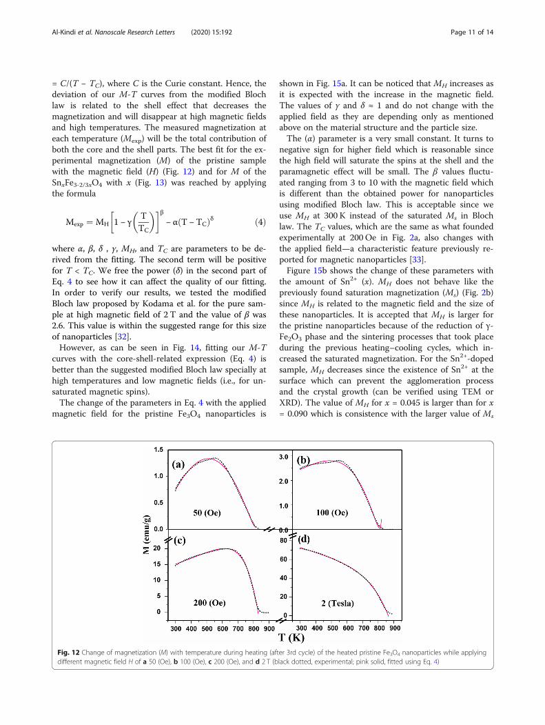
= C/(T − TC), where C is the Curie constant. Hence, thedeviation of our M-T curves from the modified Blochlaw is related to the shell effect that decreases themagnetization and will disappear at high magnetic fieldsand high temperatures. The measured magnetization ateach temperature (Mexp) will be the total contribution ofboth the core and the shell parts. The best fit for the ex-perimental magnetization (M) of the pristine samplewith the magnetic field (H) (Fig. 12) and for M of theSnxFe3-2/3xO4 with x (Fig. 13) was reached by applyingthe formula
Mexp ¼ MH 1 − γTTC
� �� �β− α T − TCð Þδ ð4Þ
where α, β, δ , γ, MH, and TC are parameters to be de-rived from the fitting. The second term will be positivefor T < TC. We free the power (δ) in the second part ofEq. 4 to see how it can affect the quality of our fitting.In order to verify our results, we tested the modifiedBloch law proposed by Kodama et al. for the pure sam-ple at high magnetic field of 2 T and the value of β was2.6. This value is within the suggested range for this sizeof nanoparticles [32].However, as can be seen in Fig. 14, fitting our M-T
curves with the core-shell-related expression (Eq. 4) isbetter than the suggested modified Bloch law specially athigh temperatures and low magnetic fields (i.e., for un-saturated magnetic spins).The change of the parameters in Eq. 4 with the applied
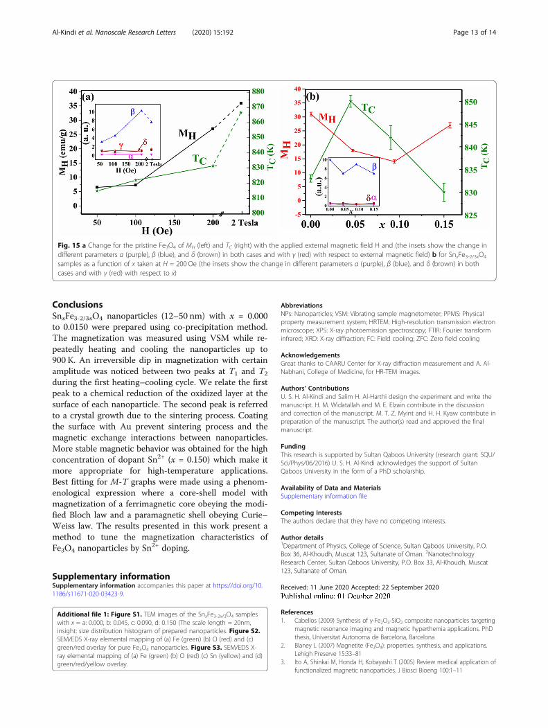
magnetic field for the pristine Fe3O4 nanoparticles is
shown in Fig. 15a. It can be noticed that MH increases asit is expected with the increase in the magnetic field.The values of γ and δ ≈ 1 and do not change with theapplied field as they are depending only as mentionedabove on the material structure and the particle size.The (α) parameter is a very small constant. It turns to
negative sign for higher field which is reasonable sincethe high field will saturate the spins at the shell and theparamagnetic effect will be small. The β values fluctu-ated ranging from 3 to 10 with the magnetic field whichis different than the obtained power for nanoparticlesusing modified Bloch law. This is acceptable since weuse MH at 300 K instead of the saturated Ms in Blochlaw. The TC values, which are the same as what foundedexperimentally at 200 Oe in Fig. 2a, also changes withthe applied field—a characteristic feature previously re-ported for magnetic nanoparticles [33].Figure 15b shows the change of these parameters with
the amount of Sn2+ (x). MH does not behave like thepreviously found saturation magnetization (Ms) (Fig. 2b)since MH is related to the magnetic field and the size ofthese nanoparticles. It is accepted that MH is larger forthe pristine nanoparticles because of the reduction of γ-Fe2O3 phase and the sintering processes that took placeduring the previous heating–cooling cycles, which in-creased the saturated magnetization. For the Sn2+-dopedsample, MH decreases since the existence of Sn2+ at thesurface which can prevent the agglomeration processand the crystal growth (can be verified using TEM orXRD). The value of MH for x = 0.045 is larger than for x= 0.090 which is consistence with the larger value of Ms
Fig. 12 Change of magnetization (M) with temperature during heating (after 3rd cycle) of the heated pristine Fe3O4 nanoparticles while applyingdifferent magnetic field H of a 50 (Oe), b 100 (Oe), c 200 (Oe), and d 2 T (black dotted, experimental; pink solid, fitted using Eq. 4)
Al-Kindi et al. Nanoscale Research Letters (2020) 15:192 Page 11 of 14

for this sample. Interestingly, for the larger NPs with x =0.150, MH increased which opposes the decrease in theirMs and this is due to the larger particle size with largerblocking temperature. The values of (α) and (δ) are con-stants with average value equals 0.3 and 0.6, respectively.This is predicted since the second part of Eq. 4 is related
to the change with the magnetic field which is now con-stant (200 Oe). The values of TC for different samplesare approximately the same as recorded experimentally.γ is a constant with a value equals 1 which is the sameas in Bloch law. β is also almost a constant since it is re-lated to the material with an average value of 8.
Fig. 14 Change of magnetization (M) with temperature during heating for the 3rd cycle of the heated pristine Fe3O4 nanoparticles whileapplying a magnetic field H = 2 (Tesla) (pink dotted, experimental; solid, fitted using the new bulk-shell expression (black) and the modified Blochlaw proposed by Kodama et al. (green)). Green arrows indicate the temperatures where the modified Bloch law proposed by Kodama et al. failedto fully fit the experimental data
Fig. 13 Change of magnetization (M) with temperature during heating (the 3rd cycles) of the heated SnxFe3-2/3xO4 nanoparticles with differentamount of the indicated x (0.000, 0.045, 0.090, 0.150) (H = 200 Oe) (black dotted, experimental; pink solid, fitted)
Al-Kindi et al. Nanoscale Research Letters (2020) 15:192 Page 12 of 14
ConclusionsSnxFe3-2/3xO4 nanoparticles (12–50 nm) with x = 0.000to 0.0150 were prepared using co-precipitation method.The magnetization was measured using VSM while re-peatedly heating and cooling the nanoparticles up to900 K. An irreversible dip in magnetization with certainamplitude was noticed between two peaks at T1 and T2
during the first heating–cooling cycle. We relate the firstpeak to a chemical reduction of the oxidized layer at thesurface of each nanoparticle. The second peak is referredto a crystal growth due to the sintering process. Coatingthe surface with Au prevent sintering process and themagnetic exchange interactions between nanoparticles.More stable magnetic behavior was obtained for the highconcentration of dopant Sn2+ (x = 0.150) which make itmore appropriate for high-temperature applications.Best fitting for M-T graphs were made using a phenom-enological expression where a core-shell model withmagnetization of a ferrimagnetic core obeying the modi-fied Bloch law and a paramagnetic shell obeying Curie–Weiss law. The results presented in this work present amethod to tune the magnetization characteristics ofFe3O4 nanoparticles by Sn2+ doping.
Supplementary informationSupplementary information accompanies this paper at https://doi.org/10.1186/s11671-020-03423-9.
Additional file 1: Figure S1. TEM images of the SnxFe3-2x/3O4 sampleswith x = a: 0.000, b: 0.045, c: 0.090, d: 0.150 (The scale length = 20nm,insight: size distribution histogram of prepared nanoparticles. Figure S2.SEM/EDS X-ray elemental mapping of (a) Fe (green) (b) O (red) and (c)green/red overlay for pure Fe3O4 nanoparticles. Figure S3. SEM/EDS X-ray elemental mapping of (a) Fe (green) (b) O (red) (c) Sn (yellow) and (d)green/red/yellow overlay.
AbbreviationsNPs: Nanoparticles; VSM: Vibrating sample magnetometer; PPMS: Physicalproperty measurement system; HRTEM: High-resolution transmission electronmicroscope; XPS: X-ray photoemission spectroscopy; FTIR: Fourier transforminfrared; XRD: X-ray diffraction; FC: Field cooling; ZFC: Zero field cooling
AcknowledgementsGreat thanks to CAARU Center for X-ray diffraction measurement and A. Al-Nabhani, College of Medicine, for HR-TEM images.
Authors’ ContributionsU. S. H. Al-Kindi and Salim H. Al-Harthi design the experiment and write themanuscript. H. M. Widatallah and M. E. Elzain contribute in the discussionand correction of the manuscript. M. T. Z. Myint and H. H. Kyaw contribute inpreparation of the manuscript. The author(s) read and approved the finalmanuscript.
FundingThis research is supported by Sultan Qaboos University (research grant: SQU/Sci/Phys/06/2016) U. S. H. Al-Kindi acknowledges the support of SultanQaboos University in the form of a PhD scholarship.
Availability of Data and MaterialsSupplementary information file
Competing InterestsThe authors declare that they have no competing interests.
Author details1Department of Physics, College of Science, Sultan Qaboos University, P.O.Box 36, Al-Khoudh, Muscat 123, Sultanate of Oman. 2NanotechnologyResearch Center, Sultan Qaboos University, P.O. Box 33, Al-Khoudh, Muscat123, Sultanate of Oman.
Received: 11 June 2020 Accepted: 22 September 2020
References1. Cabellos (2009) Synthesis of γ-Fe2O3-SiO2 composite nanoparticles targeting
magnetic resonance imaging and magnetic hyperthemia applications. PhDthesis, Universitat Autonoma de Barcelona, Barcelona
2. Blaney L (2007) Magnetite (Fe3O4): properties, synthesis, and applications.Lehigh Preserve 15:33–81
3. Ito A, Shinkai M, Honda H, Kobayashi T (2005) Review medical application offunctionalized magnetic nanoparticles. J Biosci Bioeng 100:1–11
Fig. 15 a Change for the pristine Fe3O4 of MH (left) and TC (right) with the applied external magnetic field H and (the insets show the change indifferent parameters α (purple), β (blue), and δ (brown) in both cases and with γ (red) with respect to external magnetic field) b for SnxFe3-2/3xO4
samples as a function of x taken at H = 200 Oe (the insets show the change in different parameters α (purple), β (blue), and δ (brown) in bothcases and with γ (red) with respect to x)
Al-Kindi et al. Nanoscale Research Letters (2020) 15:192 Page 13 of 14

4. Ebner D, Ritter J, James H, Ploehn A, Harry J (1999) Two-particle magnetichetero-flocculation model for nanolevel high gradient magnetic separation.In: Engineering Foundation Conference, pp 193–204
5. Rikers R, Rem P, Dalmijn W (1998) Improved method for prediction of heavymetal recoveries from soil using high intensity magnetic separation (HIMS).Int J Miner Process 54:165–182
6. Chen P, Cai Y, Wang J, Wang K, Tao Y, Xue J, Wang H (2018) Preparation ofprotonized titanate nanotubes/Fe3O4/TiO2 ternary composites and dye self-sensitization for visible-light-driven photodegradation of Rhodamine B.Powder Technol 326:272–280
7. Atabaev TS (2018) PEG-coated superparamagnetic dysprosium-doped Fe3O4
nanoparticles for potential MRI imaging. BioNanoScience 8:299–3038. Atabaev T, Kim H, Hwang Y-H (2013) Fabrication of bifunctional core-shell
Fe3O4 particles coated with ultrathin phosphor layer. Nanoscale Res Lett 8:357–363
9. Lei Y, Ding J, Yu P, He G, Chen Y, Chen H (2020) Low-temperaturepreparation of magnetically separable Fe3O4@ZnO-RGO for high-performance removal of methylene blue in visible light. J Alloys Compd821:153366–153373
10. Buschow K (2006) Handbook of magnetic materials. Elsevier B. V,Amesterdam
11. Berry F, Greaves C, Helgason O, McManus J (1999) Synthesis andcharacterisation of tin-doped iron oxides. J Mater Chem 9:223–226
12. Haneda K, Morrish A (1977) Magnetite to maghemite transformation inultrafine particles. J Phys Colloq 38:C1-321–C1-323
13. Khan U, Amanullah A, Manan N, Khan A, Mahmood A (2015) Rahim,transformation mechanism of magnetite nanoparticles. Mater Sci-Poland 33:278–285
14. Kolen’ko V, Bañobre-López M, Rodríguez-Abreu C, Carbó-Argibay E, DeepakF, Petrovyk D, Cerqueira M, Kamali S, Kovnir K, Shtansky D, Lebedev O, RivasJ (2014) High-temperature magnetism as a probe for structural andcompositional uniformity in ligand-capped magnetite nanoparticles. J PhysChem C 118:28322–28329
15. Berry F, Skinner S, Helgason Ö, Bilsborrow B, Marco J (1998) Location of tinand charge balance in materials of composition Fe3-xSnxO4 (x < 0.3).Polyhedron 17:149–152
16. Berry F, Helgason Ö, Jónsson K, Skinner S (1996) The high temperatureproperties of tin-doped magnetite. J Solid State Chem 122:353–357
17. Bhaumik A, Nori S, Sachan R, Gupta S, Kumar D, Majumdar A, Narayan J(2018) Room-temperature ferromagnetism and extraordinary hall effect innanosrtuctured Q-carbon: implication for potential spintronic devices. ACSAppl Nano Mater 1:807–819
18. Lutterotti L (2010) Total pattern fitting for the combined size-strain-stress-texture determination in thin film diffraction. Nucl Instrum Methods PhysRes B 268:334–340
19. Fairly N (1999) CasaXPS application. In: N. CasaXPS (ed.), Casa software ltd,U.K.
20. Cao X, Prozorov R, Koltypin Y, Kataby G, Felner I, Gedanken A (1997)Synthesis of pure amorphous Fe2O3. J Mater Res 12:402–406
21. Mørup S, Topsøe H, Lipka J (1976) Modified theory for Mössbauer spectra ofsuperparamagnetic particles: application to Fe3O4. J Phys Colloq 37:C6-287–C6-290
22. Rondinone AJ, Samia ACS, Zhang ZJ (1999) Superparamagnetic relaxationand magnetic anisotropy energy distribution in CoFe2O4 spinel ferritenanocrystallites. J Phys Chem B 103:6876–6880
23. Kim W, Suh C-Y, Cho S-W, Roh K-M, Kwon H, Song K, Shon I-J (2012) A newmethod for the identification and quantification of magnetite-maghemitemixture using conventional X-ray diffraction technique. Talanta 94:348–352
24. Grosvenor AP, Kobe BA, McIntyre NS (2004) Studies of the oxidation of ironby water vapour using X-ray photoelectron spectroscopy and QUASES™.Surf Sci 572:217–227
25. Grosvenor AP, Kobe BA, Biesinger MC, McIntyre NS (2004) Investigation ofmultiplet splitting of Fe 2p XPS spectra and bonding in iron compounds.Surf Interface Anal 36:1564–1574
26. Yamaura M, Camilo R, Sampaio L, Maceedo M, Nakamura M, Tomad H(2004) Preparation and characterization of (3-aminopropyl) triethoxysilane-coated magnetite nanoparticles. J Mag Mag Mat 279:210–217
27. Aliahmad M, Moghaddam N (2013) Synthesis of maghemite (γ-Fe2O3)nanoparticles by thermal-decomposition of magnetite (Fe3O4) nanoparticles.Mater Sci-Poland 31:264–268
28. Rubim JC, Sousa MH, Silva JCO, Tourinho FA (2001) Raman spectroscopy asa powerful technique in the characterization of ferrofluids. Braz J Phys 31:402–408
29. Kodama R, Berkowitz A, McNiff E, Foner S (1996) Surface spin disorder innanoparticles. Phys Rev Lett 77:394–397
30. Caizer C (2004) Deviations from Bloch law in the case of surfactednanoparticles. Appl Phys A 80:1745–1751
31. Cojocaru S, Naddeo A, Citro R (2014) Modification of the Bloch law inferromagnetic nanostructures. EPL 106:17001–17007
32. Kodama R (1999) Magnetic nanoparticles. J Magn Magn Mater 200:359–37233. Mulyukov K, Musabirov I (2010) Influence of magnetic field intensity on the
temperature dependence of magnetization of Ni2.08Mn0.96Ga0.96 Alloy.JEMAA 2:431–435
Publisher’s NoteSpringer Nature remains neutral with regard to jurisdictional claims inpublished maps and institutional affiliations.
Al-Kindi et al. Nanoscale Research Letters (2020) 15:192 Page 14 of 14


![NANO EXPRESS Open Access Propargylic substitution ... · Background Electrophilic attack on aromatic carbons is a useful ... [1-3]. Electrophilic aromatic substitution is an organic](https://img.pdfslide.us/doc/110x75/5b461b0a7f8b9a114c8b5ee0/nano-express-open-access-propargylic-substitution-background-electrophilic.jpg)