Embed Size (px)
Citation preview
u n i ve r s i t y o f co pe n h ag e n
Københavns Universitet
Influence of nanoparticles of platinum on chicken embryo development and brainmorphologyPrasek, Marta ; Saworz, Ewa; Jaworski, Slawomir; Grodzik, Marta; Ostaszewska, Teresa;Kamaszewski, Maciej; Wierzbicki, Mateusz; Chwalibog, AndréPublished in:Nanoscale Research Letters
DOI:10.1186/1556-276X-8-251
Publication date:2013
Document VersionEarly version, also known as pre-print
Citation for published version (APA):Prasek, M., Saworz, E., Jaworski, S., Grodzik, M., Ostaszewska, T., Kamaszewski, M., ... Chwalibog, A. (2013).Influence of nanoparticles of platinum on chicken embryo development and brain morphology. DOI:10.1186/1556-276X-8-251
Download date: 23. Jun. 2018
Prasek et al. Nanoscale Research Letters 2013, 8:251http://www.nanoscalereslett.com/content/8/1/251
NANO EXPRESS Open Access
Influence of nanoparticles of platinum on chickenembryo development and brain morphologyMarta Prasek1, Ewa Sawosz1, Slawomir Jaworski1, Marta Grodzik1, Teresa Ostaszewska2, Maciej Kamaszewski2,Mateusz Wierzbicki1 and Andre Chwalibog3*
Abstract
Platinum nanoparticles (NP-Pt) are noble metal nanoparticles with unique physiochemical properties that haverecently elicited much interest in medical research. However, we still know little about their toxicity and influenceon general health. We investigated effects of NP-Pt on the growth and development of the chicken embryo modelwith emphasis on brain tissue micro- and ultrastructure. The embryos were administered solutions of NP-Pt injectedin ovo at concentrations from 1 to 20 μg/ml. The results demonstrate that NP-Pt did not affect the growth anddevelopment of the embryos; however, they induced apoptosis and decreased the number of proliferating cells inthe brain tissue. These preliminary results indicate that properties of NP-Pt might be utilized in brain cancer therapy,but potential toxic side effects must be elucidated in extensive follow-up research.
Keywords: Platinum nanoparticles, Chicken embryo, Toxicity, Neurotoxicity, Brain morphology, Cancer therapy
BackgroundPlatinum (Pt) is a noble metal with unique physiologicaland chemical properties widely used in chemistry, phy-sics, biology, and medicine. Regarding the biologicalactivities of Pt, it is known that Pt compounds have theability to arrest the cell cycle [1,2] and cause DNAstrand breaks. The DNA damage is caused by Pt ions,which attach to N7 sites of DNA guanine bases and,after hydrolysis of Pt-Cl bonds, form adducts with theDNA double helix [2,3]. These properties of Pt areexploited in cancer therapy in the form of antineoplasticdrugs to treat different types of cancer such as head,neck, brain [4], testicular, bladder, ovarian, or uterinecervix carcinomas [5]. However, toxic side effects ofPt-based drugs are major drawbacks in cancer therapy[6,7].Nanotechnology has introduced possibilities for using
alternate forms of elements - nanoparticles. Nanoparticleshave unique physiochemical features because of theirsmall size (<100 nm), large surface-to-mass ratio, excep-tional quantum characteristics [8], and consequentlyunique biological properties. Smaller nanoparticles can
* Correspondence: [email protected] of Veterinary Clinical and Animal Sciences, University ofCopenhagen, Groennegaardsvej 3, Frederiksberg 1870, DenmarkFull list of author information is available at the end of the article
© 2013 Prasek et al.; licensee Springer. This is aAttribution License (http://creativecommons.orin any medium, provided the original work is p
move across cellular and also nuclear membranes and areable to penetrate cells and intracellular structures, andtarget defined points within the body [9,10]. Platinumnanoparticles (NP-Pt) have recently elicited much interestbecause of their physicochemical properties such as cata-lytic activity and high reactivity [11]. NP-Pt, as metalstructures (Pt0), differ significantly from platinum saltsand have quite different chemical properties when admin-istered to an organism. They are a very limited source ofions, and consequently, the process of forming platinumsalts is very slow and restricted. However, the solubilityand, consequently, the bioavailability of NP-Pt depend ontheir size [12]. Although it has been demonstrated thatsmall doses of NP-Pt have negligible toxic effects onchicken and zebra fish embryos [13], they might impingethe cell structures [12].It has been demonstrated that hadron cancer therapy
can be amplified by simultaneous application of NP-Pt,resulting in the production of hydroxyl radicals causinglethal DNA damage by double-strand breaks [14]. Fur-thermore, DNA damage could also be induced by theattack of OH groups linked with NP-Pt on DNA phos-phate groups [2]. NP-Pt can also cause cell cycle arrestand induction of apoptosis through the release of Pt2+
ions from the nanoparticles as a result of H2O2 gene-ration due to the low pH in endosomes [1]. It was also
n Open Access article distributed under the terms of the Creative Commonsg/licenses/by/2.0), which permits unrestricted use, distribution, and reproductionroperly cited.
Prasek et al. Nanoscale Research Letters 2013, 8:251 Page 2 of 9http://www.nanoscalereslett.com/content/8/1/251
demonstrated that DNA double-strand breaks arecaused by Pt2+ ions formed during the incubation ofNP-Pt with cancer cells [15]. However, the consequencesof introducing NP-Pt into an organism are still not welldocumented, especially when even very small amountsof nanoparticles or released ions may overcome theblood–brain barrier (BBB), enter the brain tissue, andaffect the BBB and brain function. It has also beenreported that various types of nanoparticles, in differentsizes from 20 to 300 nm and produced from differentmaterials, may cause cell death by apoptosis in the braintissue [16].In the present study, we hypothesized that NP-Pt may
affect the growth and development of embryos and, fur-thermore, can cross the BBB and penetrate the brain tis-sue, affecting brain morphology. Consequently, theobjective of this preliminary work was to investigate theeffects of NP-Pt on embryo growth and developmentwith an emphasis on brain morphology, concerning theirpotential applicability in brain cancer therapy.
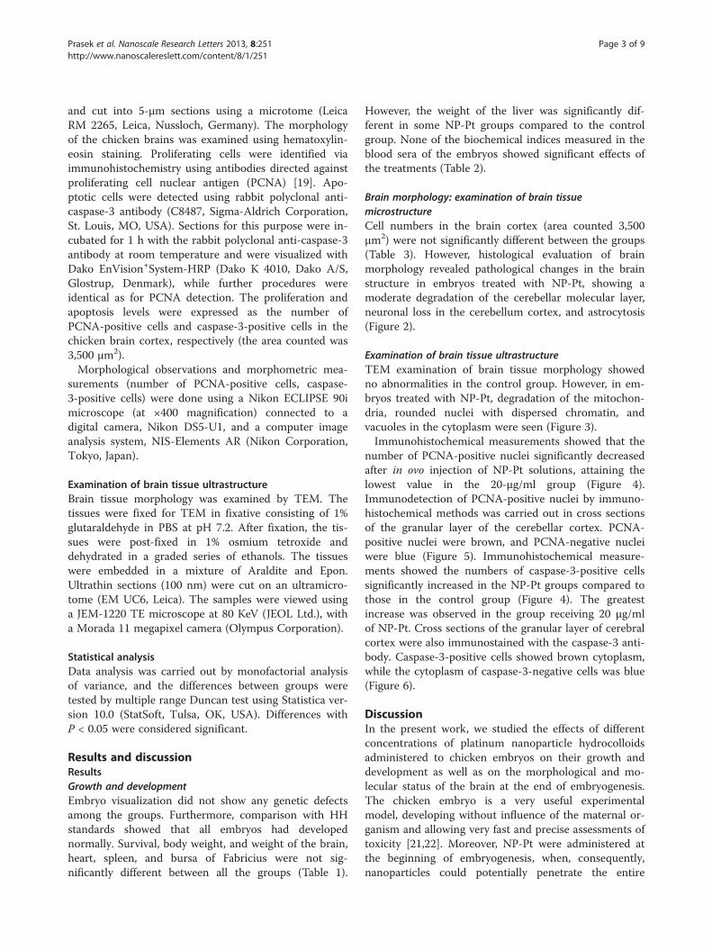
MethodsNanoparticlesHydrocolloids of NP-Pt were obtained from Nano-TechPolska (Warsaw, Poland). They were produced by a pat-ented electric nonexplosive method [17] from high pur-ity metal (99.9999%) and high purity demineralizedwater. The shape and size of the nanoparticles wereinspected by transmission electron microscopy (TEM)using a JEOL JEM-1220 TE microscope at 80 KeV (JEOLLtd., Tokyo, Japan), with a Morada 11 megapixel camera(Olympus Corporation, Tokyo, Japan) (Figure 1). The di-ameters of the Pt particles ranged from 2 to 19 nm. Asample of Pt for TEM was prepared by placing dropletsof the hydrocolloids onto Formvar-coated copper grids(Agar Scientific Ltd., Stansted, UK). Immediately afterdrying the droplets in dry air, the grids were inserted
Figure 1 TEM image of platinum nanoparticles. Bar scale100 nm.
into the TE microscope (Figure 1). The zeta potential ofthe nanoparticle hydrocolloids was measured by electro-phoretic light-scattering method, using a ZetasizerNano-ZS90 (Malvern, Worcestershire, UK). Each samplewas measured after 120 s of stabilization at 25°C in20 replicates. The mean zeta potential of the Ptnanoparticles was −9.6 mV.
Embryo modelBased on Polish law Article 2 of the act dated 21 January2005 concerning the experiments on animals (journal oflaw is dated 24 February 2005), there is no need to sub-mit an application to the local ethics committee for issu-ing an opinion about studies where the chicken embryois used. According to this act, chicken embryo is not def-inite as the animal. Fertilized eggs (n = 150; 56 ± 2.2 g)from hens of the Ross line were obtained from a com-mercial hatchery and stored at 12°C for 4 days. After 4days, the eggs were weighed and randomly divided intosix groups (n = 25 eggs per group). The control groupwas not treated, while the other groups were treatedwith 1, 5, 10, 15, or 20 μg/ml of NP-Pt solutions. Theexperimental solutions were given in ovo by injectioninto the albumen (at two-thirds of the egg's height fromthe blunt end) using a sterile 1-ml insulin syringe. Injec-tion consisted of 0.3-ml NP-Pt hydrocolloid. The injec-tion holes were sterilized, and the eggs were thenincubated at 37.5°C and 60% humidity and were turnedonce per hour for 19 days.At day 20 of incubation, the embryos were sacrificed
by decapitation. Embryos and organs (brain, heart, liver,spleen, bursa of Fabricius) were weighed and evaluatedby Hamburger and Hamilton [18] (HH) standards.
Biochemical indicesBlood serum samples were collected from the jugularvein on the 20th day of incubation. The samples werecentrifuged at 3,000 rpm for 15 min (Sorvall ST 16,Thermo Fisher Scientific, Waltham, MA, USA), andconcentrations of alanine aminotransferase (ALT), as-paragine aminotransferase, lactate dehydrogenase, alka-line phosphatase (ALP), glucose level, and blood ureanitrogen were measured in the blood serum. Biochemis-try markers were examined using a dry chemistry equip-ment Vitros DT 60 II (Johnston and Johnston, NewBrunswick, NJ, USA).
Brain morphology: examination of brain tissuemicrostructureChicken brains (n = 12), three from the control groupand nine from groups treated with 1, 10, and 20 μg/mlof NP-Pt solutions, were sampled and fixed in 10% buff-ered formalin (pH 7.2). Fixed samples were dehydratedin a graded series of ethanols, embedded in Paraplast,
Prasek et al. Nanoscale Research Letters 2013, 8:251 Page 3 of 9http://www.nanoscalereslett.com/content/8/1/251
and cut into 5-μm sections using a microtome (LeicaRM 2265, Leica, Nussloch, Germany). The morphologyof the chicken brains was examined using hematoxylin-eosin staining. Proliferating cells were identified viaimmunohistochemistry using antibodies directed againstproliferating cell nuclear antigen (PCNA) [19]. Apo-ptotic cells were detected using rabbit polyclonal anti-caspase-3 antibody (C8487, Sigma-Aldrich Corporation,St. Louis, MO, USA). Sections for this purpose were in-cubated for 1 h with the rabbit polyclonal anti-caspase-3antibody at room temperature and were visualized withDako EnVision+System-HRP (Dako K 4010, Dako A/S,Glostrup, Denmark), while further procedures wereidentical as for PCNA detection. The proliferation andapoptosis levels were expressed as the number ofPCNA-positive cells and caspase-3-positive cells in thechicken brain cortex, respectively (the area counted was3,500 μm2).Morphological observations and morphometric mea-
surements (number of PCNA-positive cells, caspase-3-positive cells) were done using a Nikon ECLIPSE 90imicroscope (at ×400 magnification) connected to adigital camera, Nikon DS5-U1, and a computer imageanalysis system, NIS-Elements AR (Nikon Corporation,Tokyo, Japan).
Examination of brain tissue ultrastructureBrain tissue morphology was examined by TEM. Thetissues were fixed for TEM in fixative consisting of 1%glutaraldehyde in PBS at pH 7.2. After fixation, the tis-sues were post-fixed in 1% osmium tetroxide anddehydrated in a graded series of ethanols. The tissueswere embedded in a mixture of Araldite and Epon.Ultrathin sections (100 nm) were cut on an ultramicro-tome (EM UC6, Leica). The samples were viewed usinga JEM-1220 TE microscope at 80 KeV (JEOL Ltd.), witha Morada 11 megapixel camera (Olympus Corporation).
Statistical analysisData analysis was carried out by monofactorial analysisof variance, and the differences between groups weretested by multiple range Duncan test using Statistica ver-sion 10.0 (StatSoft, Tulsa, OK, USA). Differences withP < 0.05 were considered significant.
Results and discussionResultsGrowth and developmentEmbryo visualization did not show any genetic defectsamong the groups. Furthermore, comparison with HHstandards showed that all embryos had developednormally. Survival, body weight, and weight of the brain,heart, spleen, and bursa of Fabricius were not sig-nificantly different between all the groups (Table 1).
However, the weight of the liver was significantly dif-ferent in some NP-Pt groups compared to the controlgroup. None of the biochemical indices measured in theblood sera of the embryos showed significant effects ofthe treatments (Table 2).
Brain morphology: examination of brain tissuemicrostructureCell numbers in the brain cortex (area counted 3,500μm2) were not significantly different between the groups(Table 3). However, histological evaluation of brainmorphology revealed pathological changes in the brainstructure in embryos treated with NP-Pt, showing amoderate degradation of the cerebellar molecular layer,neuronal loss in the cerebellum cortex, and astrocytosis(Figure 2).
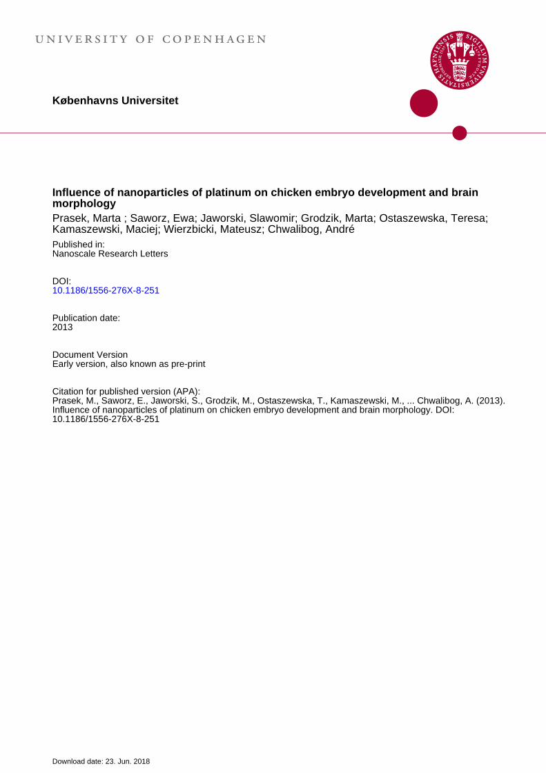
Examination of brain tissue ultrastructureTEM examination of brain tissue morphology showedno abnormalities in the control group. However, in em-bryos treated with NP-Pt, degradation of the mitochon-dria, rounded nuclei with dispersed chromatin, andvacuoles in the cytoplasm were seen (Figure 3).Immunohistochemical measurements showed that the
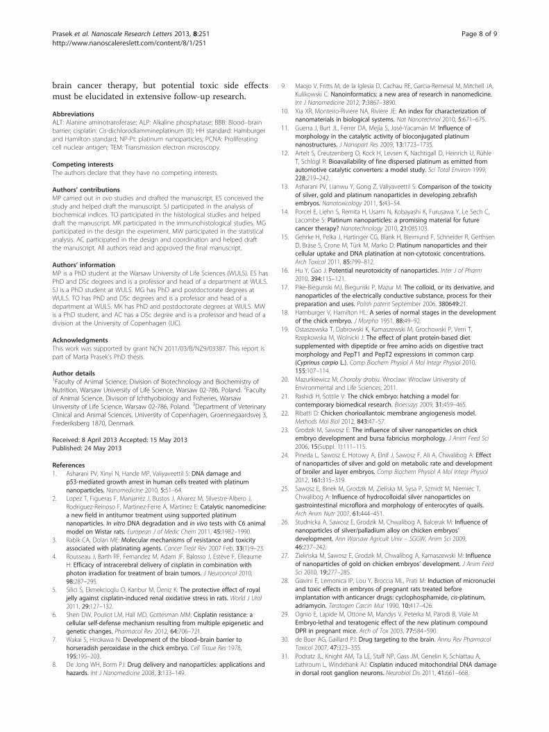
number of PCNA-positive nuclei significantly decreasedafter in ovo injection of NP-Pt solutions, attaining thelowest value in the 20-μg/ml group (Figure 4).Immunodetection of PCNA-positive nuclei by immuno-histochemical methods was carried out in cross sectionsof the granular layer of the cerebellar cortex. PCNA-positive nuclei were brown, and PCNA-negative nucleiwere blue (Figure 5). Immunohistochemical measure-ments showed the numbers of caspase-3-positive cellssignificantly increased in the NP-Pt groups compared tothose in the control group (Figure 4). The greatestincrease was observed in the group receiving 20 μg/mlof NP-Pt. Cross sections of the granular layer of cerebralcortex were also immunostained with the caspase-3 anti-body. Caspase-3-positive cells showed brown cytoplasm,while the cytoplasm of caspase-3-negative cells was blue(Figure 6).
DiscussionIn the present work, we studied the effects of differentconcentrations of platinum nanoparticle hydrocolloidsadministered to chicken embryos on their growth anddevelopment as well as on the morphological and mo-lecular status of the brain at the end of embryogenesis.The chicken embryo is a very useful experimentalmodel, developing without influence of the maternal or-ganism and allowing very fast and precise assessments oftoxicity [21,22]. Moreover, NP-Pt were administered atthe beginning of embryogenesis, when, consequently,nanoparticles could potentially penetrate the entire
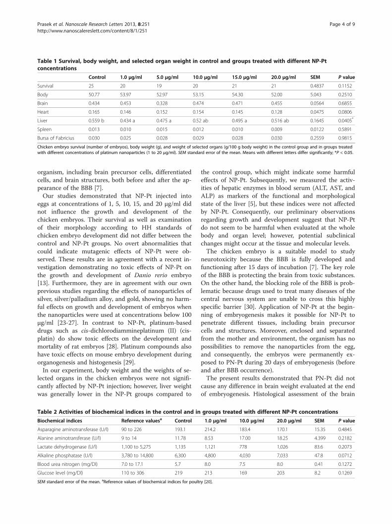
Table 1 Survival, body weight, and selected organ weight in control and groups treated with different NP-Ptconcentrations
Control 1.0 μg/ml 5.0 μg/ml 10.0 μg/ml 15.0 μg/ml 20.0 μg/ml SEM P value
Survival 25 20 19 20 21 21 0.4837 0.1152
Body 50.77 53.97 52.97 53.15 54.30 52.00 5.043 0.2510
Brain 0.434 0.453 0.328 0.474 0.471 0.455 0.0564 0.6855
Heart 0.165 0.146 0.152 0.154 0.145 0.128 0.0475 0.0806
Liver 0.559 b 0.434 a 0.475 a 0.52 ab 0.495 a 0.516 ab 0.1645 0.0405*
Spleen 0.013 0.010 0.015 0.012 0.010 0.009 0.0122 0.5891
Bursa of Fabricius 0.030 0.025 0.028 0.029 0.028 0.030 0.2559 0.9815
Chicken embryo survival (number of embryos), body weight (g), and weight of selected organs (g/100 g body weight) in the control group and in groups treatedwith different concentrations of platinum nanoparticles (1 to 20 μg/ml). SEM standard error of the mean. Means with different letters differ significantly; *P < 0.05.
Prasek et al. Nanoscale Research Letters 2013, 8:251 Page 4 of 9http://www.nanoscalereslett.com/content/8/1/251
organism, including brain precursor cells, differentiatedcells, and brain structures, both before and after the ap-pearance of the BBB [7].Our studies demonstrated that NP-Pt injected into
eggs at concentrations of 1, 5, 10, 15, and 20 μg/ml didnot influence the growth and development of thechicken embryos. Their survival as well as examinationof their morphology according to HH standards ofchicken embryo development did not differ between thecontrol and NP-Pt groups. No overt abnormalities thatcould indicate mutagenic effects of NP-Pt were ob-served. These results are in agreement with a recent in-vestigation demonstrating no toxic effects of NP-Pt onthe growth and development of Danio rerio embryo[13]. Furthermore, they are in agreement with our ownprevious studies regarding the effects of nanoparticles ofsilver, silver/palladium alloy, and gold, showing no harm-ful effects on growth and development of embryos whenthe nanoparticles were used at concentrations below 100μg/ml [23-27]. In contrast to NP-Pt, platinum-baseddrugs such as cis-dichlorodiammineplatinum (II) (cis-platin) do show toxic effects on the development andmortality of rat embryos [28]. Platinum compounds alsohave toxic effects on mouse embryo development duringorganogenesis and histogenesis [29].In our experiment, body weight and the weights of se-
lected organs in the chicken embryos were not signifi-cantly affected by NP-Pt injection; however, liver weightwas generally lower in the NP-Pt groups compared to
Table 2 Activities of biochemical indices in the control and in
Biochemical indices Reference valuesa Control
Asparagine aminotransferase (U/l) 90 to 226 193.1
Alanine aminotransferase (U/l) 9 to 14 11.78
Lactate dehydrogenase (U/l) 1,100 to 5,275 1,135
Alkaline phosphatase (U/l) 3,780 to 14,800 6,300
Blood urea nitrogen (mg/Dl) 7.0 to 17.1 5.7
Glucose level (mg/Dl) 110 to 306 219
SEM standard error of the mean. aReference values of biochemical indices for poult
the control group, which might indicate some harmfuleffects of NP-Pt. Subsequently, we measured the activ-ities of hepatic enzymes in blood serum (ALT, AST, andALP) as markers of the functional and morphologicalstate of the liver [5], but these indices were not affectedby NP-Pt. Consequently, our preliminary observationsregarding growth and development suggest that NP-Ptdo not seem to be harmful when evaluated at the wholebody and organ level; however, potential subclinicalchanges might occur at the tissue and molecular levels.The chicken embryo is a suitable model to study
neurotoxicity because the BBB is fully developed andfunctioning after 15 days of incubation [7]. The key roleof the BBB is protecting the brain from toxic substances.On the other hand, the blocking role of the BBB is prob-lematic because drugs used to treat many diseases of thecentral nervous system are unable to cross this highlyspecific barrier [30]. Application of NP-Pt at the begin-ning of embryogenesis makes it possible for NP-Pt topenetrate different tissues, including brain precursorcells and structures. Moreover, enclosed and separatedfrom the mother and environment, the organism has nopossibilities to remove the nanoparticles from the egg,and consequently, the embryos were permanently ex-posed to PN-Pt during 20 days of embryogenesis (beforeand after BBB occurrence).The present results demonstrated that PN-Pt did not
cause any difference in brain weight evaluated at the endof embryogenesis. Histological assessment of the brain
groups treated with different NP-Pt concentrations
1.0 μg/ml 10.0 μg/ml 20.0 μg/ml SEM P value
214.2 183.4 170.1 15.35 0.4845
8.53 17.00 18.25 4.399 0.2182
1,121 778 1,026 83.6 0.2073
4,800 4,030 7,033 47.8 0.0712
8.0 7.5 8.0 0.41 0.1272
213 169 203 8.2 0.1269
ry [20].
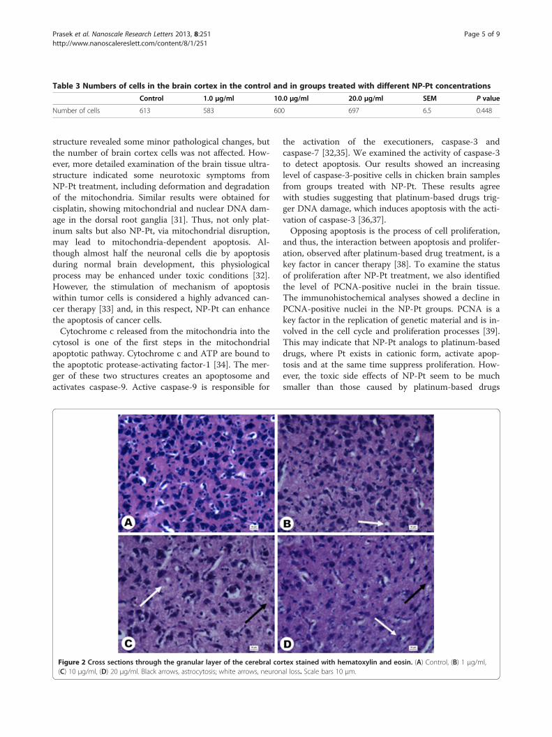
Table 3 Numbers of cells in the brain cortex in the control and in groups treated with different NP-Pt concentrations
Control 1.0 μg/ml 10.0 μg/ml 20.0 μg/ml SEM P value
Number of cells 613 583 600 697 6.5 0.448
Prasek et al. Nanoscale Research Letters 2013, 8:251 Page 5 of 9http://www.nanoscalereslett.com/content/8/1/251
structure revealed some minor pathological changes, butthe number of brain cortex cells was not affected. How-ever, more detailed examination of the brain tissue ultra-structure indicated some neurotoxic symptoms fromNP-Pt treatment, including deformation and degradationof the mitochondria. Similar results were obtained forcisplatin, showing mitochondrial and nuclear DNA dam-age in the dorsal root ganglia [31]. Thus, not only plat-inum salts but also NP-Pt, via mitochondrial disruption,may lead to mitochondria-dependent apoptosis. Al-though almost half the neuronal cells die by apoptosisduring normal brain development, this physiologicalprocess may be enhanced under toxic conditions [32].However, the stimulation of mechanism of apoptosiswithin tumor cells is considered a highly advanced can-cer therapy [33] and, in this respect, NP-Pt can enhancethe apoptosis of cancer cells.Cytochrome c released from the mitochondria into the
cytosol is one of the first steps in the mitochondrialapoptotic pathway. Cytochrome c and ATP are bound tothe apoptotic protease-activating factor-1 [34]. The mer-ger of these two structures creates an apoptosome andactivates caspase-9. Active caspase-9 is responsible for
Figure 2 Cross sections through the granular layer of the cerebral co(C) 10 μg/ml, (D) 20 μg/ml. Black arrows, astrocytosis; white arrows, neuron
the activation of the executioners, caspase-3 andcaspase-7 [32,35]. We examined the activity of caspase-3to detect apoptosis. Our results showed an increasinglevel of caspase-3-positive cells in chicken brain samplesfrom groups treated with NP-Pt. These results agreewith studies suggesting that platinum-based drugs trig-ger DNA damage, which induces apoptosis with the acti-vation of caspase-3 [36,37].Opposing apoptosis is the process of cell proliferation,
and thus, the interaction between apoptosis and prolifer-ation, observed after platinum-based drug treatment, is akey factor in cancer therapy [38]. To examine the statusof proliferation after NP-Pt treatment, we also identifiedthe level of PCNA-positive nuclei in the brain tissue.The immunohistochemical analyses showed a decline inPCNA-positive nuclei in the NP-Pt groups. PCNA is akey factor in the replication of genetic material and is in-volved in the cell cycle and proliferation processes [39].This may indicate that NP-Pt analogs to platinum-baseddrugs, where Pt exists in cationic form, activate apop-tosis and at the same time suppress proliferation. How-ever, the toxic side effects of NP-Pt seem to be muchsmaller than those caused by platinum-based drugs
rtex stained with hematoxylin and eosin. (A) Control, (B) 1 μg/ml,al loss. Scale bars 10 μm.
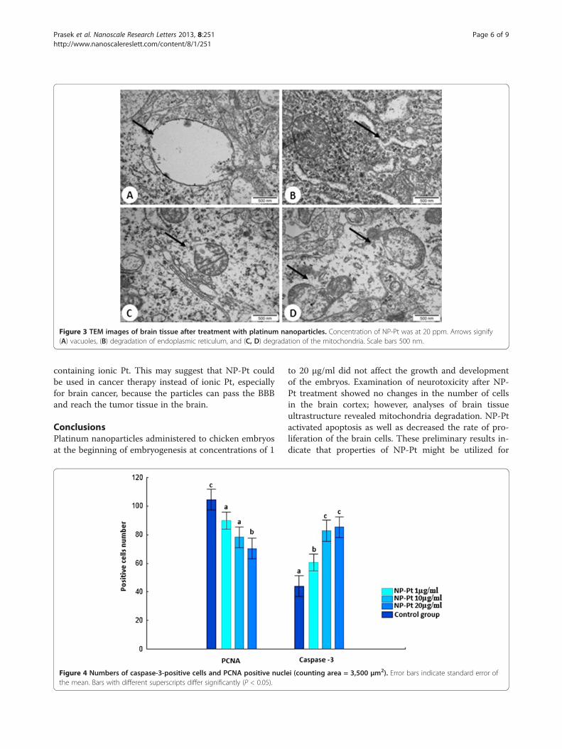
Figure 3 TEM images of brain tissue after treatment with platinum nanoparticles. Concentration of NP-Pt was at 20 ppm. Arrows signify(A) vacuoles, (B) degradation of endoplasmic reticulum, and (C, D) degradation of the mitochondria. Scale bars 500 nm.
Prasek et al. Nanoscale Research Letters 2013, 8:251 Page 6 of 9http://www.nanoscalereslett.com/content/8/1/251
containing ionic Pt. This may suggest that NP-Pt couldbe used in cancer therapy instead of ionic Pt, especiallyfor brain cancer, because the particles can pass the BBBand reach the tumor tissue in the brain.
ConclusionsPlatinum nanoparticles administered to chicken embryosat the beginning of embryogenesis at concentrations of 1
Figure 4 Numbers of caspase-3-positive cells and PCNA positive nuclthe mean. Bars with different superscripts differ significantly (P < 0.05).
to 20 μg/ml did not affect the growth and developmentof the embryos. Examination of neurotoxicity after NP-Pt treatment showed no changes in the number of cellsin the brain cortex; however, analyses of brain tissueultrastructure revealed mitochondria degradation. NP-Ptactivated apoptosis as well as decreased the rate of pro-liferation of the brain cells. These preliminary results in-dicate that properties of NP-Pt might be utilized for
ei (counting area = 3,500 μm2). Error bars indicate standard error of
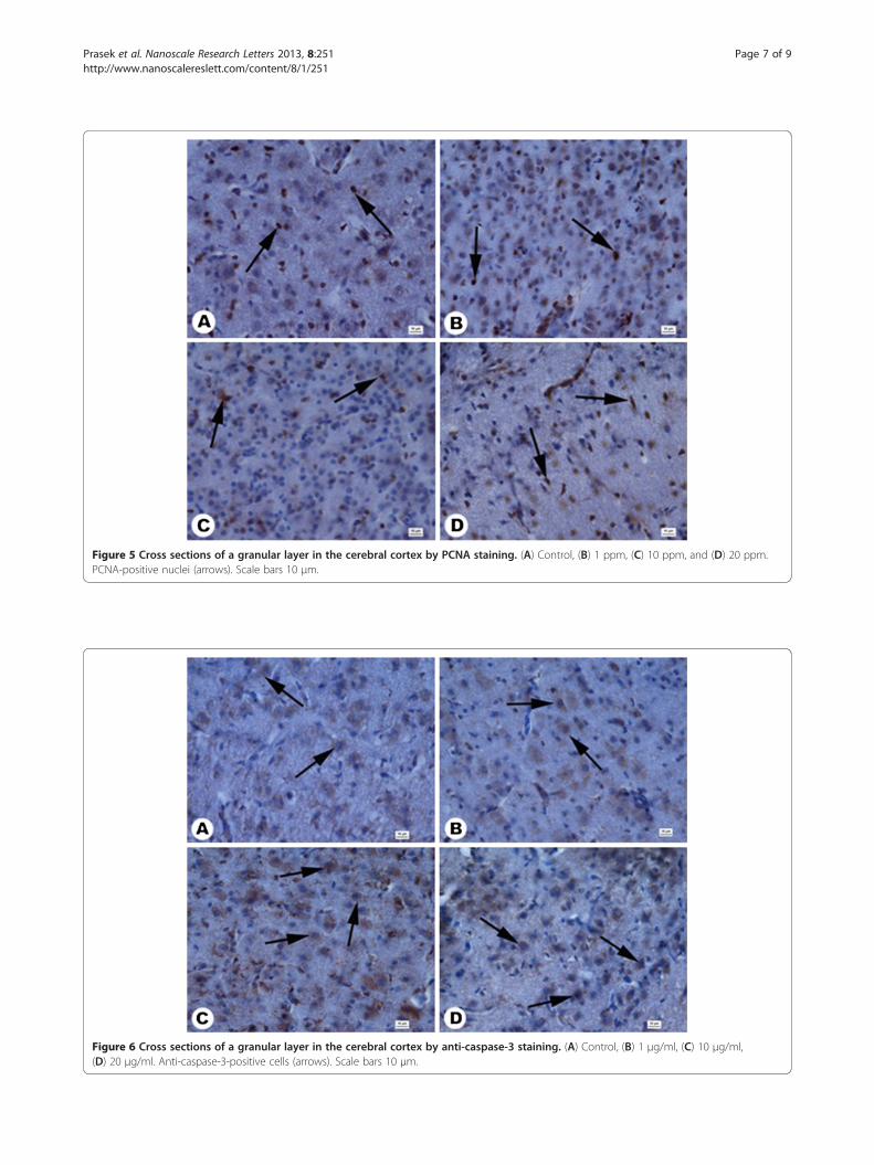
Figure 5 Cross sections of a granular layer in the cerebral cortex by PCNA staining. (A) Control, (B) 1 ppm, (C) 10 ppm, and (D) 20 ppm.PCNA-positive nuclei (arrows). Scale bars 10 μm.
Figure 6 Cross sections of a granular layer in the cerebral cortex by anti-caspase-3 staining. (A) Control, (B) 1 μg/ml, (C) 10 μg/ml,(D) 20 μg/ml. Anti-caspase-3-positive cells (arrows). Scale bars 10 μm.
Prasek et al. Nanoscale Research Letters 2013, 8:251 Page 7 of 9http://www.nanoscalereslett.com/content/8/1/251
Prasek et al. Nanoscale Research Letters 2013, 8:251 Page 8 of 9http://www.nanoscalereslett.com/content/8/1/251
brain cancer therapy, but potential toxic side effectsmust be elucidated in extensive follow-up research.
AbbreviationsALT: Alanine aminotransferase; ALP: Alkaline phosphatase; BBB: Blood–brainbarrier; cisplatin: Cis-dichlorodiammineplatinum (II); HH standard: Hamburgerand Hamilton standard; NP-Pt: platinum nanoparticles; PCNA: Proliferatingcell nuclear antigen; TEM: Transmission electron microscopy.
Competing interestsThe authors declare that they have no competing interests.
Authors’ contributionsMP carried out in ovo studies and drafted the manuscript. ES conceived thestudy and helped draft the manuscript. SJ participated in the analysis ofbiochemical indices. TO participated in the histological studies and helpeddraft the manuscript. MK participated in the immunohistological studies. MGparticipated in the design the experiment. MW participated in the statisticalanalysis. AC participated in the design and coordination and helped draftthe manuscript. All authors read and approved the final manuscript.
Authors’ informationMP is a PhD student at the Warsaw University of Life Sciences (WULS). ES hasPhD and DSc degrees and is a professor and head of a department at WULS.SJ is a PhD student at WULS. MG has PhD and postdoctorate degrees atWULS. TO has PhD and DSc degrees and is a professor and head of adepartment at WULS. MK has PhD and postdoctorate degrees at WULS. MWis a PhD student, and AC has a DSc degree and is a professor and head of adivision at the University of Copenhagen (UC).
AcknowledgmentsThis work was supported by grant NCN 2011/03/B/NZ9/03387. This report ispart of Marta Prasek's PhD thesis.
Author details1Faculty of Animal Science, Division of Biotechnology and Biochemistry ofNutrition, Warsaw University of Life Science, Warsaw 02-786, Poland. 2Facultyof Animal Science, Division of Ichthyobiology and Fisheries, WarsawUniversity of Life Science, Warsaw 02-786, Poland. 3Department of VeterinaryClinical and Animal Sciences, University of Copenhagen, Groennegaardsvej 3,Frederiksberg 1870, Denmark.
Received: 8 April 2013 Accepted: 15 May 2013Published: 24 May 2013
References1. Asharani PV, Xinyi N, Hande MP, Valiyaveettil S: DNA damage and
p53-mediated growth arrest in human cells treated with platinumnanoparticles. Nanomedicine 2010, 5:51–64.
2. Lopez T, Figueras F, Manjarrez J, Bustos J, Alvarez M, Silvestre-Albero J,Rodriguez-Reinoso F, Martinez-Ferre A, Martinez E: Catalytic nanomedicine:a new field in antitumor treatment using supported platinumnanoparticles. In vitro DNA degradation and in vivo tests with C6 animalmodel on Wistar rats. European J of Medic Chem 2011, 45:1982–1990.
3. Rabik CA, Dolan ME: Molecular mechanisms of resistance and toxicityassociated with platinating agents. Cancer Treat Rev 2007 Feb, 33(1):9–23.
4. Rousseau J, Barth RF, Fernandez M, Adam JF, Balosso J, Estève F, ElleaumeH: Efficacy of intracerebral delivery of cisplatin in combination withphoton irradiation for treatment of brain tumors. J Neurooncol 2010,98:287–295.
5. Silici S, Ekmekcioglu O, Kanbur M, Deniz K: The protective effect of royaljelly against cisplatin-induced renal oxidative stress in rats. World J Urol2011, 29:127–132.
6. Shen DW, Pouliot LM, Hall MD, Gottesman MM: Cisplatin resistance: acellular self-defense mechanism resulting from multiple epigenetic andgenetic changes. Pharmacol Rev 2012, 64:706–721.
7. Wakai S, Hirokawa N: Development of the blood–brain barrier tohorseradish peroxidase in the chick embryo. Cell Tissue Res 1978,195:195–203.
8. De Jong WH, Borm PJ: Drug delivery and nanoparticles: applications andhazards. Int J Nanomedicine 2008, 3:133–149.
9. Maojo V, Fritts M, de la Iglesia D, Cachau RE, Garcia-Remesal M, Mitchell JA,Kulikowski C: Nanoinformatics: a new area of research in nanomedicine.Int J Nanomedicine 2012, 7:3867–3890.
10. Xia XR, Monteiro-Riviere NA, Riviere JE: An index for characterization ofnanomaterials in biological systems. Nat Nanotechnol 2010, 5:671–675.
11. Guerra J, Burt JL, Ferrer DA, Mejía S, José-Yacamán M: Influence ofmorphology in the catalytic activity of bioconjugated platinumnanostructures. J Nanopart Res 2009, 13:1723–1735.
12. Artelt S, Creutzenberg O, Kock H, Levsen K, Nachtigall D, Heinrich U, RühleT, Schlögl R: Bioavailability of fine dispersed platinum as emitted fromautomotive catalytic converters: a model study. Sci Total Environ 1999,228:219–242.
13. Asharani PV, Lianwu Y, Gong Z, Valiyaveettil S: Comparison of the toxicityof silver, gold and platinum nanoparticles in developing zebrafishembryos. Nanotoxicology 2011, 5:43–54.
14. Porcel E, Liehn S, Remita H, Usami N, Kobayashi K, Furusawa Y, Le Sech C,Lacombe S: Platinum nanoparticles: a promising material for futurecancer therapy? Nanotechnology 2010, 21:085103.
15. Gehrke H, Pelka J, Hartinger CG, Blank H, Bleimund F, Schneider R, GerthsenD, Bräse S, Crone M, Türk M, Marko D: Platinum nanoparticles and theircellular uptake and DNA platination at non-cytotoxic concentrations.Arch Toxicol 2011, 85:799–812.
16. Hu Y, Gao J: Potential neurotoxicity of nanoparticles. Inter J of Pharm2010, 394:115–121.
17. Pike-Biegunski MJ, Biegunski P, Mazur M: The colloid, or its derivative, andnanoparticles of the electrically conductive substance, process for theirpreparation and uses. Polish patent September 2006, 380649:21.
18. Hamburger V, Hamilton HL: A series of normal stages in the developmentof the chick embryo. J Morpho 1951, 88:49–92.
19. Ostaszewska T, Dabrowski K, Kamaszewski M, Grochowski P, Verri T,Rzepkowska M, Wolnicki J: The effect of plant protein-based dietsupplemented with dipeptide or free amino acids on digestive tractmorphology and PepT1 and PepT2 expressions in common carp(Cyprinus carpio L.). Comp Biochem Physiol A Mol Integr Physiol 2010,155:107–114.
20. Mazurkiewicz M: Choroby drobiu. Wroclaw: Wroclaw University ofEnvironmental and Life Sciences; 2011.
21. Rashidi H, Sottile V: The chick embryo: hatching a model forcontemporary biomedical research. Bioessays 2009, 31:459–465.
22. Ribatti D: Chicken chorioallantoic membrane angiogenesis model.Methods Mol Biol 2012, 843:47–57.
23. Grodzik M, Sawosz E: The influence of silver nanoparticles on chickembryo development and bursa fabricius morphology. J Anim Feed Sci2006, 15(Suppl. 1):111–115.
24. Pineda L, Sawosz E, Hotowy A, Elnif J, Sawosz F, Ali A, Chwalibog A: Effectof nanoparticles of silver and gold on metabolic rate and developmentof broiler and layer embryos. Comp Biochem Physiol A Mol Integr Physiol2012, 161:315–319.
25. Sawosz E, Binek M, Grodzik M, Zieliska M, Sysa P, Szmidt M, Niemiec T,Chwalibog A: Influence of hydrocolloidal silver nanoparticles ongastrointestinal microflora and morphology of enterocytes of quails.Arch Anim Nutr 2007, 61:444–451.
26. Studnicka A, Sawosz E, Grodzik M, Chwalibog A, Balcerak M: Influence ofnanoparticles of silver/palladium alloy on chicken embryos’development. Ann Warsaw Agricult Univ – SGGW, Anim Sci 2009,46:237–242.
27. Zielińska M, Sawosz E, Grodzik M, Chwalibog A, Kamaszewski M: Influenceof nanoparticles of gold on chicken embryos’ development. J Anim FeedSci 2010, 19:277–285.
28. Giavini E, Lemonica IP, Lou Y, Broccia ML, Prati M: Induction of micronucleiand toxic effects in embryos of pregnant rats treated beforeimplantation with anticancer drugs: cyclophosphamide, cis-platinum,adriamycin. Teratogen Carcin Mut 1990, 10:417–426.
29. Ognio E, Lapide M, Ottone M, Mandys V, Peterka M, Parodi B, Viale M:Embryo-lethal and teratogenic effect of the new platinum compoundDPR in pregnant mice. Arch of Tox 2003, 77:584–590.
30. de Boer AG, Gaillard PJ: Drug targeting to the brain. Annu Rev PharmacolToxicol 2007, 47:323–355.
31. Podratz JL, Knight AM, Ta LE, Staff NP, Gass JM, Genelin K, Schlattau A,Lathroum L, Windebank AJ: Cisplatin induced mitochondrial DNA damagein dorsal root ganglion neurons. Neurobiol Dis 2011, 41:661–668.
Prasek et al. Nanoscale Research Letters 2013, 8:251 Page 9 of 9http://www.nanoscalereslett.com/content/8/1/251
32. Yakovlev AG, Ota K, Wang G, Movsesyan V, Bao WL, Yoshihara K, Faden AI:Differential expression of apoptotic protease-activating factor-1 andcaspase-3 genes and susceptibility to apoptosis during braindevelopment and after traumatic brain injury. J Neurosci 2001,21:7439–7446.
33. Kamesaki H: Mechanisms involved in chemotherapy-induced apoptosisand their implications in cancer chemotherapy. Int J Hematol 1998,1998(68):29–43.
34. Li P, Nijhawan D, Budihardjo I, Srinivasula SM, Ahmad M, Alnemri ES, WangX: Cytochrome c and dATP-dependent formation of Apaf-1/caspase-9complex initiates an apoptotic protease cascade. Cell 1997, 91:479–489.
35. Su JH, Zhao M, Anderson AJ, Srinivasan A, Cotman CW: Activatedcaspase-3 expression in Alzheimer’s and aged control brain: correlationwith Alzheimer pathology. Brain Res 2001, 898:350–357.
36. Cummings BS, Schnellmann RG: Cisplatin-induced renal cell apoptosis:caspase 3-dependent and -independent pathways. J Pharmacol Exp Ther2002, 302:8–17.
37. Hartmann A, Hunot S, Michel PP, Muriel MP, Vyas S, Faucheux BA, Mouatt-Prigent A, Turmel H, Srinivasan A, Ruberg M, Evan GI, Agid Y, Hirsch EC:Caspase-3: a vulnerability factor and final effector in apoptotic death ofdopaminergic neurons in Parkinson’s disease. Proc Natl Acad Sci USA2000, 97:2875–2880 (Agid, E.C).
38. Pisu MB, Roda E, Guioli S, Avella D, Bottone MG, Bernocchi G: Proliferationand migration of granule cells in the developing rat cerebellum:cisplatin effects. Anat Rec 2005, 287:1226–1235.
39. Louis DN, Edgerton S, Thor AD, Hedley-Whyte ET: Proliferating cell nuclearantigen and Ki-67 immunohistochemistry in brain tumors: a comparativestudy. Acta Neuropathol 1991, 81:675–679.
doi:10.1186/1556-276X-8-251Cite this article as: Prasek et al.: Influence of nanoparticles of platinumon chicken embryo development and brain morphology. NanoscaleResearch Letters 2013 8:251.
Submit your manuscript to a journal and benefi t from:
7 Convenient online submission
7 Rigorous peer review
7 Immediate publication on acceptance
7 Open access: articles freely available online
7 High visibility within the fi eld
7 Retaining the copyright to your article
Submit your next manuscript at 7 springeropen.com