Embed Size (px)
Citation preview
NANO EXPRESS Open Access
Aptamer Combined with Fluorescent SilicaNanoparticles for Detection of HepatomaCellsZixi Hu1†, Juntao Tan1†, Zongqiang Lai1†, Rong Zheng1, Jianhong Zhong2, Yiwei Wang1, Xiaoxue Li1, Nuo Yang1,Jieping Li1, Wei Yang1, Yong Huang1, Yongxiang Zhao1* and Xiaoling Lu1,3*
Abstract
Purpose: The purpose of this study is to develop a simple, effective method to label hepatoma cells with aptamersand then detect them using fluorescent silica nanoparticles (FSNPs).
Method: Streptavidin was conjugated to carboxyl-modified fluorescein isothiocyanate (FITC)-doped silica nanoparticleswhich were prepared by the reverse microemulsion method. The resulting streptavidin-conjugated fluorescentsilica nanoparticles (SA-FSNPs) were mixed with hepatoma cells that had been labeled with biotin-conjugatedaptamer TLS11a (Bio-TLS11a). The specificity and sensitivity of the nanoprobes were assessed using flow cytometry andfluorescence microscopy. Their toxicity was assessed in normal human liver cell cultures using the MTT assay, as well asin nude mice using immunohistochemistry.
Results: SA-FSNPs showed uniform size and shape, and fluorescence properties of them was similar to the free FITCdye. SA-FSNPs were able to detect aptamer-labeled hepatoma cells with excellent specificity and good sensitivity, andthey emitted strong, photobleach-resistant fluorescent signal. SA-FSNPs showed no significant toxic effects in vitro or invivo.
Conclusion: The combination of biotin-conjugated aptamers and SA-FSNPs shows promise for sensitive detection ofhepatoma cells, and potentially of other tumor cell types as well.
Keywords: Aptamer, Fluorescent nanoparticles, Hepatoma, Cancer
BackgroundEarly diagnosis of cancer is key to improving the survivaland prognosis of cancer patients [1]. Most cancer detectionmethods, including blood biochemistry, genetic analysis,and imaging have disadvantages such as low sensitivity,high false-positive rates, high cost or complex procedures[2, 3]. Thus, researchers continue to investigate ways todetect tumor cells simply and effectively in early stages ofcancer.While traditional antibodies against tumor markers can
aid in cancer diagnosis, recently developed “chemical
antibodies”, which are short sequences of single-strandedDNA or RNA known as aptamers, may prove to be super-ior. Aptamers specifically recognize targets such as smallmolecules, protein, virus, bacteria, and whole cells [4, 5].Aptamers can show higher selectivity and affinity, as well aslower immunogenicity, than traditional antibodies; apta-mers are also easier to synthesize, and they can penetratetissue more rapidly with fewer toxic effects [5–7].Hundreds of aptamers against tumor cells, most of them la-beled with organic dyes, have been described for tumor celldetection [8–13]. One disadvantage of using these fluores-cent dye labeled aptamers on their own is that they arerapidly photobleached, severely hindering their clinical use-fulness [14].Recently, the functionalized silica nanoparticles for bio-
sensing have attracted the interest of many researchers[15–18]. And one way to reduce photobleaching of
* Correspondence: [email protected]; [email protected]†Equal contributors1National Center for International Research of Biological Targeting Diagnosisand Therapy, Guangxi Key Laboratory of Biological Targeting Diagnosis andTherapy Research, Collaborative Innovation Center for Targeting TumorDiagnosis and Therapy, Guangxi Medical University, Nanning, Guangxi, ChinaFull list of author information is available at the end of the article
© The Author(s). 2017 Open Access This article is distributed under the terms of the Creative Commons Attribution 4.0International License (http://creativecommons.org/licenses/by/4.0/), which permits unrestricted use, distribution, andreproduction in any medium, provided you give appropriate credit to the original author(s) and the source, provide a link tothe Creative Commons license, and indicate if changes were made.
Hu et al. Nanoscale Research Letters (2017) 12:96 DOI 10.1186/s11671-017-1890-6
fluorescent-dye labeled aptamers is to conjugate aptamersto the surface of fluorescent silica nanoparticles (FSNPs)[19–21]. With their unique core-shell structure, FSNPsshow good biocompatibility, chemical stability, and photo-stability [22]. Many aptamer-functionalized FSNPs havebeen reported that they detect tumor cells and showclinical potential for cancer diagnosis [23–25]. However,linking aptamers directly to the nanoparticle surface maydestabilize the nanoparticles by making them so large thatthey are cleared from the circulation [26]. It may also limitthe specificity and selectivity of aptamer targeting becauseof steric hindrance between the target tumor cells and thenanoparticles, such as when aptamer DNA “lies down” onthe nanoparticle surface [27]. This is indeed the case withanti-tumor antibodies, which lose much of their sensi-tivity and specificity after being conjugated to nano-particles [28].To avoid these potential problems arising from conju-
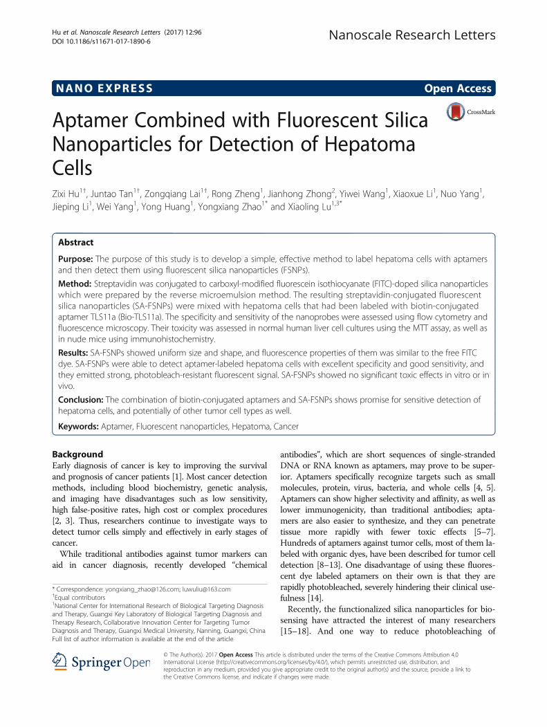
gating aptamers directly to FSNPs, we have developed analternative approach in which the aptamer and FSNP arephysically separate but interact via extremely strongbiotin-streptavidin interaction (Fig. 1). HepG2 cells are in-cubated first with biotin-labeled TLS11a aptamer (Bio-TLS11a) and then with streptavidin-conjugated FSNPs(SA-FSNPs). The SA-FSNPs then bind and interact withcells where the biotin-labeled aptamer has bound. Thisapproach avoids the limitations intrinsic to nanoparticlesurface modification, and it may allow efficient, sensitivedetection of cancer cells in vitro.
MethodsCells and AnimalsHuman hepatoma cell line HepG2, human normal liver cellline L02, and human embryonic kidney cell line 293T werepurchased from the Cell Bank of the Chinese Academy of
Sciences (Shanghai, China). All cell lines were culturedat 37 °C under 5% CO2 in DMEM supplemented with10% fetal bovine serum (FBS, Hyclone) and penicillin-streptomycin (Gibco, Grand Island, NY, USA).Female BALB/c nude mice aged 4–6 weeks were obtained
from the Guangxi Laboratory Animal Center (Guangxi,China) and housed in laminar flow cabinets underpathogen-free conditions. All experimental protocols wereapproved by the Animal Ethics Committee of GuangxiMedical University (Nanning, Guangxi, China).
ReagentsFluorescein isothiocyanate (FITC), cyclohexane, TritonX-100, n-hexanol, bovine serum albumin (BSA), acetone,tetraethyl orthosilicate (TEOS), (3-aminopropyl) triethoxysilane (APTES), 3-aminopropylmethyldimethoxysilane(APTMS), 1-ethyl-3-(3-dimethylaminopropyl) carbodiimide hydrochloride (EDC) N-hydroxysulfosuccinimide so-dium salt (sulfo-NHS) and polyoxymethylene were boughtfrom Sigma (St. Louis, MO, USA). Ethanol, dimethyl sulf-oxide (DMSO), 3-(4,5-dimethylthiazol-2-yl)-2,5-diphenyl-tetrazolium bromide (MTT), and hematoxylin-eosin (HE)were purchased from Solarbio (Beijing, China). The nu-clear dye 4′,6-diamidino-2-phenylindole (DAPI) was pur-chased from Life Technologies (USA). The biotin-labeledaptamer 5′-bio-(CH2)6-AGTAATGCCCGGTAGTTATTCAAAGATGAGTAGGAAAAGA-3′ (Bio-TLS11a) andFITC-labeled aptamer 5′-FITC-AGTAATGCCCGGTAGTTATTCAAAGATGAGTAGGAAAAGA-3′ (FITC-TLS11a) were synthesized by Shanghai Sangon Biotechnology(Shanghai, China).
Preparation and Characterization of SA-FSNPsFITC-doped, carboxyl-modified FSNPs were synthesized asdescribed [14, 29, 30]. Briefly, a water-in-oil microemulsion
Fig. 1 Schematic illustration of highly sensitive detection of HepG2 hepatoma cells using a biotin-conjugated aptamer (Bio-TLS11a) andstreptadivin-conjugated fluorescent silica nanoparticles (FSNPs)
Hu et al. Nanoscale Research Letters (2017) 12:96 Page 2 of 8
was prepared with FITC, cyclohexane, Triton X-100, n-hex-anol, and distilled water, giving rise to FITC-doped silicananoparticles. These FSNPs were amine-modified usingTEOS and APTES; the flocculent precipitate was collectedby centrifugation and washed with acetone, followed bydeionized water. The precipitate (2 mg) was dissolved in1 mL of 0.1-M phosphate-buffered saline (PBS, pH 7.4)containing EDC (1 mg) and sulfo-NHS (2.5 mg). When thereaction was complete, 50 μl of streptavidin diluted in PBSwas added to the solution, which was incubated at roomtemperature for 4 h with gentle shaking. The nanoparticleswere washed with PBS and then resuspended in 1 ml of0.05% BSA for 30 min to block free carboxylates, generatingSA-FSNPs. The SA-FSNPs were washed three times withPBS and stored at 4 °C. For subsequent experiments, theSA-FSNPs were resuspended in PBS as needed.The morphology and size distribution of SA-FSNPs
were assessed using transmission electron microscopy(TEM; H-7650, Japan). Their photoluminescence wasmeasured using a fluorescence spectrophotometer (FL-7000, Perkin Elmer, USA).
Flow Cytometry of Aptamer-Labeled Cells Mixed with SA-FSNPsHepG2 or L02 cells (3.0 × 10⁵ cells/ml) were harvested,washed three times with PBS, then incubated with for30 min either with SA-FSNPs (ca. 0.1 mg, 1 ml) at roomtemperature or with FITC-TLS11a (100 nM) on ice. Ineither case, the cells and labeling agents were suspendedin binding buffer (200 μl) prepared by supplementingPBS with 4.5 g/L of glucose and 5 mM of MgCl2. Othercell suspensions were incubated with Bio-TLS11a(100 nM) at 4 °C for 30 min, followed by SA-FSNPs (ca.0.1 mg, 1 ml) at 37 °C for 60 min with gentle shaking.All suspensions were washed three times with PBS, sus-pended in 500 μl of binding buffer, and then analyzed byflow cytometry (Epics XL, Beckman Coulter, USA) usingFLOWJO 7.6 software.
Fluorescence Microscopy of Aptamer-Labeled Cells Mixedwith SA-FSNPsHepG2 and L02 cells were cultured for 12 h in 6-well plates (3 × 10⁵ cells per well). Cells were washedthree times with cold PBS, fixed for 15 min with 4%polyoxymethylene, washed with PBS, and then incu-bated with SA-FSNPs or FITC-TLS11a, or the se-quential combination of Bio-TLS11a followed by SA-FSNPs as described above. Finally, cells were stainedwith DAPI for 90 s, washed with PBS, and analyzedby fluorescence microscopy (DS-Ri1; Nikon Corporation,Tokyo, Japan). Fluorescence intensity was quantitatedusing Image Pro (Media Cybernetics, Bethesda, MD,USA).
In Vitro Toxicity of SA-FSNPsToxicity of SA-FSNPs against 293T or L02 cells wasassessed using the MTT assay. Cells (2 × 10⁵ /ml) werecultured overnight in 96-well plates, then treated withSA-FSNPs (0.1, 0.5, or 1.0 mg/ml) for 12, 24, or 48 h.Control cells were treated with PBS. At specific timepoints, 10 μl of MTT (5 mg/ml) was added to wells, andplates were incubated at room temperature for 4 h inthe dark. The medium was discarded, 150 μl of DMSOwas added to each well, and plates were incubated for10 min. Optical density (OD) at 570 nm was measuredusing an ELISA microplate reader (Thermo Scientific,USA). Cell viability was calculated using the formula:
Viability %ð Þ ¼ ODexperimental=ODcontrol � 100 %:
In Vivo Toxicity of SA-FSNPsNude mice received a single tail vein injection of 200-μlPBS or SA-FSNPs (1 mg/ml) (n = 3 animals per group).After 1 week, the animals were sacrificed, and the majortissues (heart, lung, liver, spleen, kidney) were immersedin 10% formaldehyde solution, dehydrated, and paraffin-embedded. Paraffin sections (4 μm thick) were processedusing routine methods and stained with HE.
Statistical AnalysesStatistical analysis was performed using Student’s t testand analysis of variance (ANOVA) in GraphPad Prismsoftware (San Diego, CA, USA), with P < 0.05 defined asthe significance threshold. Data were shown as mean ±SD or as median (range).
Results and DiscussionHere, we explored the possibility of detecting humanhepatoma HepG2 cells, a common cell model for livercancer studies, using aptamer TLS11a, which was originallyselected through the SELEX method to bind specifically toHepG2 cells and which shows promise for targeteddiagnostics and therapy of hepatocellular carcinoma[10, 31–33]. In contrast to previous approaches in whichthe aptamer was conjugated to the surface of FSNPs,potentially limiting the sensitivity of aptamer-based detec-tion, we kept the aptamer and FSNPs physically separatebut we conjugated the former to biotin and the latter tostreptavidin to allow for strong, specific interaction. Separ-ating aptamer binding to target cells from FSNP bindingto aptamer may allow a larger number of aptamers to bindto each target cell, amplifying the fluorescence signal.
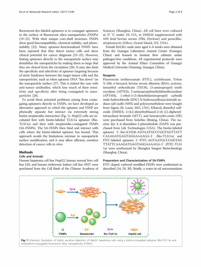
Characterization of SA-FSNPsTransmission electron microscopy showed SA-FSNPs tobe nearly monodisperse and spherical, with an averagediameter of 75.47 ± 2.52 nm (Fig. 2a). The core-shell
Hu et al. Nanoscale Research Letters (2017) 12:96 Page 3 of 8
structure of silica nanoparticles allows fluorescent dyessuch as FITC to be trapped inside [34, 35]. Using rhoda-mine B in ethanol solution as a reference [36], the fluor-escence quantum yields of FITC dye-doped silicananoparticles were about 0.52. The maximum emission
wavelength of free FITC dye and SA-FSNPs was 522 and525 nm, respectively (Fig. 2b). The emission peak of SA-FSNPs is slightly red-shifted from FITC, which may bedue to the loss of energy due to the interaction of silicasubstrate with the dye [37].
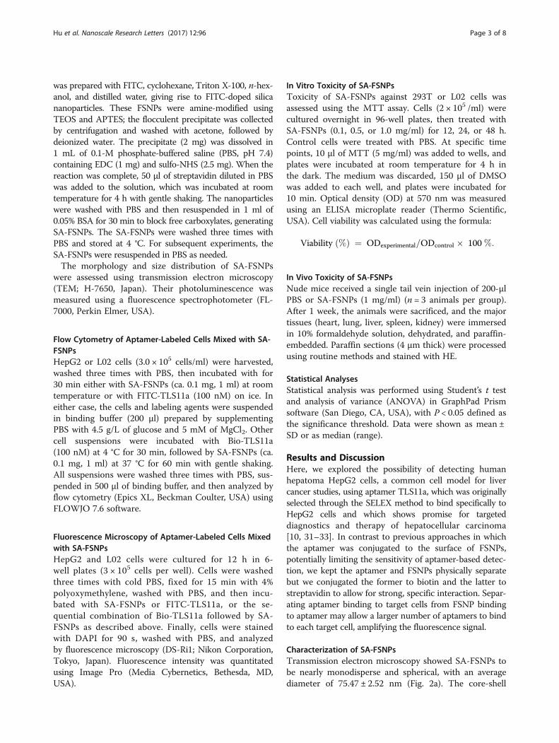
Fig. 3 a Flow cytometric detection of HepG2 cells (a) or L02 cells (b) after incubation with SA-FSNPs or FITC-TLS11a or the combination of Bio-TLS11a and SA-FSNPs. b Quantitative analysis of HepG2 cells (a) or L02 cells (b). NS not significant. **P < 0.01, ***P < 0.001
Fig. 2 Characterization of SA-FSNPs. a Transmission electron micrograph of SA-FSNPs. b Fluorescence emission spectra of FITC dye and SA-FSNPs
Hu et al. Nanoscale Research Letters (2017) 12:96 Page 4 of 8

Flow Cytometry of aptamer-Labeled Cells Mixed with SA-FSNPsTo determine whether the synthesized SA-FSNPs can beused as a detection probe for aptamer-labeled cells, HepG2cells were firstly reacted with Bio-TLS11a. After washing,HepG2 cells was then incubated with SA-FSNPs. L02 cells
served as negative cells, and FITC-TLS11a was used as acontrol probe. For the detection of HepG2 cells, strongerfluorescence intensity was found on Bio-TLS11a combinedwith SA-FSNPs (Bio-TLS11a + SA-FSNPs) and FITC-TLS11a, while no obvious fluorescence signal was observedon SA-FSNPs alone (Fig. 3a, panel a). Statistical graph of
Fig. 4 Fluorescence micrographs of HepG2 and L02 cells after incubation with SA-FSNPs or FITC-TLS11a or the combination of Bio-TLS11a andSA-FSNPs. SA-FSNPs and FITC were examined in the green channel, while DAPI-stained nuclei were examined in the blue channel
Fig. 5 Photostability of FITC-TLS11a and of the combination of Bio-TLS11a with SA-FSNPs. a Fluorescence micrographs of HepG2 cells labeled with thecombination of Bio-TLS11a and SA-FSNPs (upper row) or with FITC-TLS11a alone (lower row) and then continuously irradiated for the indicated periods. bQuantitative analysis of fluorescence intensity after different irradiation periods. ***P< 0.001
Hu et al. Nanoscale Research Letters (2017) 12:96 Page 5 of 8
the binding rate of HepG2 cells showed the similar results(Fig. 3a, panel b). Additionally, there was no fluorescencesignal on L02 cells after treating with SA-FSNPs alone,FITC-TLS11a and Bio-TLS11a + SA-FSNPs, respectively(Fig. 3b, panel a), in accordance with the results of statis-tical graph of the binding rate of L02 cells (Fig. 3b, panel b).These results suggest that the sequential combination ofBio-TLS11a with SA-FSNPs can detect HepG2 cells withhigher specificity than FITC-TLS11a.
Fluorescence Microscopy of Aptamer-Labeled Cells Mixedwith SA-FSNPsTo allow a more direct visualization of HepG2 detectionusing our system, we used fluorescence microscopy toexamine HepG2 cells incubated with SA-FSNPs or FITC-TLS11a or Bio-TLS11a + SA-FSNPs. As can be seendistinctly in fluorescence images, both FITC-TLS11a andBio-TLS11a + SA-FSNPs showed green fluorescence onperiphery of HepG2 cells, while SA-FSNPs did not. Fur-thermore, the fluorescence intensity of Bio-TLS11a + SA-FSNPs was stronger than FITC-TLS11a (Fig. 4a). No greenfluorescence was observed on L02 cells after incubation
with SA-FSNPs alone, FITC-TLS11a and Bio-TLS11a +SA-FSNPs, respectively (Fig. 4b), which was consistentwith the analysis of flow cytometry. Therefore, we couldagree that aptamer TLS11a could recognize and bindHepG2 cells with high affinity and specificity. Moreover,fluorescence signal from HepG2 cells is owing to theinteraction between Bio-TLS11a labeled HepG2 cells andthe SA-FSNPs. The SA-FSNPs display stronger fluores-cent signals than the FITC-labeled aptamer probably dueto the special core-shell structure of silica nanoparticleswhich allow the fluorescent dyes entrapped inside to pre-vent them from photodamaging oxidation [38–42].
Photostability of SA-FSNPsFluorescent dye molecules can quench easily after irradi-ation, limiting their usefulness. Doping fluorophores withinporous silica nanoparticles can improve their photostabilitywhile maintaining their strong fluorescence emission[34, 35]. We measured the photostability of SA-FSNPsby mixing them with aptamer-labeled HepG2 cells andimaging the cells by fluorescence microscopy aftercontinuous illumination lasting 0, 1, 5, and 10 min. In
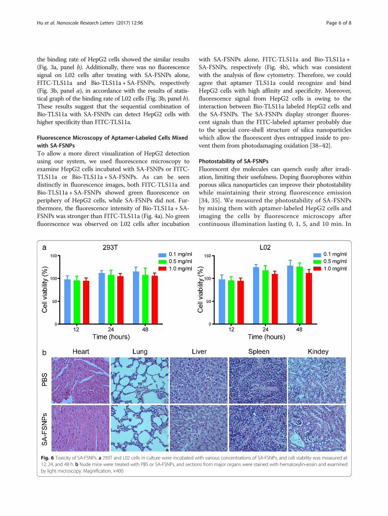
Fig. 6 Toxicity of SA-FSNPs. a 293T and L02 cells in culture were incubated with various concentrations of SA-FSNPs, and cell viability was measured at12, 24, and 48 h. b Nude mice were treated with PBS or SA-FSNPs, and sections from major organs were stained with hematoxylin-eosin and examinedby light microscopy. Magnification, ×400
Hu et al. Nanoscale Research Letters (2017) 12:96 Page 6 of 8
parallel, cells treated with FITC-aptamer alone wereimaged in the same way. Green fluorescence from SA-FSNPs remained clearly visible even after intense irradi-ation for 10 min, whereas fluorescence from FITC-TLS11a had nearly disappeared after 2 min (Fig. 5). Theseresults are consistent with the idea that fluorescent dyemolecules are encapsulated within the silica matrix,where they are kept separate from potential quenchersand photo-oxidizers [39–42].
Toxicity of SA-FSNPsWe assessed the cytotoxicity of SA-FSNPs on cul-tures of the normal cell lines 293T and L02. Viabilityof both cell lines was high according to the MTTassay after incubation with various SA-FSNP concen-trations (Fig. 6a), suggesting that SA-FSNP showedminimal cytotoxicity. However, FSNPs have a shorthalf-life in the circulatory system, and the entry offluorescent dye molecules into the blood may in-crease the risk of systemic toxicity [43]. Therefore, itis necessary to evaluate the toxicity of SA-FSNPs invivo. We further studied the in vivo toxicity of SA-FSNPs in nude mice. After intravenous injection ofSA-FSNPs for 1 week, tissue sections of the main or-gans were stained with HE. As shown in Fig. 6b,there were no significant inflammation or necrosisobserved on tissue sections. These results confirmedthat SA-FSNPs were almost non-toxic to the mainorgans, showing the potential to be clinically usefulas a diagnostic probe.
ConclusionsWe have developed an approach to detect hepatomacells based on a biotin-labeled aptamer and streptavidin-modified FSNPs. The strong affinity and specificity ofbiotin-TLS11a for HepG2 tumor cells, coupled with theaffinity and specificity of biotin for the streptavidin inSA-FSNPs, ensure highly specific and sensitive HepG2detection. In addition, the fluorescence signal from SA-FSNPs is much stronger and more photostable than thesignal from the FITC-labeled aptamer. SA-FSNPs do notshow obvious toxic effects in vitro or in nude mice,based on the MTT assay or histology of major organs.This two-step labeling system may be adaptable to thedetection of other cancers by changing the aptamer. Inaddition, this system may become a useful platform fortargeted therapy if the nanoparticles can be loaded withanti-tumor drugs or microRNAs.
AbbreviationsAPTES: (3-aminopropyl) triethoxysilane; APTMS: 3-aminopropylmethyldimethoxysilane; Bio-TLS11a: Biotin-conjugated aptamer TLS11a; BSA: Bovineserum albumin; DAPI: 4′,6-diamidino-2-phenylindole; DMSO: Dimethylsulfoxide; EDC: 1-ethyl-3-(3-dimethylaminopropyl) carbodiimide hydrochloride;FITC: Fluorescein isothiocyanate; FSNPs: Fluorescent silica nanoparticles;HE: Hematoxylin-eosin; MTT: 3-(4,5-dimethylthiazol-2-yl)-2,5-diphenyltetrazolium
bromide; OD: Optical density; SA-FSNPs: Streptavidin-conjugated fluorescentsilica nanoparticles; Sulfo-NHS: N-hydroxysulfosuccinimide sodium salt;TEM: Transmission electron microscopy; TEOS: Tetraethyl orthosilicate
AcknowledgementsThe authors acknowledge support from the Key Project of National NaturalScience Foundation of China (No. 81430055), the Program for ChangjiangScholars and Innovative Research Teams at University (Ministry of Educationof China, IRT_15R13), the International Cooperation Project of the Ministry ofScience and Technology of China (20 15DFA31320), and the Project forInnovative Research Teams (Guangxi Natural Science Foundation,2015GXNSFFA139001).
Authors’ ContributionsZXH, YXZ, and XLL designed the experiments. ZXH, ZQL, and YWW preparedthe nanoparticles. RZ, JHZ, XXL, and NY performed the experiments. JPL, WY,and YH analyzed the data. ZXH and JTT wrote the manuscript. All authorsread and approved the final manuscript.
Competing InterestsThe authors declare that they have no competing interests.
Author details1National Center for International Research of Biological Targeting Diagnosisand Therapy, Guangxi Key Laboratory of Biological Targeting Diagnosis andTherapy Research, Collaborative Innovation Center for Targeting TumorDiagnosis and Therapy, Guangxi Medical University, Nanning, Guangxi, China.2Surgery Oncology Department, Affiliated Tumor Hospital of GuangxiMedical University, Nanning, China. 3The Department of Immunology,Guangxi Medical University, Nanning, Guangxi, China.
Received: 1 December 2016 Accepted: 12 January 2017
References1. Etzioni R, Urban N, Ramsey S, McIntosh M, Schwartz S, Reid B, Radich J,
Anderson G, Hartwell L (2003) The case for early detection. Nat Rev Cancer3:243–2452
2. Valente K, Yacoub G, Cappellari JO, Parks G (2016) Metastatic pancreaticacinar cell carcinoma in a younger male with marked AFP production: Apotential pitfall on fine needle aspiration biopsy. Diagn Cytopathol doi. doi:10.1002/dc.23610
3. Sabour L, Sabour M, Ghorbian S (2016) Clinical applications of next-generation sequencing in cancer diagnosis. Pathol Oncol Res doi. doi:10.1007/s12253-016-0124-z
4. Ganji A, Varasteh A, Sankian M (2016) Aptamer: new arrows to targetdendritic cells. J Drug Target 24:1–12
5. Groff K, Brown J, Clippinger AJ (2015) Modern affinity reagents: recombinantantibodies and aptamers. Biotechnol Adv 33:1787–1798
6. Meng H, Liu H, Kuai H, Peng R, Mo L, Zhang X (2016) Aptamer-integratedDNA nanostructures for biosensing, bioimaging and cancer therapy. ChemSoc Rev 45:2583–2602
7. Tan W, Donovan M, Jiang J (2013) Aptamers from cell-based selection forbioanalytical applications. Chem Rev 113:2842–2862
8. Tang Z, Shangguan D, Wang K, Shi H, Sefah K, Mallikratchy P, Chen HW, LiY, Tan W (2007) Selection of aptamers for molecular recognition andcharacterization of cancer cells. Anal Chem 79:4900–4907
9. Chen H, Medley CD, Sefah K, Shangguan D, Tang Z, Meng L, Smith JE, TanW (2008) Molecular recognition of small-cell lung cancer cells usingaptamers. ChemMedChem 3:991–1001
10. Shangguan D, Meng L, Cao Z, Xiao Z, Fang X, Li Y, Cardona D, Witek RP, LiuC, Tan W (2008) Identification of liver cancer-specific aptamers using wholelive cells. Anal Chem 80:721–728
11. Li W, Bing T, Wei J, Chen Z, Shangguan D, Fang J (2014) Cell-SELEX-basedselection of aptamers that recognize distinct targets on metastaticcolorectal cancer cells. Biomaterials 35:6998–7007
12. Wang Y, Luo Y, Bing T, Chen Z, Lu M, Zhang N, Shangguan D, Gao X (2014)DNA aptamer evolved by cell-SELEX for recognition of prostate cancer.PLoS One 9, e100243
Hu et al. Nanoscale Research Letters (2017) 12:96 Page 7 of 8

13. Sefah K, Bae KM, Phillips JA, Siemann DW, Su Z, McClellan S, Vieweg J, TanW (2013) Cell-based selection provides novel molecular probes for cancerstem cells. Int J Cancer 132:2578–2588
14. Tan J, Yang N, Hu Z, Su J, Zhong J, Yang Y, Yu Y, Zhu J, Xue D, Huang Y, Lai Z,Huang Y, Lu X, Zhao Y (2016) Aptamer-functionalized fluorescent silicananoparticles for highly sensitive detection of leukemia cells. Nanoscale ResLett 11:298
15. Wu Y, Chen C, Liu S (2009) Enzyme-functionalized silica nanoparticles assensitive labels in biosensing. Anal Chem 81:1600–1607
16. Qian J, Zhang C, Cao X, Liu S (2010) Versatile immunosensor using aquantum dot coated silica nanosphere as a label for signal amplification.Anal Chem 82:6422–6429
17. Yuan L, Xu L, Liu S (2012) Integrated tyramide and polymerization-assisted signalamplification for a highly-sensitive immunoassay. Anal Chem 84:10737–10744
18. Chen L, Chen C, Li R, Li Y, Liu S (2009) CdTe quantum dot functionalizedsilica nanosphere labels for ultrasensitive detection of biomarker. ChemCommun (Camb) 21:2670–2672
19. Wang K, He X, Yang X, Shi H (2013) Functionalized silica nanoparticles: aplatform for fluorescence imaging at the cell and small animal levels.Acc Chem Res 46:1367–1376
20. Smith JE, Medley CD, Tang Z, Shangguan D, Lofton C, Tan W (2007)Aptamer-conjugated nanoparticles for the collection and detection ofmultiple cancer cells. Anal Chem 79:3075–3082
21. Herr JK, Smith JE, Medley CD, Shangguan D, Tan W (2006) Aptamer-conjugatednanoparticles for selective collection and detection of cancer cells.Anal Chem 78:2918–2924
22. Geng J, Liu J, Liang J, Shi H, Liu B (2013) A general approach to prepareconjugated polymer dot embedded silica nanoparticles with aSiO2@CP@SiO2 structure for targeted HER2-positive cellular imaging.Nanoscale 5:8593–8601
23. Özalp VC, Çam D, Hernandez FJ, Hernandez LI, Schäfer T, Öktem HA (2016)Small molecule detection by lateral flow strips via aptamer-gated silicananoprobes. Analyst 141:2595–2599
24. Li H, Mu Y, Lu J, Wei W, Wan Y, Liu S (2014) Target-cell-specific fluorescencesilica nanoprobes for imaging and theranostics of cancer cells. Anal Chem86:3602–3609
25. Hu H, Dai A, Sun J, Li X, Gao F, Wu L, Fang Y, Yang H, An L, Wu H, Yang S(2013) Aptamer-conjugated Mn3O4@SiO2 core-shell nanoprobes for targetedmagnetic resonance imaging. Nanoscale 5:10447–10454
26. Wu C, Liu J, Zhang P, Li J, Ji H, Yang X, Wang K (2015) A recognition-before-labeling strategy for sensitive detection of lung cancer cells with a quantumdot-aptamer complex. Analyst 140:6100–6107
27. Algar WR, Krull UJ (2006) Adsorption and hybridization of oligonucleotideson mercaptoacetic acid-capped CdSe/ZnS quantum dots and quantumdot-oligonucleotide conjugates. Langmuir 22:11346–11352
28. Pathak S, Davidson MC, Silva GA (2007) Characterization of the functional bindingproperties of antibody conjugated quantum dots. Nano Lett 7:1839–1845
29. Wang Q, Kang Y (2016) Bioprobes based on aptamer and silica fluorescentnanoparticles for bacteria salmonella typhimurium detection. Nanoscale ResLett 11:150
30. Bagwe RP, Yang C, Hilliard LR, Tan W (2004) Optimization of dye-dopedsilica nanoparticles prepared using a reverse microemulsion method.Langmuir 20:8336–8342
31. Sun D, Lu J, Zhong Y, Yu Y, Wang Y, Zhang B, Chen Z (2016) Sensitiveelectrochemical aptamer cytosensor for highly specific detection of cancercells based on the hybrid nanoelectrocatalysts and enzyme for signalamplification. Biosens Bioelectron 75:301–307
32. Chen X, Pan Y, Liu H, Bai X, Wang N, Zhang B (2016) Label-free detection ofliver cancer cells by aptamer-based microcantilever biosensor. BiosensBioelectron 79:353–358
33. Qu L, Xu J, Tan X, Liu Z, Xu L, Peng R (2014) Dual-aptamer modificationgenerates a unique interface for highly sensitive and specific electrochemicaldetection of tumor cells. ACS Appl Mater Interfaces 6:7309–7315
34. Lee JE, Lee N, Kim H, Kim J, Choi SH, Kim JH, Kim T, Song IC, Park SP, MoonWK, Hyeon T (2010) Uniform mesoporous dye-doped silica nanoparticlesdecorated with multiple magnetite nanocrystals for simultaneous enhancedmagnetic resonance imaging, fluorescence imaging, and drug delivery.J Am Chem Soc 132:552–557
35. Lee JE, Lee N, Kim T, Kim J, Hyeon T (2011) Multifunctional mesoporous silicananocomposite nanoparticles for theranostic applications. Acc Chem Res 44:893–902
36. Karstens T, Kobs K (2002) Rhodamine B and rhodamine 101 as referencesubstances for fluorescence quantum yield measurements. Journal ofPhysical Chemistry 84:1871–1872
37. Taton TA, Mirkin CA, Letsinger RL (2000) Scanometric DNA array detectionwith nanoparticle probes. Science 289:1757–1760
38. Cai L, Chen Z, Chen M, Tang H, Pang D (2013) MUC-1 aptamer conjugated dye-doped silica nanoparticles for MCF-7 cells detection. Biomaterials 34:371–381
39. Liu A, Wu L, He Z, Zhou J (2011) Development of highly fluorescent silicananoparticles chemically doped with organic dye for sensitive DNAmicroarray detection. Anal Bioanal Chem 401:2003–2011
40. Burns A, Sengupta P, Zedayko T, Baird B, Wiesner U (2006) Core/shellfluorescent silica nanoparticles for chemical sensing: towards single-particlelaboratories. Small 2:723–726
41. Zhao X, Bagwe RP, Tan W (2004) Development of organic-dye-doped silicananoparticles in a reverse microemulsion. Adv Mater 16:173–176
42. Liu F, Ni ASY, Lim Y, Mohanram H, Bhattacharjya S, Xing B (2012)Lipopolysaccharide neutralizing peptide-porphyrin conjugates for effectivephotoinactivation and intracellular imaging of gram-negative bacteriastrains. Bioconjugate Chem 23:1639–1647
43. Jo H, Her J, Ban C (2015) Dual aptamer-functionalized silica nanoparticles forthe highly sensitive detection of breast cancer. Biosens Bioelectron 71:129–136
Submit your manuscript to a journal and benefi t from:
7 Convenient online submission
7 Rigorous peer review
7 Immediate publication on acceptance
7 Open access: articles freely available online
7 High visibility within the fi eld
7 Retaining the copyright to your article
Submit your next manuscript at 7 springeropen.com
Hu et al. Nanoscale Research Letters (2017) 12:96 Page 8 of 8