Embed Size (px)
Citation preview
The Muscular SystemThe Muscular System
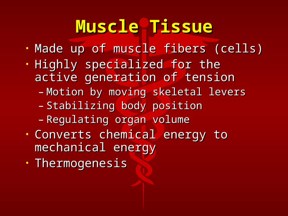
Muscle TissueMuscle Tissue• Made up of muscle fibers (cells)Made up of muscle fibers (cells)• Highly specialized for the active Highly specialized for the active
generation of tensiongeneration of tension– Motion by moving skeletal leversMotion by moving skeletal levers– Stabilizing body positionStabilizing body position– Regulating organ volumeRegulating organ volume
• Converts chemical energy to Converts chemical energy to mechanical energymechanical energy
• ThermogenesisThermogenesis
Functions of Muscle Functions of Muscle TissueTissue
• Movement FacilitationMovement Facilitation• ThermogenesisThermogenesis• Postural SupportPostural Support• Regulation of Organ VolumeRegulation of Organ Volume• Protects Internal Organs Protects Internal Organs • Pumps Blood Pumps Blood (HEART)(HEART)
Characteristics of Characteristics of Muscle TissueMuscle Tissue
• ContractilityContractility• Extensibility (Flexibility)Extensibility (Flexibility)• ElasticityElasticity• Excitability (Irritability)Excitability (Irritability)
Skeletal MuscleSkeletal Muscle
• Attached to bonesAttached to bones• Striated appearance under a microscopeStriated appearance under a microscope• Voluntary control (conscious control)Voluntary control (conscious control)• MultinucleatedMultinucleated• Sarcolemma - muscle cell membraneSarcolemma - muscle cell membrane• Sarcoplasm - muscle cell cytoplasmSarcoplasm - muscle cell cytoplasm• Myofilaments - contractile elements of Myofilaments - contractile elements of
each muscle fibereach muscle fiber
Connective Tissue Connective Tissue Components of Skeletal Components of Skeletal
Muscle Muscle• Epimysium - surrounds entire muscleEpimysium - surrounds entire muscle• Perimysium - surrounds a bundle of Perimysium - surrounds a bundle of
muscle fibers (fascicles)muscle fibers (fascicles)• Endomysium - surrounds each Endomysium - surrounds each
individual muscle fiberindividual muscle fiber• Epimysium, Perimysium, Endomysium Epimysium, Perimysium, Endomysium
converge at the end of a muscle to form converge at the end of a muscle to form a tendon or aponeurosisa tendon or aponeurosis
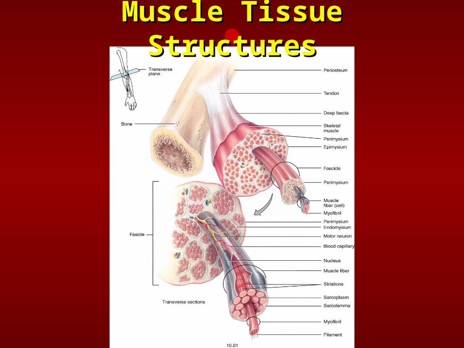
Muscle Tissue Muscle Tissue StructuresStructures
Muscle Tissue Muscle Tissue StructuresStructures
Muscle Attachments to Muscle Attachments to BoneBone
• Tendon - round, cord like fibrous Tendon - round, cord like fibrous connective tissueconnective tissue
• Aponeurosis - broad, flat sheet of Aponeurosis - broad, flat sheet of connective tissueconnective tissue
• Tendon Sheaths - specialized tendons Tendon Sheaths - specialized tendons that are enclosed tubes of fibrous that are enclosed tubes of fibrous connective tissueconnective tissue– Contain a film of synovial fluidContain a film of synovial fluid– Similar in function to bursaeSimilar in function to bursae
Muscle Tissue Muscle Tissue HistologyHistology
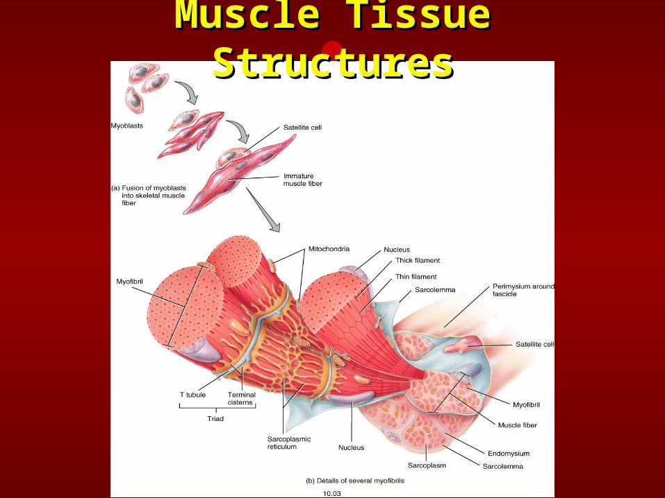
• Muscle Fiber - elongated cylindrical cellMuscle Fiber - elongated cylindrical cell• Sarcolemma - muscle cell membraneSarcolemma - muscle cell membrane• Sarcoplasm - muscle cell cytoplasmSarcoplasm - muscle cell cytoplasm• Myofibrils - groups of contractile Myofibrils - groups of contractile
myofilaments that run longitudinally myofilaments that run longitudinally within the musclewithin the muscle
• Sarcomere - a small section or Sarcomere - a small section or compartment within a myofibril which compartment within a myofibril which serves as the unit of contractionserves as the unit of contraction
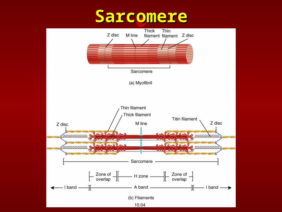
SarcomereSarcomere
Muscle Tissue HistologyMuscle Tissue Histology
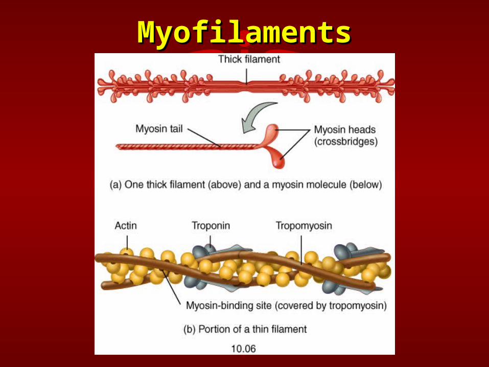
• Myofilaments - structural Myofilaments - structural components of myofibrilscomponents of myofibrils– Myosin - thick myofilamentsMyosin - thick myofilaments– Actin - thin myofilamentsActin - thin myofilaments
• Z Line - connective tissue that Z Line - connective tissue that separates individual sarcomeresseparates individual sarcomeres
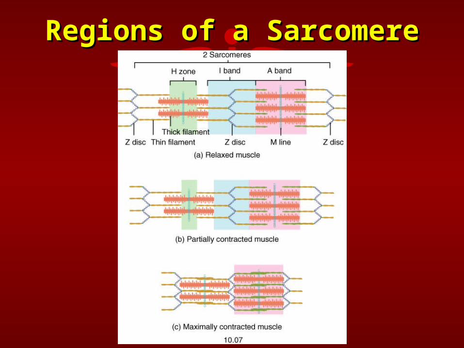
Regions of a SarcomereRegions of a Sarcomere
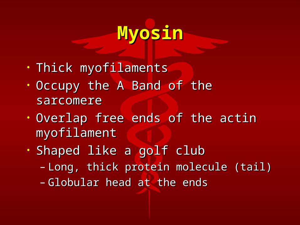
MyosinMyosin
• Thick myofilamentsThick myofilaments• Occupy the A Band of the Occupy the A Band of the
sarcomeresarcomere• Overlap free ends of the actin Overlap free ends of the actin
myofilamentmyofilament• Shaped like a golf clubShaped like a golf club
– Long, thick protein molecule (tail)Long, thick protein molecule (tail)– Globular head at the endsGlobular head at the ends
ActinActin• Thin myofilamentsThin myofilaments• Anchored to the Z LineAnchored to the Z Line• Two stranded protein molecule Two stranded protein molecule
intertwined around each otherintertwined around each other• Associated with two regulatory proteinsAssociated with two regulatory proteins
– Tropomyosin - long stranded protein Tropomyosin - long stranded protein molecule that follows the contour of actinmolecule that follows the contour of actin
– Troponin - protein located at regular interval Troponin - protein located at regular interval along the tropomyosin that covers the along the tropomyosin that covers the active sites on actin. Has three subunitsactive sites on actin. Has three subunits
MyofilamentsMyofilaments
Muscle Nerve Muscle Nerve InteractionInteraction
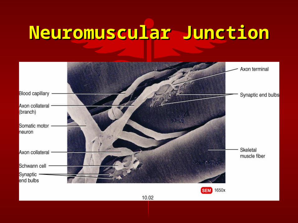
• Neuron - nerve cellNeuron - nerve cell• Axon - long, threadlike process that Axon - long, threadlike process that
transmits impulse away from cell transmits impulse away from cell body (may be up to 1 meter in length)body (may be up to 1 meter in length)
• Axon Terminal - branches of the axon Axon Terminal - branches of the axon after it enters the muscle tissueafter it enters the muscle tissue
• Motor Unit - motor neuron and all the Motor Unit - motor neuron and all the muscle fibers it innervatesmuscle fibers it innervates
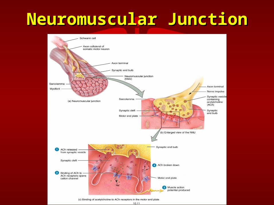
• Neuromuscular Junction - junction Neuromuscular Junction - junction between axon terminal and muscle between axon terminal and muscle fiberfiber
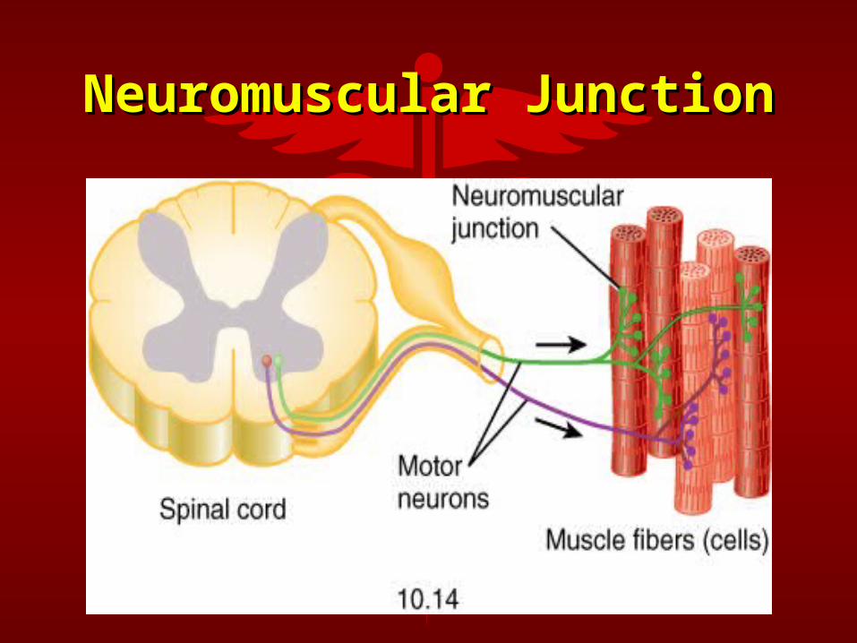
Neuromuscular JunctionNeuromuscular Junction
Neuromuscular JunctionNeuromuscular Junction
Neuromuscular JunctionNeuromuscular Junction
Muscle Nerve Muscle Nerve InteractionInteraction
• Motor End Plate - location on the Motor End Plate - location on the muscle fiber at the end of an axon muscle fiber at the end of an axon terminalterminal
• Synaptic End Bulb - distal end of Synaptic End Bulb - distal end of axon terminalaxon terminal
• Synaptic Vesicles - membrane Synaptic Vesicles - membrane enclosed sacs within the synaptic enclosed sacs within the synaptic end bulbs that store end bulbs that store neurotransmittersneurotransmitters
Muscle Nerve Muscle Nerve InteractionInteraction
• Synaptic Cleft - space between axon Synaptic Cleft - space between axon terminal and motor end plateterminal and motor end plate
• Subneural Clefts - folds in sarcolemma Subneural Clefts - folds in sarcolemma along the synaptic gutter along the synaptic gutter
• Acetylcholine (Ach) - neurotransmitter Acetylcholine (Ach) - neurotransmitter released from synaptic vesicles that released from synaptic vesicles that initiates an action potential in a initiates an action potential in a musclemuscle
Sarcoplasmic Reticulum Sarcoplasmic Reticulum and Transverse Tubulesand Transverse Tubules• Sarcoplasmic Reticulum - storage site Sarcoplasmic Reticulum - storage site
for calcium within the musclefor calcium within the muscle• Transverse (T) Tubules - invaginations Transverse (T) Tubules - invaginations
of the sarcolemma deep within the of the sarcolemma deep within the muscle muscle
• Terminal Cisternae - dilated end sacs of Terminal Cisternae - dilated end sacs of sarcoplasmic reticulum next to the T-sarcoplasmic reticulum next to the T-tubulestubules
• Triad - a T-tubule and the terminal Triad - a T-tubule and the terminal cisternae on both sides of itcisternae on both sides of it
Muscle Action PotentialMuscle Action Potential
An electrical impulse that An electrical impulse that originates at the motor end originates at the motor end plate, travels along the plate, travels along the length of the sarcolemma, length of the sarcolemma, down a transverse tubule, down a transverse tubule, and causes the muscle to and causes the muscle to contract.contract.
Sliding Filament Theory Sliding Filament Theory of Muscular Contractionof Muscular Contraction• Due to an action potential, the actin Due to an action potential, the actin
and myosin myofilaments slide past and myosin myofilaments slide past one another shortening the sarcomereone another shortening the sarcomere
• No change in length of myofilamentsNo change in length of myofilaments• H Zone narrows or disappearsH Zone narrows or disappears• I Band narrows or may disappearI Band narrows or may disappear• A Band remains the same lengthA Band remains the same length
Muscle Response to Muscle Response to Nervous StimuliNervous Stimuli
• All or None PrincipleAll or None Principle– Once a threshold stimulus is applied Once a threshold stimulus is applied
to a motor unit the muscle fibers to a motor unit the muscle fibers innervated by that motor unit will innervated by that motor unit will contract to their fullest potentialcontract to their fullest potential
• Threshold Stimulus - the weakest Threshold Stimulus - the weakest stimulus from a neuron that will stimulus from a neuron that will initiate a muscular contractioninitiate a muscular contraction
Events Leading to Events Leading to Muscular ContractionMuscular Contraction
• An action potential travels down the An action potential travels down the motor neuron. When it arrives at motor neuron. When it arrives at the synaptic knob, the membrane of the synaptic knob, the membrane of the nerve at the synaptic cleft is the nerve at the synaptic cleft is depolarized, thereby increasing the depolarized, thereby increasing the Ca++ permeability of the Ca++ permeability of the membrane.membrane.
• Ca++ diffuses from outside of the Ca++ diffuses from outside of the synaptic knob to inside the synaptic synaptic knob to inside the synaptic knob.knob.
• The influx of Ca++ into the nerve The influx of Ca++ into the nerve causes the release of Ach.causes the release of Ach.
• Ach is ejected into the synaptic Ach is ejected into the synaptic cleft, diffuses across the cleft, and cleft, diffuses across the cleft, and depolarizes the muscle membrane.depolarizes the muscle membrane.
• This increases the permeability of This increases the permeability of the muscle membrane to Na+.the muscle membrane to Na+.
• Na+ rushes into the muscle cell, Na+ rushes into the muscle cell, depolarizing the membrane as it depolarizing the membrane as it travels away from the motor end travels away from the motor end plate thus initiating an action plate thus initiating an action potential.potential.
• Ach is quickly broken down in the Ach is quickly broken down in the cleft by Ach-ase so that each action cleft by Ach-ase so that each action potential arriving from the nerve potential arriving from the nerve initiates only one action potential initiates only one action potential within the muscle.within the muscle.
• The action potential spreads across The action potential spreads across the muscle membrane and down the muscle membrane and down the T-tubules deep into the muscle the T-tubules deep into the muscle cell.cell.
• The action potential of the T-tubules The action potential of the T-tubules depolarizes the membrane of the depolarizes the membrane of the nearby sarcoplasmic reticulum nearby sarcoplasmic reticulum which results in the release of Ca++ which results in the release of Ca++ into the sarcoplasm.into the sarcoplasm.
• Ca++ is very quickly removed out of Ca++ is very quickly removed out of the sarcoplasm by the sarcoplasmic the sarcoplasm by the sarcoplasmic reticulum so the effects of one action reticulum so the effects of one action potential are very short lived and potential are very short lived and produce a very small contraction.produce a very small contraction.
• Many action potentials are necessary Many action potentials are necessary to produce enough force to produce a to produce enough force to produce a strong or prolonged muscle strong or prolonged muscle contraction.contraction.
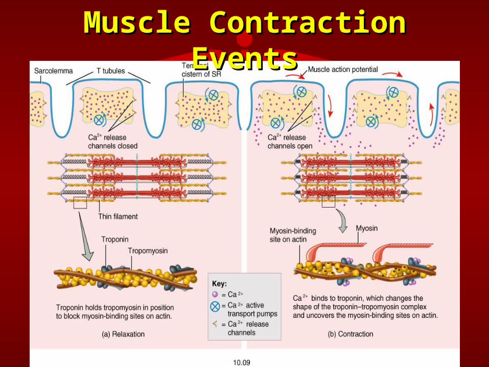
• The Ca++ released from the The Ca++ released from the sarcoplasmic reticulum binds with sarcoplasmic reticulum binds with troponin and cause troponin to troponin and cause troponin to change shape.change shape.
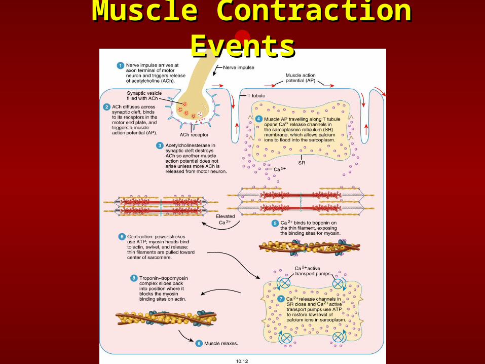
Muscle Contraction Muscle Contraction EventsEvents
• When troponin changes shape, it When troponin changes shape, it physically moves the other physically moves the other regulatory protein, tropomyosin, out regulatory protein, tropomyosin, out of the way exposing the active sites of the way exposing the active sites on the actin myofilament.on the actin myofilament.
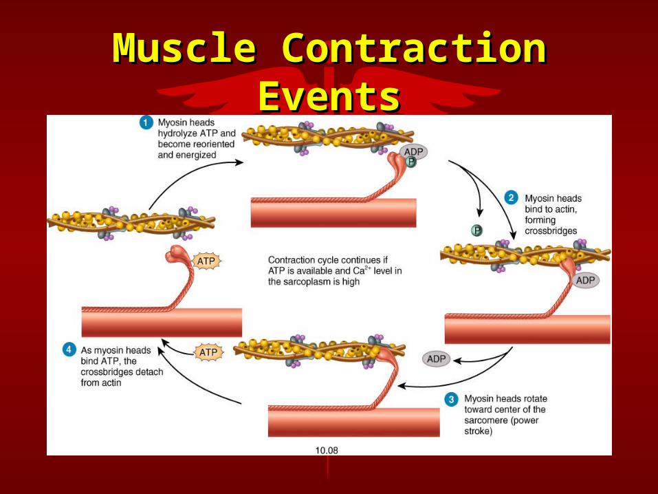
• Since the heads or cross-bridges of Since the heads or cross-bridges of myosin have a very strong affinity myosin have a very strong affinity for the active sites on actin, they for the active sites on actin, they make contact immediately after the make contact immediately after the active sites have been exposed.active sites have been exposed.
• The acto-myosin complex has The acto-myosin complex has ATPase activity and ATP is split into ATPase activity and ATP is split into ADP + P and energy is released.ADP + P and energy is released.
• The energy released by the splitting The energy released by the splitting of ATP is used to produce of ATP is used to produce movement of the cross-bridges, movement of the cross-bridges, sliding the actin and myosin sliding the actin and myosin filaments past one another which filaments past one another which causes the sarcomere to shorten causes the sarcomere to shorten and the muscle to contract and and the muscle to contract and produce force.produce force.
• The myosin cross-bridge has a low The myosin cross-bridge has a low affinity for ADP but a very high affinity for ADP but a very high affinity for ATP.affinity for ATP.
• It discards the ADP and becomes It discards the ADP and becomes recharged with a new ATP.recharged with a new ATP.
Muscle Contraction Muscle Contraction EventsEvents
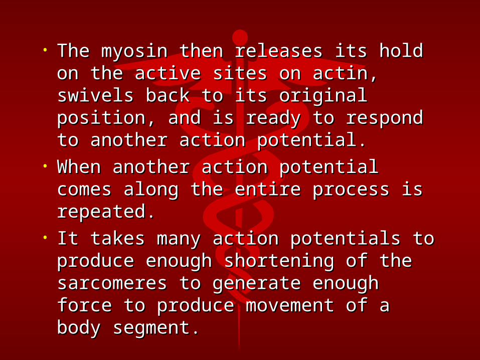
• The myosin then releases its hold on The myosin then releases its hold on the active sites on actin, swivels back the active sites on actin, swivels back to its original position, and is ready to to its original position, and is ready to respond to another action potential.respond to another action potential.
• When another action potential comes When another action potential comes along the entire process is repeated.along the entire process is repeated.
• It takes many action potentials to It takes many action potentials to produce enough shortening of the produce enough shortening of the sarcomeres to generate enough force sarcomeres to generate enough force to produce movement of a body to produce movement of a body segment.segment.
Muscle Contraction Muscle Contraction EventsEvents
Cardiac MuscleCardiac Muscle• Forms the bulk of heart wall (Myocardium)Forms the bulk of heart wall (Myocardium)• StriatedStriated• Involuntary (typically)Involuntary (typically)• Fibers are quadrangular and branchingFibers are quadrangular and branching• Cardiac fibers typically have a centrally Cardiac fibers typically have a centrally
located nucleuslocated nucleus• Sarcolemmas connected by intercalated Sarcolemmas connected by intercalated
discsdiscs– Strengthens cardiac muscle tissueStrengthens cardiac muscle tissue– Propagates an action potential from cell to cell Propagates an action potential from cell to cell
through specialized structures on the through specialized structures on the intercalated discs called gap junctionsintercalated discs called gap junctions
Smooth (Visceral) Smooth (Visceral) MuscleMuscle
• Located in walls of hollow internal Located in walls of hollow internal surfaces such as:surfaces such as:– blood vesselsblood vessels - stomach- stomach– urinary bladderurinary bladder- intestines- intestines
• Non-striated in appearanceNon-striated in appearance• Involuntary (typically)Involuntary (typically)• Can be stretched to great lengthsCan be stretched to great lengths• Allows for tremendous size variabilityAllows for tremendous size variability
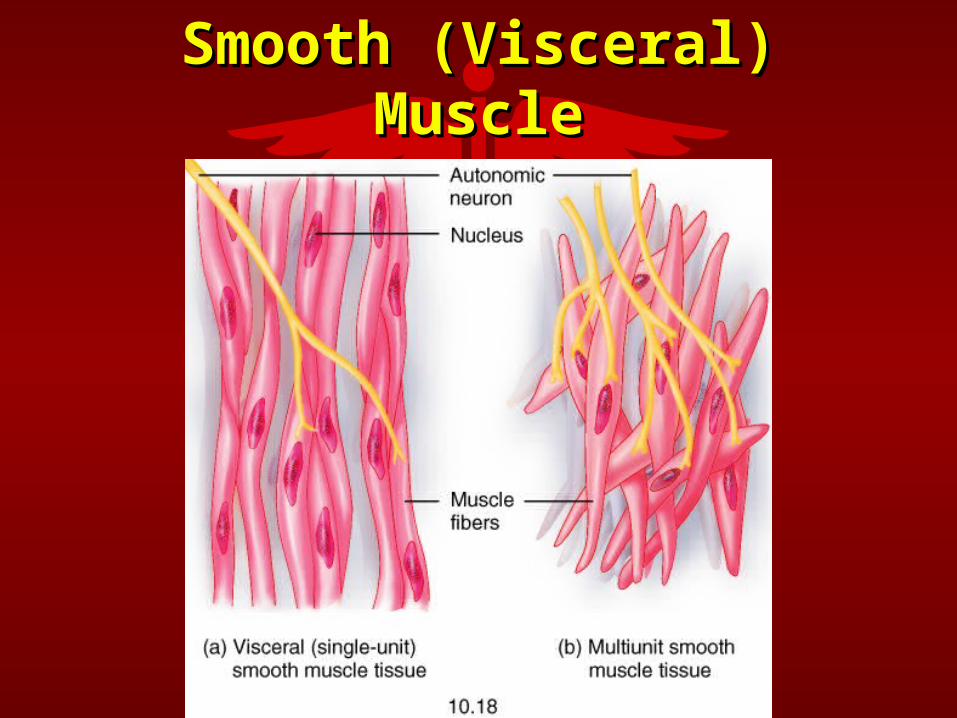
Smooth (Visceral) Smooth (Visceral) MuscleMuscle
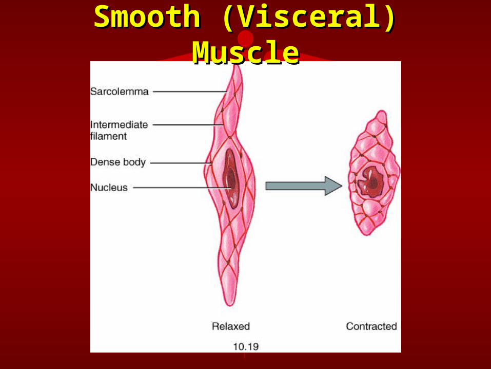
Smooth (Visceral) Smooth (Visceral) MuscleMuscle
Muscles and MovementMuscles and Movement
• Produce movement by contracting, Produce movement by contracting, creating tension on tendons, which pull on creating tension on tendons, which pull on bones or other structures.bones or other structures.
• Most muscles cross over at least one joint.Most muscles cross over at least one joint.• Attempt to pull bones together.Attempt to pull bones together.• Since muscles cross over a joint, the joint Since muscles cross over a joint, the joint
serves as the axis of rotation and the serves as the axis of rotation and the shortening muscle produces angular shortening muscle produces angular rotation.rotation.
Muscle Origin and Muscle Origin and InsertionInsertion
• OriginOrigin– Body segment with most massBody segment with most mass– Usually more proximally locatedUsually more proximally located– Usually larger surface area of attachmentUsually larger surface area of attachment
• InsertionInsertion– Body segment with least massBody segment with least mass– Usually more distally locatedUsually more distally located– Usually smaller surface area of attachmentUsually smaller surface area of attachment
• Gaster (Belly)Gaster (Belly)– Fleshy portion of the muscle between the Fleshy portion of the muscle between the
tendons of the origin and insertiontendons of the origin and insertion
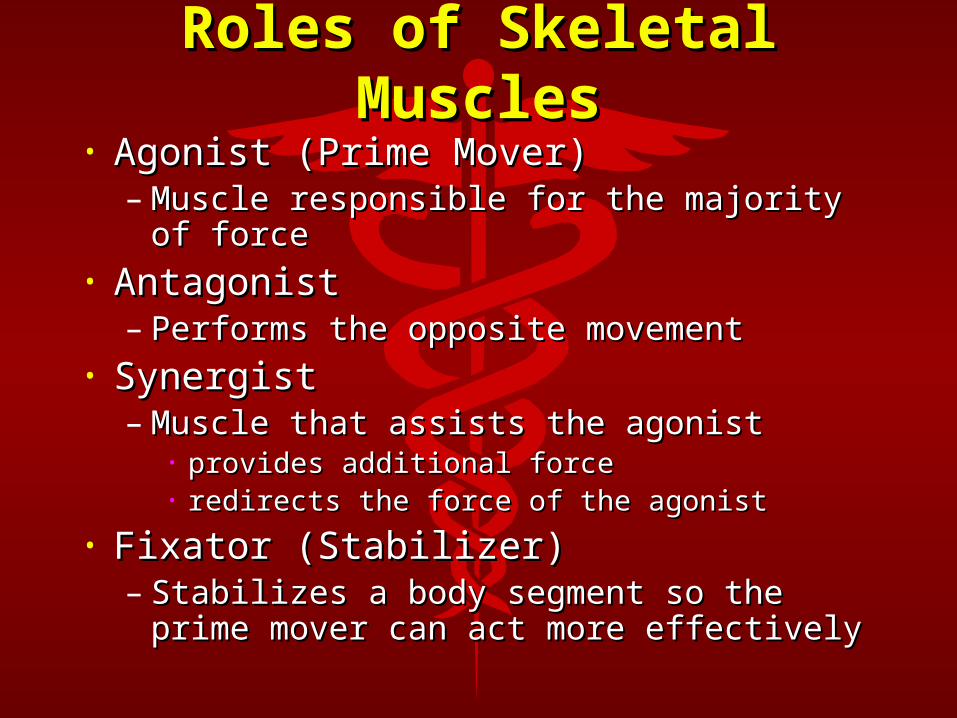
Roles of Skeletal Roles of Skeletal MusclesMuscles
• Agonist (Prime Mover)Agonist (Prime Mover)– Muscle responsible for the majority of Muscle responsible for the majority of
forceforce• AntagonistAntagonist
– Performs the opposite movement Performs the opposite movement • SynergistSynergist
– Muscle that assists the agonistMuscle that assists the agonist• provides additional forceprovides additional force• redirects the force of the agonistredirects the force of the agonist
• Fixator (Stabilizer)Fixator (Stabilizer)– Stabilizes a body segment so the prime Stabilizes a body segment so the prime
mover can act more effectivelymover can act more effectively
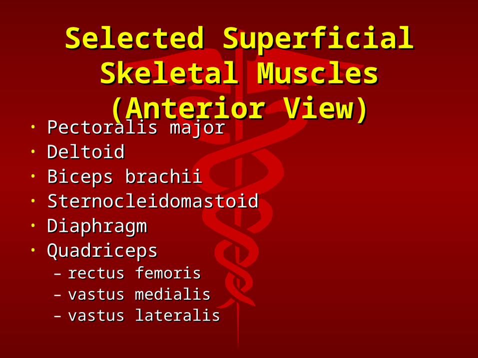
Selected Superficial Selected Superficial Skeletal Muscles Skeletal Muscles (Anterior View)(Anterior View)
• Pectoralis majorPectoralis major• DeltoidDeltoid• Biceps brachiiBiceps brachii• SternocleidomastoidSternocleidomastoid• DiaphragmDiaphragm• QuadricepsQuadriceps
– rectus femorisrectus femoris– vastus medialisvastus medialis– vastus lateralisvastus lateralis
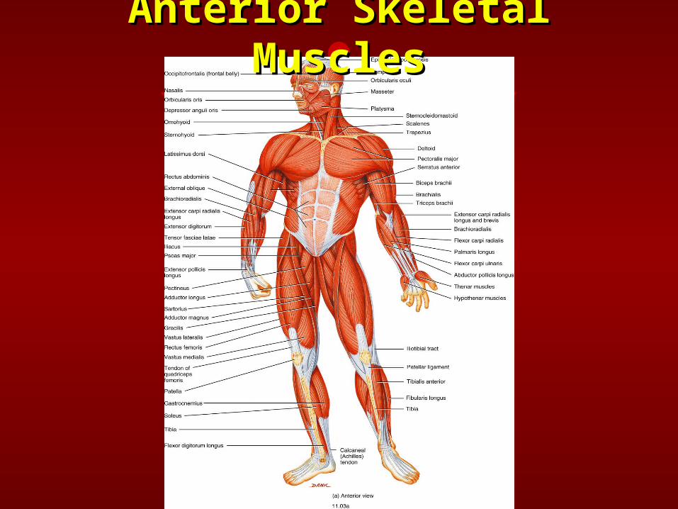
Anterior Skeletal Anterior Skeletal MusclesMuscles
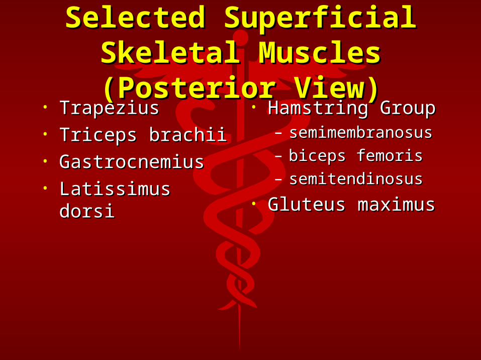
Selected Superficial Selected Superficial Skeletal Muscles Skeletal Muscles (Posterior View)(Posterior View)
• TrapeziusTrapezius• Triceps brachiiTriceps brachii• GastrocnemiusGastrocnemius• Latissimus dorsiLatissimus dorsi
• Hamstring GroupHamstring Group– semimembranosusemimembranosu
ss– biceps femorisbiceps femoris– semitendinosussemitendinosus
• Gluteus maximusGluteus maximus
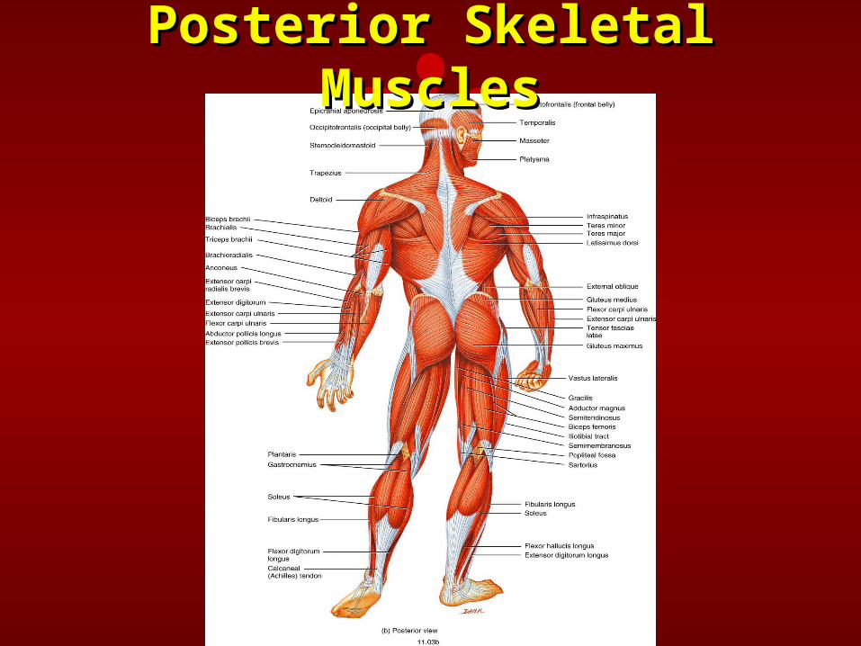
Posterior Skeletal Posterior Skeletal MusclesMuscles
Muscle Diseases and Muscle Diseases and DisordersDisorders
Myalgia (Fibromyalgia)Myalgia (Fibromyalgia)
• Painful disorders of muscles, Painful disorders of muscles, tendons, and surrounding soft tendons, and surrounding soft tissuetissue
Muscular DystrophiesMuscular Dystrophies
• Muscle destroying diseases Muscle destroying diseases characterized by the degeneration characterized by the degeneration of individual muscle fibersof individual muscle fibers
• Leads to progressive atrophy of Leads to progressive atrophy of skeletal musclesskeletal muscles
• Due to a genetic defectDue to a genetic defect
Shin SplintsShin Splints• Pain in the lower legPain in the lower leg• Tendonitis of the tibialis posterior muscleTendonitis of the tibialis posterior muscle• Inflammation of the periosteumInflammation of the periosteum• Stress fracture of the tibiaStress fracture of the tibia• Exaggerated enlargement of muscles Exaggerated enlargement of muscles
within the epimysium within the epimysium • Pulling away of the periosteum from the Pulling away of the periosteum from the
underlying boneunderlying bone• Treatment:Treatment:
– RICERICE– strengthen tibialis anterior musclestrengthen tibialis anterior muscle
SprainsSprains• the forcible wrenching or twisting the forcible wrenching or twisting
of a joint with partial or complete of a joint with partial or complete rupture or injury to joint rupture or injury to joint attachments without dislocationattachments without dislocation
• 1st Degree Sprain = stretching of 1st Degree Sprain = stretching of ligamentsligaments
• 2nd Degree Sprain = partial 2nd Degree Sprain = partial tearing of ligamentstearing of ligaments
• 3rd Degree Sprain = complete tear 3rd Degree Sprain = complete tear of ligamentsof ligaments
StrainsStrains
• pulling or overstretching a muscle pulling or overstretching a muscle • soft tissue (Muscle) injurysoft tissue (Muscle) injury