Embed Size (px)
Citation preview
Strömberg et al. Skeletal Muscle 2013, 3:12http://www.skeletalmusclejournal.com/content/3/1/12
RESEARCH Open Access
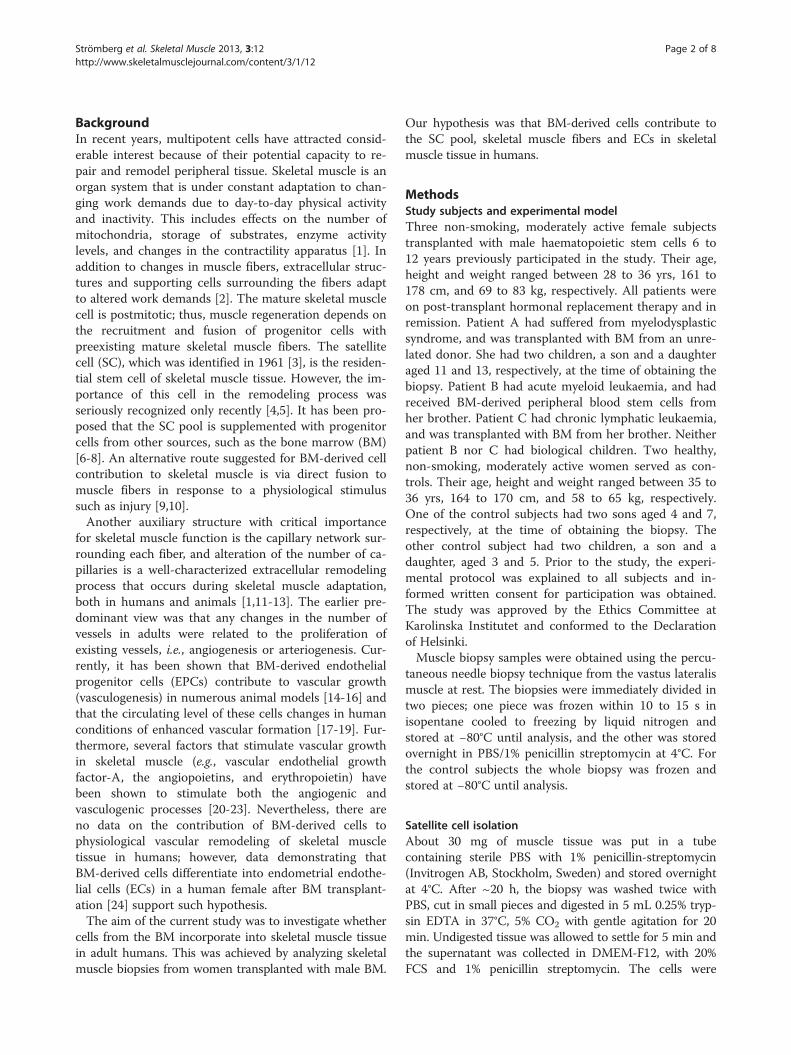
Bone marrow derived cells in adult skeletalmuscle tissue in humansAnna Strömberg1*, Monika Jansson2, Helene Fischer1, Eric Rullman1, Hans Hägglund3 and Thomas Gustafsson1
Abstract
Background: During the past decade, several animal studies have demonstrated that in addition to local cells, cellsfrom the bone marrow (BM) possess the ability to contribute to regeneration of injured skeletal muscle tissue. Inaddition, in mice, regular physical activity has been displayed to be a sufficient stimulus for BM-derived cellcontribution to the muscle, indicating that this is part of the ongoing physiological remodeling of skeletal muscle.However, whether BM-derived cells participate in human skeletal muscle remodeling is not known. To this end, weanalyzed the incorporation of BM-derived cells in healthy human skeletal muscle in women transplanted with maleBM.
Methods: Skeletal muscle biopsies were obtained from the m. vastus lateralis of women transplanted with maledonor hematopoietic stem cells 6 to 12 years earlier. Healthy women served as controls. Immunohistochemicalstaining for skeletal muscle fibers, satellite cells (SCs) or endothelial cells (ECs) combined with fluorescent in situhybridization (FISH) of X and Y chromosomes was used to identify cells of BM origin within the biopsies. Threedimensional confocal imaging was performed to demonstrate colocalization of Y chromosome and DAPI withinmuscle fibers. To further investigate whether BM-derived cells incorporate into the SC niche, myoblasts wereextracted from the biopsies from the transplanted women, cultured, and analyzed using XY FISH andimmunocytochemistry.
Results: Three dimensional confocal imaging indisputably demonstrated colocalization of Y chromosome and DAPIwithin muscle fibers. Some Y chromosomes were found within centrally located nuclei. No Y chromosomes weredetected in CD56+ SCs in the tissue sections nor in the myoblasts cultured from the extracted SCs. Y chromosome+ECs were found in all sections from the transplanted subjects. No Y chromosomes were found in the skeletal musclebiopsies obtained from healthy control women.
Conclusions: We demonstrate that BM-derived cells contribute to skeletal muscle fibers and ECs. Our results supportthat BM contribution to skeletal muscle occurs via direct fusion to muscle fibers, and that the contributing cells derivefrom the hematopoietic lineage. Thus, the present findings encourage further studies of the importance of this processfor the physiological adaptation occurring throughout life.
Keywords: Fluorescent in situ hybridization (FISH), Needle biopsies, Satellite cell niche, Vasculogenesis
* Correspondence: [email protected] of Laboratory Medicine, Division of Clinical Physiology,Karolinska Institutet, Karolinska University Hospital Huddinge, 141 86,Stockholm, SwedenFull list of author information is available at the end of the article
© 2013 Strömberg et al.; licensee BioMed Central Ltd. This is an Open Access article distributed under the terms of theCreative Commons Attribution License (http://creativecommons.org/licenses/by/2.0), which permits unrestricted use,distribution, and reproduction in any medium, provided the original work is properly cited.
Strömberg et al. Skeletal Muscle 2013, 3:12 Page 2 of 8http://www.skeletalmusclejournal.com/content/3/1/12
BackgroundIn recent years, multipotent cells have attracted consid-erable interest because of their potential capacity to re-pair and remodel peripheral tissue. Skeletal muscle is anorgan system that is under constant adaptation to chan-ging work demands due to day-to-day physical activityand inactivity. This includes effects on the number ofmitochondria, storage of substrates, enzyme activitylevels, and changes in the contractility apparatus [1]. Inaddition to changes in muscle fibers, extracellular struc-tures and supporting cells surrounding the fibers adaptto altered work demands [2]. The mature skeletal musclecell is postmitotic; thus, muscle regeneration depends onthe recruitment and fusion of progenitor cells withpreexisting mature skeletal muscle fibers. The satellitecell (SC), which was identified in 1961 [3], is the residen-tial stem cell of skeletal muscle tissue. However, the im-portance of this cell in the remodeling process wasseriously recognized only recently [4,5]. It has been pro-posed that the SC pool is supplemented with progenitorcells from other sources, such as the bone marrow (BM)[6-8]. An alternative route suggested for BM-derived cellcontribution to skeletal muscle is via direct fusion tomuscle fibers in response to a physiological stimulussuch as injury [9,10].Another auxiliary structure with critical importance
for skeletal muscle function is the capillary network sur-rounding each fiber, and alteration of the number of ca-pillaries is a well-characterized extracellular remodelingprocess that occurs during skeletal muscle adaptation,both in humans and animals [1,11-13]. The earlier pre-dominant view was that any changes in the number ofvessels in adults were related to the proliferation ofexisting vessels, i.e., angiogenesis or arteriogenesis. Cur-rently, it has been shown that BM-derived endothelialprogenitor cells (EPCs) contribute to vascular growth(vasculogenesis) in numerous animal models [14-16] andthat the circulating level of these cells changes in humanconditions of enhanced vascular formation [17-19]. Fur-thermore, several factors that stimulate vascular growthin skeletal muscle (e.g., vascular endothelial growthfactor-A, the angiopoietins, and erythropoietin) havebeen shown to stimulate both the angiogenic andvasculogenic processes [20-23]. Nevertheless, there areno data on the contribution of BM-derived cells tophysiological vascular remodeling of skeletal muscletissue in humans; however, data demonstrating thatBM-derived cells differentiate into endometrial endothe-lial cells (ECs) in a human female after BM transplant-ation [24] support such hypothesis.The aim of the current study was to investigate whether
cells from the BM incorporate into skeletal muscle tissuein adult humans. This was achieved by analyzing skeletalmuscle biopsies from women transplanted with male BM.
Our hypothesis was that BM-derived cells contribute tothe SC pool, skeletal muscle fibers and ECs in skeletalmuscle tissue in humans.
MethodsStudy subjects and experimental modelThree non-smoking, moderately active female subjectstransplanted with male haematopoietic stem cells 6 to12 years previously participated in the study. Their age,height and weight ranged between 28 to 36 yrs, 161 to178 cm, and 69 to 83 kg, respectively. All patients wereon post-transplant hormonal replacement therapy and inremission. Patient A had suffered from myelodysplasticsyndrome, and was transplanted with BM from an unre-lated donor. She had two children, a son and a daughteraged 11 and 13, respectively, at the time of obtaining thebiopsy. Patient B had acute myeloid leukaemia, and hadreceived BM-derived peripheral blood stem cells fromher brother. Patient C had chronic lymphatic leukaemia,and was transplanted with BM from her brother. Neitherpatient B nor C had biological children. Two healthy,non-smoking, moderately active women served as con-trols. Their age, height and weight ranged between 35 to36 yrs, 164 to 170 cm, and 58 to 65 kg, respectively.One of the control subjects had two sons aged 4 and 7,respectively, at the time of obtaining the biopsy. Theother control subject had two children, a son and adaughter, aged 3 and 5. Prior to the study, the experi-mental protocol was explained to all subjects and in-formed written consent for participation was obtained.The study was approved by the Ethics Committee atKarolinska Institutet and conformed to the Declarationof Helsinki.Muscle biopsy samples were obtained using the percu-
taneous needle biopsy technique from the vastus lateralismuscle at rest. The biopsies were immediately divided intwo pieces; one piece was frozen within 10 to 15 s inisopentane cooled to freezing by liquid nitrogen andstored at −80°C until analysis, and the other was storedovernight in PBS/1% penicillin streptomycin at 4°C. Forthe control subjects the whole biopsy was frozen andstored at −80°C until analysis.
Satellite cell isolationAbout 30 mg of muscle tissue was put in a tubecontaining sterile PBS with 1% penicillin-streptomycin(Invitrogen AB, Stockholm, Sweden) and stored overnightat 4°C. After ~20 h, the biopsy was washed twice withPBS, cut in small pieces and digested in 5 mL 0.25% tryp-sin EDTA in 37°C, 5% CO2 with gentle agitation for 20min. Undigested tissue was allowed to settle for 5 min andthe supernatant was collected in DMEM-F12, with 20%FCS and 1% penicillin streptomycin. The cells were
Strömberg et al. Skeletal Muscle 2013, 3:12 Page 3 of 8http://www.skeletalmusclejournal.com/content/3/1/12
cultured until reaching ~70% confluence when a fractionof the cells was obtained for cytocentrifugation.
CytocentrifugationThe cells were suspended at a concentration of 105 cells/mL and 100 μL were spun onto each glass slide(Superfrost/Plus microscope slides, Fisher Scientific,Pittsburgh, PA, USA). The slides were fixed in −20°Cacetone for 10 min and then washed in PBS. Slides wereblocked in PBS/4% BSA for 30 min in a humid chamber.A mouse anti-human desmin antibody (M0760, cloneD33, Dako, Glostrup, Denmark) was added at a concen-tration of 1:200 in 1% BSA/PBS and the slides were in-cubated overnight in the cold room. The slides werewashed in PBS before staining with the secondaryrabbit-anti mouse Alexa488 antibody (A11059, Molecu-lar Probes, Eugene, OR, USA) at 1:1000 in 1% BSA/PBSfor 60 min in the humid chamber. After washing in PBS,the slides were mounted in Vectashield DAPI (406-diamidine-2-phenylidole-dihydrochloride)/antifade (Vec-tor Laboratories Inc. Burlingame, CA, USA) and thenanalyzed for percentage of desmin positive cells.
ImmunohistochemistryThe frozen muscle biopsies were cut into 4 μm sectionsand placed on Superfrost/Plus microscope slides (FisherScientific). For serial sections, one section was put oneach glass slide in a series of slides. The sections werefixed for 10 min in −20°C acetone, and then dried beforethree 3 min washes in PBS. The slides were blocked inPBS/4% BSA for 30 min in a humid chamber, and werethen incubated with the primary antibody overnight at4°C in the humid chamber. The antibodies used weremouse anti-human CD31 at 1:500 (M0823, Dako),mouse anti-human CD56 (NCAM) at 1:50, (347740,Becton Dickinson, San Jose, CA, USA), sheep anti-human laminin at 1:10000 (PC128, The Binding Site,Birmingham, UK), mouse anti-human Caveolin-3 (A-3)at 1:500 (sc-5310, Santa Cruz Biotechnology Inc. SantaCruz, CA, USA) and mouse anti-human CD68 at 1:500(M0718, Dako). After washing the slides in PBS, they wereincubated in the secondary rabbit-anti mouse Alexa488antibody (A11059, Molecular Probes) at 1:500 and/or thedonkey anti-goat TexasRed at 1:300 (ab6883, Abcam,Cambridge, UK) for 60 min in room temperature, washedagain and mounted in VectaShield with or without DAPI(Vector Laboratories). The staining was evaluated beforefluorescent in situ hybridization.
Fluorescent in situ hybridization (FISH)After antibody staining, the glass slides were put in 2×saline sodium-citrate (SSC) buffer, and the cover slipswere allowed to fall off. The chromosome enumerationprobes (CEP) for the X and Y chromosomes (Vysis CEP
X (DXZ1) SpectrumGreen or SpectrumOrange Probeand CEP Y (DYZ1) SpectrumAqua or SpectrumGreenProbe), were mixed with hybridization buffer accordingto the manufacturer (Abbott-Vysis Inc Downers Grove,IL, USA); 1.5 μL of the probe solution were added to around cover slip (Thermo Fischer Scientific, MenzelGmbH & Co KG, Braunschweig, Germany), and theglass slide was then put on top of the cover slip. Thecover slip was sealed around the edges with rubber ce-ment, to prevent the probe solution from drying outduring hybridization. Glass slides and probe mixturewere denatured together in a Vysis HYBrite (AbbotDiagnostics). The melt temperature was set to 73°C for 2min and hybridization temperature 38°C for 20 hours.When the hybridization was complete, the rubber ce-ment was removed from the cover slips, and the slideswere immersed in 2×SSC until the cover slips fell off.The slides were then washed in 0.4×SSC/0.3% Igepal,72°C for 2 min and then 2×SSC/0.1% Igepal at roomtemperature for 30 s, and subsequently allowed to air-dry in the dark before mounting in VectaShield antifade(Vector Laboratories) with DAPI or propidium iodide asnuclear staining.
Evaluation of immunohistochemistry and FISH stainingEvaluation and cell identification was done using anOlympus fluorescence microscope BH60 with appropriatefilter set equipped with a CCD camera and connected to aCyto-Vision image system (Applied Imaging Corp., SanJose, CA, USA) in which the results also were docu-mented. Contribution of Y+ nuclei to skeletal musclefibers was analyzed in sections stained with caveolin-3,while sections double-stained with laminin-1 and CD56were analyzed for contribution to the SC pool. A cellstained with the marker CD56 containing a nucleus lyinginside laminin-1 positive lamina was identified as an SC.Three sections from each subject, at least 80 μm apart,were completely examined by two independent investiga-tors using an oil immersion objective with magnification100×1.3. Some of the skeletal muscle fibers containing Ychromosomes were also evaluated using confocal micros-copy. For evaluation of Y+ nuclei within ECs, two sectionsfrom each subject stained with CD31 were examined. Toexclude for the possibility of the Y chromosome belongingto a leukocyte travelling within the vessel, serial sectionsstained with CD68 were performed and examined. Afterevaluating the sections for Y chromosomes, the sectionswere photographed using a Leica DMLA microscopeequipped with a Leica DFC 420 C digital camera(Leica Microsystems AB, Sweden). The amount ofmyofibers and ECs per section was then calculated usingthe software Leica Qwin V. For myofibers, the tissue sec-tions were photographed using the 5× objective. For ECs,the 20× objective was used and a picture was taken in a
A B C
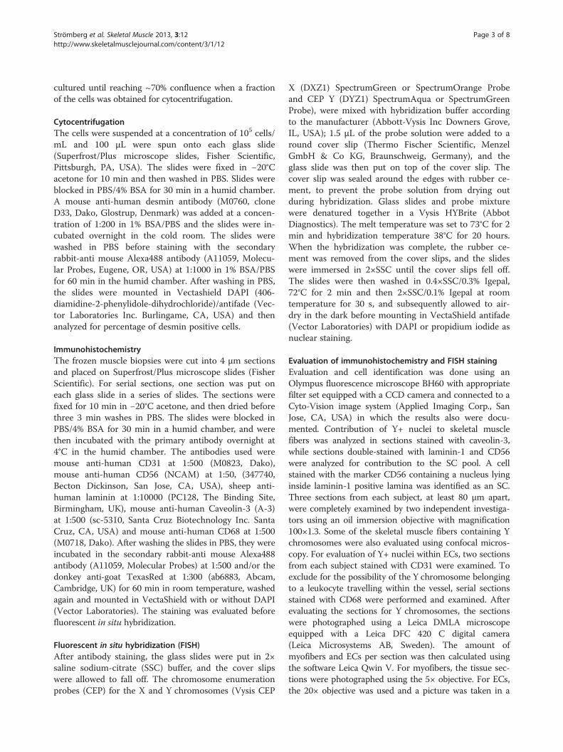
Figure 1 Y chromosome-positive nuclei are incorporated into host muscle fibers. Microphotographs show confocal images of combinedFISH (Y chromosome, bright green, arrow) and indirect, fluorescent immunohistochemistry for caveolin-3 (green). Nuclear DNA wascounterstained by DAPI. (A) Microphotograph of a muscle fiber section (width × height; 134.95 × 134.95 μm) depicting Y chromosome-negativehost nuclei and a single Y chromosome-positive donor nucleus (arrow). Caveolin-3 staining is used to visualize muscle fiber membranes.(B) provides a Z-stack (width × height × depth; 67.48 × 67.48 × 5.148 μm; 13 optical sections) of the muscle fiber in (A). Dashed lines markmuscle fiber membranes indicating that the DAPI/Y chromosome nucleus is localized inside the muscle fiber. Size bar = 10 μm. (C) A highmagnification Z-stack (15.83 × 15.83 × 6.007 μm; 15 optical sections) of the double-stained nucleus depicted in (B) clearly shows the integrationof the Y chromosome DNA in the nucleus.
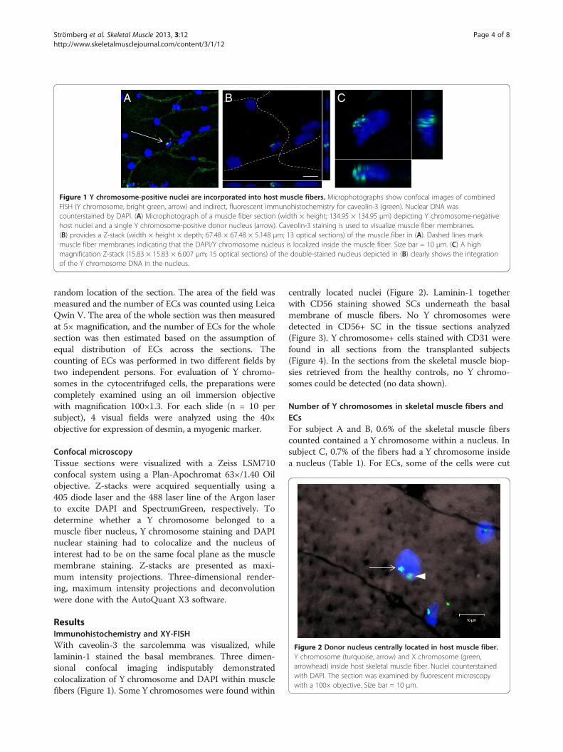
Figure 2 Donor nucleus centrally located in host muscle fiber.Y chromosome (turquoise, arrow) and X chromosome (green,arrowhead) inside host skeletal muscle fiber. Nuclei counterstainedwith DAPI. The section was examined by fluorescent microscopywith a 100× objective. Size bar = 10 μm.
Strömberg et al. Skeletal Muscle 2013, 3:12 Page 4 of 8http://www.skeletalmusclejournal.com/content/3/1/12
random location of the section. The area of the field wasmeasured and the number of ECs was counted using LeicaQwin V. The area of the whole section was then measuredat 5× magnification, and the number of ECs for the wholesection was then estimated based on the assumption ofequal distribution of ECs across the sections. Thecounting of ECs was performed in two different fields bytwo independent persons. For evaluation of Y chromo-somes in the cytocentrifuged cells, the preparations werecompletely examined using an oil immersion objectivewith magnification 100×1.3. For each slide (n = 10 persubject), 4 visual fields were analyzed using the 40×objective for expression of desmin, a myogenic marker.
Confocal microscopyTissue sections were visualized with a Zeiss LSM710confocal system using a Plan-Apochromat 63×/1.40 Oilobjective. Z-stacks were acquired sequentially using a405 diode laser and the 488 laser line of the Argon laserto excite DAPI and SpectrumGreen, respectively. Todetermine whether a Y chromosome belonged to amuscle fiber nucleus, Y chromosome staining and DAPInuclear staining had to colocalize and the nucleus ofinterest had to be on the same focal plane as the musclemembrane staining. Z-stacks are presented as maxi-mum intensity projections. Three-dimensional render-ing, maximum intensity projections and deconvolutionwere done with the AutoQuant X3 software.
ResultsImmunohistochemistry and XY-FISHWith caveolin-3 the sarcolemma was visualized, whilelaminin-1 stained the basal membranes. Three dimen-sional confocal imaging indisputably demonstratedcolocalization of Y chromosome and DAPI within musclefibers (Figure 1). Some Y chromosomes were found within
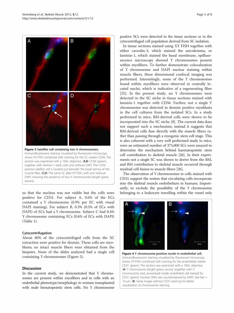
centrally located nuclei (Figure 2). Laminin-1 togetherwith CD56 staining showed SCs underneath the basalmembrane of muscle fibers. No Y chromosomes weredetected in CD56+ SC in the tissue sections analyzed(Figure 3). Y chromosome+ cells stained with CD31 werefound in all sections from the transplanted subjects(Figure 4). In the sections from the skeletal muscle biop-sies retrieved from the healthy controls, no Y chromo-somes could be detected (no data shown).
Number of Y chromosomes in skeletal muscle fibers andECsFor subject A and B, 0.6% of the skeletal muscle fiberscounted contained a Y chromosome within a nucleus. Insubject C, 0.7% of the fibers had a Y chromosome insidea nucleus (Table 1). For ECs, some of the cells were cut
C D
BA
Figure 3 Satellite cell containing two X chromosomes.Immunofluorescent staining visualized by fluorescent microscopyshows XY-FISH combined with staining for the SC marker CD56. Thesection was examined with a 100× objective. (A,B) CD56 (green),together with laminin-1 (red), with and without DAPI. The CD56positive satellite cell is located just beneath the basal lamina of themuscle fiber. (C,D) The same SC after XY-FISH, with and withoutDAPI, showing the presence of two X chromosomes (bright green,arrows).
BA
Strömberg et al. Skeletal Muscle 2013, 3:12 Page 5 of 8http://www.skeletalmusclejournal.com/content/3/1/12
so that the nucleus was not visible but the cells werepositive for CD31. For subject A, 0.6% of the ECscontained a Y chromosome (0.9% per EC with visualDAPI staining). For subject B, 0.3% (0.5% of ECs withDAPI) of ECs had a Y chromosome. Subject C had 0.4%Y chromosome containing ECs (0.6% of ECs with DAPI)(Table 1).
Figure 4 Y chromosome-positive nuclei in endothelial cell.Immunofluorescent staining visualized by fluorescent microscopy
CytocentrifugationAbout 80% of the cytocentrifuged cells from the SCextraction were positive for desmin. These cells are myo-blasts; no intact muscle fibers were obtained from thebiopsies. None of the slides analyzed had a single cellcontaining Y chromosomes (Figure 5).
shows XY-FISH combined with staining for the endothelial markerCD31 (green). The section was examined with a 100× objective.(A) Y chromosome (bright green, arrow), together with Xchromosome (red, arrowhead) inside endothelial cell stained forCD31 (green). Nuclear DNA was counterstained by DAPI. Size bar =10 μm. (B) Same image without CD31-staining for bettervisualization of chromosome staining.
DiscussionIn the current study, we demonstrated that Y chromo-somes are present within myofibers and in cells with anendothelial phenotype/morphology in women transplantedwith male hematopoietic stem cells. No Y chromosome
positive SCs were detected in the tissue sections or in thecytocentrifuged cell population derived from SC isolation.In tissue sections stained using XY FISH together with
either caveolin-3, which stained the sarcolemma, orlaminin-1, which stained the basal membrane, epifluor-escence microscopy showed Y chromosomes presentwithin myofibers. To further demonstrate colocalizationof Y chromosome and DAPI nuclear staining withinmuscle fibers, three dimensional confocal imaging wasperformed. Interestingly, some of the Y chromosomesfound within myofibers were observed in centrally lo-cated nuclei, which is indicative of a regenerating fiber[25]. In the present study, no Y chromosomes weredetected in the SC niche in tissue sections stained withlaminin-1 together with CD56. Further, not a single Ychromosome was detected in desmin positive myoblastsin the cell cultures from the isolated SCs. In a studyperformed in mice, BM-derived cells were shown to beincorporated into the SC niche [8]. The current data doesnot support such a mechanism; instead it suggests thatBM-derived cells fuse directly with the muscle fibers ra-ther than passing through a myogenic stem cell stage. Thisis also coherent with a very well performed study in mice,were an estimated number of 375,000 SCs were assayed todetermine the mechanism behind haematopoietic stemcell contribution to skeletal muscle [26]. In their experi-ments not a single SC was shown to derive from the BM,and BM contribution to skeletal muscle occurred throughmyeloid cell fusion to muscle fibers [26].The observation of Y chromosomes in cells stained with
CD31 support the notion that circulating cells incorporateinto the skeletal muscle endothelium in humans. Import-antly, to exclude the possibility of the Y chromosomebelonging to a leukocyte travelling within the vessel only
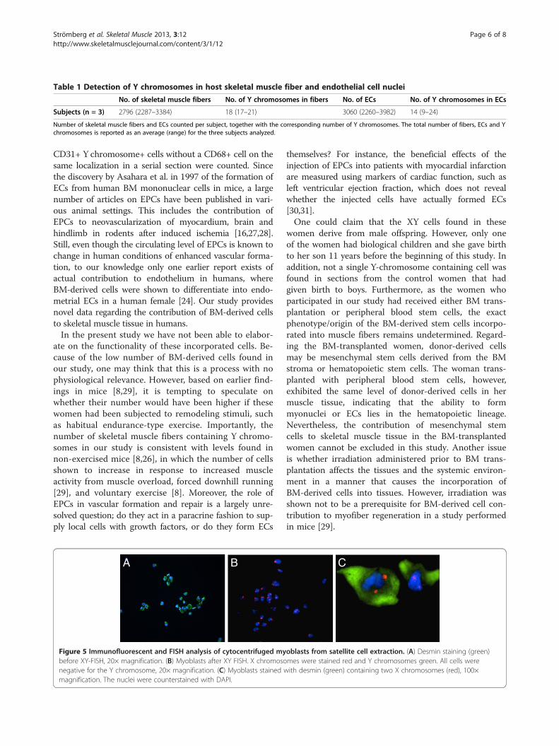
Table 1 Detection of Y chromosomes in host skeletal muscle fiber and endothelial cell nuclei
No. of skeletal muscle fibers No. of Y chromosomes in fibers No. of ECs No. of Y chromosomes in ECs
Subjects (n = 3) 2796 (2287–3384) 18 (17–21) 3060 (2260–3982) 14 (9–24)
Number of skeletal muscle fibers and ECs counted per subject, together with the corresponding number of Y chromosomes. The total number of fibers, ECs and Ychromosomes is reported as an average (range) for the three subjects analyzed.
Strömberg et al. Skeletal Muscle 2013, 3:12 Page 6 of 8http://www.skeletalmusclejournal.com/content/3/1/12
CD31+ Y chromosome+ cells without a CD68+ cell on thesame localization in a serial section were counted. Sincethe discovery by Asahara et al. in 1997 of the formation ofECs from human BM mononuclear cells in mice, a largenumber of articles on EPCs have been published in vari-ous animal settings. This includes the contribution ofEPCs to neovascularization of myocardium, brain andhindlimb in rodents after induced ischemia [16,27,28].Still, even though the circulating level of EPCs is known tochange in human conditions of enhanced vascular forma-tion, to our knowledge only one earlier report exists ofactual contribution to endothelium in humans, whereBM-derived cells were shown to differentiate into endo-metrial ECs in a human female [24]. Our study providesnovel data regarding the contribution of BM-derived cellsto skeletal muscle tissue in humans.In the present study we have not been able to elabor-
ate on the functionality of these incorporated cells. Be-cause of the low number of BM-derived cells found inour study, one may think that this is a process with nophysiological relevance. However, based on earlier find-ings in mice [8,29], it is tempting to speculate onwhether their number would have been higher if thesewomen had been subjected to remodeling stimuli, suchas habitual endurance-type exercise. Importantly, thenumber of skeletal muscle fibers containing Y chromo-somes in our study is consistent with levels found innon-exercised mice [8,26], in which the number of cellsshown to increase in response to increased muscleactivity from muscle overload, forced downhill running[29], and voluntary exercise [8]. Moreover, the role ofEPCs in vascular formation and repair is a largely unre-solved question; do they act in a paracrine fashion to sup-ply local cells with growth factors, or do they form ECs
BA
Figure 5 Immunofluorescent and FISH analysis of cytocentrifuged mybefore XY-FISH, 20× magnification. (B) Myoblasts after XY FISH. X chromosonegative for the Y chromosome, 20× magnification. (C) Myoblasts stained wmagnification. The nuclei were counterstained with DAPI.
themselves? For instance, the beneficial effects of theinjection of EPCs into patients with myocardial infarctionare measured using markers of cardiac function, such asleft ventricular ejection fraction, which does not revealwhether the injected cells have actually formed ECs[30,31].One could claim that the XY cells found in these
women derive from male offspring. However, only oneof the women had biological children and she gave birthto her son 11 years before the beginning of this study. Inaddition, not a single Y-chromosome containing cell wasfound in sections from the control women that hadgiven birth to boys. Furthermore, as the women whoparticipated in our study had received either BM trans-plantation or peripheral blood stem cells, the exactphenotype/origin of the BM-derived stem cells incorpo-rated into muscle fibers remains undetermined. Regard-ing the BM-transplanted women, donor-derived cellsmay be mesenchymal stem cells derived from the BMstroma or hematopoietic stem cells. The woman trans-planted with peripheral blood stem cells, however,exhibited the same level of donor-derived cells in hermuscle tissue, indicating that the ability to formmyonuclei or ECs lies in the hematopoietic lineage.Nevertheless, the contribution of mesenchymal stemcells to skeletal muscle tissue in the BM-transplantedwomen cannot be excluded in this study. Another issueis whether irradiation administered prior to BM trans-plantation affects the tissues and the systemic environ-ment in a manner that causes the incorporation ofBM-derived cells into tissues. However, irradiation wasshown not to be a prerequisite for BM-derived cell con-tribution to myofiber regeneration in a study performedin mice [29].
C
oblasts from satellite cell extraction. (A) Desmin staining (green)mes were stained red and Y chromosomes green. All cells wereith desmin (green) containing two X chromosomes (red), 100×
Strömberg et al. Skeletal Muscle 2013, 3:12 Page 7 of 8http://www.skeletalmusclejournal.com/content/3/1/12
ConclusionsIn conclusion, the contribution of BM-derived cells toskeletal muscle fibers and ECs was demonstrated byobtaining skeletal muscle biopsies from women trans-planted with male BM. The extent of contributionto muscle fibers was similar to the levels seen intransplanted mice not exposed to injury or exercise[8,26]. Our results support that BM contribution to skel-etal muscle occurs via direct fusion to muscle fibers, andthat the contributing cells derive from the hematopoieticlineage. The present study encourages further studies ofthe importance of this process for the physiologicaladaptation occurring throughout life.
ConsentWritten informed consent was obtained from the subjectsfor publication of this manuscript. A copy of the writtenconsent is available for review by the Editor-in-Chief of thisjournal.
AbbreviationsBM: Bone marrow; BSA: Bovine serum albumin; DAPI: 406-diamidine-2-phenylidole-dihydrochloride; EC: Endothelial cell; EPCs: Endothelialprogenitor cells; FCS: Fetal calf serum; FISH: Fluorescent in situ hybridization;PBS: Phosphate buffered saline; SC: Satellite cell.
Competing interestsThe authors declare that they have no competing interests.
Authors’ contributionsAS designed the study, collected and analyzed the material, interpreted thedata and drafted the manuscript. MJ contributed to the study design, andparticipated in the acquisition and interpretation of data. HF analyzed andinterpreted the data. ER obtained the muscle biopsies and helped to draftthe manuscript. HH recruited the study subjects and contributed to thestudy design. TG conceived the study, and contributed to the design andcoordination of the study and drafted the manuscript. All authors read andapproved the final manuscript.
AcknowledgmentsWe thank associate professor Gunnar Schulte, Karolinska Institutet whoperformed the confocal imaging. His work is supported by grants from theKnut & Alice Wallenberg Foundation, the Swedish Medical Research Counciland Karolinska Institutet.
GrantsThis study was supported by grants from the Swedish Medical ResearchCouncil, the Wallenberg foundation, the Swedish National Centre forResearch in Sports, the Swedish Medical Association and the KarolinskaInstitutet Foundation.
Author details1Department of Laboratory Medicine, Division of Clinical Physiology,Karolinska Institutet, Karolinska University Hospital Huddinge, 141 86,Stockholm, Sweden. 2Department of Medicine, Center for Hematology andRegenerative Medicine, Karolinska Institutet, Karolinska University HospitalHuddinge, 141 86, Stockholm, Sweden. 3Department of Medicine,Hematology Center, Karolinska Institutet, Karolinska University HospitalHuddinge, 141 86, Stockholm, Sweden.
Received: 17 December 2012 Accepted: 10 April 2013Published: 16 May 2013
References1. Saltin B, Gollnick PD: Skeletal muscle adaptability: significance for
metabolism and performance. In Handbook of Physiology. Section 10,Skeletal Muscle. Edited by Peachey LD. Bethesda: American PhysiologicalSociety; 1983:555–631.
2. Timmons JA, Jansson E, Fischer H, Gustafsson T, Greenhaff PL, Ridden J,Rachman J, Sundberg CJ: Modulation of extracellular matrix genesreflects the magnitude of physiological adaptation to aerobic exercisetraining in humans. BMC Biol 2005, 3:19.
3. Mauro A: Satellite cell of skeletal muscle fibers. J Biophys Biochem Cytol1961, 9:493–495.
4. Sherwood RI, Christensen JL, Conboy IM, Conboy MJ, Rando TA, WeissmanIL, Wagers AJ: Isolation of adult mouse myogenic progenitors: functionalheterogeneity of cells within and engrafting skeletal muscle. Cell 2004,119:543–554.
5. Kadi F, Charifi N, Denis C, Lexell J, Andersen JL, Schjerling P, Olsen S, KjaerM: The behaviour of satellite cells in response to exercise: what have welearned from human studies? Pflugers Arch 2005, 451:319–327.
6. Wagers AJ, Sherwood RI, Christensen JL, Weissman IL: Little evidence fordevelopmental plasticity of adult hematopoietic stem cells. Science 2002,297:2256–2259.
7. Torrente Y, Belicchi M, Sampaolesi M, Pisati F, Meregalli M, D’Antona G,Tonlorenzi R, Porretti L, Gavina M, Mamchaoui K, Pellegrino MA, Furling D,Mouly V, Butler-Browne GS, Bottinelli R, Cossu G, Bresolin N: Humancirculating AC133(+) stem cells restore dystrophin expression andameliorate function in dystrophic skeletal muscle. J Clin Invest 2004,114:182–195.
8. LaBarge MA, Blau HM: Biological progression from adult bone marrow tomononucleate muscle stem cell to multinucleate muscle fiber inresponse to injury. Cell 2002, 111:589–601.
9. Ferrari G, Cusella-De Angelis G, Coletta M, Paolucci E, Stornaiuolo A, CossuG, Mavilio F: Muscle regeneration by bone marrow-derived myogenicprogenitors. Science 1998, 279:1528–1530.
10. Gussoni E, Soneoka Y, Strickland CD, Buzney EA, Khan MK, Flint AF, KunkelLM, Mulligan RC: Dystrophin expression in the mdx mouse restored bystem cell transplantation. Nature 1999, 401:390–394.
11. Hudlicka O, Brown M, Egginton S: Angiogenesis in skeletal and cardiacmuscle. Physiol Rev 1992, 72:369–417.
12. Gustafsson T, Kraus WE: Exercise-induced angiogenesis-related growthand transcription factors in skeletal muscle, and their modification inmuscle pathology. Front Biosci 2001, 6:D75–D89.
13. Gustafsson T: Vascular remodelling in human skeletal muscle.Biochem Soc Trans 2011, 39:1628–1632.
14. Asahara T, Murohara T, Sullivan A, Silver M, van der Zee R, Li T,Witzenbichler B, Schatteman G, Isner JM: Isolation of putative progenitorendothelial cells for angiogenesis. Science 1997, 275:964–967.
15. Kalka C, Masuda H, Takahashi T, Kalka-Moll WM, Silver M, Kearney M, Li T,Isner JM, Asahara T: Transplantation of ex vivo expanded endothelialprogenitor cells for therapeutic neovascularization. Proc Natl Acad Sci USA2000, 97:3422–3427.
16. Kawamoto A, Gwon HC, Iwaguro H, Yamaguchi JI, Uchida S, Masuda H,Silver M, Ma H, Kearney M, Isner JM, Asahara T: Therapeutic potential ofex vivo expanded endothelial progenitor cells for myocardial ischemia.Circulation 2001, 103:634–637.
17. Gill M, Dias S, Hattori K, Rivera ML, Hicklin D, Witte L, Girardi L, Yurt R, HimelH, Rafii S: Vascular trauma induces rapid but transient mobilization ofVEGFR2(+)AC133(+) endothelial precursor cells. Circ Res 2001, 88:167–174.
18. Shintani S, Murohara T, Ikeda H, Ueno T, Honma T, Katoh A, Sasaki K,Shimada T, Oike Y, Imaizumi T: Mobilization of endothelial progenitor cellsin patients with acute myocardial infarction. Circulation 2001,103:2776–2779.
19. Vasa M, Fichtlscherer S, Aicher A, Adler K, Urbich C, Martin H, Zeiher AM,Dimmeler S: Number and migratory activity of circulating endothelialprogenitor cells inversely correlate with risk factors for coronary arterydisease. Circ Res 2001, 89:E1–E7.
20. Moore MA, Hattori K, Heissig B, Shieh JH, Dias S, Crystal RG, Rafii S:Mobilization of endothelial and hematopoietic stem and progenitor cellsby adenovector-mediated elevation of serum levels of SDF-1, VEGF, andangiopoietin-1. Ann N Y Acad Sci 2001, 938:36–45. Discussion 45–37.
21. Hiratsuka S: Vasculogenensis, angiogenesis and special features of tumorblood vessels. Front Biosci 2011, 16:1413–1427.

Strömberg et al. Skeletal Muscle 2013, 3:12 Page 8 of 8http://www.skeletalmusclejournal.com/content/3/1/12
22. Heeschen C, Aicher A, Lehmann R, Fichtlscherer S, Vasa M, Urbich C,Mildner-Rihm C, Martin H, Zeiher AM, Dimmeler S: Erythropoietin is apotent physiologic stimulus for endothelial progenitor cell mobilization.Blood 2003, 102:1340–1346.
23. Hattori K, Dias S, Heissig B, Hackett NR, Lyden D, Tateno M, Hicklin DJ, ZhuZ, Witte L, Crystal RG, Moore MA, Rafii S: Vascular endothelial growthfactor and angiopoietin-1 stimulate postnatal hematopoiesis byrecruitment of vasculogenic and hematopoietic stem cells. J Exp Med2001, 193:1005–1014.
24. Mints M, Jansson M, Sadeghi B, Westgren M, Uzunel M, Hassan M, PalmbladJ: Endometrial endothelial cells are derived from donor stem cells in abone marrow transplant recipient. Hum Reprod 2008, 23:139–143.
25. Charge SB, Rudnicki MA: Cellular and molecular regulation of muscleregeneration. Physiol Rev 2004, 84:209–238.
26. Camargo FD, Green R, Capetanaki Y, Jackson KA, Goodell MA: Singlehematopoietic stem cells generate skeletal muscle through myeloidintermediates. Nat Med 2003, 9:1520–1527.
27. Zhang ZG, Zhang L, Jiang Q, Chopp M: Bone marrow-derived endothelialprogenitor cells participate in cerebral neovascularization after focalcerebral ischemia in the adult mouse. Circ Res 2002, 90:284–288.
28. Yamahara K, Sone M, Itoh H, Yamashita JK, Yurugi-Kobayashi T, Homma K,Chao TH, Miyashita K, Park K, Oyamada N, Sawada N, Taura D, Fukunaga Y,Tamura N, Nakao K: Augmentation of neovascularization [corrected] inhindlimb ischemia by combined transplantation of human embryonicstem cells-derived endothelial and mural cells. PLoS One 2008, 3:e1666.
29. Palermo AT, Labarge MA, Doyonnas R, Pomerantz J, Blau HM: Bone marrowcontribution to skeletal muscle: a physiological response to stress.Dev Biol 2005, 279:336–344.
30. Yousef M, Schannwell CM, Kostering M, Zeus T, Brehm M, Strauer BE: TheBALANCE study: clinical benefit and long-term outcome afterintracoronary autologous bone marrow cell transplantation in patientswith acute myocardial infarction. J Am Coll Cardiol 2009, 53:2262–2269.
31. Leistner DM, Fischer-Rasokat U, Honold J, Seeger FH, Schachinger V,Lehmann R, Martin H, Burck I, Urbich C, Dimmeler S, Zeiher AM, Assmus B:Transplantation of progenitor cells and regeneration enhancement inacute myocardial infarction (TOPCARE-AMI): final 5-year results suggestlong-term safety and efficacy. Clin Res Cardiol 2011, 100:925–934.
doi:10.1186/2044-5040-3-12Cite this article as: Strömberg et al.: Bone marrow derived cells in adultskeletal muscle tissue in humans. Skeletal Muscle 2013 3:12.
Submit your next manuscript to BioMed Centraland take full advantage of:
• Convenient online submission
• Thorough peer review
• No space constraints or color figure charges
• Immediate publication on acceptance
• Inclusion in PubMed, CAS, Scopus and Google Scholar
• Research which is freely available for redistribution
Submit your manuscript at www.biomedcentral.com/submit