Embed Size (px)
Citation preview

10:32 1
Muscle Tissue
• Alternating contraction and relaxation of cells
• Chemical energy changed into mechanical energy

10:32 2
Properties of Muscle Tissue• Excitability
– responds to chemical messengers (neurotransmitters) released from nerve cells
• Contractility– can shorten and generate force
• Extensibility– can be stretched without damaging the tissue
• Elasticity– can return to original shape after being stretched

10:32 3
3 Types of Muscle Tissue• Skeletal muscle
– attaches to bone, skin or fascia– is striated with light & dark bands visible with
the microscope – contraction & relaxation is under voluntary
control
10:32 4
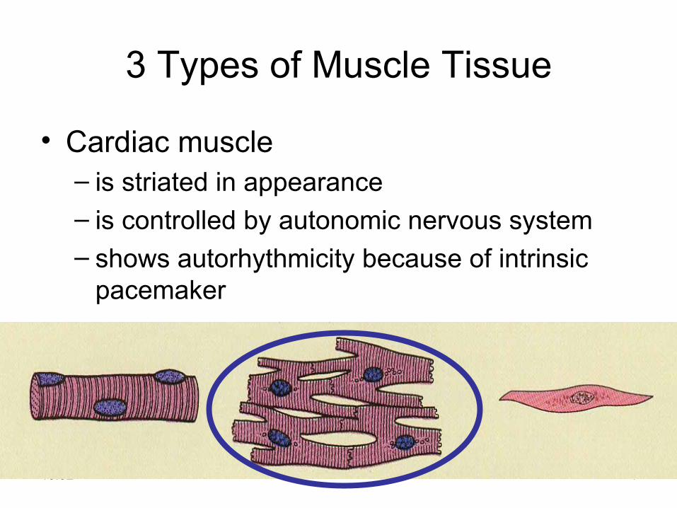
3 Types of Muscle Tissue
• Cardiac muscle– is striated in appearance– is controlled by autonomic nervous system– shows autorhythmicity because of intrinsic
pacemaker

10:32 5
3 Types of Muscle Tissue• Smooth muscle
– forms erector pili muscles attached to hair follicles in skin
– forms the muscle tunics in the walls of hollow organs
– shows a nonstriated appearance– is involuntary

10:32 6
Functions of Muscle Tissue• Producing body movements• Stabilizing body positions• Regulating organ volumes
– bands of circularly arranged smooth muscle are sphincters
• Movement of substances within the body– blood, lymph, urine, air, food and fluids, sperm
• Producing heatcontractions of skeletal muscle

10:32 7
Skeletal Muscle -- Connective Tissue• Superficial fascia is loose connective tissue & fat
underlying the skin• Deep fascia: dense irregular connective tissue
around muscle• Connective tissue components of the muscle include
– epimysium: surrounds the whole muscle – perimysium: surrounds bundles (fascicles) of 10-
100 muscle fibers– endomysium: separates individual muscle cells
• All of these connective tissue layers extend beyond the muscle belly to form the tendon

10:32 8
Connective Tissue Components
10:32 9
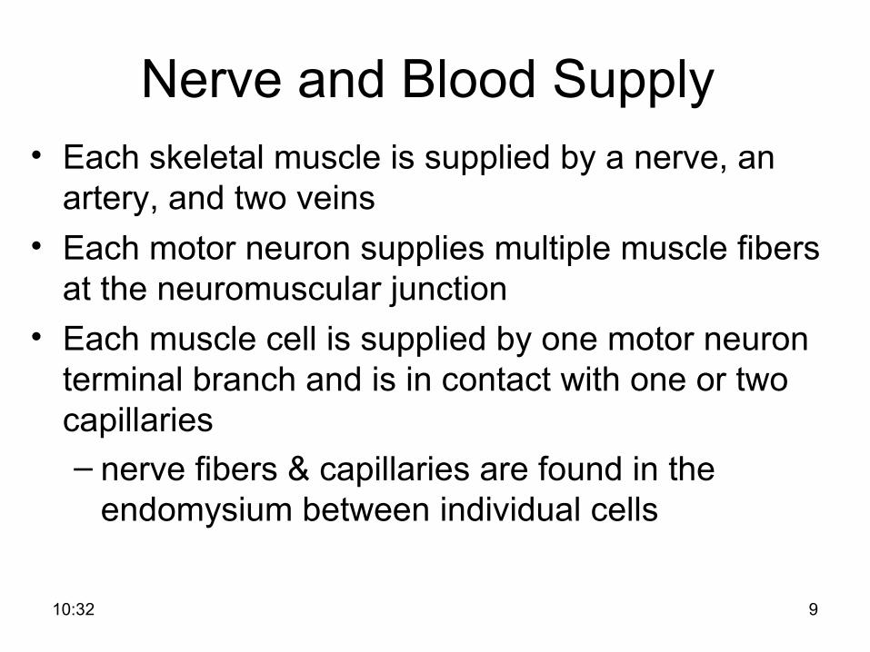
Nerve and Blood Supply • Each skeletal muscle is supplied by a nerve, an
artery, and two veins• Each motor neuron supplies multiple muscle fibers
at the neuromuscular junction• Each muscle cell is supplied by one motor neuron
terminal branch and is in contact with one or two capillaries– nerve fibers & capillaries are found in the
endomysium between individual cells

10:32 10
The Muscle Fiber
• Muscle cells (fibers) are long, cylindrical, & multinucleated
• Sarcolemma: muscle cell membrane• Sarcoplasm filled with tiny threads called myofibrils &
myoglobin (red-colored, oxygen-binding protein)

10:32 11
Myofibrils & Myofilaments
• Muscle fibers are filled with threads called myofibrils separated by sarcoplasmic reticulum
• Myofilaments (thick & thin filaments) are the contractile proteins of muscle

10:32 12
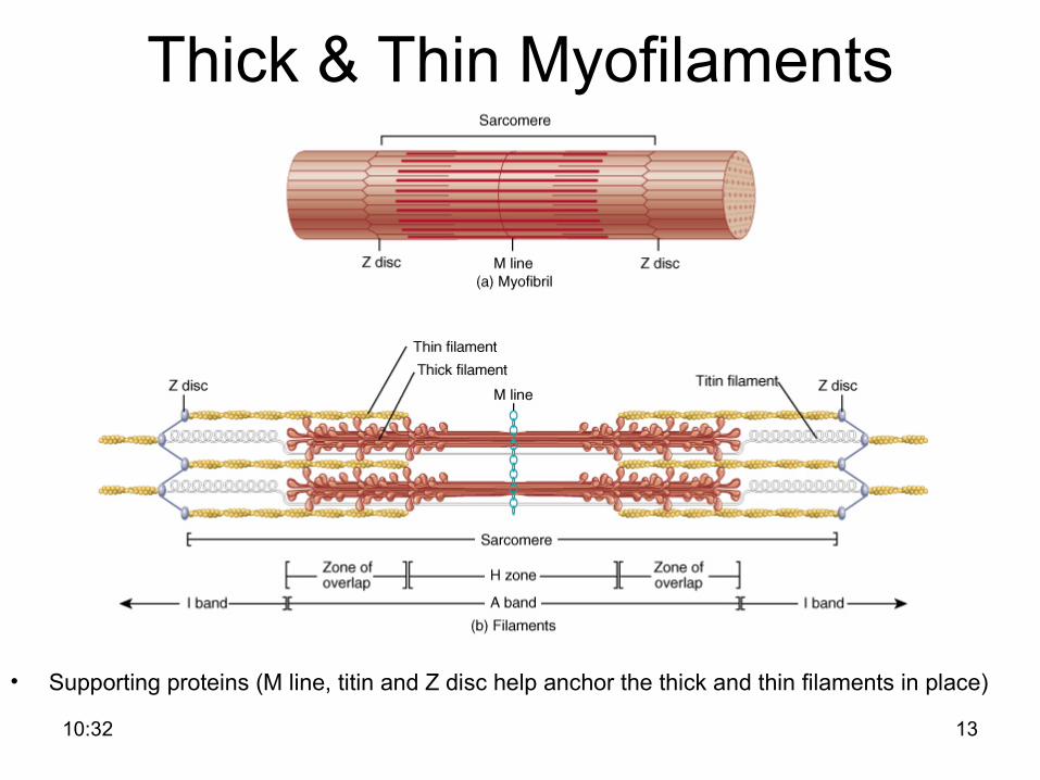
Filaments and the Sarcomere• Thick and thin filaments overlap each other in a
pattern that creates striations (light I bands and dark A bands)
• The I band region contains only thin filaments.• They are arranged in compartments called
sarcomeres, separated by Z discs/lines.• In the overlap region, six thin filaments surround
each thick filament
10:32 13
Thick & Thin Myofilaments
• Supporting proteins (M line, titin and Z disc help anchor the thick and thin filaments in place)

10:32 14
Overlap of Thick & Thin Myofilaments within a Myofibril
Overlap of Thick & Thin Myofilaments within a Myofibril
Dark (A) and light (I) bands visible with an electron microscope

10:32 15
Transverse Tubules
• T (transverse) tubules are invaginations of the sarcolemma into the center of the cell– filled with extracellular fluid– carry muscle action potentials down into cell
• Mitochondria lie in rows throughout the cell– near the muscle proteins that use ATP during contraction

10:32 16
Sarcoplasmic Reticulum (SR)
• System of tubular sacs similar to smooth ER in nonmuscle cells
• Triad: T tubule + terminal cisterns/lateral sacs. Lateral sacs store Ca+2 in a relaxed muscle

10:32 17
The Proteins of Muscle• Myofibrils are built of three kinds of protein
– contractile proteins• myosin and actin
– regulatory proteins which turn contraction on & off• troponin and tropomyosin
– structural proteins which provide proper alignment, elasticity, and extensibility• titin, myomesin, nebulin and dystrophin

10:32 18
The Proteins of Muscle -- Myosin
• Thick filaments are composed of myosin – each molecule resembles two golf clubs twisted together– myosin heads (cross bridges) extend toward the thin
filaments• Held in place by the M line proteins
10:32 19
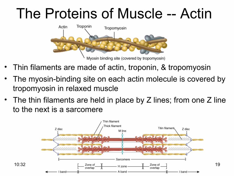
The Proteins of Muscle -- Actin
• Thin filaments are made of actin, troponin, & tropomyosin • The myosin-binding site on each actin molecule is covered by
tropomyosin in relaxed muscle• The thin filaments are held in place by Z lines; from one Z line
to the next is a sarcomere

10:32 20
Atrophy and Hypertrophy• Atrophy
– wasting away of muscles– caused by disuse (disuse atrophy) or severing of
the nerve supply (denervation atrophy), diet– the transition to connective tissue cannot be
reversed• Hypertrophy
– increase in the diameter of muscle fibers – resulting from very forceful, repetitive muscular
activity and an increase in myofibrils, SR, & mitochondria

10:32 21
Neuromuscular Junction (NMJ)
• NMJ – end of axon nears the surface of a muscle fiber at its motor end plate region (they remain separated by synaptic cleft or gap)

10:32 22
Structures of NMJ Region
• Synaptic end bulbs are swellings of axon terminals
• End bulbs contain synaptic vesicles filled with acetylcholine (ACh)
• Motor end plate membrane contains 30 million ACh receptors.

10:32 23
Pharmacology of the NMJ• Botulinum toxin blocks release of neurotransmitter at
the NMJ so muscle contraction can not occur– bacteria found in improperly canned food– death occurs from paralysis of the diaphragm
• Curare (plant poison from poison arrows)– causes muscle paralysis by blocking the ACh receptors – used to relax muscle during surgery
• Neostigmine (anticholinesterase agent)– blocks removal of ACh from receptors so strengthens
weak muscle contractions of myasthenia gravis– also an antidote for curare after surgery is finished
10:32 24
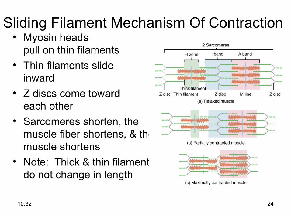
Sliding Filament Mechanism Of Contraction • Myosin heads
pull on thin filaments• Thin filaments slide
inward• Z discs come toward
each other• Sarcomeres shorten, the
muscle fiber shortens, & the muscle shortens
• Note: Thick & thin filaments do not change in length
10:32 25
How Does Contraction Begin?• Nerve impulse reaches an axon terminal;
synaptic vesicles release acetylcholine (ACh)• ACh diffuses to receptors on the sarcolemma • A muscle action potential (membrane
potential change) spreads over sarcolemma and down into the transverse tubules
• SR/Triad releases Ca+2 into the sarcoplasm• This is excitation

10:32 26
Excitation - Contraction Coupling
• The steps that occur from the muscle action potential reaching the T tubule to contraction of the muscle fiber

10:32 27
Contraction Cycle - 1• Repeating sequence of events that cause the
thick & thin filaments to move past each other• 4 steps to contraction cycle
– ATP hydrolyzed by myosin ATPase; ADP and P
i remain attached to myosin binding site;
energy is stored in cross-bridge– Ca+2 released at excitation results in
disinihibition of actin; myosin and actin bind– Power stroke of cross-bridge; ADP and P
i
released

10:32 28
Contraction Cycle - 2•
– Fresh ATP binds to myosin ATP-binding site; myosin detaches from actin
• Cycle keeps repeating as long as there is ATP available & high Ca+2 level near thin filament

10:32 29
Steps in the Contraction Cycle
• Notice how the myosin head attaches and pulls on the thin filament with the energy released from ATP

10:32 30
ATP and Myosin• Myosin heads are “cocked” by ATP• Activated heads attach to actin & pull (power stroke)• Thin filaments slide past the thick filaments• ADP & P
i are released (hydrolysis of ATP releases P
i,
ADP, & energy)• New ATP binds to myosin head & allows myosin head
to detach from actin• All of these steps repeat over and over
– if ATP is available and– Ca+2 level near the troponin-tropomyosin complex is
high

10:32 31
Relaxation• Acetylcholinesterase (AChE) breaks down ACh
within the synaptic cleft• Muscle action potential ceases• Active transport pumps Ca2+ back into storage in the
lateral sacs• Calcium-binding protein (calsequestrin) helps hold
Ca+2 in SR (Ca+2 concentration 10,000 times higher than in cytosol)
• Tropomyosin-troponin complex recovers binding site on the actin

10:32 32
Rigor Mortis• Rigor mortis is a state of muscular rigidity
that begins 3-4 hours after death and lasts about 24 hours
• After death, Ca+2 ions leak out of the SR and allow myosin heads to bind to actin
• Since ATP synthesis has ceased, cross bridges cannot detach from actin until proteolytic enzymes begin to digest the decomposing cells

10:32 33
• Optimal overlap of thick & thin filaments– produces greatest number of cross bridges and
the greatest amount of tension• Overstretched muscle (past optimal length)
– fewer cross bridges exist & less force is produced • If muscle is overly shortened (less than optimal)
– fewer cross bridges exist & less force is produced– thick filaments crumpled by Z discs
Length of Muscle Fibers

10:32 34
The Motor Unit
• Motor unit: one somatic motor neuron & all the skeletal muscle cells (fibers) it stimulates – One nerve cell supplies on average 150 muscle cells
that all contract in unison.• Total strength of a contraction depends on how many
motor units are activated & how large the motor unit is
10:32 35
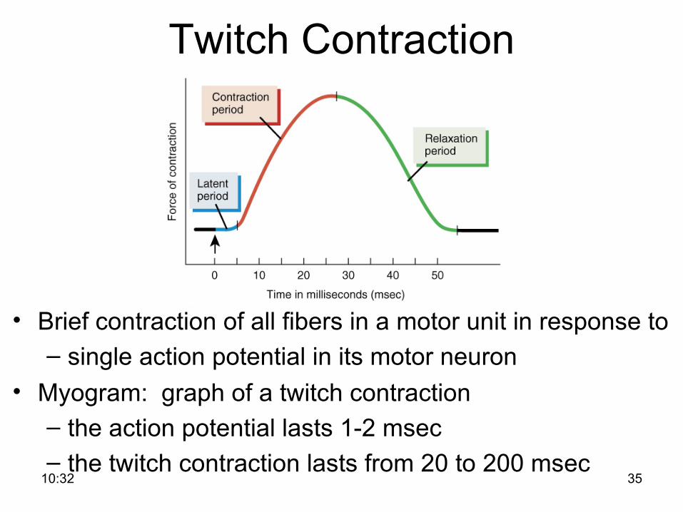
Twitch Contraction
• Brief contraction of all fibers in a motor unit in response to – single action potential in its motor neuron
• Myogram: graph of a twitch contraction– the action potential lasts 1-2 msec– the twitch contraction lasts from 20 to 200 msec

10:32 36
Parts of a Twitch Contraction• Latent Period – 2 msec
– Ca+2 is being released from SR• Contraction Period
– 10 to 100 msec– filaments slide past each other
• Relaxation Period– 10 to 100 msec – active transport of Ca+2 into SR
• Refractory Period– muscle cannot respond

10:32 37
Wave Summation• If second stimulation applied after the refractory period but
before complete muscle relaxation—second contraction is stronger than first

10:32 38
Complete and Incomplete Tetanus
• Unfused/incomplete tetanus– if stimulate at 20-30 times/second, there will be only partial
relaxation between stimuli• Fused/complete tetanus
– if stimulate at 80-100 times/second, a sustained contraction with no relaxation between stimuli will result

10:32 39
Explanation of Summation & Tetanus• Wave summation & both types of tetanus
result from Ca+2 remaining in the sarcoplasm
• Force of 2nd contraction is easily added to the first, because the elastic elements remain partially contracted and do not delay the beginning of the next contraction

10:32 40
Motor Unit Recruitment• Motor units in a whole muscle fire asynchronously
– some fibers are active others are relaxed – delays muscle fatigue so contraction can be
sustained• Produces smooth muscular contraction
– not a series of jerky movements• Precise movements require smaller contractions
– motor units must be smaller (fewer fibers/nerve)• Large motor units are active when large tension is
needed

10:32 41
Muscle Tone• Involuntary contraction of a small number of motor
units (alternately active and inactive in a constantly shifting pattern)– keeps muscles firm even though relaxed– does not produce movement
• Essential for maintaining posture (head upright)• Important in maintaining blood pressure
– tone of smooth muscles in walls of blood vessels

10:32 42
Muscle MetabolismProduction of ATP in Muscle Fibers
• Muscle uses ATP at a great rate when active• Sarcoplasmic ATP only lasts for few seconds• 3 sources of ATP production within muscle
– creatine phosphate– anaerobic cellular respiration– aerobic cellular respiration

10:32 43
Creatine Phosphate• Excess ATP within resting muscle used to form creatine
phosphate• Creatine phosphate 3-6
times more plentiful than ATP within muscle
• Its quick breakdownprovides energy for creation of ATP
• Sustains maximal contraction for 15 sec (used for 100 meter dash)
• Athletes tried creatine supplementation – gain muscle mass but shut down bodies own synthesis
(for safety?)
10:32 44
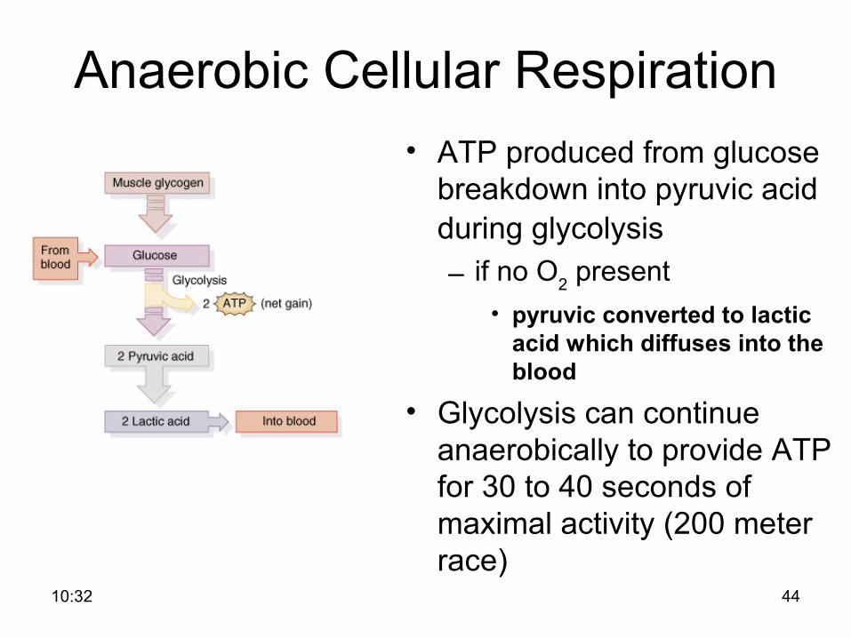
Anaerobic Cellular Respiration• ATP produced from glucose
breakdown into pyruvic acid during glycolysis – if no O2 present
• pyruvic converted to lactic acid which diffuses into the blood
• Glycolysis can continue anaerobically to provide ATP for 30 to 40 seconds of maximal activity (200 meter race)

10:32 45
Aerobic Cellular Respiration
• ATP for any activity lasting over 30 sec – if sufficient oxygen is available, pyruvic acid enters the
mitochondria to generate ATP, water and heat– fatty acids and amino acids can also be used by the
mitochondria• Provides 90% of ATP if activity lasts more than 10 min

10:32 46
Muscle Fatigue• Inability to contract after prolonged activity
– central fatigue is feeling of tiredness and a desire to stop (protective mechanism)
– depletion of creatine phosphate– decline of Ca+2 within the sarcoplasm
• Factors that contribute to muscle fatigue– insufficient oxygen or glycogen– buildup of lactic acid and ADP– insufficient release of acetylcholine from
motor neurons

10:32 47
Classification of Muscle Fibers
• Slow oxidative (slow-twitch)– red in color (lots of mitochondria, myoglobin, & blood vessels)– prolonged, sustained contractions for maintaining posture
• Fast oxidative-glycolytic (fast-twitch A)– red in color (lots of mitochondria, myoglobin, & blood vessels)– split ATP at very fast rate; used for walking and sprinting
• Fast glycolytic (fast-twitch B)– white in color (few mitochondria & BV, low myoglobin)– anaerobic movements for short duration; used for weight-lifting

10:32 48
Fiber Types within a Whole Muscle• Most muscles contain a mixture of all three fiber
types• Proportions vary with the usual action of the
muscle– neck, back and leg muscles have a higher
proportion of postural, slow oxidative fibers– shoulder and arm muscles have a higher
proportion of fast glycolytic fibers• All fibers of any one motor unit are same.

10:32 49
Muscular Dystrophies • Inherited, muscle-destroying diseases • Sarcolemma tears during muscle contraction• Mutated gene is on X chromosome so problem is
with males almost exclusively• Appears by age 5 in males and by 12 may be unable
to walk• Degeneration of individual muscle fibers produces
atrophy of the skeletal muscle• Gene therapy is hoped for with the most common
form, Duchenne muscular dystrophy

10:32 50
Abnormal Contractions• Spasm: involuntary contraction of single
muscle• Cramp: a painful spasm• Tic: involuntary twitching of muscles
normally under voluntary control--eyelid or facial muscles
• Tremor: rhythmic, involuntary contraction of opposing muscle groups
• Fasciculation: involuntary, brief twitch of a motor unit visible under the skin

10:32 51
Isotonic and Isometric Contraction
• Concentric (isotonic) contraction: a load is moved• Eccentric (isotonic) contraction • Isometric contraction: no movement occurs
– tension is generated without muscle shortening– maintaining posture & supporting objects in a fixed position