Embed Size (px)
Citation preview

IOSR Journal of Dental and Medical Sciences (IOSR-JDMS)
e-ISSN: 2279-0853, p-ISSN: 2279-0861.Volume 15, Issue 8 Ver. II (August. 2016), PP 129-142
www.iosrjournals.org
DOI: 10.9790/0853-150802129142 www.iosrjournals.org 129 | Page
Morphology of Sacrum and its Variations
1Dr.A.K.Manicka Vasuki,
2 Dr.M.Nirmaladevi,
3Mrs.Deborah Joy Hebzibah,
4Prof.M.Jamuna,
5Dr.K.Kalyana Sundaram,
6Dr.V.Vimala,
1,3Assistant Professors, Department of Anatomy, PSG Institute Of Medical Sciences And Research,
Coimbatore - 641004. 2Associate Professor, Department of Anatomy, PSG Institute Of Medical Sciences And Research,
Coimbatore - 641004 4Prof. & Hod, Department of Anatomy, PSG Institute Of Medical Sciences And Research,
Coimbatore - 641004. 5Associate Professor, Department of Anaesthesiology, Coimbatore Medical College, Coimbatore.
6Assistant Professor, Department of Anatomy, Coimbatore Medical College, Coimbatore.
Abstract: Sacrum is formed by the fusion of five Sacral vertebrae and forms the lower part of Vertebral
column. The opening present at the lower end of Sacral canal is known as Sacral hiatus. Anatomical variations,
Morphology and Morphometry of Sacral hiatus are important clinically as well as surgically1. This study was
carried out on 75 dry human sacra of unknown sex to know different features of Sacrum, anatomical variations
of sacrum and to study the variations of sacral hiatus. Anatomical variations – failure of formation of superior
articular process and lamina of left first sacral vertebra, incomplete development and fusion of second sacral
vertebra, multiple foramens on either side of spinous process above the sacral hiatus, multiple foramens in the
dorsal surface of base of the sacrum behind the auricular surface, incomplete median crest, bilateral five pelvic
and dorsal foramens were identified. Various shapes of sacral hiatus were observed which included inverted u,
inverted v, irregular, dumbbell and bifid2. The apex of sacral hiatus was commonly found at the level of fourth
sacral vertebra. The mean length of sacral hiatus was measured. The mean anteroposterior diameter of sacral
canal and narrowing of sacral canal at the apex of sacral hiatus was measured1&2
.
The knowledge of anatomical variations of Sacrum and variations of Sacral hiatus are clinically important for
Caudal epidural block in Pediatric, Obsteric, Orthopedic, Urologic and Surgical practice.
The reliability and success of Caudal epidural block depends upon Knowledge of Variations of Sacrum and
Variations of Sacral hiatus.
Keywords: Sacrum, lamina, variation, sacral hiatus, caudal epidural block
I. Introduction Sacrum is a large, triangular bone formed by fusion of five vertebrae present between the two hip
(innominate) bones. It presents concave anterior or pelvic surface and convex posterior surface. The broad base
is directed above and the apex is at the lower end. The base is divided into central part consisting of body of first
sacral vertebra and lateral mass or ala on either side. By its base the sacrum articulates with the fifth lumbar
vertebra and by its apex it articulates with the coccyx. The base presents the upper opening of sacral canal. The
superolateral margin of the body of first sacral vertebra projects forwards as the sacral promontory, which is
useful in measuring the diameters of the pelvis.
The triangular sacral canal is formed by sacral vertebral foramina. The opening present at the caudal
end of sacral canal is known as Sacral hiatus. The lamina and spinous process of the fifth and /or fourth sacral
vertebrae fail to meet in the midline creating a deficiency known as the hiatus in the posterior wall of the sacral
canal3. It is located inferior to the fourth or third fused sacral spines or lower end of median sacral crest. The
remnants elongate downwards on both sides of sacral hiatus. These two bony processes are called the sacral
cornua and define important landmarks during caudal epidural block (CEB). Sacral hiatus is identified by
palpation of sacral cornua. Sacral cornua are felt at the upper end of natal cleft 5cm above the tip of coccyx.
Structures emerge from sacral hiatus are the filum terminale, fifth sacral nerves and coccygeal nerves. The
hiatus provides access to the extradural space in the sacral canal.
Sacral hiatus is covered posteriorly by skin, a subcutaneous fatty tissue and the sacrococcygeal
membrane when the needle has passed through the sacrococcygeal ligament, the hiatus communicates with the
epidural space. The dural sac ends at the level of second piece of sacrum. Sacral canal below this level contains
extradural fat, vertebral venous plexus, lower sacral nerve roots and the filum terminale. Sacral hiatus has been
utilized for administration of epidural anaesthesia in obstetrics as well as in Orthopedic practice for
Transpedicular and lateral mass screw placement.

Morphology Of Sacrum And Its Variations
DOI: 10.9790/0853-150802129142 www.iosrjournals.org 130 | Page
The anterior surface of Sacrum bears four anterior sacral foramina which give passage to ventral rami
of upper four sacral spinal nerves and lateral sacral arteries4. The dorsal surface of sacrum bears four posterior
sacral foramina which give passage to posterior rami of the upper four sacral spinal nerves.
The upper surface of the lateral mass of Sacrum is termed the Ala of Sacrum4.
Sacral canal contains the cauda equina, duramater and arachnoid mater5&7
. At the lower margin of second sacral
vertebrae, the subarachnoid and subdural spaces terminate. The fifth sacral roots, coccygeal roots and filum
terminale pierce the blind end of the dural tube. Beyond the dural tube, there is roomy extradural space in the
sacral canal (capacity is 25 – 30 ml).
Reliability and success of Caudal epidural block depends on anatomical variations of sacral hiatus as
observed by many authors.Caudal epidural block has been widely used for treatment of chronic back pain6.
Sacral hiatus functions as a landmark when caudal anaesthesia is administered in Urology, Proctology,General
surgery and Obsterics and Gynaecology practice. The present study was undertaken to find out the different
features of Sacrum, variations of Sacrum and also variations of sacral hiatus which will be useful for Caudal
anaesthesia.
II. Materials And Methods The materials for the present study consists of Seventy five dry adult Sacra of unknown sex obtained
from Anatomy department, PSG IMS & R,Coimbatore. The measurements were taken with the help of Vernier
caliper on the intact parts of normal bone. Anatomical variations of Sacrum was observed. Each sacrum was
studied for different features with regards to:
A.Observations :
1.Sacral composition
2.Shape of Sacral hiatus
3.Level of apex of hiatus
4.Level of base of hiatus
5.Level of maximum curvature of sacrum at S3,S4 &S5 vertebrae
B. Measurements :
1.Length of sacral hiatus from apex to midpoint of base
2. Anteroposterior diameter at the sacral hiatus
3. Base of sacral hiatus / intercornual distance
4. Median distance between the level of lower margin of S2 foramen and apex of sacral hiatus
5. Distance between midpoint of base of sacral hiatus to 2nd
sacral foramen
6. Transverse distance between right and left lateral sacral crest at the level of first sacral foramen
7. Distance between right superolateral crest and between the apex
8. Distance between left superolateral crest and between the apex
9. Angle between the base and left side of the triangle
10. Angle between the base and right side of the triangle
C. Results:
Study on the variation in anatomical features of sacral hiatus and the dorsal wall of the sacral canal is
related with regards to its clinical application in Caudal epidural block in perineal surgery and painless delivery.
Sacral approach to epidural space produces reliable and effective block of sacral nerves. Epidural
injection of Corticosteroids and local anaesthetic agents were widely used for symptomatic relief in low back
disorder. Bony irregularities, different shapes of hiatus and defects in the dorsal wall of sacral canal have to be
considered before undertaking Caudal epidural block.
1. Sacral composition was found to be higher in 42 Sacra among 75 dry Sacra which was similar to the
previous studies. Fifth lumbar sacralisation and Coccygeal ankylosis with Sacrum was also observed.
Table 1: Sacral composition (n=75) SERIAL NUMBER
SACRAL COMPOSITION NUMBER PERCENTAGE
1. 5 segments 42 56%
2. 4 segments 12 16%
3. Fifth lumbar sacralisation 11 15%
4. Coccygeal fusion 10 13%
TOTAL 75 100%

Morphology Of Sacrum And Its Variations
DOI: 10.9790/0853-150802129142 www.iosrjournals.org 131 | Page
2. Shape of Sacral hiatus – Inverted U shape was about 27 Sacra, inverted V shape was about 15 Sacra, Dumb
bell shape was about 16 Sacra, Irregular shape was about 13 sacra and Bifid shape was about 3 Sacra and
absence of hiatus with deficient doral wall in one Sacrum.
Table 2: Shape of Sacral hiatus (n=75) SERIAL NUMBER SHAPE OF SACRAL
HIATUS NUMBER PERCENTAGE
1. Inverted ‘U’shaped 27 36
2. Inverted ‘V’ shaped 15 20
3. Irregular 55 33
4. Dumbbell 16 23
5. Bifid 3 3
6. Deficient dorsal wall 1 1
Total 75 100
Fig.1:Inverted ‘V’ shaped sacral hiatus Fig.2: Inverted ‘U’ shaped sacral hiatus
Fig.3:Dumbbell shaped sacral hiatus Fig.4: Irregular sacral hiatus
Fig.5 Bifid Sacral hiatus Fig.6 Complete dorsal wall agenesis

Morphology Of Sacrum And Its Variations
DOI: 10.9790/0853-150802129142 www.iosrjournals.org 132 | Page
3. Level of Apex of Sacral hiatus was seen in S3,S4 and S5 foramina and was found to be 4th
Sacral
vertebra in 36 dry human Sacra. High apex is associated with high chances of dural puncture and the
lower apex needs longer needle.
Table 3: Location of Apex of Sacral hiatus (no=75) SERIAL NUMBER LOCATION OF APEX NUMBER PERCENTAGE
1. 4th Sacral vertebrae 36 48
2. 3rd Sacral vertebrae 32 43
3. 5th Sacral vertebrae 4 5
4. 2nd Sacral vertebrae 2 3
5. Deficient dorsal wall without apex
1 1
Total 75 100
4. .Location of Base was observed at S4,S5 and Coccyx and was found to be more in S5 in 48 dry
Sacra. The lowest location of base at Coccyx 20% in the present study. However base when present
at Coccyx was little narrower than at the sacral level.
Table 4: Location of Base (n=75) SERIAL NUMBER LOCATION OF BASE NUMBER PERCENTAGE
1. Coccyx 15 20
2. 5th Sacral vertebrae 48 64
3. 4th Sacral vertebrae 12 16
Total 75 100
5. Maximum curvature of Sacrum was observed at S3,S4 and S5 levels and was found to be S3 in 47 dry
Sacra.
Table 5: Maximum curvature of Sacrum (n=75) SERIAL NUMBER MAXIMUM CURVATURE
OF SACRUM
NUMBER PERCENTAGE
1. S3 47 62
2. S4 22 29
3. S5 6 8
Total 75 100
Measurements
1. Length of Sacral hiatus from apex to midpoint of base – maximum length was found to be 11 – 20 mm in
32 dry sacra. Increase in length of the hiatus is influenced by the defect of nonunion of 2nd
and 3rd
pair of
sacral lamina and also by coccygeal fusion.
Table 6: Length of Sacral hiatus from apex to base SERIAL NUMBER LENGTH IN MILLIMETRE NUMBER PERCENTAGE
1. 0 – 10 6 8
2. 11 – 20 32 42
3. 21 – 30 25 33
4. 31 – 40 5 6
5. 41 – 50 5 6
6. >51 3 4
7. Deficient dorsal wall 1 1
Total 75 100
2. AP diameter at the sacral hiatus was found to be 0 – 3 mm. in 35 dry sacra. APdiameter of <3mm indicates
that there would be difficulty in inserting the needle.
Table 7: Anteroposterior diameter of Sacral canal at the level of apex (n=75) SERIAL NUMBER ANTEROPOSTERIOR
DIAMETER IN
MILLIMETRE
NUMBER PERCENTAGE
1. 0 – 3 35 47
2. 4 -6 31 42
3. 7 – 9 7 9
4. >9 1 1
5. Deficient dorsal wall 1 1
Total 75 100

Morphology Of Sacrum And Its Variations
DOI: 10.9790/0853-150802129142 www.iosrjournals.org 133 | Page
3. Base (Transverse diameter) was found to be 11 – 15 mm in 36 dry sacra.<10 mm Base is sufficient to insert
the needle.
Table 8: Transverse diameter (Width) at the level of cornua (n=75) SERIAL NUMBER TRANSVERSE DIAMETER
IN MILLIMETRE
NUMBER PERCENTAGE
1. 0 – 5 0 0
2. 6 -10 7 9
3. 11 -15 36 48
4. >15 32 43
Total 75 100
4. Median distance between the level of lower margin of S2 and apex of sacral hiatus was 21 – 30 mm in 25
Sacra. Dural sac terminates at S2 level. Hence distance between apex of hiatus and S2 level decides the
length of the needle that can be safely introduced into the canal.
Table 9: Median distance between the level of lower margin of S2 and apex of Sacral hiatus SERIAL NUMBER MEDIAN DISTANCE IN
MILLIMETRE
NUMBER PERCENTAGE
1. 10 6 8
2. 11 -20 17 23
3. 21 -30 25 33
4. 31 – 40 20 27
5. >40 6 8
6. Deficient dorsal wall 1 1
Total 75 100
5. Distance between midpoint of Base to S2 foramen was found to be >40 mm in 52 Sacra and that accounts
for 69 %. The needle should be advanced only a few mm after penetrating the Sacrococcygeal membrane
in adults to reduce the risk of dural puncture.
Table 10: Distance between midpoint of base to S2 foramen SERIAL NUMBER DISTANCE IN
MILLIMETRE
NUMBER PERCENTAGE
1. <10 0 0
2. 11 – 20 2 2
3. 21 – 30 3 4
4. 31 – 40 17 23
5. >40 52 69
6. Defient dorsal wall 1 1
Total 75 100
6. Distance between right superolateral sacral crest and left superolateral sacral crest was found to be 51 – 60
mm in 40 dry sacra and that accounts for 53%.
Table 11: Distance between right superolateral Sacral crest (SLSC) and left Superolateral Sacral crest
(SLSC) SERIAL NUMBER DISTANCE IN
MILLIMETRE NUMBER PERCENTAGE
1. <40 0 0
2. 41 – 50 3 4
3. 51 -60 40 53
4. 61 – 70 30 40
5. 71 -80 2 2
Total 75 100
7. Distance between right superolateral sacral crest and apex was found to be 51 – 60 mmin 32 dry sacra and
that accounts for 43%
Table 12: Distance between right superolateral Sacral crest and apex SERIAL NUMBER DISTANCE IN
MILLIMETRE NUMBER PERCENTAGE
1. <40 7 9
2. 41 – 50 18 24
3. 51 – 60 32 43
4. 61 – 70 12 16
5. 71 – 80 3 4
6. >80 3 4
Total 75 100

Morphology Of Sacrum And Its Variations
DOI: 10.9790/0853-150802129142 www.iosrjournals.org 134 | Page
8. Distance between left superolateral sacral crest and apex was found to be 51 – 60 mm in 29 dry sacra and
that accounts for 29 %
Table 13: Distance between left superolateral Sacral crest and apex SERIAL NUMBER DISTANCE IN
MILLIMETRE
NUMBER PERCENTAGE
1. <40 6 8
2. 41 – 50 15 20
3. 51 – 60 29 39
4. 61 – 70 19 25
5. 71 – 80 4 5
6. >80 2 3
Total 75 100
9. Angle between the base and left side of the triangle and angle between the base and right side of the triangle
was found to be equal. The distance from right and left superolateral crest to sacral hiatus apex is almost
equal. The angles formed on the right and left sides are almost same indicating equilateral triangle. This
suggested that this is an important landmark to detect sacral hiatus for Caudal Epidural Block.
Table – 14 : Measurements Of Sacrum : SERIAL NUMBER PARAMETERS MEAN±STD.DEVIATION
1. Length of Sacral hiatus 28.1±8.2 (8 – 54)
2. Anteroposterior diameter of Sacral
hiatus
5.3±1.28 (2 – 10)
3. Base of Sacral hiatus 13.2±3.67 (8 -20)
4. S2 to apex of Sacral hiatus 34.6±1.21 (6 – 52)
5. S2 to base of Sacral hiatus 51.2±7.83 (15 – 58)
D.Anatomical variations :
1. Failure of formation of right first sacral lamina and superior articular process with abnormal bony growth
near the first dorsal foramina.(Fig.7)
Fig.7: Failure of formation of first sacral lamina
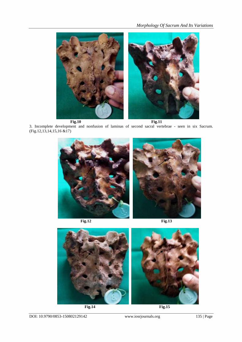
2. Nonfusion of first sacral lamina - seen in four Sacrum.( Fig.8,9,10 &11)
Fig.8 Fig.9
Morphology Of Sacrum And Its Variations
DOI: 10.9790/0853-150802129142 www.iosrjournals.org 135 | Page
Fig.10 Fig.11
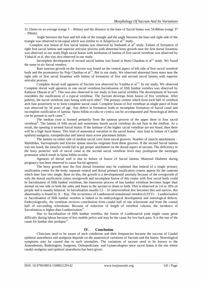
3. Incomplete development and nonfusion of laminas of second sacral vertebrae - seen in six Sacrum.
(Fig.12,13,14,15,16 &17)
Fig.12 Fig.13
Fig.14 Fig.15

Morphology Of Sacrum And Its Variations
DOI: 10.9790/0853-150802129142 www.iosrjournals.org 136 | Page
Fig.16 Fig.17
4. Absence of median crest with nonfusion of laminas of first and second sacral vertebrae.(Fig.18)
Fig.18
6. Incomplete median crest. (Fig.19,20,21 ,22 & 23)
Fig.19 Fig.20

Morphology Of Sacrum And Its Variations
DOI: 10.9790/0853-150802129142 www.iosrjournals.org 137 | Page
Fig.21 Fig.22
Fig.23
7. A foramina in the right side of lamina of second sacral vertebrae which indicates incomplete development
of right side of second sacral vertebrae.(Fig.24 & 25)
Fig.24 Fig.25

Morphology Of Sacrum And Its Variations
DOI: 10.9790/0853-150802129142 www.iosrjournals.org 138 | Page
7.Foramens on either side below the first spinous process and at the level of second and third spinous process
with absence of median crest.(Fig.26 &27)
Fig.26 Fig.27
8. Foramen just above the apex of sacral hiatus on either side.(Fig.28.29,30 &31)
Fig.28 Fig.29
Fig.30 Fig.31

Morphology Of Sacrum And Its Variations
DOI: 10.9790/0853-150802129142 www.iosrjournals.org 139 | Page
9. Multiple foramen in the dorsal surface of base of sacrum behind the auricular surface in the ala for the
attachment of Interosseous ligament and Dorsal sacroiliac ligament.(Fig.32)
Fig.32
10. Complete dorsal wall agenesis.(Fig.33)
Fig.33
11. Sacralisation of fifth lumbar vertebrae.(Fig.34,35,36 &37)
Fig.34 Fig.35

Morphology Of Sacrum And Its Variations
DOI: 10.9790/0853-150802129142 www.iosrjournals.org 140 | Page
Fig.36 Fig.37
12. High sacral hiatus.(Fig.38 &39 )
Fig.38 Fig.39
III. Discussion Sacral composition was five segments and was about 56% which was higher. This was similar to study
done by Anupriya et al15
.
Shape of Sacral hiatus was inverted ‘U’ shaped among 27 Sacra which was higher, followed by
Inverted ‘V’shaped, irregular, Dumbbell, bifid in our study which was similar to study done by, Seema et al1,
Nagar.S.K2, Dipali Rani Pal
6 et al and Jadhav Mayuri et al.
Location of apex of Sacral hiatus was at fourth Sacral vertebrae in 36 dry Sacral vertebrae in our
study.This was similar to Seema1, Nagar S.K
2, Jadhav Mayuri
8 studies.
Location of Base was at fifth Sacral vertebrae in 48 dry Sacra in our study which was similar to Seema
et al1, Nagar S.K
2, Dipali Rani Pal
6 et al studies.
Maximum curvature of Sacrum was observed to be at S3 vertebrae level which was similar to study
done by A.Anupriya et al15
.
Anteroposterior diameter at the sacral hiatus was about 0 – 3 mm in 35 dry Sacra in our study. In the
other studies by Seema et al1, Nagar.S.K
2, Dipali Rani Pal
6 et al and Jadhav Mayuri et al
8 4 – 6 mm was
observed as higher Anteroposterior diameter.
Transverse diameter was found to be 11 – 15 mm in 36 Sacra in our study. This was similar to Seema
et al1, Nagar.S.K
2, Dipali Rani Pal
6 et al studies.
The distance between S2 foramen and apex of Sacral hiatus was 34.6mm on an average (range 6 –
52mm ) and the distance to the base of the Sacral hiatus was 51.2mm (range 15 – 58mm) in our study. It has
reported by Senoglu N et al16
that the distance between S2 foramen and apex of Sacral hiatus was 35.37mm on
an average (range 11 – 62mm) and the distance to the base of the Sacral hiatus was 65.25mm (range 39 –
85mm). It also has reported by Dipali Rani Pal6 et al that the distance between S2 and apex of Sacral hiatus was
Morphology Of Sacrum And Its Variations
DOI: 10.9790/0853-150802129142 www.iosrjournals.org 141 | Page
31.33mm on an average (range 5 – 60mm) and the distance to the base of Sacral hiatus was 54.88mm (range 37
– 39mm).
Angle between the base and left side of the triangle and the angle between the base and right side of the
triangle was observed to be equal which was similar to A.Anupriya et al15
study.
Complete non fusion of first sacral lamina was observed by Sushanth et al9 study. Failure of formation of
right first sacral lamina and superior articular process with abnormal bony growth near the first dorsal foramina
was observed in our study.High sacral hiatus with nonfusion of lamina of first sacral vertebrae was observed by
Vishal.K et al. this was also observed in our study.
Incomplete development of second sacral lamina was found in Renu Chauhan et al11
study. We found
the same in six Sacral vertebra.
Rare osseous growth on the Sacrum was found on the ventral aspect of left side of first sacral vertebral
body and the promontory by Puja Chauhan et al12
. But in our study, We observed abnormal bony mass near the
right side of first sacral foramina with failure of formation of first and second sacral lamina with superior
articular process.
Complete dorsal wall agenesis of Sacrum was observed by Vanitha et al13
. In our study, We observed
Complete dorsal wall agenesis in one sacral vertebrae.Sacralisation of fifth lumbar vertebra was observed by
Kubavat Dharati et al14
. This was also observed in our study in four sacral vertebra.The development of Sacrum
resembles the ossification of a typical vertebrae. The Sacrum develops from fusion of five vertebrae. After
puberty, the sacral vertebrae start fusing with each other6. The primary centres which form each half of vertebral
arch fuse posteriorly to to form complete sacral canal. Complete fusion of five vertebrae as single piece of bone
was observed by 34 years of age. Any defect in formation leads to incomplete formation of Sacral canal and
incomplete ossification of lamina. Spina bifida occulta or cystica can be accompanied and Neurological defects
can be present in such cases14
.
The median crest is formed primarily from the spinous process of the upper three to four sacral
vertebrae8. The lamina of fifth sacral and sometimes fourth sacral vertebrae do not fuse in the midline. As a
result, the opening is termed Sacral hiatus. If the laminae of the higher sacral vertebrae are not fused, then there
will be a high Saral hiatus. This kind of anatomical variation in the sacral hiatus7 may lead to failure of Caudal
epidural analgesia, transpedicular and lateral mass screw placement failure.
The lamina on either side of median sacral crest form sacral grooves. Number of muscle attachments –
Multifidus, Sacrospinalis and Erector spinae muscles originate from these grooves. If the second Sacral lamina
was not fused, the muscles would fail to get proper attachment on the dorsal aspect of sacrum. The deficiency in
the bony posterior wall of sacral canal at the second sacral vertebrae level may predispose the meninges
protrusion which result in Spina bifida occulta.
Agenesis of dorsal wall is due to failure of fusion of Sacral lamina. Maternal Diabetes during
pregnancy has been observed to cause Sacral agenesis.
The bony growth near the first dorsal foramina may be explained that instesd of a single primary
ossification centre for the body, separate ventral and dorsal primary ossification centres appear for the centrum
which later fuse into single. Base on this, the growth is a developmental anomaly because of the overgrowth of
only the dorsal ossification centre overgrowth and incomplete fusion of this centre with first sacral body could
In Sacralisation of fifth lumbar vertebrae, the transverse process of last lumbar vertebrae becomes larger than
normal on one side or both the sides and fuses to the sacrum or ilium or both. This is observed in 3.6 to 18% of
people and is usually bilateral. In Sacralisation usually L5 – S1 intervertebral disc becomes thin and narrow, this
abnormality is found by X – Ray. The occurrence of Lumbosacral transitional vertebra (LSTV) – Lumborisation
or Sacralisation of fifth lumbar vertebra is linked to its embryological development and osteological defects.
Embryologically, the vertebrae receives contribution from caudal half of one sclerotome and from the cranial
half of succeeding sclerotome. Because of reduction of length of vertebral column, the incidence of
Sacralisation is higher than Lumborisation11
.
Due to Sacralisation of fifth lumbar vertebra, the fusion of Lumbosacral joint might cause great
difficulty during labour because of less mobile pelvis and may be the cause for low back pain. It is the one of the
cause for lumbar disc prolapse11
.
IV. Conclusion Clinicians need to be aware of such conditions and their frequencies because the success of Caudal
epidural anaesthesia and analgesia depends on the anatomical variations of Sacrum and the hiatus. Neurological
symptoms may be caused due to such anomalies. The variations of sacrum need to be known to the
Anaesthetists, Radiologists, Surgeons, Orthopedicians and Gynaecologists since sacral hiatus is the site where
caudal analgesia and epidural anaesthesia has been given.

Morphology Of Sacrum And Its Variations
DOI: 10.9790/0853-150802129142 www.iosrjournals.org 142 | Page
Knowledge of this type of variation may be helpful to the Radiologists in interpreting the radiographs of
Sacral spine.This variation may also benefit Orthopedicians in diagnosing the cause of Lowbackpain. It is useful
in diagnosing the cause of neurological involvement of bladder, rectum and lower limbs8.
The sacrum has anatomical variations and these variations are important for Caudal epidural anaesthesia. In
the present study, many variations of Sacrum like incomplete formation of first Sacral lamina and superior
articular process with bony growth near the first dorsal foramina, nonfusion of first sacral lamina with bony
growth near the first dorsal foramina, nonfusion of second sacral lamina, absence of median crest with
nonfusion of lamina of first and second sacral lamina, complete dorsal wall agenesis, Sacralisation of fifth
lumbar vertebrae, elongated hiatus and narrowing of Sacral canal at the apex of the hiatus were found in higher
percentage1&5
. These variations should be kept in mind while giving Caudal anaesthesia.
Exact localization of Sacral hiatus would help in easy passage of needle into the sacral canal.
Variations in the shape and level of the hiatus may lead to failure of Caudal epidural anaesthesia. This study will
be helpful to the Anaesthesiologists in identifying the variations of Sacrum and to locate the sacral hiatus during
Caudal epidural anaesthesia.
References [1]. Seema,Singh,Mahajan. An anatomical study of variations of sacral hiatusin sacra of North Indian origin and its clinical
significance.Int.J.Morphol 2013:31(1);110 - 14
[2]. Nagar S.K. A study of sacral hiatus in dry human sacra. J.Anat.Soc.India2004;53(2)18 – 21 [3]. Standring S. The Back.In: Gray’s Anatomy’s Sacrum.40th ed.Edinburg,UK: Churchill Livingstone 2008 : 724
[4]. Neeta V.Kulkarni. Bones of Abdomen and Pelvis.In:Clinical Anatomy,2nd edition,Jaypee Brothers.Medical publishers(P)Ltd.2012.632
[5]. Janusz piontek. Variation of level of closure of the sacral canal in man. Folia morphologica,1971,459 -64
[6]. Dipali Rani Pal, Md.ashfagur Rahman et al. Morphometric study of sacral hiatus: A basis for successful Caudal epidural block, Bangaladesh J.Anat.2012;10(1):5 – 10
[7]. Santanu Bhattacharya, Sudeshna Majumdar et al. A morphometric study of sacral hiatus for caudal epidural block among the
population of West Bengal.I.J.Basic & Applied Med.Research:Jun 2013:issue – 7,vol -2,p.660 – 67 [8]. Jadhav Mayuri, Ghorpadi vijay et al, Anatomical study of Sacral hiatus in Dry isolated sacra, J.Rearch in Medical & dental Sci,
vol.2,issue2, April – June 2014
[9]. Sushanth, Shishirkumar, Complete nonfusion of sacral lamina – A case study, I.J.S.R 2014,vol.3,issue7, 737 – 38 [10]. Vishal K, Vinay K.V, High sacral hiatus with Nonfusion of lamina of first sacral vertebrae: A case report, Nitte university, J.Health
Sci,Dec 2012,vol.2, No.4, 60 – 62
[11]. Renu chauhan, Jugesh Khanna, incomplete development of second sacral lamina: a case report, I.J.Research in Med.Sci,2013,vol.1,issue3,278 -80
[12]. Puja Chauhan, Sumita Kalra, Arare osseous growth on sacrum, I.J.A.V,2010;3:218 – 19
[13]. Vanitha, Taqdees Fatima, H.S.Kadlimatti, Complete dorsal wall agesis of sacrum: A case report, I.J.Dental &Medical Sciences,2014, vol.13,issue 4,pp80 – 81
[14]. Kubavat Dharati, Nagar et al, A study of Sacralisation of fifth lumbar vertebra in Gujarat, N.J.Medical
Research,2012,vol.2,issue2,211 – 13. [15]. A.Anupriya, M.Mahima Sophia, Anatomical study of Sacral hiatus in South Indian population and its clinical significance in Caudal
epidural anaesthesia, N.J.C.Anatomy, July 2014, vol.3,issue3,pp128 – 36.
[16]. Senoglu N, SenogluM, Oksuz H et al, Landmarks of the sacral hiatus for Caudal epidural block: an anatomical study. Br.Janaes,2005; 95(5):692 – 95
[17]. A.K.Datta, Essentials of Human Embryology, 6th ed.2010; 97,278 – 79.