Embed Size (px)
Citation preview
Journal of Medical Science 2020;89(3) 167
Morphology of rat brain neurons in subtotal ischaemia and introduction of L-NAME and omega-3 polyunsaturated fatty acids
O R I G I N A L PA P E R
Lizaveta BonDepartment of Pathological Physiology named after D.A. Maslakov, Grodno State Medical University
https://orcid.org/0000-0002-2380-1586
Corresponding author: [email protected]
Natalia Ye. MaksimovichDepartment of Pathological Physiology named after D.A. Maslakov, Grodno State Medical University
https://orcid.org/0000-0003-3181-9513
Sergey M. ZimatkinChair of Histology, Cytology and Embryology, Grodno State Medical University
https://orcid.org/0000-0001-5728-2588
DOI: https://doi.org/10.20883/medical.e423
Keywords: cerebral ischaemia, parietal cortex, hippocampus, omega-3 polyunsaturated fatty acids, Nω-nitro-L-Arginine Methyl Ester
Published: 2020-09-30
How to cite: Bon L, Maksimovich NY, Zimatkin SM. Morphol-ogy of rat brain neurons in subtotal ischaemia and introduc-tion of L-NAME and omega-3 polyunsaturated fatty acids. JMS [Internet]. 2020 Sep 30;89(3):e423. doi:10.20883/medi-cal.e423
© 2020 by the author(s). This is an open access article distributed under the terms and conditions of the Creative Commons Attri-bution (CC BY-NC) licencse. Published by Poznan University of Medical Sciences
AbstRAct
Introduction. Cerebral ischaemia leads to the development of numerous morphofunctional disorders of the cerebral cortex, which can be exacerbated by the introduction of a Nω-nitro-L-arginine methyl ester (L-NAME), which is a non-selective inhibitor of nitric oxide synthase (NOS).Aim. To study the morphological features of rat brain neurons in subtotal ischaemia during the administra-tion of L-NAME and naturally occurring omega-3 polyunsaturated docosahexaenoic acid (DHA).Material and Methods. Subtotal cerebral ischaemia was modelled in rats by ligation of both common carot-id arteries. L-NAME and DHA were given individually or in combination to separate groups of rats. L-NAME (5 mg/kg) was administered intramuscularly immediately before ligation and DHA (5 mg/kg) intragastrically during the week before ligation.Results. The introduction of DHA alone had a corrective effect on the hippocampus under conditions of sub-total ischaemia, reducing the number of shadow cells and hyperchromic wrinkled neurons, without signifi-cantly affecting the size and shape of the neurons of the parietal cortex. However, the previous administra-tion of DHA to rats with cerebral ischaemia receiving a NOS inhibitor did not abrogate the negative effects on the state of the neurons in the cerebral cortex.Conclusion. The administration of DHA can modulate the morphological disorder of the hippocampus which occurs in subtotal cerebral ischaemia.
Introduction
Stroke is one of the most pressing problems in modern medicine [1,2]. Cerebrovascular diseases
of ischaemic origin tend to grow, rejuvenate and are associated with a severe clinical course, high rates of disability, and mortality. The frequency of acute cerebrovascular accidents varies from 1 to
Journal of Medical Science 2020;89(3)168
4 cases per 1,000 population per year. Cerebro-vascular injuries are critical, requiring the con-centrated efforts of different specialists, as well as the search for new approaches for the treat-ment of the disorders of ischaemic brain gene-sis [2]. One of the promising directions for solv-ing this issue is to inhibit the formation of nitric oxide (NO). NO acts as a free radical therefore can exhibit both anti- and prooxidant proper-ties, affecting vascular tone, haemostasis, and inflammation, showing dual effects. The non-se-lective inhibitor of NO synthase (NOS), Nω-nitro-L-arginine methyl ester (L-NAME) inhibits all iso-forms of the enzyme, including endothelial, which leads to a decrease in antihypoxic resistance, an increase in platelet aggregation, and a decrease in cerebral blood flow [3,4].
Omega-3 polyunsaturated fatty acids (Ome-ga-3) can have a corrective effect on the state of the endothelium, reducing the severity of oxi-dative stress by activating antioxidant enzymes (catalase, superoxide dismutase, glutathione per-
oxidase, and glutathione transferase), as well as acting as free radical scavengers [5,6]. The action of Omega-3 is associated with their effect on the function of cell membranes, transmembrane ion channels and the regulation of physiological pro-cesses. Furthermore, Omega-3 is involved in the implementation of basic neuronal functions, such as impulse transmission and receptor activation. Brain neurons, being electrically active cells rich in ion channels, are most sensitive to Omega-3 deficiency [7].
This study aimed to investigate the morpho-logical characteristics of neurons of the parietal cortex and hippocampus of rats with subtotal cerebral ischaemia against the background of the administration of Omega-3 and L-NAME.
Material and Methods
Studies were performed on 30 male outbred white rats weighing 210±20 g according to the require-ments of the Directive of the European Parliament and the Council No. 2010/63 / EU of 09.22.2010 on the protection of animals used for scientific purposes. Protocols were reviewed and approved by the Ethical Committee of the Grodno State Medical University (protocol No 1, 14.04.2013). The animals were kept in the vivarium in a ven-tilated room at 22°C, 50% humidity and illumina-tion not more than 25 luxin, with no more than five rats per cage. The animals were fed cereals and legumes (oats, millet, barley, peas) and suc-culent feed (carrots, beets, cabbage) three times a day and had easy access to food and water.
The rats were divided into five groups, with six rats per group as described in Table 1. Sub-total cerebral ischaemia (SCI) was modelled by ligation of both common carotid arteries under conditions of intravenous thiopental anaesthe-
sia (40-50 mg/kg). L-NAME was administered intramuscularly immediately before SCI at a dose of 5 mg/kg. Docosahexaenoic acid (DHA) in the form of a microalgae Schizochytrium sp. (Polski Lek S.A., Poland) preparation at a dose of 0,5g/kg was given intragastrically during the week before SCI. The control group consisted of false-operat-ed rats treated with 0.5 ml isotonic NaCl solution.
The duration of the surgery to induct SCI was 60 minutes, after which the rats were decapitat-ed. Samples of the anterior cortex of the cere-bral hemispheres were quickly fixed in Carnoy’s fluid to study the morphological changes in the neurons of the parietal cortex and CA1 field of the hippocampus in rats after SCI. Serial paraf-
table 1. Experimental scheme
Animal group Experimental schemeControl false-operated rats received 0.5 ml of isotonic NaCl solutionGroup 1: SCI ligation of both common carotid arteries under conditions of intravenous thiopental
anaesthesia (40-50 mg/kg)Group 2: SCI + L-NAME L-NAME was administered intramuscularly at a dose of 5 mg/kg immediately before SCIGroup 3: SCI + L-NAME + DHA L-NAME was administered intramuscularly at a dose of 5 mg/kg immediately before SCI
and DHA (5 mg/kg) administered intragastrically during the week before surgeryGroup 4: SCI + DHA were additionally given intragastrically during the week before SCI with DHA at a dose
of 5 mg/kg body weight
Journal of Medical Science 2020;89(3) 169
fin sections were stained with 0.1% toluidine blue according to the Nissl method.
The microphotography, morphometry, and densitometry of chromogen sediment in histo-logical preparations were performed using an Axioscop 2 plus microscope (Zeiss, Germany), a digital video camera (LeicaDFC 320, Germany) and ImageWarp image analysis programme (Bit-flow, USA). The localisation of the parietal cor-tex and the hippocampus cortex was determined using a stereotactic atlas [9]. At least 30 neu-rons of the fifth layer of the parietal cortex and the pyramidal layer of the CA1 field of the hip-pocampus were evaluated in each animal, which ensured a sufficient sample size for subsequent analysis. The number of large pyramidal neurons per unit area of paraffin sections of the cerebral cortex was determined. Cells were distinguished by the intensity of cytoplasm (chromatophilia) staining: normochromic – moderately stained, hyperchromic – dark, hyperchromic shrunken – very dark, with deformed perikaryons, hypochro-mic – lightly stained and shadow cells – almost transparent. The number of cells of each type per 1 mm2 of brain tissue was counted. The size and shape of neuronal perikaryons were determined by area, form factor, and elongation factor.
Non-parametric statistical methods were used for data analysis (Statistica 10.0 software for Windows, StatSoft, Inc., USA) and the results are presented as Me (LQ; UQ), where Me is the median, LQ is the boundary of the lower quartile and UQ is the boundary of the upper quartile. The comparison between analysed groups was performed by Kruskal-Wallis test with post-hoc Dunn’s tests. All tests were considered signifi-cant at p<0.05.
Results
The dimensions and shape of perikaryon neu-rons of the parietal cortex and hippocampus are shown in Table 2. The morphometry of the neurons in group 1 (SCI) revealed a significant decrease in the area of their perikaryons by 53% and 49%, with a 20% increase in elongation in each of the studied sections of the cerebral cor-tex, and an 11% and 22% decrease in roundness, respectively. It is assumed that these changes in the size and shape of neurons are due to water-
electrolyte abnormalities, as well as the denatur-ation of the protein inside the cell, which forms the basis of the neurofibrils.
In group 2 (SCI + L-NAME), the form factor decreased by 22% in the neurons of the parietal cortex compared to group 1 (SCI). Compared with the control group, the area of neurons decreased by 52%, the form factor by 22%, and the elonga-tion factor of neurons increased by 29% in the parietal cortex. No changes were detected in the hippocampus, and compared with the control group, there was a 52% decrease in perikaryon area and 11% decrease in the factor form, with a 29% increase in the elongation factor.
Compared to the control, in the parietal cortex of group 3, the area of neurons decreased by 53%, the form factor by 22%, and the elongation fac-tor of neurons increased by 20%, whereas in the hippocampus, the area of neurons decreased by 48%, the form factor by 11%, and the elongation factor of neurons increased by 20%.
In the group 4 (SCI + L-NAME + DHA), there were no significant different area of neurons decreased by 53%, the form factor by 22%, and the elongation factor of neurons increased by 20% compared with those in the group 1 (SCI) and group 2 (SCI + L-NAME).
table 2. Dimensions and shape of perikaryon neurons of the parietal cortex and hippocampus of rats
Animal group Brain departmentsparietal cortex hippocampusarea, mkm2
Control 145(130; 154)b 109(100; 122)b
SCI 69(67; 74)a 56(55; 57)a
SCI + L-NAME 69 (59; 79)a 52(38; 58)a
SCI + DHA 68(50; 84)a 58(53; 84)a
SCI + L-NAME + DHA 68 (54; 80)a 57(40; 60)a
form factor, units Control 0,9(0,9; 0,9)b 0,9(0,9; 0,9)b
SCI 0,8(0,8; 0,8)a 0,7(0,7; 0,8)a
SCI + L-NAME 0,7(0,6; 0,7)a 0,8(0,8; 0,8)a
SCI + DHA 0,7(0,7; 0,8)a 0,8(0,6; 0,8)a
SCI + L-NAME + DHA 0,7(0,7; 0,8)a 0,8(0,7; 0,8)a
elongation factor, units Control 1,2(1,1; 1,3)a 1,2(1,1; 1,3)a
SCI 1,5(1,4; 1,5)b 1,5(1,4; 1,6)b
SCI + L-NAME 1,7(1,5; 1,8)b 1,7(1,6; 1,8)b
SCI + DHA 1,4(1,4; 1,5)b 1,4(1,4; 1,4)b
SCI + L-NAME + DHA 1,5(1,5; 1,5)b 1,5(1,4; 1,6)b
a, b – groups followed by the same letter do not differ statistically significantly
Journal of Medical Science 2020;89(3)170
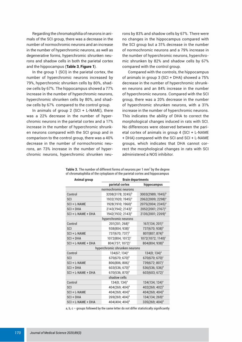
Regarding the chromatophilia of neurons in ani-mals of the SCI group, there was a decrease in the number of normochromic neurons and an increase in the number of hyperchromic neurons, as well as degenerative forms, hyperchromic shrunken neu-rons and shadow cells in both the parietal cortex and the hippocampus (Table 3, Figure 1).
In the group 1 (SCI) in the parietal cortex, the number of hyperchromic neurons increased by 79%, hyperchromic shrunken cells by 80%, shad-ow cells by 67%. The hippocampus showed a 77% increase in the number of hyperchromic neurons, hyperchromic shrunken cells by 80%, and shad-ow cells by 67% compared to the control group.
In animals of group 2 (SCI + L-NAME), there was a 22% decrease in the number of hyper-chromic neurons in the parietal cortex and a 17% increase in the number of hyperchromic shrunk-en neurons compared with the SCI group and in comparison to the control group, there was a 40% decrease in the number of normochromic neu-rons, an 73% increase in the number of hyper-chromic neurons, hyperchromic shrunken neu-
rons by 83% and shadow cells by 67%. There were no changes in the hippocampus compared with the SCI group but a 31% decrease in the number of normochromic neurons and a 79% increase in the number of hyperchromic neurons, hyperchro-mic shrunken by 82% and shadow cells by 67% compared with the control group.
Compared with the controls, the hippocampus of animals in group 3 (SCI + DHA) showed a 75% decrease in the number of hyperchromic shrunk-en neurons and an 84% increase in the number of hyperchromic neurons. Compared with the SCI group, there was a 20% decrease in the number of hyperchromic shrunken neurons, with a 31% increase in the number of hyperchromic neurons. This indicates the ability of DHA to correct the morphological changes induced in rats with SCI. No differences were observed between the pari-etal cortex of animals in group 4 (SCI + L-NAME + DHA) compared with the SCI and SCI + L-NAME groups, which indicates that DHA cannot cor-rect the morphological changes in rats with SCI administered a NOS inhibitor.
table 3. The number of different forms of neurons per 1 mm2 by the degree of chromatophilia of the cytoplasm of the parietal cortex and hippocampus
Animal group Brain departmentsparietal cortex hippocampus
normochromic neuronsControl 3208(3178; 3245)b 3003(2989; 1945)b
SCI 1932(1920; 1945)a 2062(2009; 2298)a
SCI + L-NAME 1928(1910; 1960)a 2075(2004; 2345)a
SCI + DHA 2143(1942; 2143)a 2052(2001; 2167)a
SCI + L-NAME + DHA 1942(1932; 2143)a 2135(2001; 2269)a
hyperchromic neuronsControl 201(201; 268)a 167(134; 201)a
SCI 938(804; 938)c 737(670; 938)b
SCI + L-NAME 737(670; 737)b 807(807; 874)b
SCI + DHA 1072(804; 1072)c 1072(1072; 1140)c
SCI + L-NAME + DHA 804(737; 1072)c 804(804; 938)b
hyperchromic shrunken neuronsControl 134(67; 134)a 134(0; 134)a
SCI 670(670; 670)b 670(670; 670)c
SCI + L-NAME 806(806; 806)c 739(672; 807)c
SCI + DHA 603(536; 670)b 536(536; 536)b
SCI + L-NAME + DHA 670(536; 870)b 603(603; 672)c
shadow cellsControl 134(0; 134)a 134(134; 134)a
SCI 404(269; 404)b 402(269; 402)b
SCI + L-NAME 404(269; 404)b 404(269; 404)b
SCI + DHA 269(269; 404)b 134(134; 269)a
SCI + L-NAME + DHA 404(404; 404)b 335(269; 404)b
a, b, c – groups followed by the same letter do not differ statistically significantly
Journal of Medical Science 2020;89(3) 171
Discussion
Subtotal cerebral ischaemia induces significant morphological changes in the parietal cortex such as a decrease in the size and deformation of the perikaryons of neurons, the appearance of a large number of hyperchromic neurons, as well as the appearance of degenerative forms (hyperchromic shrunken neurons and shadow
cells). Hyperchromic neurons are often regarded as markers of ischaemia [9, 10] and the intense staining of their cytoplasm is due to a significant predominance of the fraction of free ribosomes forming large clusters. The fixation of ribosomes to the membranes of the granular endoplasmic reticulum is an energy-dependent process pro-vided by the ribophorin protein. Therefore, the degranulation of cisterns of the granular endo-
Figure 1. Neurons of the pyramidal layer CA1 of the rat hippocampus stained by Nissl. A – control, B – SCI, C – SCI + L-NAME, D – SCI + L-NAME + DHA, E – SCI + DHA. Scale bars and magnifications: A - 20 µm, X400
Journal of Medical Science 2020;89(3)172
plasmic reticulum indicates increasing energy deficiency due to hypoxia. Degenerative chang-es in the granular endoplasmic reticulum lead to the accumulation of proteins in the cytoplasm, which denature under the influence of develop-ing hypoxia and acidosis. Shrinkage of neurons in the cerebral cortex is the result of water loss due to energy and ionic disorders that decrease the size and increases the deformation of per-ikaryons. Hyperchromic shrunken neurons lose their functional activity and are subsequently phagocytosed by microglia [9]. The appearance of shadow cells is the next stage of hypochromia, the cause of which is oedema due to electrolyte changes caused by energy deficiency [11].
The introduction of a non-selective inhibitor of NO synthase L-NAME exacerbated the histologi-cal disorders of neurons that occur during SCI: an increase in the number of hyperchromic shrunk-en neurons, a decrease in size and deformation of their perikaryons. This effect may be due to an increasing degree of ischaemia due to decreased formation of NO, primarily in the endothelium and neurons, which impedes the development of vasodilator compensatory reactions [3,4]. This leads to the progression of disturbances in cellu-lar metabolism, aggravation of water-electrolyte imbalance, manifested by deformation of neuron bodies, their wrinkling, and swelling [1,10].
Hippocampal neurons, as a phylogenetical-ly more ancient part of the cerebral cortex, are less sensitive to hypoxia, therefore in this part of the brain, DHA had a corrective effect in rats with SCI, reducing the number of pathological forms of neurons (hyperchromic shrunken and shadow cells). A favourable effect on the state of hippocampal neurons in conditions of subtotal cerebral ischaemia may be due to a decrease in the production of thromboxane A2 by platelets, an increase in the level of tissue plasminogen acti-vator, and improved erythrocyte membrane flu-idity, which leads to a decrease in viscosity and improved rheological properties of blood and cerebral circulation in general. DHA also exerts anti-inflammatory effects due to the incorpora-tion of monocytes, leukocytes, endothelial cells into the phospholipid layer of the cell membranes, which is accompanied by a decrease in the pro-duction of inflammatory mediators and adhesion of leukocytes to the endothelial wall. In addition, Omega-3 affects the synthesis of prostaglandins
that regulate vascular tone and inhibit vasocon-striction of blood vessels under the influence of catecholamines [5–7, 12–15].
Docosahexaenoic acid is involved in the bio-synthesis of tissue hormones such as resolvin, which inhibits inflammation and neuroprotec-tin D1, an endogenous neuroprotector with anti-apoptotic activity [7]. However, the administration of DHA to rats with SCI treated with a NOS inhibi-tor did not have a corrective effect on the neurons of the parietal cortex and hippocampus of rats.
The limitations of this study are due to the impossibility of following the dynamics of the adaptation of the brain during ischaemia and the metabolic changes of neurons [7].
Conclusion
Subtotal cerebral ischaemia leads to the devel-opment of morphofunctional disorders of the cerebral cortex, which can be modulated with the administration of DHA, reducing the number of shadow cells and hyperchromic wrinkled neu-rons, without significantly affecting the size and shape of the neurons of the parietal cortex.
Acknowledgements
conflict of interest statementThe authors declare no conflict of interest.
Funding sourcesThere are no sources of funding to declare.
ReferencesMaksimovich NY, Pronko TP, Maksimovich YN, Yer-1. mak VV. Epidemiology of ischemic strokes in the Grodno region (Belarus). Cerebrovascular Diesases. 2015 Jan 170-170;39.Igorevna Bon L. Effects of Experemental Cerebral 2. Ishemia on Metabolic Characteristics of Parietal Cortex Neurons. Bioprocess Engineering. 2018;2(1):1. https://doi.org/10.11648/j.be.20180201.11Maksimovich N, Zinchuk V, Maslacov D. The degree 3. of oxidative stress in the rat brain during ischemia and reperfusion in conditions of correction of the L-arginine-NO system. Neuroscience and Behavioral Physiol. 2006;36(4):373-8.Maksimovich N. Tolerance of hypoxic hypoxia in rats 4. with cerebral ischaemia treated by NO-synthase modulators. Hypoxia Medical. 2004;V(1-2):20-3.Kaliannan K, Li X, Wang B, Pan Q, Chen C, Hao L, Xie 5. S, Kang JX. Multi-omic analysis in transgenic mice implicates omega-6/omega-3 fatty acid imbalance as a risk factor for chronic disease. Communications
Journal of Medical Science 2020;89(3) 173
Biology. 2019 Jul 26;2(1). https://doi.org/10.1038/s42003-019-0521-4Khunt D, Shrivas M, Polaka S, Gondaliya P, Misra M. 6. Role of Omega-3 Fatty Acids and Butter Oil in Tar-geting Delivery of Donepezil Hydrochloride Microe-mulsion to Brain via the Intranasal Route: a Compar-ative Study. AAPS PharmSciTech. 2020 Jan 3;21(2). https://doi.org/10.1208/s12249-019-1585-7Kangari H, Eftekhari MH, Sardari S, Hashemi H, 7. Salamzadeh J, Ghassemi-Broumand M, Khabaz-khoob M. Short-term Consumption of Oral Ome-ga-3 and Dry Eye Syndrome. Ophthalmology. 2013 Nov;120(11):2191-2196. https://doi.org/10.1016/j.ophtha.2013.04.006Wu B, Song Q, Zhang Y, Wang C, Yang M, Zhang J, Han 8. W, Jiang P. Antidepressant activity of ω-3 polyunsat-urated fatty acids in ovariectomized rats: role of neu-roinflammation and microglial polarization. Lipids in Health and Disease. 2020 Jan 8;19(1). https://doi.org/10.1186/s12944-020-1185-2Paxinos G, Watson C. The Rat Brain in Stereotaxic 9. Coordinates. 6. Australia: Academic Press; 1998.Gallyas F, Pál J, Bukovics P. Supravital microwave 10. experiments support that the formation of “dark” neu-
rons is propelled by phase transition in an intracellu-lar gel system. Brain Research. 2009 May;1270:152-156. https://doi.org/10.1016/j.brainres.2009.03.020Zimatkin SM, Bon EI. Dark Neurons of the Brain. 11. Neuroscience and Behavioral Physiology. 2018 Oct;48(8):908-912. https://doi.org/10.1007/s11055-018-0648-7Shahidi F, Ambigaipalan P. Omega-3 Polyunsatu-12. rated Fatty Acids and Their Health Benefits. Annual Review of Food Science and Technology. 2018 Mar 25;9(1):345-381. https://doi.org/10.1146/annurev-food-111317-095850Grosso G, Galvano F, Marventano S, Malaguarnera M, 13. Bucolo C, Drago F, Caraci F. Omega-3 Fatty Acids and Depression: Scientific Evidence and Biological Mech-anisms. Oxidative Medicine and Cellular Longevity. 2014;2014:1-16. https://doi.org/10.1155/2014/313570Serini S, Calviello G. Long-chain omega-3 fatty acids 14. and cancer. Current Opinion in Clinical Nutrition & Metabolic Care. 2018 Mar;21(2):83-89. https://doi.org/10.1097/mco.0000000000000439Rogers T, Seehusen D. Omega-3 Fatty Acids 15. and Cardiovascular Disease. Am Fam Physician. 2018;97(9):562-4.


![Myocardial Ischaemia - national audit project [MINAP] 2011 - UCL](https://img.pdfslide.us/doc/110x75/620349a224f6b61e9c664083/myocardial-ischaemia-national-audit-project-minap-2011-ucl.jpg)