Embed Size (px)
Citation preview
Case Series
Ischaemia and Infarction in Patients with Non-obstructive Coronary ArteriesRobert Sykes1,2*, Andrew Morrow1,2*, Chiara Bucciarelli-Ducci3, Colin Berry1,2,4
1 West of Scotland Heart and Lung Centre, Golden Jubilee National Hospital, Glasgow, UK2 Institute of Cardiovascular and Medical Sciences, University of Glasgow, UK3 Department of Cardiology, Bristol Heart Institute, University Hospitals Bristol and Weston NHS Trust and University of Bristol, UK
4 Department of Cardiology, Queen Elizabeth University Hospital, NHS Greater Glasgow and Clyde Health Board, Glasgow, UK
* contributed equally
Case
1, F
igur
e 1
siemens-healthineers.com/magnetom-world
IntroductionMyocardial ischaemia and infarction are complex patholo-gies caused by transient or sustained interruptions in myocardial blood flow, respectively. Myocardial ischaemia is caused by metabolic supply:demand imbalance which may be epicardial, microcirculatory, structural, arrhythmo-genic or systemic in origin. The absence of obstructive coronary artery disease in patients with myocardial infarction or objective myocardial ischaemia requires further assessment as patient outcomes and quality of life are impaired.
The following case examples illustrate the utilisation and yield of cardiovascular imaging in patients with MINOCA or INOCA during daily clinical practice. Cardiac MRI images were obtained including T1 and T2 parametric maps with gadolinium contrast, at the Golden Jubilee National Hospital, Clydebank, UK using a MAGNETOM Avanto 1.5 Tesla MRI scanner (Siemens Healthcare, Erlangen, Germany). Computed tomography coronary angiography is obtained using a SOMATOM 128 slice-dual source scanner (Siemens Healthcare, Forchheim, Germany).
A 51-year-old Caucasian female presented acutely with spontaneous central chest pain in association with stress. The symptoms lasted thirty-minutes and were associated with inferolateral T-wave inversion on a 12-lead ECG. The patient was a former smoker, otherwise well and active with a body mass index of 23 kg/m2. Haematology and renal function, and a chest x-ray were normal. The initial high-sensitivity troponin-I concentration was 2,566 ng/L, increasing to 24,639 ng/L at six hours after symptom onset. Echocardiography revealed mild left ventricular dysfunction in association with an infero-lateral wall motion abnormality.
The patient was treated with aspirin, ticagrelor and fondaparinux. She had uncomplicated progress in the coronary care unit and coronary angiography was per-formed within 72 hours of admission. The angiogram revealed a dominant right coronary anatomy with a <25% stenosis in the 1st obtuse marginal coronary artery. The
Case 1: Non-ST-elevated myocardial infarction with non-obstructive coronary arteries
angiogram was otherwise normal. Coronary physiology or intracoronary imaging were not performed. The diagnosis was MINOCA but the aetiology was uncertain. The patient was prescribed dual antiplatelet and secondary prevention therapies and discharged.
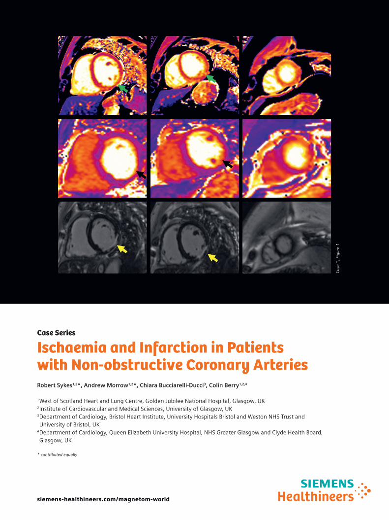
Cardiac MRI was performed on day 13 (Fig. 1). The scan revealed a discrete area of subendocardial late gadolinium enhancement (LGE) within the mid-basal inferolateral left ventricular wall involving up to 50% wall thickness. Overall, there was mild LVSD with biatrial dilatation. There was evidence of myocardial oedema with raised areas of native T1 and T2 co-localizing with the area of LGE. The findings were consistent with a convalescent inferolateral myocardial infarction presumed secondary to plaque rupture in the obtuse marginal coronary artery. She continued with cardiac rehabilitation and secondary prevention medication.
1 Cardiac MRI revealing a discrete area of subendocardial late gadolinium enhancement (yellow arrows) within the mid-basal inferolateral left ventricular wall co-localising with raised areas of native T1 (green arrows) and T2 (black arrows). Findings are consistent with a convalescent lateral myocardial infarction.
Cardiovascular Imaging
2 siemens-healthineers.com/magnetom-world
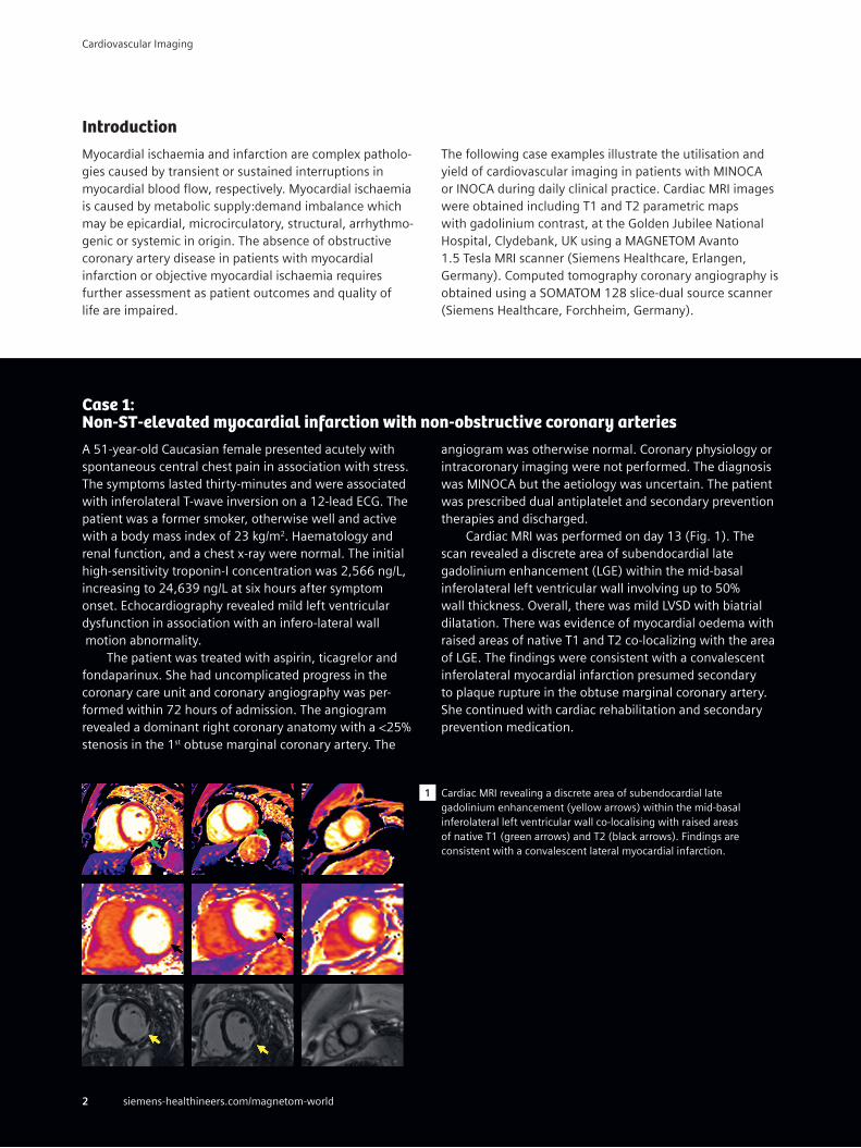
Case 2: Acute myopericarditisA 29-year-old Caucasian male with a family history of ischaemic heart disease but no other medical history presented with heavy central chest pain that worsened on inspiration and had increased over the preceding week. On admission cardiac enzymes were elevated (hs-TnI 4.944 ng/L) and 12-lead electrocardiogram demonstrated no abnormality. Echocardiogram revealed normal biventricular function and no pericardial fluid or enhancement. He was initially managed as a suspected non-ST-elevated myocardial infarction with non-obstructive coronary arteries but underwent inpatient cardiac MRI due to young age and relatively lacking risk factors for coronary artery disease (Fig. 2).
The MRI scan revealed preserved an inferior left ventricular wall motion abnormality associated with a mid-wall pattern of late gadolinium enhancement in the basal inferior wall. These features co-localized with areas of myocardial foci with increased native T1, T2 and ECV consistent with evi-dence of myocardial oedema. The diagnosis was revised to acute myopericarditis. There was no preceding prodromal illness, pathogen or substance was identified in this case. Due to the pericarditic features of the chest pain this patient was treated with oral colchicine and discharged home with advice to avoid strenuous exercise.
2 Cardiac MRI revealing a mid-wall pattern of late gadolinium enhancement in the basal inferior LV wall (red arrow) that co-localize with areas of raised native T1 (green arrow) and ECV (blue arrow) indicative of myocardial oedema.
3siemens-healthineers.com/magnetom-world
Cardiovascular Imaging
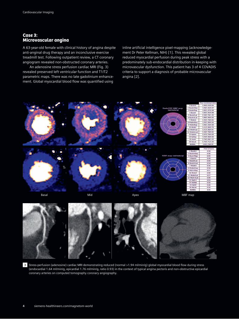
Case 3: Microvascular anginaA 63-year-old female with clinical history of angina despite anti-anginal drug therapy and an inconclusive exercise treadmill test. Following outpatient review, a CT coronary angiogram revealed non-obstructed coronary arteries.
An adenosine stress perfusion cardiac MRI (Fig. 3) revealed preserved left ventricular function and T1/T2 parametric maps. There was no late gadolinium enhance-ment. Global myocardial blood flow was quantified using
inline artificial intelligence pixel-mapping (acknowledge-ment Dr Peter Kellman, NIH) [1]. This revealed global reduced myocardial perfusion during peak stress with a predominately sub-endocardial distribution in-keeping with microvascular dysfunction. This patient has 3 of 4 COVADIS criteria to support a diagnosis of probable microvascular angina [2].
3 Stress-perfusion (adenosine) cardiac MRI demonstrating reduced (normal >1.94 ml/min/g) global myocardial blood flow during stress (endocardial 1.64 ml/min/g, epicardial 1.76 ml/min/g, ratio 0.93) in the context of typical angina pectoris and non-obstructive epicardial coronary arteries on computed tomography coronary angiography.
Basal Mid Apex MBF map
4 siemens-healthineers.com/magnetom-world
Cardiovascular Imaging
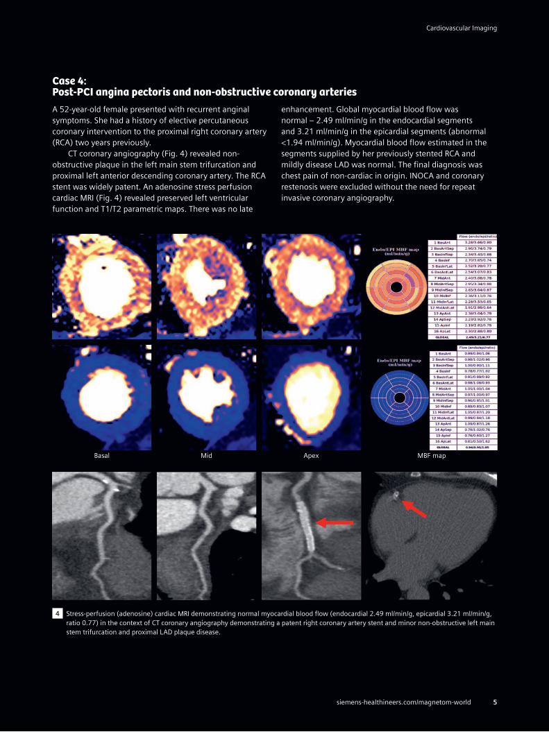
A 52-year-old female presented with recurrent anginal symptoms. She had a history of elective percutaneous coronary intervention to the proximal right coronary artery (RCA) two years previously.
CT coronary angiography (Fig. 4) revealed non- obstructive plaque in the left main stem trifurcation and proximal left anterior descending coronary artery. The RCA stent was widely patent. An adenosine stress perfusion cardiac MRI (Fig. 4) revealed preserved left ventricular function and T1/T2 parametric maps. There was no late
enhancement. Global myocardial blood flow was normal – 2.49 ml/min/g in the endocardial segments and 3.21 ml/min/g in the epicardial segments (abnormal <1.94 ml/min/g). Myocardial blood flow estimated in the segments supplied by her previously stented RCA and mildly disease LAD was normal. The final diagnosis was chest pain of non-cardiac in origin. INOCA and coronary restenosis were excluded without the need for repeat invasive coronary angiography.
4 Stress-perfusion (adenosine) cardiac MRI demonstrating normal myocardial blood flow (endocardial 2.49 ml/min/g, epicardial 3.21 ml/min/g, ratio 0.77) in the context of CT coronary angiography demonstrating a patent right coronary artery stent and minor non-obstructive left main stem trifurcation and proximal LAD plaque disease.
Basal Mid Apex MBF map
Case 4: Post-PCI angina pectoris and non-obstructive coronary arteries
5siemens-healthineers.com/magnetom-world
Cardiovascular Imaging
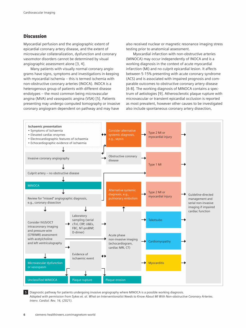
DiscussionMyocardial perfusion and the angiographic extent of epicardial coronary artery disease, and the extent of microvascular collateralization, dysfunction and coronary vasomotor disorders cannot be determined by visual angiographic assessment alone [3, 4].
Many patients with visually normal coronary angio-grams have signs, symptoms and investigations in-keeping with myocardial ischemia – this is termed ischemia with non-obstructive coronary arteries (INOCA). INOCA is a heterogenous group of patients with different disease endotypes – the most common being microvascular angina (MVA) and vasospastic angina (VSA) [5]. Patients presenting may undergo computed tomography or invasive coronary angiogram dependent on pathway and may have
also received nuclear or magnetic resonance imaging stress testing prior to anatomical assessment.
Myocardial infarction with non-obstructive arteries (MINOCA) may occur independently of INOCA and is a working diagnosis in the context of acute myocardial infarction (MI) and no culprit epicardial lesion. It affects between 5-15% presenting with acute coronary syndrome (ACS) and is associated with impaired prognosis and com-parable outcomes to obstructive coronary artery disease [6-8]. The working diagnosis of MINOCA contains a spec-trum of aetiologies [9]. Atherosclerotic plaque rupture with microvascular or transient epicardial occlusion is reported as most prevalent, however other causes to be investigated also include spontaneous coronary artery dissection,
5 Diagnostic pathway for patients undergoing invasive angiography where MINOCA is a possible working diagnosis. Adapted with permission from Sykes et. al. What an Interventionalist Needs to Know About MI With Non-obstructive Coronary Arteries. Interv. Cardiol. Rev. 16, (2021).
Ischaemic presentation• Symptoms of ischaemia• Elevated cardiac enzymes• Electrocardiographic features of ischaemia• Echocardiographic evidence of ischaemia
Consider IVUS/OCT intracoronary imaging and pressure-wire (CFR/IMR) assessment with acetylcholine and left ventriculography
Plaque rupture
Evidence of ischaemic event
Laboratory sampling (serial cTnl, CRP, U&Es, FBC, NT-proBNP, D-dimer)
Unclassified MINIOCA
Microvascular dysfunction or vasospasm
Invasive coronary angiography
Culprit artery – no obstructive disease
Review for “missed” angiographic diagnosis, e.g., coronary dissection
MINOCAAlternative systemic diagnosis, e.g., pulmonary embolism
Obstructive coronary disease
Consider alternative systemic diagnosis, e.g., sepsis
Plaque erosion
Acute phase non-invasive imaging (echocardiogram, cardiac MRI, CT)
Type 1 MI
Type 2 MI or myocardial injury
Type 2 MI or myocardial injury Guideline-directed
management and serial non-invasive imaging if impaired cardiac function
Myocarditis
Cardiomyopathy
Takotsubo
6 siemens-healthineers.com/magnetom-world
Cardiovascular Imaging
coronary artery embolism, microvascular and vasomotor disorders as in INOCA, arrhythmogenic or valvular precipitants and systemic causes of supply:demand mismatch. Stress cardiomyopathy and myopericarditis may also be diagnosed during the diagnostic pathway of MINOCA (Fig. 5).
Guidelines in MINOCA advocate for the consideration of adjunctive tests both during angiography and post- procedurally, with noninvasive imaging strategies such as cardiac magnetic resonance imaging and T1/T2 parametric mapping in addition to late gadolinium enhancement able to improve detection of the underlying pathophysiology [10]. Intravascular ultrasound or ocular coherence-tomog-raphy during coronary angiography may also be of value to assess for plaque erosion or rupture and presence of dissection [9]. Further invasive investigations for patients with MINOCA or INOCA include coronary pressure-wire as-sessment of physiological parameters during hyperaemia, including index of microvascular resistance (abnormal ≥25) and coronary flow reserve (abnormal <2.0), vasoreactivity
assessment using acetylcholine may also be performed to identify epicardial vasospasm (>90% reduction in epicardial coronary diameter with electrical or symptomatic angina) or microvascular spasm (<90% reduction in epicardial coronary diameter but with dynamic ischaemic electrocar-diogram features or symptomatic angina).
Non-invasive perfusion imaging may be clinically useful for the diagnostic evaluation of patients with suspected INOCA, especially when invasive measurements of microvascular function are not available. It may also provide objective confirmation of diagnosis in the presence of symptoms of angina, reduced myocardial perfusion and non-obstructed epicardial arteries. Hence, stratified medicine, involving a combinatory approach of anatomical and functional testing and linked therapy, can lead to improvements in patient symptoms, satisfaction and diagnostic certainty, as originally shown in the CorMicA trial [11]. Our cases illustrate how diagnostic imaging with CTCA and MRI is clinically useful in patients with known or suspected INOCA and MINOCA.
Contact Professor Colin Berry Institute of Cardiovascular and Medical Sciences University of Glasgow 126 University Place Glasgow G12 8TA UK Tel: +44 1413303325 [email protected]
References
1 Knott, K. D. et al. Quantitative myocardial perfusion in coronary artery disease: A perfusion mapping study. J. Magn. Reson. Imaging 50, 756–762 (2019).
2 Ong, P. et al. International standardization of diagnostic criteria for microvascular angina. Int. J. Cardiol. 250, 16–20 (2018).
3 Gould, K. L., Lipscomb, K. & Hamilton, G. W. Physiologic basis for assessing critical coronary stenosis: Instantaneous flow response and regional distribution during coronary hyperemia as measures of coronary flow reserve. Am. J. Cardiol. 33, 87–94 (1974).
4 Naya, M. et al. Quantitative Relationship Between the Extent and Morphology of Coronary Atherosclerotic Plaque and Downstream Myocardial Perfusion. J. Am. Coll. Cardiol. 58, 1807–1816 (2011).
5 Ford, T. J. et al. Stratified Medical Therapy Using Invasive Coronary Function Testing in Angina: The CorMicA Trial. J. Am. Coll. Cardiol. 72, 2841–2855 (2018).
6 Collet, J.-P. et al. 2020 ESC Guidelines for the management of acute coronary syndromes in patients presenting without persistent ST-segment elevation. Eur. Heart J. 42, 1289–1367 (2021).
7 Ibanez, B. et al. 2017 ESC Guidelines for the management of acute myocardial infarction in patients presenting with ST-segment elevation: The Task Force for the management of acute myocardial infarction in patients presenting with ST-segment elevation of the European Society of Cardiology (ESC). Eur. Heart J. 39, 119–177 (2018).
8 Nordenskjöld, A. M., Baron, T., Eggers, K. M., Jernberg, T. & Lindahl, B. Predictors of adverse outcome in patients with myocardial infarction with non-obstructive coronary artery (MINOCA) disease. Int. J. Cardiol. 261, 18–23 (2018).
9 Sykes R, Doherty D, Mangion K, Morrow A & C, B. What an Interventionalist Needs to Know About MI With Non-obstructive Coronary Arteries. Interv. Cardiol. Rev. 16, e10 (2021).
10 Agewall, S. et al. ESC working group position paper on myocardial infarction with non-obstructive coronary arteries. Eur. Heart J. 38, 143–153 (2017).
11 Ford, T. J. et al. Stratified Medical Therapy Using Invasive Coronary Function Testing in Angina: The CorMicA Trial. J. Am. Coll. Cardiol. 72, 2841–2855 (2018).
7siemens-healthineers.com/magnetom-world
Cardiovascular Imaging

Published by Siemens Healthcare GmbH · Order No. A91MR-1000-148C-7600 · 8705 1120 · © Siemens Healthcare GmbH, 2021
Siemens Healthineers HeadquartersSiemens Healthcare GmbH Henkestr. 127 91052 Erlangen, Germany Phone: +49 9131 84-0 siemens-healthineers.com
Cardiovascular Imaging