Embed Size (px)
Citation preview
A Molecular Map of G Protein a Chains in Microdissected Rat Nephron Segments
Stuart I. Senkfor, Gary L. Johnson, and Tomas BerlDepartment of Medicine, University of Colorado School of Medicine, Denver, Colorado 80262; and National Jewish Center forImmunology and Respiratory Medicine, Denver, Colorado 80206
Abstract
Membrane-associated guanine nucleotide binding proteins reg-ulate many receptor-mediated signals. Heterogeneity of bio-chemical and functional properties in nephron segments couldbe due to differences in G protein expression. To ascertainwhether such heterogeneity of Gproteins is present in variousnephron segments, this study examines the distribution andrelative abundance of Gprotein a chains in microdissected med-ullary thick ascending limb, cortical collecting tubules, outermedullary collecting tubules, proximal inner medullary tu-bules, and distal inner medullary tubules. Reverse transcriptionand polymerase chain reactions were employed using oligonu-cleotides encoding highly conserved regions of all known achains. The cDNA was sequenced for a chain identification.The a12 versus a, distribution was different in the outer medul-lary collecting tubules, when compared with the medullarythick ascending limb (P < 0.001) or the cortical collecting tu-bule, the proximal inner medullary tubules, and the distal innermedullary tubules (P < 0.05). These latter four segments didnot significantly differ from each other. A similar analysis wasapplied to the frequently used line of kidney cells, LLC-PK1,whose exact cellular origin remains unclear. Interestingly, wedetected both a12 and a%, while only ai2 was detected in the ratdistal nephron. No a. or a. reverse transcription PCRproductswere detected. In contrast all and al4 members of the morerecently described aq family were detected in the outer medul-lary collecting tubules and the proximal inner medullary tu-bules, respectively. Weconclude that the majority of nephronsegments have a relatively constant distribution of Gprotein achains. (J. Clin. Invest. 1993. 92:786-790.) Key words: (5)Gproteins * microdissected * rat* nephron * PCR
Introduction
Membrane-associated guanine nucleotide binding proteins (Gproteins) act as regulatory elements for many receptor me-diated signals (1, 2). The a component of the heterotrimericprotein appears to convey specificity for enzymatic and iontransport processes. A role for Gproteins in physiologic eventsand pathologic states is being increasingly recognized ( 1, 3).
The various segments of the mammalian nephron have a
multitude of receptors and respond to a large number of effec-tors, thereby allowing for diverse and complex responses that
Address correspondence to Dr. Tomas Berl, C28 1, University of Colo-rado School of Medicine, 4200 E. 9th Avenue, Denver, CO80262.
Receivedfor publication 28 September 1992 and in revisedform 8March 1993.
subserve its many functions (4). Of great interest, however, isthe demonstration that even a single hormone can bring abouta variety of effects in any given nephron segment (5-7). Forexample, in the inner medullary collecting tubules, vasopressinby its V2 receptor stimulates adenylyl cyclase. This results inincreased water permeability, and in its most terminal seg-ment, also in increased urea permeability. Likewise, in thissame segment the hormone also increases cell Ca2" by V2 re-ceptor-mediated pathways (8). A possible explanation for thisobservation is the presence of V2 receptor subtypes for whichevidence is as yet not forthcoming but could well emerge whenthe newly cloned vasopressin receptor is investigated further.An alternative explanation of the various responses to a singlehormone is divergent coupling of a single receptor to variouseffectors. Such a diversity of responses could be in part attrib-uted to G protein a chain heterogeneity. Such a mechanismmay well be operant in the response of LLC-PKI cells to calci-tonin where, in a cell cycle-dependent fashion, the hormonestimulates cyclase via Gs or activates the protein kinase Cpath-way via Gi (9). The heterogeneity in functional and biochemi-cal properties on the various regions of the nephron could thusrelate to differences in the a chain subunits of the guaninenucleotide binding proteins. In this regard, recent attemptshave been undertaken to localize G proteins along variousnephron segments using immunocytochemical methods withvariable results (10, 11). In contrast, we have chosen to iden-tify the specific a chain mRNAexpressed in vivo using a re-verse transcription PCRprotocol in vasopressin-sensitive re-gions of the nephron.
Methods
Preparation of tissue. Studies were performed on male Sprague-Daw-ley rats (Sasco Inc., Omaha, NE) weighing between 250 and 300 g. Theanimals were fed a commercially available diet (ICN Nutritional Bio-chemicals, Cleveland, OH) and unrestricted water. Microdissectionwas performed as previously described ( 12), modified by the additionof 10 mMVanadyl ribonucleoside complex to the dissection bath toinhibit RNAase. The following segments were dissected: medullarythick ascending limb of the loop of Henle (MTAL),' cortical collectingtubule (CCT), outer medullary collecting tubule (OMCT), proximalinner medullary collecting tubule (PIM), and distal inner medullarycollecting tubule (DIM).
Preparation of RNA extraction in cultured cells. LLC-PKl pur-chased from American Type Culture Collection, Rockville, MD, weregrown to confluence in Dulbecco's modified Eagle's media supple-mented with newborn calf and bovine calf serum. RNAwas extractedusing 1 ml of a 6 Murea and 3 MLiCl solution followed by sonicationfor 30 s. After 2 d of 4VC refrigeration, the sample was centrifuged at10,000 rpm for 15 min and the RNApellet resuspended in 500 ,l of a
1. Abbreviations used in this paper: CCT, cortical collecting tubule;DIM, distal inner medullary collecting tubule; MTAL, medullary thickascending limb; OMCT,outer medullary collecting tubule; PIM, prox-imal inner medullary collecting tubule.
786 S. I. Senkfor, G. L. Johnson, and T. Berl
J. Clin. Invest.© The American Society for Clinical Investigation, Inc.0021-9738/93/08/0786/05 $2.00Volume 92, August 1993, 786-790
buffer containing 10 mMTris (pH 7.5), 5 mMEDTA, and 0.1% wt/vol SDS. The mixture was extracted with phenol/chloroform (1:1)and chloroform followed by ethanol extraction. RNAquality was con-firmed by agarose gel electrophoresis in the presence of formaldehyde.
Reverse transcription. The dissected tubules as well as the RNAextracted from the cultured cells were subjected to reverse transcriptionusing a synthetic antisense oligonucleotide (29 mer) (vide infra asprimer 1). Specifically, the microdissected tubules were centrifuged fora few seconds ( 10,000 rpm) to pellet the sample. To this was added 9 Mlof a mixture containing 2% Triton X-100, 1 U/Ml placental RNAaseinhibitor and 5 mMDTT. After addition of this mixture, 11 .l of asecond mixture containing 2 LI of lOX amplification buffer (500 mMKCI, 100 mMTris-HCl pH 8.3, 15 mMMgCI2, and 0.1% wt/ volgelatin), 1 Ml 25 U/Ml placental RNAase inhibitor, 2 Ml of 10 mMdeoxynucleotides (dNTP's), 2 id of 50 MMprimer 1, 1 Ml of 50 mMMgC12, 0.5 Ml of avian myeloblastoma virus reverse transcriptase(Boehringer Mannheim, Corp., Indianapolis, IN) and 2.5 Ml of doublydeionized water was added. The samples and appropriate controls wereincubated at 37'C for 1 h followed by 95'C for 5 min.
For reverse transcription of the RNAobtained from cultured cells,the RNAwas combined with 10 Ml of a mixture containing 2 Ml ofdeoxynucleotides (10 mMeach dNTP), 1 Ml of placental RNAase in-hibitor, 1 ML of 50 mMMgCi2, 2 Ml of 50MMprimer 1, 0.5 Ml of avianmyeloblastosis virus reverse transcriptase (Boehringer MannheimCorp.) plus doubly deionized water to a final volume of 20 Ml. Sampleswere incubated as above.
Polymerase chain reaction. PCRreaction was performed using theDNA Thermal Cycler (Perkin-Elmer Cetus Instruments, Norwalk,CT). The antisense oligonucleotide was designed from a highly con-served region near the COOHterminus. Inosine was used at points ofhigh divergence. The sequence of primer 1 was as follows: 5'-CCAGCA-AGCTTIGTRTCIRYIGCRCAIGT-3'. Primer 2 was designed from asecond highly conserved region roughly 150 amino acids toward theNH2 terminus (a,) and was as follows: 5'-CCAGCGGTACCGAYG-TIGGIGSICARBG-3'. In these primers, R = A or G, Y = Cor T, S = Gor C, B = A or C, H = A, C, or T.
After reverse transcription and heat inactivation, the 20-Ml reactionmixture was combined with 80 Ml of the PCRreaction mixture. Thisincluded 10 Ml of lOx reaction buffer ( 100 mMTris-Hcl, pH 8.3, 500mMKCI, 0.01% [wt/vol] gelatin), 16 Ml of( 1.25 mMeach) deoxynu-cleotides (dNTP's), 10 Ml of 10MMprimer 1, 10 Ml of 10MMprimer 2,0.5 Ml of Amplitaq DNApolymerase (5 U/Ml; Perkin-Elmer Cetus)and 33.5 Ml of doubly distilled, sterile water. Lastly, 100 Ml of mineraloil was added to prevent evaporation. The first setting was 94'C for 2min, followed by 72'C for 2 min, and annealing temp of 60°C for 2min. The total number of cycles was 30. Upon completion of the reac-tion, 50 Ml of the PCRproduct was placed in an agarose gel along withappropriate standards. The area of product, based on expected size, wasextracted and DNArecovered using glass milk silica matrix techniqueprovided by GeneClean II Kit (BIO 101, Inc., La Jolla, CA). The DNAwas recovered in 20 Ml doubly deionized water and subjected to a sec-ond round of PCRusing the same PCRmixture and reaction condi-tions. The final product was subjected to phenol chloroform and chloro-form extraction followed by ethanol precipitation.
Restriction digestion. The oligonucleotides were designed withHindIll and KpnI sites on the 5' end of primer 1 and primer 2, respec-tively. Therefore, the final PCRproducts were subjected to restrictiondigestion with Hindill and KpnI (New England Biolabs Inc., Beverly,MA) as well as the plasmid vector PUC18. After digestion, the prod-ucts were run on an agarose gel and the areas of interest were extractedas described above.
Ligation. Ligation was carried out using 3 Ml of doubly digestedPUC18 and 5 Ml of similarly digested PCRproduct. The mixture wasincubated at 68'C for 10 min, placed on ice, and 2 Ml of lOX ligationbuffer (500 mMTris pH 7.4, 100 mMMgC12, 100 mMDTT, 10 mMspermidine, 10 mMATP, and 1 mg/ml BSA), 2 MI of 1:10 diluted T4polynucleotide ligase, and 8 Ml of doubly deionized water were added.The samples were incubated at 12'C for 12 h.
Bacterial transformation. Competent HB101 Escherichia coli weretransformed with the ligation product. 5-ml cultures were grown inLuria Bertan medium plus 50 Mug/ml ampicillin overnight. Next, thebacteria was pelleted by a 2-min, 3,000 rpm centrifugation, and thesupernatant discarded. The pellet was resuspended in 0.3 ml of 400Mg/ ml RNAase; 50 mMTris-HCI; 10 mMEDTA. Next, 0.3 ml of 200mMNaOH; 1%SDSwas added and incubated at room temperature for5 min. Lastly, 0.3 ml of 2.55 KAc (pH 4.8) was added and the entiremixture was centrifuged for 20 min at 10,000g. The supernatant wasrecovered and 0.7 vol of isopropanol added. The sample was recentri-fuged at 10,000g and the pellet washed with 70% ethanol, dried, andresuspended in 25 MAl of water. A small sample was removed for restric-tion digest with KpnI and HindIII to confirm the presence of an appro-priate size insert.
Sequencing. Sequencing of the double stranded PUC18 plus insertwas performed using the Sequenase version 2.0 system supplied byUnited States Biochemical Corp. (Cleveland, OH) and the Ml 3 orReverse Ml 3 primer. Sequences were analyzed using the computerprogram Macvector.
Statistical analyses. The data were subjected to chi square analysiswith statistical significance defined as P < 0.05 (13).
Results
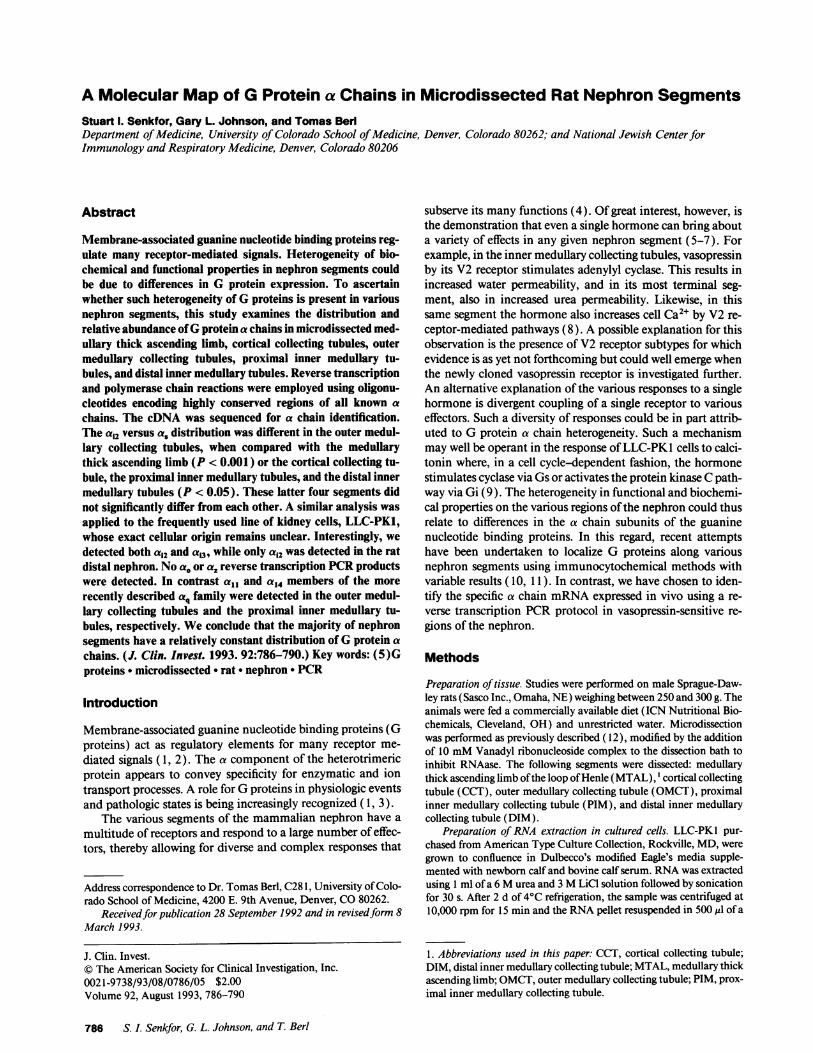
To ensure that the above procedures in general and the PCR, inparticular, did not preferentially amplify one cDNA a chainsubunit over another, known amounts of as and ai2 cDNAs atratios of 1:1, 1:10, and 10:1 were subjected to PCRand agarosegel analysis. As shown in Fig. 1, PCRsuccessfully amplified thea chain cDNA subunit without distortion of their relativeamounts of the two a chain cDNAs.
G proteins a chain in LLC-PK1 cells. LLC-PKl cells ofporcine renal origin have been extensively studied yet theirprecise nephronal origin remains unclear. Wedecided to iden-tify the pattern of a chain subunit mRNAin this cell line topotentially identify its cellular origin. Reverse transcription
10:11:1
10:10 --1:10
10:0
LMWSFigure 1. PCRamplification of various ratios of a, andai2- On a 1.5%agarose gel, it is easy to identify the heavier a, cDNA. Stock solutionswere not exactly the same concentration but the relative ratios of a.and ai2 were preserved. (LMWS, low molecular weight standard).
GProtein a Chains in Renal Collection Tubules 787
and PCRamplification of the LLC-PKl cells provided a highyield of cDNA. RNAwas prepared on two separate occasionsfrom confluent 10-cm dishes. No PCRcontamination was ob-served in the control samples. 36 PCR products were se-quenced for identification. As is depicted in Fig. 2, ai2 was thepredominant a chain cDNA, comprising 53% of all samplessequenced. The rest of the cDNAswere approximately equallydivided between as, aU3, and aq. a"3 contributed 19% and aqadded 17%. a, was present in 1 1% of the PCRproducts. Noother forms of a chains were identified.
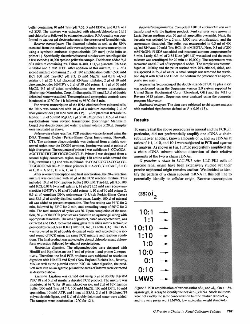
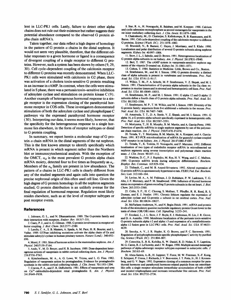
Gprotein a chains in dissected nephron segments. Fig. 3demonstrates the final PCRproduct from the proximal innermedullary collecting duct tubule and the distal inner medullarycollecting duct tubule. Note the paucity of signal at the regionwhich the heavier as control runs. Sequencing confirmed thelow percentage of as in these two segments. The relative abun-dance of a chains in the five dissected segments of the nephronis shown in Fig. 4. In all instances, sequenced samples wereobtained from tissues from at least two different rats. In micro-dissected MTAL, 41 samples were sequenced and 34 identified(83%). The great majority of these were ai2 (94%) while theremaining were a, (6%). No other forms of a chains were de-tected. A similar pattern was obtained in dissected CCT. 41samples were sequenced and only 14 were identifiable (33%).Ofthese, 13 were ai (93%) and 1 sample was a, (7%). Whereasin all other segments examined no more than 22% of insertsproved to be nonsense sequence, 67% of those in the CCTsegment proved unidentifiable.
The pattern of a chains prevalent in the OMCTproved tobe different. In this segment, 33 products were sequenced and32 identified (97%). Of these the predominant a chain was a.,16 samples accounting for 50% while only 12 were ai2 (37%).Of particular interest is the finding of four cDNAsof an a chainthat is identical to the one designated as aII ( 13%) by Strath-man et al. ( 14), an a chain in the recently described q family.
Because the PIM and DIM appear to have distinct func-tional and structural characteristics, we microdissected thesetwo areas and studied them separately. The Gprotein a chaindistribution was, however, not markedly different. In the PIM,55 samples were sequenced and 43 were identified (78%) com-pared with 41 out of 49 in the DIM (84%). Specifically, bothhad a predominance of a2, 33 of 43 products (77%) in the PIMand 36 of 41 products (88%) in the distal or terminal innermedulla. In the former, 9 of 43 samples were a, (21%) and inthe latter 5 of 41 samples were of this nature ( 12%). OnecDNA
100
80
%of 60Clones
Sequenced 40
20
0
xs control
PIMCT
DIMCT
LMWS
Figure 3. Agarose gel electrophoresis (1%) of the final PCRproductfrom two regions of the nephron. A known as control was used forPCRcontrol and size identification. The predominant band is slightlylighter and represents ai2. Sequencing data confirmed the agarose gelobservation. (LMWS, low molecular weight standard; DIMCT, distalinner medullary collecting tubule; and PIMCT, proximal inner med-ullary collecting tubule).
of a Gprotein identical in sequence to a14 (15) was found in thePIM, another member of the recently described q family.
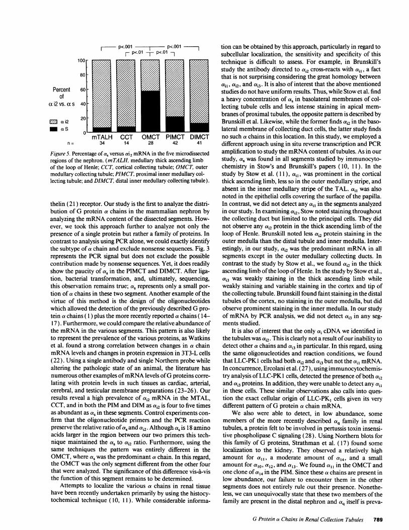
A statistical comparison of the ratio of ai2 and a, in thevarious segments is depicted in Fig. 5. By chi square analysisthe OMCTstands out as being different (P < 0.01 ) from allother segments. However, the other regions (MTAL, CCT,PIM, and DIM) do not statistically differ from each other.
Discussion
Gproteins play a pivotal role in a number of cellular processesincluding hormonal responses, transport of ions, and cell divi-sion. It has been well recognized that the protein is markedlyheterogeneous, particularly in the structure of its a chain as anincreasing number of such chains are being described andcloned ( 14-17). While Gproteins are rather ubiquitous thereappears to be differences in the tissue distribution. For exam-ple, a., is more prevalent in the brain and a16 in hematopoietictissue (18). Microdissection and PCRanalysis has been ap-plied to the detection of aldose reductase ( 19) and more re-cently to localize the atrial natriuretic peptide (20) and endo-
100l
%ofClones
Sequenced
aS al2 ai3 aq
Figure 2. Distribution of G protein a chains mRNAin culturedLLC-PKI cells.
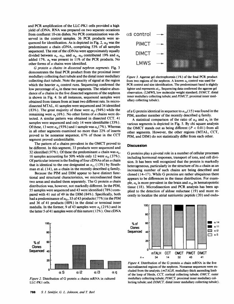
- a14- allM ai2a aS
mTALH CCT OMCT PIMCT DIMCTno= 34 14 32 43 41
Figure 4. Distribution of the G protein a chain mRNAin the fivemicrodissected regions of the nephron. Nonsense sequences were ex-cluded from the analysis. (mTALH, medullary thick ascending limbof the loop of Henle, CCT, cortical collecting tubule; OMCT, outermedullary collecting tubule; PIMCT, proximal inner medullary col-lecting tubule; and DIMCT, distal inner medullary collecting tubule).
788 S. I. Senkfor, G. L. Johnson, and T. Berl
p<0O01 p<.00 IF P<01 TF P<'.01-100
80-
Percent 60-Of
cxu2 vs. cXs 40
ED ai2 20
-asmTALH COT OMCT PIMOT DIMCT
n==34 14 28 42 41
Figure 5. Percentage of a. versus ai2 mRNAin the five microdissectedregions of the nephron. (mTALH, medullary thick ascending limbof the loop of Henle; CCT, cortical collecting tubule; OMCT, outermedullary collecting tubule; PIMCT, proximal inner medullary col-lecting tubule; and DIMCT, distal inner medullary collecting tubule).
thelin (21 ) receptor. Our study is the first to analyze the distri-bution of G protein a chains in the mammalian nephron byanalyzing the mRNAcontent of the dissected segments. How-ever, we took this approach further to analyze not only thepresence of a single protein but rather a family of proteins. Incontrast to analysis using PCRalone, we could exactly identifythe subtype of a chain and exclude nonsense sequences. Fig. 3represents the PCR signal but does not exclude the possiblecontribution made by nonsense sequences. Yet, it does readilyshow the paucity of a, in the PIMCT and DIMCT. After liga-tion, bacterial transformation, and, ultimately, sequencing,this observation remains true; as represents only a small por-tion of a chains in these two segment. Another example of thevirtue of this method is the design of the oligonucleotideswhich allowed the detection of the previously described Gpro-tein a chains ( 1) plus the more recently reported a chains ( 14-17). Furthermore, we could compare the relative abundance ofthe mRNAin the various segments. This pattern is also likelyto represent the prevalence of the various proteins, as Watkinset al. found a strong correlation between changes in a chainmRNAlevels and changes in protein expression in 3T3-L cells(22). Using a single antibody and single Northern probe whilealtering the pathologic state of an animal, the literature hasnumerous other examples of mRNAlevels of Gproteins corre-lating with protein levels in such tissues as cardiac, arterial,cerebral, and testicular membrane preparations (23-26). Ourresults reveal a high prevalence ofai mRNAin the MTAL,CCT, and in both the PIM and DIM as ai2 is four to five timesas abundant as as in these segments. Control experiments con-firm that the oligonucleotide primers and the PCR reactionpreserve the relative ratio of as and ai2. Although as is 18 aminoacids larger in the region between our two primers this tech-nique maintained the as toac2 ratio. Furthermore, using thesame techniques the pattern was entirely different in theOMCT,where as was the predominant a chain. In this regard,the OMCTwas the only segment different from the other fourthat were analyzed. The significance of this difference vis-A-visthe function of this segment remains to be determined.
Attempts to localize the various a chains in renal tissuehave been recently undertaken primarily by using the histocy-tochemical technique (10, I1). While considerable informa-
tion can be obtained by this approach, particularly in regard tosubcellular localization, the sensitivity and specificity of thistechnique is difficult to assess. For example, in Brunskill'sstudy the antibody directed to ai2 cross-reacts with ail, a factthat is not surprising considering the great homology betweenail, ai2, andai3.3 It is also of interest that the above mentionedstudies do not have uniform results. Thus, while Stow et al. finda heavy concentration of a, in basolateral membranes of col-lecting tubule cells and less intense staining in apical mem-branes of proximal tubules, the opposite pattern is described byBrunskill et al. Likewise, while the former finds ai2 in the baso-lateral membrane of collecting duct cells, the latter study findsno such a chains in this location. In this study, we employed adifferent approach using in situ reverse transcription and PCRamplification to study the mRNAcontent of tubules. As in our
study, a, was found in all segments studied by immunocyto-chemistry in Stow's and Brunskill's papers (10, 11). In thestudy by Stow et al. (1 1),ail, was prominent in the corticalthick ascending limb, less so in the outer medullary stripe, andabsent in the inner medullary stripe of the TAL. ail was alsonoted in the epithelial cells covering the surface of the papilla.In contrast, we did not detect any ail in the segments analyzedin our study. In examining ai2, Stow noted staining throughoutthe collecting duct but limited to the principal cells. They didnot observe any ai2 protein in the thick ascending limb of theloop of Henle. Brunskill noted less ai2 protein staining in theouter medulla than the distal tubule and inner medulla. Inter-estingly, in our study, ai2 was the predominant mRNAin allsegments except in the outer medullary collecting ducts. Incontrast to the study by Stow et al., we found ai2 in the thickascending limb of the loop of Henle. In the study by Stow et al.,ai3 was weakly staining in the thick ascending limb whileweakly staining and variable staining in the cortex and tip ofthe collecting tubule. Brunskill found faint staining in the distaltubules of the cortex, no staining in the outer medulla, but didobserve prominent staining in the inner medulla. In our studyof mRNAby PCRanalysis, we did not detect ai3 in any seg-ments studied.
It is also of interest that the only ai cDNAwe identified inthe tubules wasai2. This is clearly not a result of our inability todetect other a chains and ai3 in particular. In this regard, usingthe same oligonucleotides and reaction conditions, we foundthat LLC-PKl cells had both ai2 andai3 but not the ail mRNA.In concurrence, Ercolani et al. (27), using immunocytochemis-try analysis of LLC-PK1 cells, detected the presence of both ai2andai3 proteins. In addition, they were unable to detect any ailin these cells. These similar observations also calls into ques-tion the exact cellular origin of LLC-PK1 cells given its verydifferent pattern of G protein a chain mRNA.
We also were able to detect, in low abundance, somemembers of the more recently described a. family in renaltubules, a protein felt to be involved in pertussis toxin insensi-tive phospholipase C signaling (28). Using Northern blots forthis family of G proteins, Strathman et al. ( 17) found somelocalization to the kidney. They observed a relatively highamount for all, a moderate amount of a14, and a smallamount for alo, a12, and a13. Wefound aI, in the OMCTandone clone of a14 in the PIM. Since these a chains are present inlow abundance, our failure to encounter them in the othersegments does not entirely rule out their presence. Nonethe-less, we can unequivocally state that these two members of thefamily are present in the distal nephron and aq it self is preva-
GProtein a Chains in Renal Collection Tubules 789
lent in LLC-PKl cells. Lastly, failure to detect other alphachains does not rule out their existence but rather suggests theirpotential abundance compared to the observed Gprotein al-pha chain mRNA's.
Taken together, our data reflect a high degree of homologyin the pattern of G protein a chains in the distal nephron. Itwould not seem very plausible, therefore, that the different cel-lular responses to a given hormone or ligand is a consequenceof divergent coupling of a single receptor to different G pro-teins. However, such a system has been shown by others (9, 29,30). Cell cycle-dependent coupling of the calcitonin receptorto different Gproteins was recently demonstrated. WhenLLC-PKl cells were stimulated with calcitonin in G2 phase, therewas activation of a cholera toxin-sensitive Gprotein resultingin an increase in cAMP. In contrast, when the cells were stimu-lated in S phase, there was a pertussis toxin-sensitive inhibitionof adenylate cyclase and stimulation on protein kinase C (9).Another example of heterogenous hormonal response to a sin-gle receptor is the expression cloning of the parathyroid hor-mone receptor in COScells. These investigators demonstratedstimulation of both the adenylate cyclase and phospholipase Cpathways via the expressed parathyroid hormone receptor(30). Interpreting our data, it seems more likely, however, thatthe specificity for the heterogeneous response to a single hor-mone lies elsewhere, in the form of receptor subtypes or distalto Gprotein coupling.
In summary, we report herein a molecular map of G pro-tein a chain mRNAin the distal segments of the rat nephron.This is the first known attempt to identify specifically whichmRNAis present in which segment rather than the Northernblot or immunocytochemical analysis. With the exception ofthe OMCT, ai2 is the most prevalent G protein alpha chainmRNAmoiety, detected four to five times as frequently as a8.Members of the aq family are present in low abundance. Thepattern of a chains in LLC-PKl cells is clearly different fromany of the studied segments and again calls into question theprecise nephronal origin of this often used cell line. Given thehigh degree of Gprotein homology in all the nephron segmentsstudied, G protein distribution is an unlikely avenue for thefinal regulation of hormonal response. Regulation most likelyresides elsewhere, such as at the level of receptor subtypes orpost receptor events.
References
1. Johnson, G. L., and N. Dhanasekaran. 1989. The G-protein family andtheir interaction with receptors. Endocr. Rev. 10:317-331.
2. Casey, P. J., and A. G. Gilman. 1988. Gprotein involvement in receptor-ef-fector coupling. J. Biol. Chem. 263:2577-2580.
3. Landis, C. A., S. B. Masters, A. Spada, A. M. Pace, H. R. Bourne, and L.Vallar. 1989. GTPase inhibiting mutations activate the alpha chain of Gs andstimulate adenylyl cyclase in human pituitary tumors. Nature (Lond.). 340:692-696.
4. Morel, F. 1981. Sites of hormone action in the mammalian nephron. Am. J.Physiol. 240:F159-F164.
5. Ando, Y., M. D. Breyer, and H. B. Jacobson. 1989. Dose-dependent heter-ogenous actions of vasopressin in rabbit cortical collecting ducts. Am. J. Physiol.256:F556-F562.
6. Kirschenbaum, M. A., A. G. Lowe, W. Trizna, and L. G. Fine. 1982.Regulation of vasopressin action by prostaglandins. Evidence for prostaglandinsynthesis in the rabbit cortical collecting tubule. J. Clin. Invest. 70:1193-1204.
7. Craven, P. A., and F. R. DeRubertis. 1981. Effects of vasopressin and ureaon Ca2+-calmodulin-dependent renal prostaglandin E. Am. J. Physiol.241:F649-F658.
8. Star, R. A., H. Nonoguchi, R. Balaban, and M. Knepper. 1988. Calciumand cyclic adenosine monophosphate as second messengers for vasopressin in therat inner medullary collecting duct. J. Clin. Invest. 81:1879-1888.
9. Chakraborty, M., D. Chattedjee, S. Kellokumpu, R. B. Rasmussen, and R.Baron. 1991. Cell cycle-dependent coupling of the calcitonin receptor to differentGproteins. Science (Wash. DC). 251:1078-1082.
10. Brunskill, N., B. Bastani, C. Hayes, J. Morrissey, and S. Klahr. 1990.Localization and polar distribution of several G-protein subunits along nephronsegments. Kidney Int. 40:997-1006.
1 1. Stow, J. L., I. Sabolic, and D. Brown. 1991. Heterogeneous localization ofGprotein alpha-subunits in rat kidney. Am. J. Physiol. 261:F831-F840.
12. Berl, T. 1987. The cAMPsystem in vasopressin-sensitive nephron seg-ments of the vitamin D-treated rat. Kidney Int. 31:1065-1071.
13. Colton, T. 1984. Statistics in Medicine. Little, Brown and Co., Boston.14. Strathmann, M., and M. I. Simon. 1990. Gprotein diversity: a distinct
class of alpha subunits is present in vertebrates and invertebrates. Proc. Natl.Acad. Sci. USA. 87:9113-9117.
15. Wilkie, T. M., P. A. Scherle, M. P. Strathmann, V. Z. Slepak, and M. I.Simon. 1991. Characterization of G-protein alpha subunits in the Gq class: ex-pression in murine tissues and in stromal and hematopoietic cell lines. Proc. Nail.Acad. Sci. USA. 88:10049-10053.
16. Strathmann, M. P., and M. I. Simon. 1991. Galpha 12 and Galpha 13subunits define a fourth class of Gprotein alpha subunits. Proc. Natl. Acad. Sci.USA. 88:5582-5586.
17. Strathmann, M. P., T. M. Wilkie, and M. I. Simon. 1989. Diversity of theG-protein family: sequences from five additional a subunits in the mouse. Proc.Natl. Acad. Sci. USA. 86:7407-7409.
18. Amatruda, T. T., D. A. Steele, V. Z. Slepak, and M. I. Simon. 1991. Galpha 16, a Gprotein alpha subunit specifically expressed in hematopoietic cells.Proc. Natl. Acad. Sci. USA. 88:5587-5591.
19. Moriyama, T., H. R. Murphy, B. M. Martin, and A. Garcia-Perez. 1990.Detection of specific mRNAsin single nephron segments by use of the polymer-ase chain reaction. Am. J. Physiol. 258:F1470-F1474.
20. Terada, Y., T. Moriyama, B. M. Martin, M. A. Knepper, and A. Garcia-Perez. 1991. RT-PCR microlocalization of mRNAfor guanylyl cyclase-coupledANFreceptor in rat kidney. Am. J. Physiol. 261:F1080-F1087.
21. Terada, Y., K. Tomita, H. Nonoguchi, and F. Marumo. 1992. Differentlocalization of two types of endothelin receptor mRNAin microdissected ratnephron segments using reverse transcription and polymerase chain reactionassay. J. Clin. Invest. 90:107-1 12.
22. Watkins, D. C., P. J. Rapiejko, M. Ros, H. Y. Wang, and C. C. Malbon.1989. G-protein mRNAlevels during adipocyte differentiation. Biochem.Biophys. Res. Commun. 165:929-934.
23. Thibault, C., and M. B. Anand-Srivastava. 1992. Altered expression ofG-protein mRNAin spontaneously hypertensive rats. FEBS(Fed. Eur. Biochem.Soc.) Lett. 313:160-164.
24. Levine, M. A., A. M. Feldman, J. D. Robishaw, P. W. Ladenson, T. G.Ahn, J. F. Moroney, and P. M. Smallwood. 1990. Influence of thyroid hormonestatus on expression of genes encoding Gprotein subunits in the rat heart. J. Biol.Chem. 265:3553-3560.
25. Colin, S. F., H. C. Choong, S. Mollner, T. Pfeuffer, R. R. Reed, R. S.Duman, and E. J. Nestler. 1991. Chronic lithium regulates the expression ofadenylate cyclase and Gi-protein a subunit in rat cerebral cortex. Proc. Natl.Acad. Sci. USA. 88:10634-10637.
26. McFarlane-Anderson, N., and N. Begin-Heick. 1991. mRNAand proteinlevels of the stimulatory guanine nucleotide regulatory protein Gsare lower in thetestis of obese (OB/OB) mice. Cell. Signalling. 3:233-241.
27. Ercolani, L., J. L. Stow, J. F. Boyle, E. J. Holtzman, H. Lin, J. R. Grove,and D. A. Ausiello. 1990. Membrane localization of the pertussis toxin-sensitiveG-protein subunits alpha i-2 and alpha i-3 and expression of a metallothionein-alpha i-2 fusion gene in LLC-PKl cells. Proc. Nail. Acad. Sci. USA. 87:4635-4639.
28. Smrcka, A. V., J. R. Hepler, K. 0. Brown, and P. C. Sternweis. 1991.Regulation of polyphosphoinositide-specific phospholipase Cactivity by purifiedGq. Science (Wash. DC). 251:804-807.
29. Cotecchia, S., B. K. Kobilka, K. W. Daniel, R. D. Nolan, E. Y. Lapetina,M. G. Caron, R. J. Lefkowitz, andJ. W. Regan. 1990. Multiple second messengerpathways of alpha-adrenergic receptor subtypes expressed in eukaryotic cells. J.Biochem. 265:63-69.
30. Abou-Samra, A.-B., H. Juppner, T. Force, M. W. Freeman, X.-F. Kong,E. Schipani, P. Urena, J. Richards, J. V. Bonventre, J. T. Potts, Jr., H. J. Kronen-berg, and G. V. Segre. 1992. Expression cloning of a commonreceptor for para-thyroid hormone and parathyroid hormone-related peptide from rat osteoblast-like cells: a single receptor stimulates intracellular accumulation of both cAMPand inositol trisphosphates and increases intracellular free calcium. Proc. Natl.Acad. Sci. USA. 89:2732-2736.
790 S. I. Senkfor, G. L. Johnson, and T. Berl