Embed Size (px)
Citation preview
Impairment of Cardiac Function and Energetics in Experimental Renal FailureA. E. G. Raine, A.-M. L. Seymour,* A. F. C. Roberts, G. K. Radda, * and J. G. G. LedinghamNuffield Department of Clinical Medicine, John Radcliffe Hospital, *Department of Biochemistry,South Parks Road, Oxford OX3 9DU, England
Abstract
Cardiac function and energetics in experimental renal failure inthe rat (5/6 nephrectomy) have been investigated by means ofan isolated perfused working heart preparation and an isomet-ric Langendorff preparation using 31P nuclear magnetic reso-
nance (31P NMR). 4 wk after nephrectomy cardiac output ofisolated hearts perfused with Krebs-Henseleit buffer was signif-icantly lower (P < 0.0001 ) at all levels of preload and afterloadin the renal failure groups than in the pair-fed sham operatedcontrol group. In control hearts, cardiac output increased withincreases in perfusate calcium from 0.73 to 5.61 mmol/ literwhereas uremic hearts failed in high calcium perfusate.
Collection of 31p NMRspectra from hearts of renal failureand control animals during 30 min normoxic Langendorff per-
fusion showed that basal phosphocreatine was reduced by 32%to 4.7 ,mol/g wet wt (P < 0.01) and the phosphocreatine toATP ratio was reduced by 32% (P < 0.01) in uremic hearts.During low flow ischemia, there was a substantial decrease inphosphocreatine in the uremic hearts and an accompanyingmarked increase in release of inosine into the coronary effluent(14.9 vs 6.1 jsM, P < 0.01).
Weconclude that cardiac function is impaired in experimen-tal renal failure, in association with abnormal cardiac ener-
getics and increased susceptibility to ischemic damage. Disor-dered myocardial calcium utilization may contribute to thesederangements. (J. Clin. Invest. 1993. 92:2934-2940.) Keywords: uremia * heart failure * calcium * 31p nuclear magneticresonance * myocardial ischemia
Introduction
More than half of all deaths in end-stage renal failure are fromcardiovascular events. Of these, death from cardiac causes isespecially common, accounting for - 40% of all deaths in pa-tients maintained on hemodialysis (1). Many factors havebeen proposed to contribute to the cardiac complications ofchronic renal failure, including hypertension, fluid overload,pericardial disease, anemia, and coronary atherosclerosis (2).Previous experimental studies have also suggested that meta-bolic abnormalities characteristic of uremia, such as elevatedblood urea (3) and secondary hyperparathyroidism (4), might
Address correspondence to Prof. A. E. G. Raine, Department of Ne-phrology, St. Bartholomew's Hospital, West Smithfield, London EC A7BE, England.
Received for publication 2 July 1992 and in revised form 8 June1993.
adversely affect cardiac function. Furthermore, in dialysis pa-tients, Rostand and colleagues observed that the prevalence ofsymptomatic ischemic heart disease greatly exceeded the pres-ence of significant coronary artery narrowing (5), raising thepossibility that myocardial metabolism and oxygen demandmay be altered in chronic renal failure.
The hypothesis the present study aimed to investigate wasthat in chronic renal failure, cardiac energetics may be abnor-mal, resulting in impaired cardiac performance and increasedsusceptibility to ischemia. Previous studies of cardiac functionin vivo have given conflicting results. Acute uremia of 24-48 hduration led to increased myocardial contractility in rats (6,7), and dogs (8), whereas myocardial function was unchangedfrom controls after 7 d of uremia in both species (9, 10). Asinterpretation of studies performed in vivo may be compli-cated by reflex neural and hormonal effects, in the presentstudy, cardiac function, energetics, and susceptibility to isch-emia have been investigated in experimental chronic uremia invitro, by means of an isolated working heart preparation and31P nuclear magnetic resonance (31PNMR)' of hearts perfusedby a modified Langendorff technique.
Methods
Experimental modelMale Wistar rats weighing 200-240 g were used. Renal impairment wasproduced by subtotal nephrectomy ( 11 ). Rats were anesthetized with 1ml/ 100 g 5% chloral in 0.9% NaCI, a midline incision was made, theleft kidneys were isolated and approximately two thirds (500-600 mgtissue) of the renal parenchyma cut away. Sham-operated control ratswere anesthetized, and the left kidneys were decapsulated. 7 d later, therats were again anesthetized with 5% chloral and the right kidney wasremoved through a flank incision, with preservation of the adrenalgland. Sham-operated controls were anesthetized, and the right kidneyswere exposed, and perinephric fat removed from the otherwise intactkidney.
1 d after completion of surgery, all animals were housed in individ-ual cages, and were pair fed. Daily food intake of uremic rats wasmeasured and the same quantity of food was given to the allocatedpaired sham operated control the following day. The diet contained16.7% protein and 0.2 1%sodium (Beekay Feeds, Hull, England). Ani-mals had free access to tap water and were weighed daily. For measure-ment of systolic blood pressure without anesthesia, rats were acclima-tised to restraining cages for 2 d, and the pressure was recorded using atail cuff and sphygmomanometer (Narco Bio-Systems, Inc., Houston,TX) on the 3rd d, 21 d after surgery. Four to five measurements ofsystolic blood pressure were made over a 60-min period, and theirmean was calculated. Hematocrit and plasma urea, creatinine, andelectrolytes were measured on the 7th, 14th and 28th d after operationby taking a 1-ml sample from the tail artery under ether anesthesia.Hematocrit was measured by microcentrifugation, and sodium, potas-
1. Abbreviations used in this paper: MANOVA,multiple analysis ofvariance; 31P NMR, 31P nuclear magnetic resonance.
2934 Raine, Seymour, Roberts, Radda, and Ledingham
J. Clin. Invest.©) The American Society for Clinical Investigation, Inc.0021-9738/93/12/2934/07 $2.00Volume 92, December 1993, 2934-2940
sium, urea, and creatinine were measured by automated analyzer.Plasma ionized calcium was measured by Nova 2 analyzer. (V. A.Howe and Co., Banbury, United Kingdom)
Isolated perfused working heart28 d after right nephrectomy, rats were anesthetised intraperitoneallywith pentobarbital 100 mg/kg, 100 Uheparin was given intravenously,the hearts were removed and immediately placed in ice-cold saline.The aorta was cannulated and perfused with oxygenated Krebs Hense-leit bicarbonate saline solution containing 2.5 mMCa2" and 10 mMglucose substrate as previously described ( 12, 13). Hearts were kept atconstant temperature (370C) via a water-jacketed perfusion chamberthroughout the experiments.
Perfusion protocol. Single-pass perfusion was begun through theaortic cannula at a hydrostatic pressure of 80 cm H20, and continuedfor 3-4 min, to wash all blood from the coronary circulation. Duringthis time, the left atrium was cannulated. Recirculating perfusion viathe left atrium was then commenced, and left atrial reservoir fillingpressure (preload) and aortic pressure head (afterload) were indepen-dently varied to enable determination of left ventricular functioncurves ( 13). Left atrial filling pressure was initially held constant at17.5 cm H20, and aortic overflow height (aortic pressure) was set suc-cessively at 5-min intervals at 70, 100, 130, and 160 cm H20 to docu-ment the relationship between afterload and cardiac output. Aorticpressure was then held constant at 100 cm H20 and left atrial fillingpressure set at 7.5, 12.5, 17.5, and finally 22.5 cmH20 to determine thepreload cardiac output relationship (Starling curve). At each pressuresetting, cardiac output and coronary flow were measured by timedcollection of perfusate as previously described ( 12). Heart rate andaortic pressure were recorded continuously through a side arm in theaortic cannula, using a fluid-filled system with a transducer (PDCR75;Druck Ltd., Groby, Leicester, United Kingdom) attached to a recorderand 4820 preamplifier (MX216 and 4820, respectively; LectromedLtd., Letchworth, Herts, United Kingdom).
After determination of function curves, the perfusate calcium con-centration was reduced to 0.7 mM, cardiac output and heart rate weremeasured, and then at 5-min intervals, the perfusate calcium concen-tration was increased to 1.5, 2.9, and 5.6 mM, with repeated measure-ments of cardiac function. At the end of the protocol, hearts were blot-ted and dried in a tissue oven at 70°C until constant weight wasachieved, to determine dry weight.
Acute effects of urea and creatinine on cardiac functionHearts from control male Wistar rats weighing 320-340 g were per-fused as described above, with a left atrial pressure of 17.5 cmH20 andaortic pressure of 100 cm H20. After 20 min of control observations,urea (n = 6) or creatinine (n = 6) was added to the perfusion reservoirat 10-min intervals to achieve final concentrations of 75 and 150mmol/liter (urea) and 1,000 and 2,000 Mmol/liter (creatinine). Car-diac output, coronary flow, and heart rate were recorded at 2-min inter-vals.
31PNMRstudiesSeven male Wistar rats ( 180-200 g) were made uremic by subtotalnephrectomy, as described above, with sham-operated pair-fed animalsas controls. One control animal was lost during the experimental proto-col. After 21 d, animals were anesthetized with diethyl ether and in-jected intravenously with heparin ( 1,000 U/mml), - 1.0 ml/kg body wtvia the femoral vein. Hearts were then rapidly excised and placed inice-cold phosphate-free Krebs-Henseleit bicarbonate buffer. The perfu-sion medium consisted of 118.5 mMNaCI, 25 mMNaHCO3, 6.0 mMKCI, 1.25 mMCaCI2, and 1.25 mMMgSO4with 11.0 mMglucose assubstrate. Hearts were suspended on a glass cannula via the aorta andperfused in Langendorff isometric mode at 70 cm H20 pressure com-patible with the 3'P-NMR technique as described previously ( 14, 15).The apex of the heart was attached to a semiconductor strain gauge(Kulite Sensors Ltd., Basingstoke, Hants, United Kingdom), and car-diac function was monitored continuously by measurement of devel-
oped tension via the strain gauge. Initially, the tension was set at 10-15g. The bundle of His was severed and hearts were paced at 300 bpmusing platinum electrodes, one located in the aortic cannula and theother delicately inserted into the ventricle. Coronary flow was mea-sured by the timed collection of perfusate.
Concentrations of phosphocreatine, ATP, and inorganic phosphatewere determined by 3"P-NMR. Spectra were collected at a 31p fre-quency of 73.836 MHzusing a 4.2-T superconducting magnet with an8-cm bore and spectrometer (Biospec; Bruker Ltd., Coventry, UnitedKingdom). The probe consisted of a vertically mounted Helmholtzcoil (2.8 cm in diameter) tuned to the phosphorus frequency. During a20-min equilibration period, the magnetic field homogeneity was opti-mized by shimming on the proton signal of the heart and the surround-ing water contained within the sensitive volume of the NMRprobe.Quantitative data were obtained from a fully relaxed spectrum accu-mulated using a 900 pulse and 7-s interpulse delay. Concentrationswere measured by integrating the areas under the resonance peaks ofinterest and comparing them with that of a standard (methylene di-phosphonate) of known concentration contained in a capillary withinthe radiofrequency coil. Where appropriate peak areas were correctedfor partial saturation effects using correction factors determined fromspectra acquired using a 7-s delay and a 15-s delay. Intracellular pHwasdetermined from the chemical shift of the inorganic phosphate reso-nance peak relative to that of phosphocreatine ( 16). Subsequently,spectra were collected in 5-min time blocks using a 70° pulse and 1-sinterpulse delay. After a further 20-min of normoxic perfusion, lowflow ischemia was produced in four pairs of animals by lowering theperfusion pressure to 15-20 cmH20, to reduce the coronary flow to 1.0ml/min for 30 min. Hearts were then reperfused at a pressure of 70 cmH20 for an additional 20 min. Spectra were acquired throughout thisperiod in 5-min time blocks. Percentage changes in phosphocreatineand ATPwere determined throughout the period of low flow ischemiaand reperfusion by comparing peak heights with control spectra col-lected during normoxia (taken as 100%). 100%was considered equiva-lent to the metabolite concentrations measured using the 900 pulse and7-s delay. Coronary flow was monitored at the midpoint of each spec-trum. At the same time, a sample of coronary effluent was collected viathe sampling line for analysis of inosine release, as a marker of ischemicinjury ( 17 ). These samples were analyzed using the HPLCmethod ofHarmsen et al. (18).
At the end of the experimental protocol, hearts were freezeclamped, weighed, extracted with 8% perchloric acid, neutralized, andanalyzed for total creatine (creatine plus phosphocreatine) using anHPLCion exchange method (19).
Free cytosolic ADPwas calculated from the creatine kinase equilib-rium reaction (Koq = 1.7 X 10⁹ M-1, assuming a free Mg2+ concentra-tion of I mM)(20),
where Kq = [A TP] [Cr]~'[PCr][H+J[ADPJ
StatisticsResults are expressed as mean±SEM. Statistical comparison betweenuremic and paired control hearts was by means of multiple analysis ofvariance (MANOVA), using the Stats graphics version 4.0 statisticalprogram, followed by Student's t test for unpaired samples for compari-son of individual data points. Comparisons for 31P NMRstudies wereperformed by an unpaired Student's t test.
Results
Renal failure model. Table I shows the average food intake,final heart and body weight, systolic blood pressure, hemato-crit, and plasma biochemistry in the renal failure and controlanimals after 28 d. Despite pair feeding, uremic animalsweighed less than controls (P < 0.05). Both the heart dryweight and the heart weight/body weight ratios were increased
Cardiac Function in Experimental Renal Failure 2935
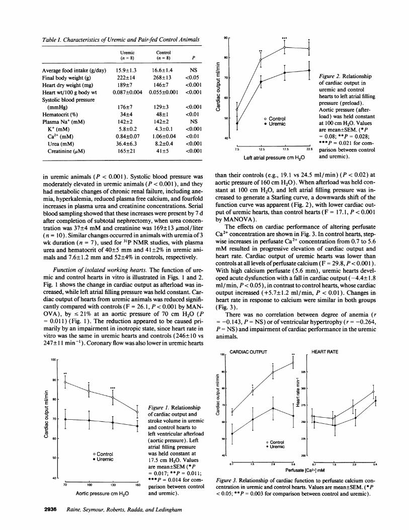
Table I. Characteristics of Uremic and Pair-fed Control Animals
Uremic Control(n= 8) (n= 8) P
Average food intake (g/day) 15.9±1.3 16.6±1.4 NSFinal body weight (g) 222±14 268±13 <0.05Heart dry weight (mg) 189±7 146±7 <0.00 1Heart wt/ 100 g body wt 0.087±0.004 0.055±0.001 <0.001Systolic blood pressure
(mmHg) 176±7 129±3 <0.001Hematocrit (%) 34±4 48±1 <0.01Plasma Na' (mM) 142±2 142±2 NS
K+ (mM) 5.8±0.2 4.3±0.1 <0.001Ca2" (mM) 0.84±0.07 1.06±0.04 <0.01Urea (mM) 36.4±6.3 8.2±0.4 <0.00 1Creatinine (tIM) 165±21 41±5 <0.001
in uremic animals (P < 0.001). Systolic blood pressure wasmoderately elevated in uremic animals (P < 0.001), and theyhad metabolic changes of chronic renal failure, including ane-mia, hyperkalemia, reduced plasma free calcium, and fourfoldincreases in plasma urea and creatinine concentrations. Serialblood sampling showed that these increases were present by 7 dafter completion of subtotal nephrectomy, when urea concen-tration was 37±4 mMand creatinine was 169±13,umol/liter(n = 10). Similar changes occurred in animals with uremia of 3wk duration (n = 7), used for 31P NMRstudies, with plasmaurea and hematocrit of 40±5 mmand 41±2% in uremic ani-mals and 7.6±1.2 mmand 52±4% in controls, respectively.
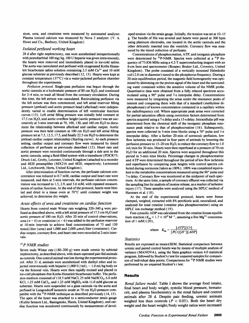
Function of isolated working hearts. The function of ure-mic and control hearts in vitro is illustrated in Figs. 1 and 2.Fig. 1 shows the change in cardiac output as afterload was in-creased, while left atrial filling pressure was held constant. Car-diac output of hearts from uremic animals was reduced signifi-cantly compared with controls (F = 26.1, P < 0.001 by MAN-OVA), by < 21% at an aortic pressure of 70 cm H20 (P= 0.011) (Fig. 1). The reduction appeared to be caused pri-marily by an impairment in inotropic state, since heart rate invitro was the same in uremic hearts and controls (246±10 vs247± 11 min '). Coronary flow was also lower in uremic hearts
80
c
E
E70
0.
50C450
40L
Figure 2. Relationshipof cardiac output inuremic and controlhearts to left atrial fillingpressure (preload).Aortic pressure (after-
o Control load) was held constant* Uremic at 100 cmH20. Values
are mean±SEM. (*P= 0.08; **P = 0.028;***P = 0.021 for com-
7.5 12.5 17.5 22.5 parison between control
Left atrial pressure cm H20 and uremic).
than their controls (e.g., 19.1 vs 24.5 ml/min) (P < 0.02) ataortic pressure of 160 cm H20) . Whenafterload was held con-stant at 100 cm H20, and left atrial filling pressure was in-creased to generate a Starling curve, a downwards shift of thefunction curve was apparent (Fig. 2), with lower cardiac out-put of uremic hearts, than control hearts (F = 17.1, P < 0.001by MANOVA).
The effects on cardiac performance of altering perfusateCa2+ concentration are shown in Fig. 3. In control hearts, step-wise increases in perfusate Ca2+ concentration from 0.7 to 5.6mMresulted in progressive elevation of cardiac output andheart rate. Cardiac output of uremic hearts was lower thancontrols at all levels of perfusate calcium (F = 29.8, P< 0.001) .With high calcium perfusate (5.6 mm), uremic hearts devel-oped acute dysfunction with a fall in cardiac output (-4.4±1.8ml/ min, P< 0.05 ), in contrast to control hearts, whose cardiacoutput increased (+5.7± 1.2 ml/min, P < 0.01). Changes inheart rate in response to calcium were similar in both groups(Fig. 3).
There was no correlation between degree of anemia (r--0.143, P = NS) or of ventricular hypertrophy (r = -0.264,P = NS) and impairment of cardiac performance in the uremicanimals.
Figure 1. Relationshipof cardiac output andstroke volume in uremicand control hearts toleft ventricular afterload(aortic pressure). Leftatrial filling pressure
o Control was held constant at* Uremic 17.5 cm H20. Values
are mean±SEM(*P= 0.017; **P = 0.011;
0***P = 0.014 for com-70 100 130 160 parison between control
Aortic pressure cm H20 and uremic).
100
90c
E0._, 80
:30
60
50
CARDIACOUTPUT
325
o Control* Uremic
HEARTRATE
300 [
275 -
250 .
225 [
2001
0.7 1.5 2.9 5.6 0.7 1.5 2.9 51
Perfusate [Ca2+] mM
Figure 3. Relationship of cardiac function to perfusate calcium con-centration in uremic and control hearts. Values are mean±SEM. (* P< 0.05; **P = 0.003 for comparison between control and uremic).
2936 Raine, Seymour, Roberts, Radda, and Ledingham
100
90
Ef 80-
0 70
C.)
CO c-o
Cu r2
50 F
40
90r
60
40
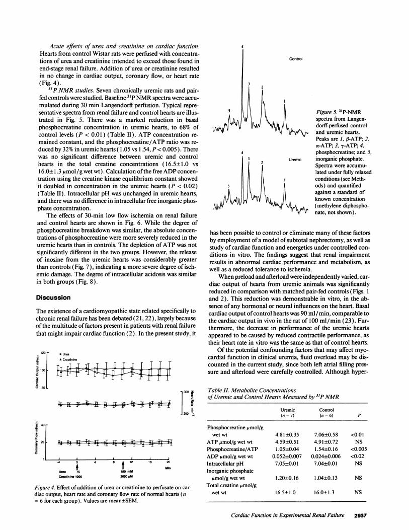
Acute effects of urea and creatinine on cardiac function.Hearts from control Wistar rats were perfused with concentra-tions of urea and creatinine intended to exceed those found inend-stage renal failure. Addition of urea or creatinine resultedin no change in cardiac output, coronary flow, or heart rate(Fig. 4).
31P NMRstudies. Seven chronically uremic rats and pair-fed controls were studied. Baseline 31P NMRspectra were accu-mulated during 30 min Langendorff perfusion. Typical repre-sentative spectra from renal failure and control hearts are illus-trated in Fig. 5. There was a marked reduction in basalphosphocreatine concentration in uremic hearts, to 68% ofcontrol levels (P < 0.01) (Table II). ATP concentration re-mained constant, and the phosphocreatine/ATP ratio was re-duced by 32% in uremic hearts ( 1.05 vs 1.54, P< 0.005 ). Therewas no significant difference between uremic and controlhearts in the total creatine concentrations ( 16.5±1.0 vs16.0±1.3 ,umol/g wet wt). Calculation of the free ADPconcen-tration using the creatine kinase equilibrium constant showedit doubled in concentration in the uremic hearts (P < 0.02)(Table II). Intracellular pH was unchanged in uremic hearts,and there was no difference in intracellular free inorganic phos-phate concentration.
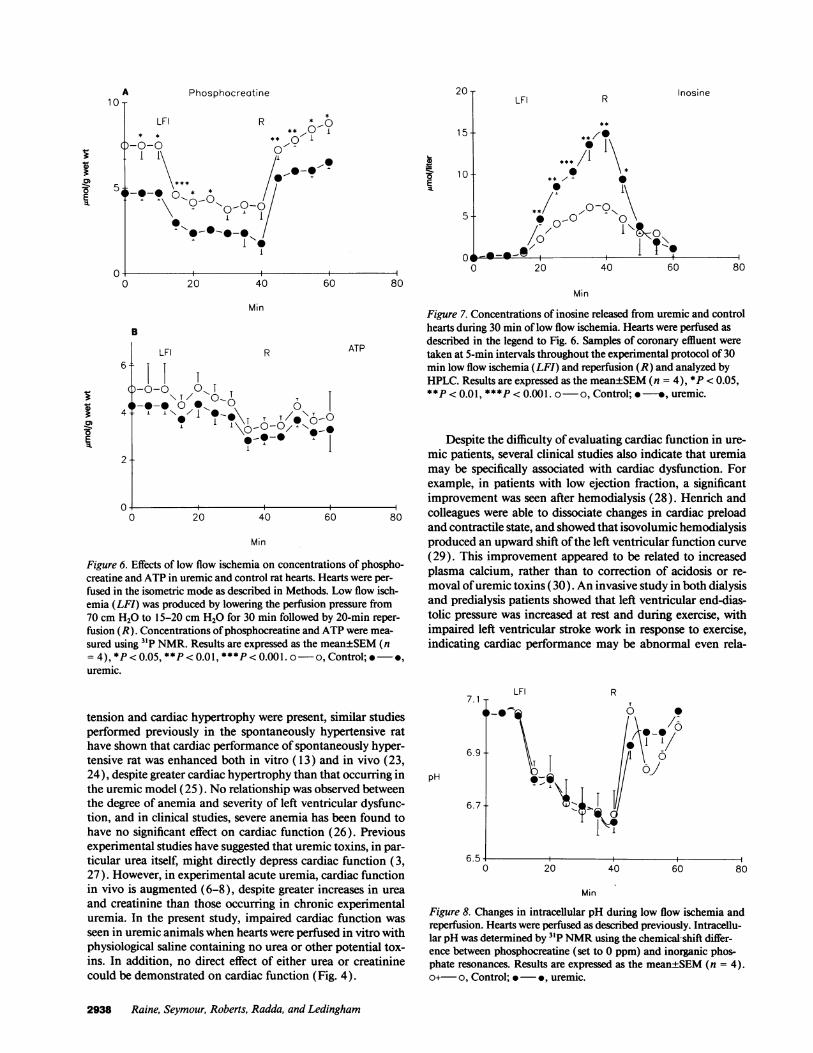
The effects of 30-min low flow ischemia on renal failureand control hearts are shown in Fig. 6. While the degree ofphosphocreatine breakdown was similar, the absolute concen-trations of phosphocreatine were more severely reduced in theuremic hearts than in controls. The depletion of ATPwas notsignificantly different in the two groups. However, the releaseof inosine from the uremic hearts was considerably greaterthan controls (Fig. 7), indicating a more severe degree of isch-emic damage. The degree of intracellular acidosis was similarin both groups (Fig. 8).
Discussion
The existence of a cardiomyopathic state related specifically tochronic renal failure has been debated (21, 22), largely becauseof the multitude of factors present in patients with renal failurethat might impair cardiac function (2). In the present study, it
120 r .UreaE A Creatinine
El
80
] 300 i1
LAz. 200 R
f 40 _E
I2 0
83
I;-F xIz- IqIT I f ~ '
4 0
Uma 75Cmatinine 1000
4 8 f 12
150 mm2000 pM
16 20
Min
Figure 4. Effect of addition of urea or creatinine to perfusate on car-diac output, heart rate and coronary flow rate of normal hearts (n= 6 for each group). Values are mean±SEM.
3
2
5
4
Control
Figure 5. 3'P-NMRspectra from Langen-dorff-perfused controland uremic hearts.Peaks are 1, fl-ATP; 2,a-ATP; 3, y-ATP; 4,phosphocreatine; and 5,
Uremic inorganic phosphate.Spectra were accumu-lated under fully relaxedconditions (see Meth-ods) and quantifiedagainst a standard ofknown concentration
< (methylene diphospho-nate, not shown).
has been possible to control or eliminate many of these factorsby employment of a model of subtotal nephrectomy, as well asstudy of cardiac function and energetics under controlled con-ditions in vitro. The findings suggest that renal impairmentresults in abnormal cardiac performance and metabolism, aswell as a reduced tolerance to ischemia.
Whenpreload and afterload were independently varied, car-diac output of hearts from uremic animals was significantlyreduced in comparison with matched pair-fed controls (Figs. 1and 2). This reduction was demonstrable in vitro, in the ab-sence of any hormonal or neural influences on the heart. Basalcardiac output of control hearts was 90 ml/ min, comparable tothe cardiac output in vivo in the rat of 100 ml/min (23). Fur-thermore, the decrease in performance of the uremic heartsappeared to be caused by reduced contractile performance, astheir heart rate in vitro was the same as that of control hearts.
Of the potential confounding factors that may affect myo-cardial function in clinical uremia, fluid overload may be dis-counted in the current study, since both left atrial filling pres-sure and afterload were carefully controlled. Although hyper-
Table II. Metabolite Concentrationsof Uremic and Control Hearts Measured by 31P NMR
Uremic Control(n = 7) (n = 6) P
Phosphocreatine gmol/gwet wt 4.81±0.35 7.06±0.58 <0.01
ATP zmol/g wet wt 4.59±0.51 4.91±0.72 NSPhosphocreatine/ATP 1.05±0.04 1.54±0.16 <0.005ADPzmol/g wet wt 0.052±0.007 0.024±0.006 <0.02Intracellular pH 7.05±0.01 7.04±0.01 NSInorganic phosphate
umol/g wet wt 1.20±0.16 1.04±0.13 NSTotal creatine gmol/g
wet wt 16.5±1.0 16.0±1.3 NS
Cardiac Function in Experimental Renal Failure 2937
A PhosphocreatineR Inosine
LFI R ONT** ** w0I
*-0-0 01~~~ ~~~~~1\ A1
-* **0-* 1,
-'I0-0
I -I.-1
E-
15
10
5
0 20 40
R
60 80
Min
B
6
4
LFI ATP
| T
0 @-@0 ,
i'./ I
,
0 20 40 60
Min
Figure 6. Effects of low flow ischemia on concentrations of phospho-creatine and ATP in uremic and control rat hearts. Hearts were per-fused in the isometric mode as described in Methods. Low flow isch-emia (LFI) was produced by lowering the perfusion pressure from70 cm H20 to 15-20 cm H20 for 30 min followed by 20-min reper-fusion (R). Concentrations of phosphocreatine and ATPwere mea-
sured using 31P NMR. Results are expressed as the mean±SEM(n= 4), *P < 0.05, **P < 0.01, ***P < 0.001. o-o, Control; @*,uremic.
Min
Figure 7. Concentrations of inosine released from uremic and controlhearts during 30 min of low flow ischemia. Hearts were perfused asdescribed in the legend to Fig. 6. Samples of coronary effluent weretaken at 5-min intervals throughout the experimental protocol of 30min low flow ischemia (LFI) and reperfusion (R) and analyzed byHPLC. Results are expressed as the mean±SEM(n = 4), *P < 0.05,**P < 0.01, ***P < 0.001. o-o, Control; *-*, uremic.
Despite the difficulty of evaluating cardiac function in ure-mic patients, several clinical studies also indicate that uremiamay be specifically associated with cardiac dysfunction. Forexample, in patients with low ejection fraction, a significantimprovement was seen after hemodialysis (28). Henrich andcolleagues were able to dissociate changes in cardiac preloadand contractile state, and showed that isovolumic hemodialysisproduced an upward shift of the left ventricular function curve(29). This improvement appeared to be related to increasedplasma calcium, rather than to correction of acidosis or re-moval of uremic toxins (30). An invasive study in both dialysisand predialysis patients showed that left ventricular end-dias-tolic pressure was increased at rest and during exercise, withimpaired left ventricular stroke work in response to exercise,indicating cardiac performance may be abnormal even rela-
tension and cardiac hypertrophy were present, similar studiesperformed previously in the spontaneously hypertensive rathave shown that cardiac performance of spontaneously hyper-tensive rat was enhanced both in vitro ( 13) and in vivo (23,24), despite greater cardiac hypertrophy than that occurring inthe uremic model (25). No relationship was observed betweenthe degree of anemia and severity of left ventricular dysfunc-tion, and in clinical studies, severe anemia has been found tohave no significant effect on cardiac function (26). Previousexperimental studies have suggested that uremic toxins, in par-ticular urea itself, might directly depress cardiac function (3,27). However, in experimental acute uremia, cardiac functionin vivo is augmented (6-8), despite greater increases in urea
and creatinine than those occurring in chronic experimentaluremia. In the present study, impaired cardiac function was
seen in uremic animals when hearts were perfused in vitro withphysiological saline containing no urea or other potential tox-ins. In addition, no direct effect of either urea or creatininecould be demonstrated on cardiac function (Fig. 4).
7.1
6.9
pH
6.7
LFI
I-.'
20
R
T
40 60 80
Min
Figure 8. Changes in intracellular pH during low flow ischemia andreperfusion. Hearts were perfused as described previously. Intracellu-lar pH was determined by 3'P NMRusing the chemical-shift differ-ence between phosphocreatine (set to 0 ppm) and inorganic phos-phate resonances. Results are expressed as the mean±SEM(n = 4).o+- o, Control; * -*, uremic.
2938 Raine, Seymour, Roberts, Radda, and Ledingham
1 0 T
5-
'5
0)0)
E
80
0)0)E
2
0
20-LFI
tively early in the development of renal failure in man (31).These changes were unrelated to anemia or hypertension.
Assessment of cardiac metabolism in uremic hearts by 31PNMRin the present study demonstrated associated changes inbasal cardiac cellular bioenergetics. There were marked reduc-tions in phosphocreatine content and phosphocreatine/ATPratio, and an increase in calculated free cytosolic ADPconcen-trations. A decrease in phosphocreatine/ATP ratio impliesthat in the basal state there is a reduced myocardial energysupply in uremia. Similar reductions in phosphocreatine/ATPratio have been documented in certain animal models of car-diac hypertrophy, such as hyperthyroidism ( 15 ) and develop-ment of failure in the aging spontaneous hypertensive rat (32).However, cardiac hypertrophy accompanying experimentalchronic anemia does not lead to any change in phosphocrea-tine content (33). Interestingly, recent clinical studies haveshown that in patients with aortic valve disease, it is those whohave coexistent heart failure in whom the phosphocreatine/ATP ratio is depressed (34).
The mechanism of these changes remains uncertain, but isnot because of a reduction in creatine kinase activity. A prelimi-nary study (35) has demonstrated no change in total creatinekinase activity in uremic hearts, in contrast to observations inexperimental (32) and clinical (36) cardiac hypertrophy andheart failure. Reduced myocardial creatine content, an alterna-tive mechanism ( 15, 32, 36), was also excluded as an explana-tion in the present study (Table II). In addition, the decrease inphosphocreatine may imply an impairment of ATP synthesisor utilization in the uremic myocardium with resulting mainte-nance of cytosolic ATP concentration via the creatine kinaseequilibrium (37), despite a decrease of phosphocreatine. Theincrease in free cytosolic ADPmay itself affect cardiac functionadversely, since high concentrations of ADP inhibit myosinATPase activity (K1 - 200 MM) (38) and might exacerbateheart muscle failure (39).
During low flow ischemia, there was a more marked releaseof inosine from the uremic hearts. Inosine, as an ATP break-down product, has been widely used as a marker of ischemicdamage in vitro ( 17, 40) and thus this observation suggests thatmyocardial susceptibility to ischemia may be increased inchronic uremia. The differences in inosine release observedcannot be attributed to varying degrees of ischemia in uremicand control hearts, since use of the isometric paced preparationensures that the output is equivalent in both groups.
Analogy with other settings in which the myocardial phos-phocreatine/ATP ratio is reduced (32, 34) implies that in thismodel of experimental renal impairment, the heart is in incipi-ent failure. In keeping with this suggestion, both myocardialfunction and tolerance of ischemia were impaired. Develop-ment of cardiac ischemia itself leads to a reduction in phospho-creatine/ATP ratio (41), together with reduced myocardialconcentrations of both metabolites. It is possible that there is abasal reduction in myocardial energy reserve in renal failure,which increases the vulnerability to ischemic damage and toirreversible loss of ATP. The greatly enhanced release of ino-sine from the uremic heart during ischemia has similarities tothe findings of Ingwall et al. (32) in hyperthyroid hearts. In thelatter model, there was also a reduced basal phosphocreatine/ATP ratio and greater susceptibility to hypoxic stress, with lossof ATP and subsequent failure of recovery of function.
The basis of the myocardial contractile impairment andreduction in energy reserve observed in uremic hearts remains
unknown. However, the observation that uremic hearts failedin high calcium perfusate, in contrast to control hearts, raisesthe possibility that disordered calcium utilization may be rele-vant. Recent studies of congestive heart failure in other experi-mental models and in patients have shown that abnormalitiesof intracellular calcium homeostasis are a consistent feature(42). It may be that in chronic renal failure, cytosolic calciumcontrol is impaired, and that the normal relationship betweenintracellular free calcium and contractile function of the myo-filaments is disturbed, with an associated reduction in energyreserves. These possibilities merit further investigation.
Acknowledaments
A. E. G. Raine was the recipient of a British Heart Foundation SeniorResearch Fellowship. The support of the British Heart Foundation(BHF NMRResearch Group) and the Medical Research Council(United Kingdom) is gratefully acknowledged.
References
1. Raine, A. E. G. 1988. Cardiovascular complications after renal transplanta-tion. In Kidney Transplantation: Principles and Practice. P. J. Morris, editor.Grune & Stratton, Inc., NewYork. 575-599.
2. Capelli, J. P., and H. Kasparian. 1977. Cardiac work demands and leftventricular function in end-stage renal disease. Ann. Intern. Med. 86:261-267.
3. Scheuer, J., and S. W. Stezoski. 1973. The effects of uremic compounds oncardiac function and metabolism. J. Moi. Cell Cardiol. 5:287-300.
4. Massry, S. G. 1984. Parathyroid hormone and uremic myocardiopathy.Contrib. Nephrol. 41:231-239.
5. Rostand, S. G., K. A. Kirk, and E. A. Rutsky. 1984. Dialysis-associatedischemic heart disease: insights from coronary angiography. Kidney Int. 25:653-659.
6. Nivatpumin, T., T. Yipintsoi, S. Penpargkul, and J. Scheuer. 1975. In-creased cardiac contractility in acute uraemia: interrelationship with hyperten-sion. Am. J. Physiol. 229:501-505.
7. Hennemann, H., G. Hevendehl, H. Arnold, and G. Kissing. 1976. Uremiccardiomyopathy. In Renal insufficiency. A. Heidland, editor. Georg ThiemePublishers, Stuttgart, Germany. 274-280.
8. Zebe, H., B. Rauch, E. Ritz, W. Hasselbach, and W. Goy. 1976. Myocardialmetabolism in experimental uremia. In Renal Insufficiency. A. Heidland, editor.Georg Thieme Publishers, Stuttgart, Germany, 268-273.
9. Scheuer, J., T. Nivatpumin, and T. Yipintsoi. 1975. Effects of moderateuremia on cardiac contractile responses. Proc. Soc. Exp. Biol. Med. 150:471-477.
10. Kreusser, W., J. Mann, M. Rambausek, P. Klooker, D. Mehls, and E.Ritz. 1983. Cardiac function in experimental uremia. Kidney Int. 24(Suppl.15 ) :S83-S88.
I 1. Ormrod, D., and T. Miller. 1980. Experimental uremia-description of amodel producing varying degrees of stable uremia. Nephron. 26:249-254.
12. Taegtmeyer, H., A. F. C. Roberts, and A. E. G. Raine. 1985. Energymetabolism in reperfused heart muscle: metabolic correlates to return of func-tion. J. Am. Coll. Cardiol. 6:864-870.
13. Raine, A. E. G., A. F. C. Roberts, B. S. Manley, J. V. Jones, and J. G. G.Ledingham. 1983. Calcium sensitivity and cardiac performance in genetic andrenal models of hypertension. J. Hypertension. I (Suppl. 2):85-87.
14. Harmsen, E., G. Hogan, G. K. Radda, and A.-M. L. Seymour. 1985.Simultaneous monitoring of intracellular high energy phosphates by 3'P-NMRand extracellular adenosine catabolites in an isometric paced rat heart during lowflow ischemia and reperfusion. Proc. Soc. Mag. Res. Med. 1:475-476.
15. Seymour, A.-M. L., H. Eldar, and G. K. Radda. 1990. Hyperthyroidismresults in increased glycolytic capacity in the rat heart: a 3"P NMRstudy. Bio-chim. Biophys. Acta. 1055:107-116.
16. Moon, R. B., and R. H. Richards. 1973. Determination of intracellularpHby 31P magnetic resonance. J. Biol. Chem. 248:7276-7278.
17. de Jong, J. W. 1988. Diagnosis of ischemic heart disease with AMP-cata-bolites. In Myocardial Energy Metabolism. J. W. de Jong, editor. Martinus Nij-hoff, Dordrecht, Holland. pp. 237-244.
18. Harmsen, E., J. W. de Jong, and P. W. Serruys. 1981. Hypoxanthineproduction by ischaemic heart demonstrated by high pressure liquid chromatog-raphy of blood purine nucleosides and oxypurines. Clin. Chim. Acta. 1 5:73-84.
19. Harmsen, E., P. Ph. De Tombe, and J. W. De Jong. 1982. Simultaneousdetermination of myocardial adenine nucleotides and creatine phosphates byhigh pressure liquid chromatography. J. Chromatogr. 230:131-136.
Cardiac Function in Experimental Renal Failure 2939
20. Veech, R. L., J. W. R. Lawson, N. W. Cornell, and H. A. Krebs. 1979.Cytosolic phosphorylation potential. J. Biol. Chem. 254:6538-6547.
21. Prosser, D., and V. Parsons. 1975. The case fora specific uraemic myocar-diomyopathy. Nephron. 15:4-7.
22. Gueron, M., G. M. Berlyne, E. Nord, and J. Ben Ari. 1975. The caseagainst the existence of a specific uraemic myocardiopathy. Nephron. 15:2-4.
23. Lundin, S., P. Friberg, and M. Hallback-Norlander. 1982. Left ventricularhypertrophy improves cardiac performance in spontaneously hypertensive rats.Acta Physiol. Scand. 1 14:321-328.
24. Friberg, P., S. Lundin, B. Folkow, and M. Hallback-Norlander. 1983. Leftventricular function in spontaneous and renal hypertension in rats. J. Hyperten-sion. I (Suppl. 2):269-27 1.
25. Rambausek, M., E. Ritz, G. Mall, 0. Mehls, and H. Katus. 1985. Myocar-dial hypertrophy in rats with renal insufficiency. Kidney Int. 28:775-782.
26. Warrier, E. R., K. G. Balakrishnan, K. Sankaran, and G. D. Gupta. 1981.Systolic time intervals in chronic severe anaemia and effect of diuretic and digi-talis. Br. Heart J. 46:80-83.
27. Kersting, F., H. Brass, and R. Heintz. 1978. Uremic cardiomyopathy:studies on cardiac function in the guinea pig. Clin. Nephrol. 10:109-113.
28. Hung, J., P. J. Harris, R. F. Uren, D. J. Tiller, and D. T. Kelly. 1980.Uraemic cardiomyopathy: effect of haemodialysis on left ventricular function inend-stage renal failure. N. Engl. J. Med. 302:547-551.
29. Nixon, J. V., J. H. Mitchell, J. J. McPhaul, and W. L. Henrich. 1983.Effect of hemodialysis on left ventricular function. Dissociation of changes infilling volume and in contractile state. J. Clin. Invest. 71:377-384.
30. Henrich, W. L., J. M. Judson, and J. V. Nixon. 1984. Increased ionizedcalcium and left ventricular contractility during hemodialysis. N. Engl. J. Med.310:19-23.
31. Pehrsson, K., R. Jonasson, and L.-E. Lins. 1984. Cardiac performance invarious stages of renal failure. Br. Heart J. 52:667-673.
32. Ingwall, J. S., D. E. Atkinson, K. Clarke, and J. K. Fetters. 1990. Energeticcorrelates of cardiac failure: changes in the creatine kinase system in the failingmyocardium. Eur. Heart J. ii: 108-115.
33. Field, M. L., C. Thompson, C. Henderson, A.-M. L. Seymour, and G. K.Radda. 1992. Changes in the myocardial creatine kinase isozyme profile withprogression and regression of volume overload eccentric hypertrophy. Biochem.Soc. Trans. 20:172S.
34. Conway, M. A., J. Allis, R. Ouwerkerk, T. Niioka, B. Rajagopalan, andG. K. Radda. 1991. Detection of low phosphocreatine to ATP ratio in failinghypertrophied human myocardium by 31P magnetic resonance spectroscopy.Lancet. 338:973-976.
35. Field, M. L., Y. Green, G. K. Radda, C. Henderson, and A.-M. L. Sey-mour. 1992. Creatine kinase isozyme profile in myocardial pressure overload,volume overload and idiopathic hypertrophy. J. Mol. Cell. Cardiol. 24(Suppl.1). S248.
36. Ingwall, J. S., M. F. Kramer, M. A. Fifer, B. H. Lorell, R. Shemin, W.Grossman, and P. D. Allen. 1985. The creatine kinase system in normal anddiseased human myocardium. N. Engl. J. Med. 313:1050-1054.
37. Meyer, R. A., H. L. Sweeney, and M. J. Kushmenick. 1984. A simpleanalysis of the 'phosphocreatine shuttle.' Am. J. Physiol. 246:C365-377.
38. Trentham, D. R., J. F. Eccleston, and C. R. Bagshaw. 1976. Kineticanalysis of ATPase mechanism. Q. Rev. Biophysics. 9:217-281.
39. Matthews, P. M., D. J. Taylor, and G. K. Radda. 1986. Biochemicalmechanisms of acute contractile failure in the hypoxic rat heart. Cardiovasc. Res.20: 13-19.
40. Van Belle, H., F. Goossens, and S. J. Wynant. 1986. Formation andrelease of purine catabolites during hypoperfusion, anoxia and ischemia. Am. J.Physiol. 252:H886-H893.
41. Weiss, R. G., P. A. Bottomley, C. J. Hardy, and G. Gerstenblith. 1990.Regional myocardial metabolism of high-energy phosphates during isometricexercise in patients with coronary artery disease. N. Engl. J. Med. 323:1593-1600.
42. Morgan, J. P. 1991. Mechanisms of disease: abnormal intracellular modu-lation of calcium as a major cause of cardiac contractile dysfunction. N. Engl. J.Med. 325:625-632.
2940 Raine, Seymour, Roberts, Radda, and Ledingham