Embed Size (px)
Citation preview
Processing and Application of Ceramics 8 [4] (2014) 195–202
DOI: 10.2298/PAC1404195M
Modification of TiO2 nanoparticles through lanthanum doping and
PEG templating
Marija Milanovic∗, Ljubica M. NikolicDepartment of Materials Engineering, Faculty of Technology, University of Novi Sad, Bulevar cara Lazara 1,21000 Novi Sad, Serbia
Received 9 October 2014; Received in revised form 3 December 2014; Accepted 13 December 2014
Abstract
Pure and lanthanum doped titania nanopowders were synthesized through a room temperature sol-gel methodusing a template of polyethylene glycol (PEG). The progress of the synthesis in terms of phase formationand size of nanoparticles was monitored by X-ray diffraction, FTIR spectroscopy and SEM analysis. Aftercalcination at 450 °C in air, the results have shown the presence of small particles crystallized predominantlyin the form of anatase phase, with significant agglomeration. Nitrogen adsorption-desorption measurementsconfirmed that all prepared powders are mesoporous with an average pore diameter in range 3.1–3.8 nm. Theaddition of lanthanum ions leads to the nanopowders with the highest specific surface (BET) area (203 m2/g).The obtained powders were compared to TiO2 prepared without a template.
Keywords: titania nanopowder, anatase, lanthanum, PEG, mesoporous
I. Introduction
Nanostructured titania is one of the most studiedsemiconducting materials from fundamental and practi-cal point of view, due to their excellent properties usefulin many applications such as: optical, sensors and solardevices, photocatalysts, photoconductors, biomaterialsetc. These applications rely upon the intrinsic proper-ties of titania, which are governed by the extent and na-ture of its crystalline phase (anatase, rutile, brookite).Since anatase is more suitable than rutile in many fieldsof applications (monitoring some gases, pollutant pho-todegradation, etc.) it is necessary to stabilize anatasephase at higher temperatures and hinder anatase to ru-tile transformation [1,2]. The addition of different con-centrations and type of dopants can be a useful way toprevent anatase to rutile transition and to inhibit its par-ticle growth [3,4]. Lanthanum is one of the rare-earthmetal elements investigated widely for this purposes.Lanthanum ion with its 4f electron configuration can in-teract with the functional groups with its f-orbital. Thiscan increase the adsorption of organic pollutants on thesurface of photocatalyst and it is beneficial for the im-provement of the photocatalytic activity [5]. In this pa-
∗Corresponding author: tel: +381 21 485 3758fax: +381 21 450 413, e-mail: [email protected]
per, we attempted to dope lanthanum into TiO2 by sol-gel method.
In addition to the polymorph of titania, the crystal-lite size, the specific surface area and porous structureare crucial parameters for high performance applica-tions [6]. If modification of chemical and physical prop-erties of a material is possible, a better control of chemi-cal reactivity and/or stability for a given application canbe achieved. In recent years, various modifications havebeen developed to improve the morphology, surfacearea and porosity of titania nanomaterials. Amorphousmesoporous titania with a periodic ordering of the poreswas first reported in 1995 via a modified sol-gel processapplying an alkyl phosphate surfactant [7]. Since then,there have been many reports on the synthesis of meso-porous titania by addition of stabilizing and/or structuredirecting agents, such as phosphates, amines, ionic andnon-ionic surfactants and block copolymers [8–10]. Inthis paper, the preparation of such modified nano tita-nia was achieved through a room temperature sol-gelmethod, employing a polyethylene glycol, PEG as astructure directing agent. Zhang et al. [11] reported thatPEG template acted as a pore forming reagent in the sol-gel preparation of TiO2-Al2O3 binary oxides. Jiaguo et
al. [12] reported that transparent TiO2 thin films withdifferent surface structures have been prepared from
195
M. Milanovic & L.M. Nikolic / Processing and Application of Ceramics 8 [4] (2014) 195–202
solutions containing polyethylene glycol (PEG) via thesol-gel method. Preethi et al. [13] combined the conven-tional PEG with a bio-template (chitosan) in preparationof mesoporous nano titania.
In the presented paper, pure and La-doped titaniananopowders were synthesized through a room temper-ature sol-gel method. The influence of lanthanum dop-ing on the phase transformation and stability of theanatase phase was investigated. To obtain the meso-porous structure, the polyethylene glycol, PEG was en-visaged as a templating agent in both pure and lan-thanum doped titania particles. The effect of PEG tem-plate on the size, porosity and surface area of the re-sulting titania was studied against the characteristics oftitania prepared without template.
II. Experimental
2.1. Materials
Tetrabutyl-orthotitanate (TBOT), polyethylene gly-col with the molecular weight of 600 (PEG),La(NO3)3·6 H2O and isobutanol were purchased fromFluka Chemicals. All the chemicals are of analyticalgrade and were used without further purification. Dis-tilled water was used throughout the experiments.
2.2. Sample preparation
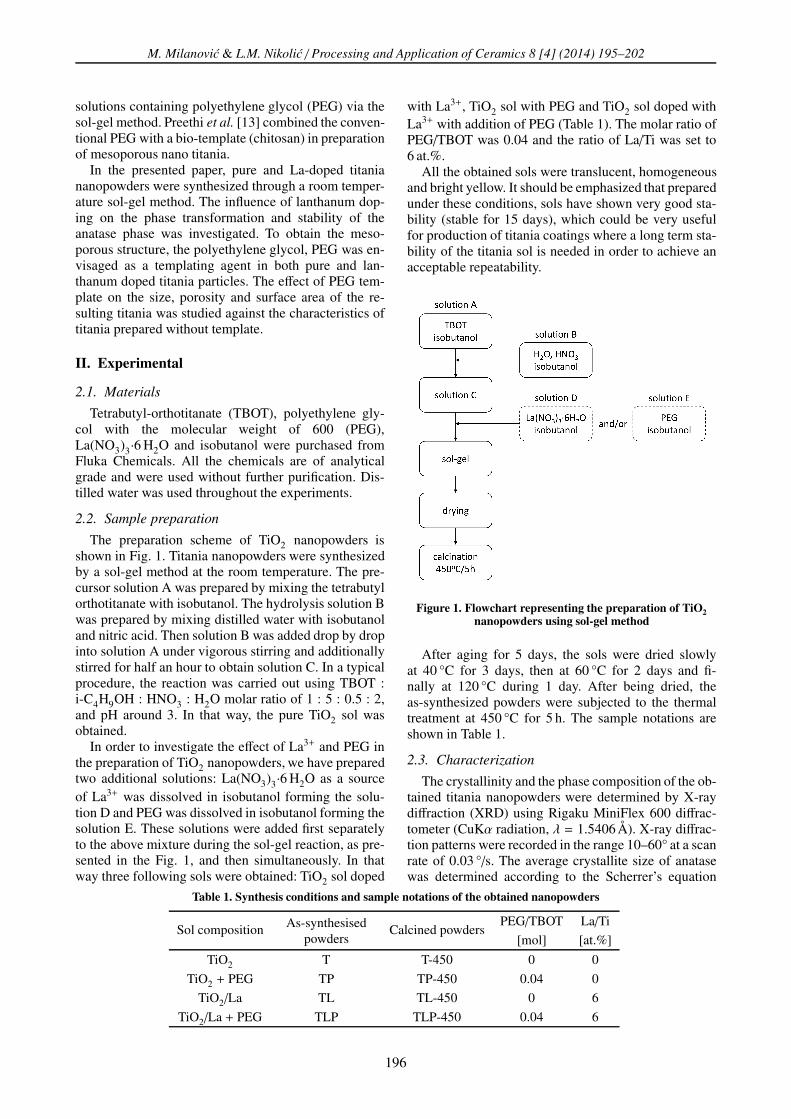
The preparation scheme of TiO2 nanopowders isshown in Fig. 1. Titania nanopowders were synthesizedby a sol-gel method at the room temperature. The pre-cursor solution A was prepared by mixing the tetrabutylorthotitanate with isobutanol. The hydrolysis solution Bwas prepared by mixing distilled water with isobutanoland nitric acid. Then solution B was added drop by dropinto solution A under vigorous stirring and additionallystirred for half an hour to obtain solution C. In a typicalprocedure, the reaction was carried out using TBOT :i-C4H9OH : HNO3 : H2O molar ratio of 1 : 5 : 0.5 : 2,and pH around 3. In that way, the pure TiO2 sol wasobtained.
In order to investigate the effect of La3+ and PEG inthe preparation of TiO2 nanopowders, we have preparedtwo additional solutions: La(NO3)3·6 H2O as a sourceof La3+ was dissolved in isobutanol forming the solu-tion D and PEG was dissolved in isobutanol forming thesolution E. These solutions were added first separatelyto the above mixture during the sol-gel reaction, as pre-sented in the Fig. 1, and then simultaneously. In thatway three following sols were obtained: TiO2 sol doped
with La3+, TiO2 sol with PEG and TiO2 sol doped withLa3+ with addition of PEG (Table 1). The molar ratio ofPEG/TBOT was 0.04 and the ratio of La/Ti was set to6 at.%.
All the obtained sols were translucent, homogeneousand bright yellow. It should be emphasized that preparedunder these conditions, sols have shown very good sta-bility (stable for 15 days), which could be very usefulfor production of titania coatings where a long term sta-bility of the titania sol is needed in order to achieve anacceptable repeatability.
Figure 1. Flowchart representing the preparation of TiO2
nanopowders using sol-gel method
After aging for 5 days, the sols were dried slowlyat 40 °C for 3 days, then at 60 °C for 2 days and fi-nally at 120 °C during 1 day. After being dried, theas-synthesized powders were subjected to the thermaltreatment at 450 °C for 5 h. The sample notations areshown in Table 1.
2.3. Characterization
The crystallinity and the phase composition of the ob-tained titania nanopowders were determined by X-raydiffraction (XRD) using Rigaku MiniFlex 600 diffrac-tometer (CuKα radiation, λ = 1.5406 Å). X-ray diffrac-tion patterns were recorded in the range 10–60° at a scanrate of 0.03 °/s. The average crystallite size of anatasewas determined according to the Scherrer’s equation
Table 1. Synthesis conditions and sample notations of the obtained nanopowders
Sol composition As-synthesisedpowders
Calcined powdersPEG/TBOT La/Ti
[mol] [at.%]
TiO2 T T-450 0 0
TiO2 + PEG TP TP-450 0.04 0
TiO2/La TL TL-450 0 6
TiO2/La + PEG TLP TLP-450 0.04 6
196
M. Milanovic & L.M. Nikolic / Processing and Application of Ceramics 8 [4] (2014) 195–202
(a) (b)
Figure 2. DTA/TG of as-synthesized titania nanopowders: a) TP and b) TLP
using a FWHM of the 101 reflection. The anatase con-tent of samples was calculated from equation [14]:
WA =KA · IA
KA · IA + IR + KB · IB
(1)
where WA represents the mass fraction of anatase andIA, IB and IR are the integrated intensities of the anatase101, brookite 121 and rutile 110 peaks and KA and KB
are two coefficients and their values are 0.886 and 2.721,respectively. The size and morphology of particles in thesamples were observed by a scanning electron micro-scope (SEM JEOL 6460LV). FTIR measurements of thesamples were carried out on Nicolet-Nexsus 670 spec-trophotometer. Simultaneous thermogravimetry and dif-ferential thermal analysis (TG-DTA, Bähr, STA 503)with a heating rate of 10 °C/min from 20 to 900 °Cin air atmosphere was used to study the thermal be-haviour of the as-synthesized (dried) powders. Specificsurface areas of TiO2 powders were measured by nitro-gen adsorption at 77 K (Quantachrom Autosorb–3B) us-ing Brunauer-Emmett-Teller (BET) method. The poresize distribution was derived from the N2 desorptionisotherm using the BJH method.
III. Results and discussion
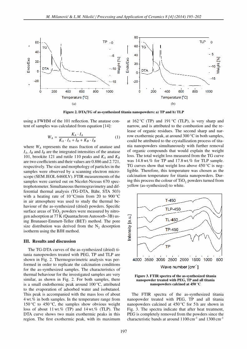
The TG-DTA curves of the as-synthesized (dried) ti-tania nanopowders treated with PEG, TP and TLP areshown in Fig. 2. Thermogravimetric analysis was per-formed in order to replicate the calcination conditionsfor the as-synthesized samples. The characteristics ofthermal behaviour for the investigated samples are verysimilar, as shown in Fig. 2. For both samples, thereis a small endothermic peak around 100 °C, attributedto the evaporation of adsorbed water and isobutanol.This peak is accompanied with the mass loss of about4 wt.% in both samples. In the temperature range from150 °C to 450 °C, the samples show obvious weightloss of about 11 wt.% (TP) and 14 wt.% (TLP). TheDTA curve shows two main exothermic peaks in thisregion. The first exothermic peak, with its maximum
at 162 °C (TP) and 191 °C (TLP), is very sharp andnarrow, and is attributed to the combustion and the re-lease of organic residues. The second sharp and nar-row exothermic peak, at around 300 °C in both samples,could be attributed to the crystallization process of tita-nia nanopowders simultaneously with further removalof organic compounds that would explain the weightloss. The total weight loss measured from the TG curvewas 14.8 wt.% for TP and 17.8 wt.% for TLP sample.TG curves show that weight loss above 450 °C is neg-ligible. Therefore, this temperature was chosen as thecalcination temperature for titania nanopowders. Dur-ing this process the colour of TiO2 powders turned fromyellow (as-synthesized) to white.
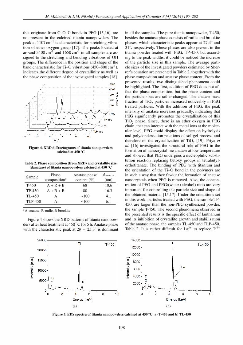
Figure 3. FTIR spectra of the as-synthesized titaniananopowder treated with PEG, TP and all titania
nanopowders calcined at 450 °C
The FTIR spectra of the as-synthesized titaniananopowder treated with PEG, TP and all titaniananopowders calcined at 450 °C for 5 h are shown inFig. 3. The spectra indicate that after heat treatment,PEG is completely removed from the powders since thecharacteristic bands at around 1100 cm-1 and 1300 cm-1
197
M. Milanovic & L.M. Nikolic / Processing and Application of Ceramics 8 [4] (2014) 195–202
that originate from C–O–C bonds in PEG [15,16], arenot present in the calcined titania nanopowders. Thepeak at 1107 cm-1 is characteristic for stretching vibra-tion of ether oxygen group [17]. The peaks located ataround 3400 cm-1 and 1650 cm-1 in all samples are as-signed to the stretching and bending vibrations of OHgroups. The difference in the position and shape of theband characteristic for Ti–O vibrations (450–800 cm-1),indicates the different degree of crystallinity as well asthe phase composition of the investigated samples [18].
Figure 4. XRD diffractograms of titania nanopowderscalcined at 450 °C
Table 2. Phase composition (from XRD) and crystallite size(danatase) of titania nanopowders calcined at 450 °C
Sample Phasecompositiona
Anatase phasecontent [%]
danatase
[nm]T-450 A + R + B 68 10.6TP-450 A + R + B 80 16.3TL-450 A ∼100 4.1TLP-450 A ∼100 6.1
a A-anatase, R-rutile, B-brookite
Figure 4 shows the XRD patterns of titania nanopow-ders after heat treatment at 450 °C for 5 h. Anatase phasewith the characteristic peak at 2θ ∼ 25.3° is dominant
in all the samples. The pure titania nanopowder, T-450,besides the anatase phase consists of rutile and brookitephases, which characteristic peaks appear at 27.4° and31°, respectively. These phases are also present in thetitania powder treated with PEG, TP-450, but accord-ing to the peak widths, it could be noticed the increaseof the particle size in this sample. The average parti-cle sizes of the investigated powders estimated by Sher-rer’s equation are presented in Table 2, together with thephase composition and anatase phase content. From thepresented results, two distinguished phenomena couldbe highlighted. The first, addition of PEG does not af-fect the phase composition, but the phase content andthe particle sizes are rather changed. The anatase massfraction of TiO2 particles increased noticeably in PEGtreated particles. With the addition of PEG, the peakintensity of anatase increases gradually, indicating thatPEG significantly promotes the crystallization of thisTiO2 phase. Since, there is an ether oxygen in PEGchain, that can interact with the metal ions at the molec-ular level, PEG could display the effect on hydrolysisand polycondensation reactions of sol-gel process andtherefore on the crystallization of TiO2 [19]. Priya et
al. [16] investigated the structural role of PEG in theformation of nanocrystalline anatase at low temperatureand showed that PEG undergoes a nucleophilic substi-tution reaction replacing butoxy groups in tetrabutyl-orthotitanate. The binding of PEG with titanium andthe orientation of the Ti–O bond in the polymers arein such a way that they favour the formation of anatasenanocrystals when PEG is removed. Also, the concen-tration of PEG and PEG/(water+alcohol) ratio are veryimportant for controlling the particle size and shape ofthe obtained material [15,17]. Under the conditions setin this work, particles treated with PEG, the sample TP-450, are larger than the non-PEG synthesized powder,the sample T-450. The second phenomena observed inthe presented results is the specific effect of lanthanumand its inhibition of crystallite growth and stabilizationof the anatase phase, the samples TL-450 and TLP-450,Table 2. It is rather difficult for La3+ to replace Ti4+
(a) (b)
Figure 5. EDS spectra of titania nanopowders calcined at 450 °C: a) T-450 and b) TL-450
198
M. Milanovic & L.M. Nikolic / Processing and Application of Ceramics 8 [4] (2014) 195–202
(a) (b)
(c) (d)
Figure 6. SEM images of titania nanopowders calcined at 450 °C: a) T-450, b) TP-450, c) TL-450 and d) TLP-450
inside the lattice, because the La3+ ionic size is muchlarger than Ti4+ one. However, it is believed that La in-corporates into titania at low temperatures by the for-mation of Ti–O–La bonds on the surface of small par-ticles [20]. This inhibits the movement of surface Tiatoms and particle coarsening by restricting direct con-tact of neighbouring crystallites, leading to the stabi-lization of the small anatase particles [21]. The averageanatase particles size in titania powders without La3+
addition are in the range 10.6–16.3 nm (the samples T-450 and TP-450), in contrast to the powders with La3+
ions where the average anatase particle sizes are 4.1–6.1 nm (the samples TL-450 and TLP-450). The pre-sented results indicate that La3+ as a dopant is efficientin hindering the crystallite growth and stabilization ofanatase phase.
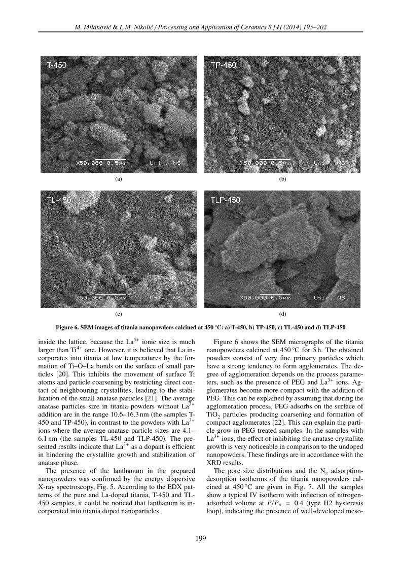
The presence of the lanthanum in the preparednanopowders was confirmed by the energy dispersiveX-ray spectroscopy, Fig. 5. According to the EDX pat-terns of the pure and La-doped titania, T-450 and TL-450 samples, it could be noticed that lanthanum is in-corporated into titania doped nanoparticles.
Figure 6 shows the SEM micrographs of the titaniananopowders calcined at 450 °C for 5 h. The obtainedpowders consist of very fine primary particles whichhave a strong tendency to form agglomerates. The de-gree of agglomeration depends on the process parame-ters, such as the presence of PEG and La3+ ions. Ag-glomerates become more compact with the addition ofPEG. This can be explained by assuming that during theagglomeration process, PEG adsorbs on the surface ofTiO2 particles producing coarsening and formation ofcompact agglomerates [22]. This can explain the parti-cle grow in PEG treated samples. In the samples withLa3+ ions, the effect of inhibiting the anatase crystallitegrowth is very noticeable in comparison to the undopednanopowders. These findings are in accordance with theXRD results.
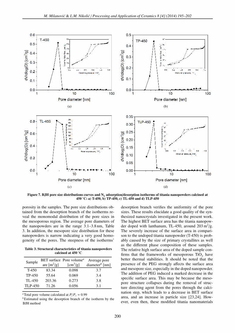
The pore size distributions and the N2 adsorption-desorption isotherms of the titania nanopowders cal-cined at 450 °C are given in Fig. 7. All the samplesshow a typical IV isotherm with inflection of nitrogen-adsorbed volume at P/P◦ = 0.4 (type H2 hysteresisloop), indicating the presence of well-developed meso-
199
M. Milanovic & L.M. Nikolic / Processing and Application of Ceramics 8 [4] (2014) 195–202
(a) (b)
(c) (d)
Figure 7. BJH pore size distributions curves and N2 adsorption/desorption isotherms of titania nanopowders calcined at450 °C: a) T-450, b) TP-450, c) TL-450 and d) TLP-450
porosity in the samples. The pore size distributions ob-tained from the desorption branch of the isotherms re-veal the monomodal distribution of the pore sizes inthe mesoporous region. The average pore diameters ofthe nanopowders are in the range 3.1–3.8 nm, Table3. In addition, the mesopore size distribution for thesenanopowders is narrow indicating a very good homo-geneity of the pores. The steepness of the isotherms’
Table 3. Structural characteristics of titania nanopowderscalcined at 450 °C
Sample BET surfaceare [m2/g]
Pore volumea
[cm3/g]Average pore
diameterb [nm]T-450 83.34 0.098 3.7
TP-450 55.64 0.069 3.4TL-450 203.36 0.273 3.8
TLP-450 71.26 0.056 3.1
a Total pore volume calculated at P/P◦ = 0.99b Estimated using the desorption branch of the isotherm by theBJH method
desorption branch verifies the uniformity of the poresizes. These results elucidate a good quality of the syn-thesized nanocrystals investigated in the present work.The highest BET surface area has the titania nanopow-der doped with lanthanum, TL-450, around 203 m2/g.The severely increase of the surface area in compari-son to the undoped titania nanopowder (T-450) is prob-ably caused by the size of primary crystallites as wellas the different phase composition of these samples.The relative high surface area of the doped sample con-firms that the frameworks of mesoporous TiO2 havebetter thermal stabilities. It should be noted that thepresence of the PEG strongly affects the surface areaand mesopore size, especially in the doped nanopowder.The addition of PEG induced a marked decrease in thespecific surface area. This may be because the meso-pore structure collapses during the removal of struc-ture directing agent from the pores through the calci-nation step, which leads to a decrease in BET surfacearea, and an increase in particle size [23,24]. How-ever, even then, these modified titania nanomaterials
200
M. Milanovic & L.M. Nikolic / Processing and Application of Ceramics 8 [4] (2014) 195–202
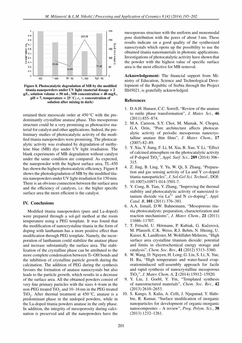
Figure 8. Photocatalytic degradation of MB by the modifiedtitania nanopowders under UV light (material dosage = 2
g/L, solution volume = 50 mL, MB concentration = 40 mg/L,pH = 7, temperature = 25 °C; co = concentration of
solution after mixing in dark)
retained their mesoscale order at 450 °C with the pre-dominantly crystalline anatase phase. This mesoporousstructure could be a very promising as photoactive ma-terial for catalyst and other applications. Indeed, the pre-liminary studies of photocatalytic activity of the modi-fied titania nanopowders were promising. The photocat-alytic activity was evaluated by degradation of methy-lene blue (MB) dye under UV light irradiation. Theblank experiments of MB degradation without catalystunder the same condition are compared. As expected,the nanopowder with the highest surface area, TL-450has shown the highest photocatalytic efficiency. Figure 8shows the photodegradation of MB by the modified tita-nia nanopowders under UV light irradiation for 150 min.There is an obvious connection between the surface areaand the efficiency of catalysts, i.e. the higher specificsurface area the more efficient is the catalyst.
IV. Conclusions
Modified titania nanopowders (pure and La-doped)were prepared through a sol-gel method at the roomtemperature using a PEG template. It was found thatthe modification of nanocrystaline titania in the form ofdoping with lanthanum has a more positive effect thanmodification through PEG template. Namely, the incor-poration of lanthanum could stabilize the anatase phaseand increase substantially the surface area. The stabi-lization of the crystalline phase can be attributed to themore complete condensation between Ti–OH bonds andthe inhibition of crystalline particle growth during thecalcination. The addition of PEG during the synthesisfavours the formation of anatase nanocrystals but alsoleads to the particle growth, which results in a decreaseof the surface area. All the obtained powders consist ofvery fine primary particles with the sizes 4–6 nm in thenon-PEG treated TiO2 and 10–16 nm in the PEG treatedTiO2. After thermal treatment at 450 °C, anatase is apredominant phase in the undoped powders, while inthe La-doped titania powders anatase in the only phase.In addition, the integrity of mesoporosity during calci-nation is preserved and all the nanopowders have the
mesoporous structure with the uniform and monomodalpore distribution with the pores of about 3 nm. Theseresults indicate on a good quality of the synthesizednanocrystals which opens up the possibility to use theobtained titania nanomaterials in photonic applications.Investigations of photocatalytic activity have shown thatthe powder with the highest value of specific surfacearea is the most effective for MB removal.
Acknowledgement: The financial support from Mi-nistry of Education, Science and Technological Deve-lopment of the Republic of Serbia through the ProjectIII45021, is gratefully acknowledged.
References
1. D.A.H. Hanaor, C.C. Sorrell, “Review of the anataseto rutile phase transformation”, J. Mater. Sci., 46
(2011) 855–874.2. M.A. Carreon, S.Y. Choi, M. Mamak, N. Chopra,
G.A. Ozin, “Pore architecture affects photocat-alytic activity of periodic mesoporous nanocrys-talline anatase thin films”, J. Mater. Chem., 17
(2007) 82–89.3. Y. Xia, Y. Jiang, F. Li, M. Xia, B. Xue, Y. Li, “Effect
of calcined atmosphere on the photocatalytic activityof P-doped TiO2”, Appl. Surf. Sci., 289 (2014) 306–315.
4. Z. Jing, B. Ling, Y. Yu, W. Qi, S. Zhang, “Prepara-tion and gas sensing activity of La and Y co-dopedtitania nanoparticles”, J. Sol-Gel Sci. Technol., DOI:10.1007/s10971-014-3501-7.
5. Y. Cong, B. Tian, Y. Zhang, “Improving the thermalstability and photocatalytic activity of nanosized ti-tanium dioxide via La3+ and N co-doping”, Appl.
Catal. B, 101 (2011) 376–381.6. A.A. Ismail, D.W. Bahnemann, “Mesoporous tita-
nia photocatalysts: preparation, characterization andreaction mechanisms”, J. Mater. Chem., 21 (2011)11686–11707.
7. T. Fröschl, U. Hörmann, P. Kubiak, G. Kucerová,M. Pfanzelt, C.K. Weiss, R.J. Behm, N. Hüsing, U.Kaiser, K. Landfester, M. Wohlfahrt-Mehrens, “Highsurface area crystalline titanium dioxide: potentialand limits in electrochemical energy storage andcatalysis”, Chem. Soc. Rev., 41 (2012) 5313–5360.
8. W. Wang, D. Nguyen, H. Long, G. Liu, S. Li, X. Yue,H. Ru, “High temperature and water-based evap-orationinduced self-assembly approach for facileand rapid synthesis of nanocrystalline mesoporousTiO2”, J. Mater. Chem. A, 2 (2014) 15912–15920.
9. Y. Liu, J. Goebl, Y. Yin, “Templated synthesisof nanostructured materials”, Chem. Soc. Rev., 42
(2013) 2610–2653.10. S. Kango, S. Kalia, A. Celli, J. Njugunad, Y. Habi-
bie, R. Kumar, “Surface modification of inorganicnanoparticles for development of organic-inorganicnanocomposites – A review”, Prog. Polym. Sci., 38
(2013) 1232–1261.
201
M. Milanovic & L.M. Nikolic / Processing and Application of Ceramics 8 [4] (2014) 195–202
11. W. Zhang, R. Li, H. He, “Synthesis of mesoporousTiO2–Al2O3 binary oxides photocatalyst by sol-gelmethod using PEG1000 as template”, Int. J. Pho-
toenergy, 2012 (2012) 1–7.12. J. Yu, X. Zhao, Q. Zhao, G. Wang, “Preparation and
characterization of superhydrophilic porous TiO2coating films”, Mater. Chem. Phys., 68 (2001) 253–259.
13. T. Preethi, B. Abarna, G.R. Rajarajeswari, “Influ-ence of chitosan–PEG binary template on the crys-tallite characteristics of sol-gel synthesized meso-porous nano-titania photocatalyst”, Appl. Surf. Sci.,317 (2014) 90–97.
14. F. Mastali Khan Tehrani, M. Rashidzadeh, A. Ne-mati, A. Irandoukht, B. Faridnia, “Characterizationand photocatalytic activities of nanosized titaniumdioxide thin films”, Int. J. Environ. Sci. Tech., 8 [3](2011) 545–552.
15. S. Rahim, M. Sasani Ghamsari, S. Radiman, “Sur-face modification of titanium oxide nanocrystalswith PEG”, Scientia Iranica F, 19 [3] (2012) 948–953.
16. S. Priya, S. Balaji, Y. Djaoued, J. Robichaud, “Struc-tural role of polyethylene glycol in the formation ofanatase nanocrystalline titania at low temperature”,J. Raman Spectrosc., 40 (2009) 1885–1894.
17. C.-H. Zhou, X.-Z. Zhao, B.-C. Yang, D. Zhang, Z.-Y.Li, K.-C. Zhou, “Effect of poly (ethylene glycol) oncoarsening dynamics of titanium dioxide nanocrys-tallites in hydrothermal reaction and the applicationin dye sensitized solar cells”, J. Colloid Interface
Sci., 374 (2012) 9–17.18. Z. He, W. Que, J. Chen, Y. He, G. Wang, “Sur-
face chemical analysis on the carbon-doped meso-porous TiO2 photocatalysts after post-thermal treat-ment: XPS and FTIR characterization”, J. Phys.
Chem. Solids, 74 (2013) 924–928.19. K. Yu, J. Zhao, X. Zhao, X. Ding, Y. Zhu, Z. Wang,
“Self-assembly and oriented organization of shape-controlled nanocrystalline TiO2”, Mater. Lett., 59
(2005) 2676–2679.20. S. Yuan, Q. Sheng, J. Zhang, F. Chen, M. Anpo, Q.
Zhang, “Synthesis of La3+ doped mesoporous titaniawith highly crystallized walls”, Micropor. Mesopor.
Mater., 79 (2005) 93–99.21. M.M. Maletin, R.R. Ðenadic, L.M. Nikolic, V.V.
Srdic, “Structural characterization of Nb and Ladoped nanostructured titania powders and coatings”,J. Optoelectron. Adv. Mater., 9 [7] (2007) 2245–2250.
22. P. Alphonse, R. Bleta, R. Soules, “Effect of PEG onrheology and stability of nanocrystalline titania hy-drosols”, J. Colloid Interface Sci., 337 (2009) 81–87.
23. S. Shamaila, A.K.L. Sajjad, F. Chen, J. Zhang,“Mesoporous titania with high crystallinity duringsynthesis by dual template system as an efficientphotocatalyst”, Catal. Today, 175 (2011) 568–575.
24. Y.-C. Lee, Y.S. Chang, L.G. Teoh, Y.L. Huang, Y.C.Shen, “The effects of the nanostructure of meso-porous TiO2 on optical band gap energy”, J. Sol-Gel
Sci. Technol., 56 (2010) 33–38.
202


![Monika Bakierska, Michał Swietosławski˛ *, Roman … · Materials 2016, 9, 696 2 of 12 these obstacles, strategies including cation or anion doping [18–20], surface modification](https://img.pdfslide.us/doc/110x75/5b904b0c09d3f2e6728ba5fd/monika-bakierska-michal-swietoslawski-roman-materials-2016-9-696-2.jpg)