Embed Size (px)
Citation preview
8/3/2019 MEU ARTIGO.pntd.0001430
http://slidepdf.com/reader/full/meu-artigopntd0001430 1/10
8/3/2019 MEU ARTIGO.pntd.0001430
http://slidepdf.com/reader/full/meu-artigopntd0001430 2/10
specificity of the tests, requiring repeated tissue sampling and a trained
laboratory staff [10]. The diagnosis of CVL, by means of ELISA,
based on Leishmania antigens has shown variable values of sensitivity
and/or specificity, mainly due to antigenic similarities between
Leishmania and other protozoa [10]. As a strategy to develop a more
specific test, several parasite antigens have been tested in prior studies
[11–14]; however, due to frequent low specificity and sensitivity in
detecting asymptomatic infections and the high variability observed in
the humoral response of individual infected dogs [15], it has been
postulated that an efficient diagnosis may require a mixture of antigens
or the use of chimerical antigens [16–19].
Proteomic approaches applied to study Leishmania protein
expression patterns offer the possibility to assign potential func-
tions for proteins, including those previously identified by geno-mics as hypothetical, new diagnostic markers, vaccine candidates,
and/or potential drug targets [20–23]. Several proteomic studies
have been performed to study stage-specific expression and
differentiation in Leishmania [24–32]. The coupling of antibodies
specific to parasite antigens generated during different stages of
disease progression in dogs will certainly contribute to refining this
analysis, which aims to identify not only differentially expressed
proteins, but also potentially new antigens identified by the
immune system during active infection. Recently, the discovery of
antigens through proteomics has been indicated as one of the main
research priorities for further development and improvement of
leishmaniasis vaccines [33].
In this work, an immunoproteomic approach, together with
two-dimensional electrophoresis (2DE) and mass spectrometry, was
carried out to analyze the protein expression profiles of promasti-gote and amastigote-like L. infantum. Aimed at identifying new
diagnostic markers and/or vaccine candidates, antibodies present in
the sera of dogs with asymptomatic and symptomatic VL were
added to this analysis, allowing for the identification of several
known, as well as hypothetical, L. infantum antigenic proteins.
Materials and Methods
Parasite cultureExperiments were carried out using the Leishmania (Leishmania)
chagasi syn. L. (L.) infantum (MHOM/BR/1970/BH46) strain. The
stationary phase of promastigote cells were grown at 24uC in
Schneider’s medium (Sigma, St. Louis, MO, USA), supplemented
with 20% inactivated fetal bovine serum (FBS, Sigma), 20 mM L-
glutamine, 200 U/mL penicillin, and 100 mg/mL streptomycin, at
pH 7.2, as previously described [34]. The amastigote-like cells
were obtained as described by Doyle et al. (1991) [35].
Sera samples
The present study used sera samples from 60 L. infantum-infecteddogs (40 clinically symptomatic and 20 asymptomatic) from Belo
Horizonte, Minas Gerais, Brazil. Animals were considered sym-
ptomatic when three or more of the following symptoms were
present: loss of weight, hepatomegaly, alopecia, adenopathy,
onychogryposis, conjunctivitis, and exfoliative dermatitis on the
nose, tail, or ear tips. The asymptomatic animals were free from
clinical symptoms. All sera samples from either symptomatic or
asymptomatic animals were positive when tested by RIFI and
ELISA, and the presence of amastigote stage of the parasite was
confirmed by microscopic observation and in vitro culture using
aspirates from popliteal and/or prescapular lymphoid nodes or
bone marrow and/or tissue fragments. The control group
consisted of sera from 20 dogs living in non-endemic areas from
VL, with no clinical signs or suspicion of leishmaniasis, and which
showed negative parasitological and serological tests. Sera samples
used in this study were kindly provided by Dr. Maria Norma Melo
(Departmento de Parasitologia, Instituto de Ciencias Biologicas,
UFMG).
Preparation of protein extractsThe protein extraction from promastigote and amastigote-like
stages L. infantum and 2DE were performed following a modified
protocol [36]. Briefly, cells from both stages (161010 cells) were
washed three times in 40 mM Tris-HCl, pH 7.2, by centrifu-
gation at 50006 g for 10 min at 4uC. The pellets were
resuspended in lyses buffer solution [7 M urea, 2 M thiourea,
4% chol-amidopropyl dimethylammonio-1-propanesulfonate
(CHAPS), 40 mM dithiothreitol (DTT), 2% IPG buffer
(pH 4–7), 40 mM Tris], and a protease inhibitor cocktail (GEHealthcare, Upsala, Sweden) was added. Samples were
incubated for 1 h at room temperature, with occasional
vortexing. Purification was carried out by protein precipitation
using a 2D Clean UpKit (GE Healthcare), according to
manufacturer instructions. Whole cell extracts were measured
by a 2D Quant-Kit (GE Healthcare), and aliquots were
immediately frozen at 280uC, until use.
Isoeletric focusing (IEF)For the first-dimension electrophoresis, 150 mg of protein
extract was added to a volume of 250 mL with a rehydration
solution [7 M urea, 2 M thiourea, 2% CHAPS, 40 mM DTT, 2%
immobilized pH gradient (IPG-buffer, pH 4–7, trace bromophe-
nol blue)]. Next, samples were applied to IPG strips (13 cm, pH 4– 7; GE Healthcare) for passive rehydration overnight at room
temperature. After in-gel rehydration for 12 h, isoeletric focusing
was performed at 500 V for 1 h, 1.000 V for 1 h, and 8.000 V for
8 h, using a Multiphor II electrophoresis unit and EPS 3500 XL
power supply (Amersham, Piscataway, NJ, USA).
SDS-PAGE After IEF, each strip was incubated for 15 min in a solution
made up of 10 mL of a 50 mM Tris-HCl buffer pH 8.8, 6 M
urea, 30% (v/v) glycerol, 2% (w/v) SDS, 0.002% bromophenol-
blue, and 125 mM DTT, followed by a second incubation step in
Author Summary
Canine visceral leishmaniasis (CVL) is an importantemerging zoonosis caused by Leishmania (Leishmania)infantum in the Mediterranean and Middle East and L. (L.)chagasi (syn. L. (L.) infantum) in Latin America. Due to theirgenotypic relationships, these species are considered iden-tical. The present study focused on comparing the proteinexpression profiles of the promastigote and amastigote-like
stages of L. infantum, by means of a protein separation bytwo-dimensional electrophoresis and identification by massspectrometry. The present study attempted to identifyproteins recognized by antibodies present in the sera of dogs with asymptomatic and symptomatic visceral leish-maniasis. A total of one hundred and four proteins wereidentified. Of these, several stage-specific proteins had beenpreviously identified as diagnosis and/or vaccine candi-dates. In addition, antibodies from infected dogs recognizedthirty-one proteins, which had been previously consideredhypothetical, indicating that these proteins are expressedduring active infection. Therefore, the present study revealsnew potential candidates for the improvement of diagnosisof CVL.
Immunoproteomic Approach in Leshmania infantum
www.plosntds.org 2 January 2012 | Volume 6 | Issue 1 | e1430
8/3/2019 MEU ARTIGO.pntd.0001430
http://slidepdf.com/reader/full/meu-artigopntd0001430 3/10
the same buffer solution, excluding DTT, which was replaced by
125 mM iodacetamide. IPG strips were transferred to a 12%
polyacrilamide and sealed with agarose solution (agarose and
bromophenol blue in a Tris-glicine cathode buffer). The protein
standard was purchased from Invitrogen (BenchMark Protein
Ladder). Electrophoresis was performed in a Mini-Protean II
system (BioRad) connected to a MultiTemp II cooling bath
(Amersham Biosciences), in a Tris/glycine/SDS buffer. Proteins
were separated at 200 V, until the dye front had reached thebottom of the gel.
Immunoblotting 2DE analysis and protein identificationTo identify the reactive spots that were recognized by the
antibodies present in the sera samples from asymptomatic and/orsymptomatic CVL, Western blot analyses were performed. Whole
cell extracts of promastigote and amastigote-like L. infantum were
separated electrophoretically and transferred onto cellulose
membranes (Schleicher & Schull, Dassel, Germany) by semi-dry
blotting for 2 h at 400 mA. Membranes were blocked in 5% (w/v)
low-fat dried milk in TBS 16 (pH 7.4) plus 0.05% Tween 20 for
2 h at room temperature. Next, the membranes were washed 6
times (10 min each) with the blocking solution and pre-incubated
in a pool of sera of symptomatic or asymptomatic CVL (1:200
diluted) for 2 h at room temperature. Then, membranes wereincubated with a peroxidase-conjugated goat anti-dog IgG
secondary antibody (1:5.000 diluted) for 2 h at room temperature.
After having been washed 3 times with TBS 16plus 0.5% Tween
20, immunoblots were developed, using a solution made up of
chloronaphtol, diaminobenzidine and H2O2. To select and
identify the spots recognized by antibodies of CVL sera, three
independent protein preparations, each obtained from indepen-
dent parasite cultures, were performed. The 2DE gels were stained
with colloidal Coomassie Brilliant Blue G-250, following proce-
dures described by Neuhoff et al. (1988) [37]. For image analysis,
the stained gels were scanned using an ImageScanner III (GE
Healthcare). Reactive spots recognized by antibodies in the serasamples of asymptomatic and/or symptomatic CVL were excised
manually from the gels for protein identification.
Protein digestion, peptide extraction, and spot handlingSpots were manually excised, and fragments were washed in
25 mM ammonium bicarbonate/50% acetonitrile until complete-
ly destained. After drying, gel fragments were placed on ice in a
50 mL protease solution (20 ng/mL of a sequence grade-modified
trypsin in a 25 mM ammonium bicarbonate) (Promega Biosci-
ences, CA, USA), for 30 min. Excess protease solution was
removed and replaced by 25 mM ammonium bicarbonate.
Digestion was performed at 37uC for 18 h. Peptide extraction
was performed twice for 15 min, using 30 mL of 50% acetonitrile/5% formic acid. Trypsin (Promega) digests were concentrated in a
Speed-Vac (Savant, USA) to approximately 10 mL and desaltedusing Zip-Tip (C18 resin; P10, Millipore Corporation, Bedford,
MA, USA). Samples were mixed with a matrix (5 mg/mlrecrystallized a-cyano-4-hydroxycinnamic acid) in a volume of
1 mL (1:1 ratio) and then spotted for MALDI-TOF/TOF
Ultraflex III (Bruker, Daltonics, Germany).
Protein identification and database searchTo determine the MS spectrum of the immunoreactive spots,
the digests were spotted onto 600 mm Anchorchips (Bruker
Daltonics). Spotting was achieved by pipetting, in duplicate,
1 mL of analyte onto the MALDI target plate, then adding 5 mg/
mL a-cyano-4-hydroxycinnamic acid diluted in 3% TFA/50%
acetonitrile, which contained 2 mM ammonium phosphate. The
Bruker peptide calibration mixture was spotted down for external
calibration. All samples were allowed to air dry at room tem-
perature, and 0.1% TFA was used for on-target washing. Allsamples were analyzed in the positive-ion, reflection mode, through
a MALDI-TOF/TOF Ultraflex III mass spectrometer (Bruker,Daltonics, Germany). Each spectrum was produced by accumulat-
ing data from 200 consecutive laser shots, with a frequency of
100 Hz, and an m/z range of 1.000–4.000. Instrument calibration
was achieved by using peptide calibration standard II (BrukerDaltonics), a mixture of angiotensin I & II, substance P, bombesin,
ACTH clip 1–17, ACTH clip 18–39 and somatostatin 28, as the
internal standard. Peptide masses were measured as mono-isotopic
masses. The MS peaks with the highest intensities were selected for
MS/MS fragmentation analyses.
The resulting spectra were processed using Flex analysis
software, version 2.4 (Bruker Daltonics), with the following settings:
peak detection algorithm set at SNAP (Sort Neaten Assign and
Place), S/N threshold at 3, precursor and product ion tolerances
were set at 0.5 Da, and quality factor threshold at 50. The
trypsin autodigestion ion peaks (842.51, 1045.56, 2211.10, and
2225.12 Da) were used as internal standards to validate the external
calibration procedure. Matrix, and/or autoproteolytic trypsin
fragments, and known contaminants ( i.e., keratins) were manually
removed. The resulting peptides list was used to search in the NCBIdatabase (http://blast.ncbi.nlm.nih.gov) for the organism option of Leishmania (taxid:5658). According to the obtained results, and using
the peptide sequences identified for each protein, the following
parameters were used as selection criteria: total score, query
coverage, and E value. Poor quality spectra were not considered for
selection in the protein sequence database.
Results
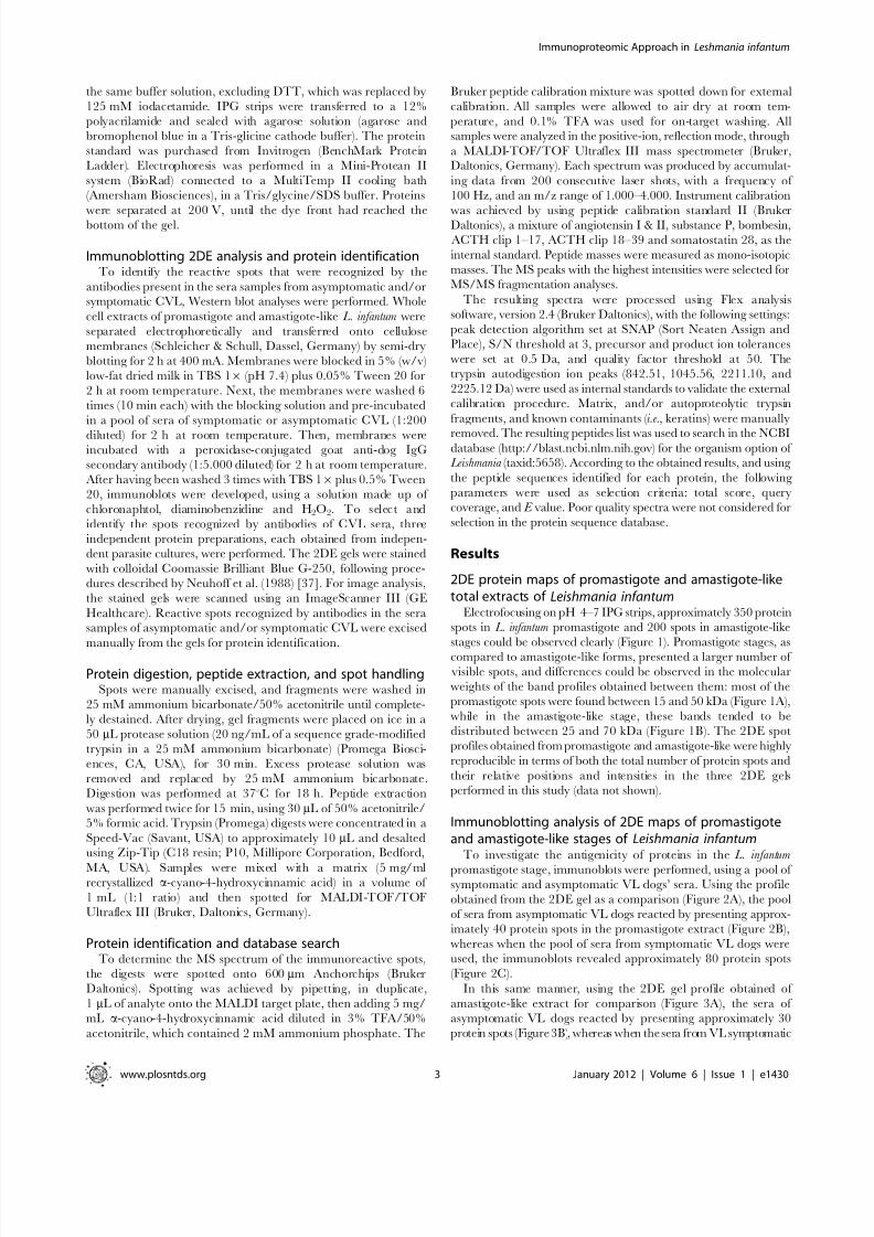
2DE protein maps of promastigote and amastigote-liketotal extracts of Leishmania infantum
Electrofocusing on pH 4–7 IPG strips, approximately 350 protein
spots in L. infantum promastigote and 200 spots in amastigote-like
stages could be observed clearly (Figure 1). Promastigote stages, as
compared to amastigote-like forms, presented a larger number of
visible spots, and differences could be observed in the molecular
weights of the band profiles obtained between them: most of the
promastigote spots were found between 15 and 50 kDa (Figure 1A),
while in the amastigote-like stage, these bands tended to be
distributed between 25 and 70 kDa (Figure 1B). The 2DE spot
profiles obtained from promastigote and amastigote-like were highly
reproducible in terms of both the total number of protein spots and
their relative positions and intensities in the three 2DE gelsperformed in this study (data not shown).
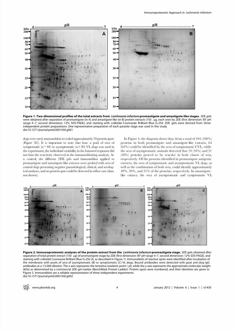
Immunoblotting analysis of 2DE maps of promastigoteand amastigote-like stages of Leishmania infantum
To investigate the antigenicity of proteins in the L. infantum
promastigote stage, immunoblots were performed, using a pool of symptomatic and asymptomatic VL dogs’ sera. Using the profile
obtained from the 2DE gel as a comparison (Figure 2A), the pool
of sera from asymptomatic VL dogs reacted by presenting approx-
imately 40 protein spots in the promastigote extract (Figure 2B),
whereas when the pool of sera from symptomatic VL dogs were
used, the immunoblots revealed approximately 80 protein spots
(Figure 2C).
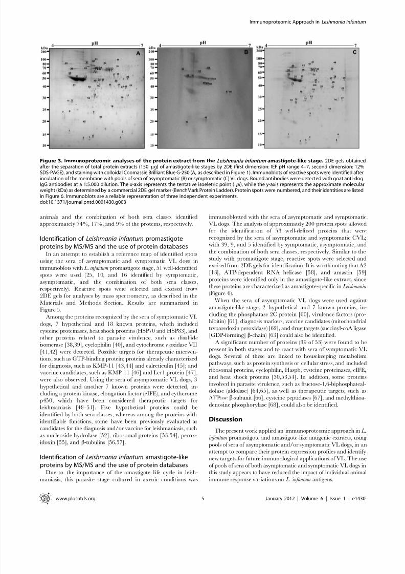
In this same manner, using the 2DE gel profile obtained of
amastigote-like extract for comparison (Figure 3A), the sera of
asymptomatic VL dogs reacted by presenting approximately 30
protein spots (Figure 3B), whereas when the sera from VL symptomatic
Immunoproteomic Approach in Leshmania infantum
www.plosntds.org 3 January 2012 | Volume 6 | Issue 1 | e1430
8/3/2019 MEU ARTIGO.pntd.0001430
http://slidepdf.com/reader/full/meu-artigopntd0001430 4/10
dogs were used, immunoblots revealed approximately 70 protein spots
(Figure 3C). It is important to note that how a pool of sera of
symptomatic (n = 40) or asymptomatic (n = 20) VL dogs was used in
the experiments, the individual variability in the humoral responses did
not bias the reactivity observed in the immunoblotting analysis. As
a control, the different 2DE gels and immunoblots applied to
promastigote and amastigote-like extracts were probed with sera of
control dogs presenting negative parasitological, clinical, and serolog-
ical analyses, and no protein spot could be detected in either case (data
not shown).
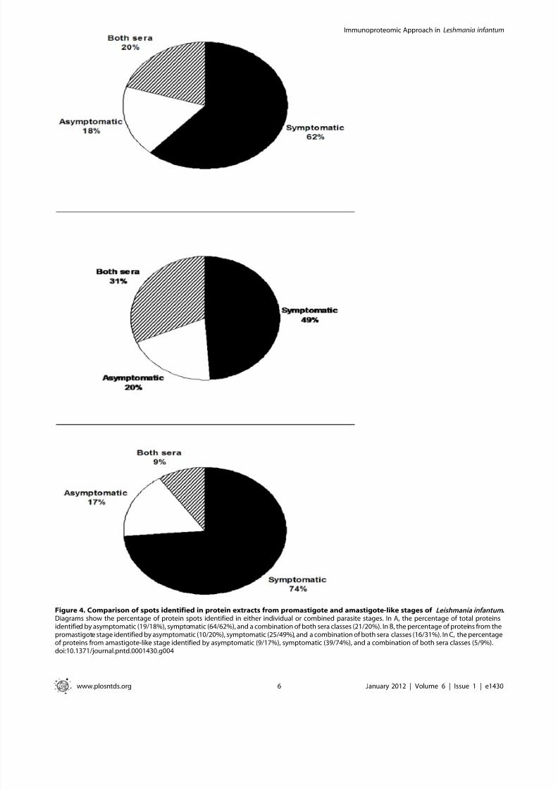
In Figure 4, the diagram shows that, from a total of 104 (100%)
proteins in both promastigote and amastigote-like extracts, 64
(62%) could be identified by the sera of symptomatic CVL, while
the sera of asymptomatic animals detected that 19 (18%) and 21
(20%) proteins proved to be reactive in both classes of sera,
respectively. Of the proteins identified in promastigote antigenic
extracts, the sera of symptomatic and asymptomatic VL dogs, as
well as the combination of both sera, could identify approximately
49%, 20%, and 31% of the proteins, respectively. In amastigote-
like extract, the sera of asymptomatic and symptomatic VL
Figure 1. Two-dimensional profiles of the total extracts from Leishmania infantum promastigote and amastigote-like stages. 2DE gelswere obtained after separation of promastigote (in A) and amastigote-like (in B) protein extracts (150 mg, each one) by 2DE (first dimension: IEF pHrange 4–7, second dimension: 12% SDS-PAGE), and staining with colloidal Coomassie Brilliant Blue G-250. 2DE gels were derived from threeindependent protein preparations. One representative preparation of each parasite stage was used in this study.doi:10.1371/journal.pntd.0001430.g001
Figure 2. Immunoproteomic analyses of the protein extract from the Leishmania infantum promastigote stage. 2DE gels obtained afterseparation of total protein extract (150 mg) of promastigote stage by 2DE (first dimension: IEF pH range 4–7, second dimension: 12% SDS-PAGE), andstaining with colloidal Coomassie Brilliant Blue G-250 (A, as described in Figure 1). Immunoblots of reactive spots were identified after incubation of the membrane with pools of sera of asymptomatic (B) or symptomatic (C) VL dogs. Bound antibodies were detected with goat anti-dog IgGantibodies at a 1:5.000 dilution. The x-axis represents the tentative isoeletric point ( pI ), while the y-axis represents the approximate molecular weight(kDa) as determined by a commercial 2DE gel marker (BenchMark Protein Ladder). Protein spots were numbered, and their identities are given inFigure 5. Immunoblots are a reliable representation of three independent experiments.doi:10.1371/journal.pntd.0001430.g002
Immunoproteomic Approach in Leshmania infantum
www.plosntds.org 4 January 2012 | Volume 6 | Issue 1 | e1430
8/3/2019 MEU ARTIGO.pntd.0001430
http://slidepdf.com/reader/full/meu-artigopntd0001430 5/10
animals and the combination of both sera classes identified
approximately 74%, 17%, and 9% of the proteins, respectively.
Identification of Leishmania infantum promastigoteproteins by MS/MS and the use of protein databases
In an attempt to establish a reference map of identified spots
using the sera of asymptomatic and symptomatic VL dogs in
immunoblots with L. infantum promastigote stage, 51 well-identified
spots were used (25, 10, and 16 identified by symptomatic,
asymptomatic, and the combination of both sera classes,
respectively). Reactive spots were selected and excised from
2DE gels for analyses by mass spectrometry, as described in the
Materials and Methods Section. Results are summarized inFigure 5.
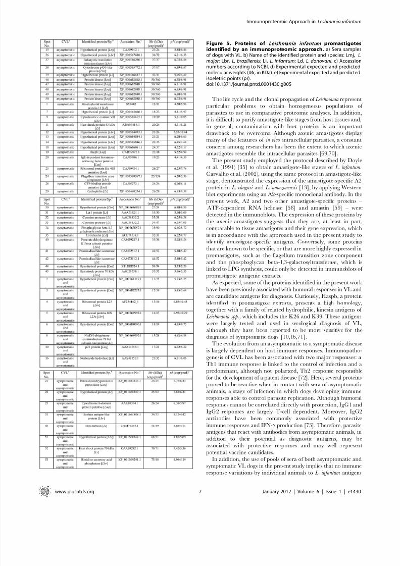
Among the proteins recognized by the sera of symptomatic VL
dogs, 7 hypothetical and 18 known proteins, which included
cysteine proteinases, heat shock proteins (HSP70 and HSP83), and
other proteins related to parasite virulence, such as disulfide
isomerase [38,39], cyclophilin [40], and cytochrome c oxidase VII
[41,42] were detected. Possible targets for therapeutic interven-
tions, such as GTP-binding protein; proteins already characterized
for diagnosis, such as KMP-11 [43,44] and calreticulin [45]; and
vaccine candidates, such as KMP-11 [46] and Lcr1 protein [47],
were also observed. Using the sera of asymptomatic VL dogs, 3
hypothetical and another 7 known proteins were detected, in-
cluding a protein kinase, elongation factor (eIFE), and cythcrome
p450, which have been considered therapeutic targets forleishmaniasis [48–51]. Five hypothetical proteins could be
identified by both sera classes, whereas among the proteins with
identifiable functions, some have been previously evaluated as
candidates for the diagnosis and/or vaccine for leishmaniasis, such
as nucleoside hydrolase [52], ribosomal proteins [53,54], perox-
idoxin [55], and b-tubulins [56,57].
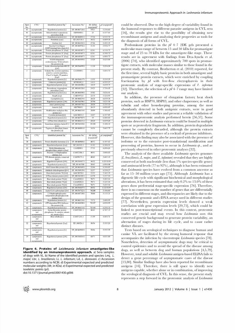
Identification of Leishmania infantum amastigote-likeproteins by MS/MS and the use of protein databases
Due to the importance of the amastigote life cycle in leish-
maniasis, this parasite stage cultured in axenic conditions was
immunoblotted with the sera of asymptomatic and symptomatic
VL dogs. The analysis of approximately 200 protein spots allowed
for the identification of 53 well-defined proteins that were
recognized by the sera of asymptomatic and symptomatic CVL;
with 39, 9, and 5 identified by symptomatic, asymptomatic, and
the combination of both sera classes, respectively. Similar to the
study with promastigote stage, reactive spots were selected and
excised from 2DE gels for identification. It is worth noting that A2
[13], ATP-dependent RNA helicase [58], and amastin [59]
proteins were identified only in the amastigote-like extract, since
these proteins are characterized as amastigote-specific in Leishmania (Figure 6).
When the sera of asymptomatic VL dogs were used againstamastigote-like stage, 2 hypothetical and 7 known proteins, in-
cluding the phosphatase 2C protein [60], virulence factors (pro-
hibitin) [61], diagnosis markers, vaccine candidates (mitochondrial
tryparedoxin peroxidase) [62], and drug targets (succinyl-coA ligase
[GDP-forming] b-chain) [63] could also be identified.
A significant number of proteins (39 of 53) were found to be
present in both stages and to react with sera of symptomatic VL
dogs. Several of these are linked to housekeeping metabolism
pathways, such as protein synthesis or cellular stress, and included
ribosomal proteins, cyclophilin, Haspb, cysteine proteinases, eIFE,
and heat shock proteins [30,53,54]. In addition, some proteins
involved in parasite virulence, such as fructose-1,6-biphosphateal-
dolase (aldolase) [64,65], as well as therapeutic targets, such as
ATPase b-subunit [66], cysteine peptidases [67], and methylthioa-
denosine phosphorylase [68], could also be identified.
Discussion
The present work applied an immunoproteomic approach in L.
infantum promastigote and amastigote-like antigenic extracts, using
pools of sera of asymptomatic and/or symptomatic VL dogs, in an
attempt to compare their protein expression profiles and identify
new targets for future immunological applications of VL. The use
of pools of sera of both asymptomatic and symptomatic VL dogs in
this study appears to have reduced the impact of individual animal
immune response variations on L. infantum antigens.
Figure 3. Immunoproteomic analyses of the protein extract from the Leishmania infantum amastigote-like stage. 2DE gels obtainedafter the separation of total protein extracts (150 mg) of amastigote-like stages by 2DE (first dimension: IEF pH range 4–7, second dimension: 12%SDS-PAGE), and staining with colloidal Coomassie Brilliant Blue G-250 (A, as described in Figure 1). Immunoblots of reactive spots were identified afterincubation of the membrane with pools of sera of asymptomatic (B) or symptomatic (C) VL dogs. Bound antibodies were detected with goat anti-dogIgG antibodies at a 1:5.000 dilution. The x-axis represents the tentative isoeletric point ( pI ), while the y-axis represents the approximate molecularweight (kDa) as determined by a commercial 2DE gel marker (BenchMark Protein Ladder). Protein spots were numbered, and their identities are listedin Figure 6. Immunoblots are a reliable representation of three independent experiments.
doi:10.1371/journal.pntd.0001430.g003
Immunoproteomic Approach in Leshmania infantum
www.plosntds.org 5 January 2012 | Volume 6 | Issue 1 | e1430
8/3/2019 MEU ARTIGO.pntd.0001430
http://slidepdf.com/reader/full/meu-artigopntd0001430 6/10
Figure 4. Comparison of spots identified in protein extracts from promastigote and amastigote-like stages of Leishmania infantum .Diagrams show the percentage of protein spots identified in either individual or combined parasite stages. In A, the percentage of total proteinsidentified by asymptomatic (19/18%), symptomatic (64/62%), and a combination of both sera classes (21/20%). In B, the percentage of proteins from thepromastigote stage identified by asymptomatic (10/20%), symptomatic (25/49%), and a combination of both sera classes (16/31%). In C, the percentageof proteins from amastigote-like stage identified by asymptomatic (9/17%), symptomatic (39/74%), and a combination of both sera classes (5/9%).doi:10.1371/journal.pntd.0001430.g004
Immunoproteomic Approach in Leshmania infantum
www.plosntds.org 6 January 2012 | Volume 6 | Issue 1 | e1430
8/3/2019 MEU ARTIGO.pntd.0001430
http://slidepdf.com/reader/full/meu-artigopntd0001430 7/10
The life cycle and the clonal propagation of Leishmania representparticular problems to obtain homogeneous populations of
parasites to use in comparative proteomic analyses. In addition,
it is difficult to purify amastigote-like stages from host tissues and,
in general, contamination with host proteins is an important
drawback to be overcome. Although axenic amastigotes display
many of the features of in vivo intracellular parasites, a constant
concern among researchers has been the extent to which axenic
amastigotes resemble the intracellular parasites [69,70].
The present study employed the protocol described by Doyle
et al. (1991) [35] to obtain amastigote-like stages of L. infantum.
Carvalho et al. (2002), using the same protocol in amastigote-like
stage, demonstrated the expression of the amastigote-specific A2
protein in L. chagasi and L. amazonensis [13], by applying Western
blot experiments using an A2-specific monoclonal antibody. In the
present work, A2 and two other amastigote-specific proteins –
ATP-dependent RNA helicase [58] and amastin [59] – were
detected in the immunoblots. The expression of these proteins by
the axenic amastigotes suggests that they are, at least in part,
comparable to tissue amastigotes and their gene expression, which
is in accordance with the approach used in the present study to
identify amastigote-specific antigens. Conversely, some proteins
that are known to be specific, or that are more highly expressed in
promastigotes, such as the flagellum transition zone component
and the phosphoglycan beta-1,3-galactosyltransferase, which is
linked to LPG synthesis, could only be detected in immunoblots of
promastigote antigenic extracts.
As expected, some of the proteins identified in the present work
have been previously associated with humoral responses in VL and
are candidate antigens for diagnosis. Curiously, Haspb, a proteinidentified in promastigote extracts, presents a high homology,
together with a family of related hydrophilic, kinesin antigens of
Leishmania spp., which includes the K26 and K39. These antigens
were largely tested and used in serological diagnosis of VL,
although they have been reported to be more sensitive for the
diagnosis of symptomatic dogs [10,16,71].
The evolution from an asymptomatic to a symptomatic disease
is largely dependent on host immune responses. Immunopatho-
genesis of CVL has been associated with two major responses: a
Th1 immune response is linked to the control of infection and a
predominant, although not polarized, Th2 response responsible
for the development of a patent disease [72]. Here, several proteins
proved to be reactive when in contact with sera of asymptomatic
animals, a stage of infection in which dogs developing immune
responses able to control parasite replication. Although humoralresponses cannot be correlated directly with protection, IgG1 and
IgG2 responses are largely T-cell dependent. Moreover, IgG2
antibodies have been commonly associated with protective
immune responses and IFN-c production [73]. Therefore, parasiteantigens that react with antibodies from asymptomatic animals, in
addition to their potential as diagnostic antigens, may beassociated with protective responses and may well represent
potential vaccine candidates.
In addition, the use of pools of sera of both asymptomatic and
symptomatic VL dogs in the present study implies that no immune
response variations by individual animals to L. infantum antigens
Figure 5. Proteins of Leishmania infantum promastigotesidentified by an immunoproteomic approach. a) Sera samplesof dogs with VL. b) Name of the identified protein and species: Lmj, L.major ; Lbr, L. braziliensis; Li, L. infantum; Ld, L. donovani . c) Accessionnumbers according to NCBI. d) Experimental expected and predictedmolecular weights (Mr, in KDa). e) Experimental expected and predictedisoeletric points (pI ).doi:10.1371/journal.pntd.0001430.g005
Immunoproteomic Approach in Leshmania infantum
www.plosntds.org 7 January 2012 | Volume 6 | Issue 1 | e1430
8/3/2019 MEU ARTIGO.pntd.0001430
http://slidepdf.com/reader/full/meu-artigopntd0001430 8/10
could be observed. Due to the high degree of variability found in
the humoral responses to different parasite antigens in CVL sera
[16], the results give rise to the possibility of obtaining new
recombinant antigens and analyzing their properties as tools for
the diagnosis of all forms of CVL.
Predominant proteins in the pI 4–7 2DE gels presented a
molecular mass range of between 15 and 50 kDa for promastigote
stage and of 25 to 70 kDa for the amastigote-like stage. These
results are in agreement with findings from Dea-Ayuela et al.(2006) [74], who identified approximately 700 spots in promas-
tigote extracts, with molecular masses similar to those found in the
present study. By contrast, Brotherton et al. (2010) reported, for
the first time, several highly basic proteins in both amastigote and
promastigote protein extracts, which were enriched by coupling
fractionation by pI with free-flow electrophoresis in their
proteomic analysis of stage-specific expressions of L. infantum[32]. Therefore, the selection of a pI 4–7 range may have limited
our analysis.
In addition, the presence of elongation factors; heat shock
proteins, such as HSP70, HSP83, and other chaperones; as well as
tubulin and other housekeeping proteins, among the most
abundantly detected in both antigenic extracts, were in good
agreement with other studies and present a reliable validation of
the immunoproteomic analysis performed herein [56,57]. Someproteins detected in Leishmania extracts could be found in multiple
spots or as proteolytic fragments. In addition, protein degradation
cannot be completely discarded, although the protein extracts
were obtained in the presence of a cocktail of protease inhibitors.
However, this finding may also be associated with the presence of
isoforms or to the extensive post-translational modification and
processing of proteins, known to occur in Leishmania sp., and as
previously observed in other proteomic analyses [32].
The analysis of the three available Leishmania species genomes
( L. braziliensis , L. major , and L. infantum ) revealed that they are highly
conserved at both nucleotide (less than 1% species-specific genes)
and aminoacid levels (77 to 92%), although it has been estimated
that Leishmania species have evolved from a common ancestor as
far as 15–50 million years ago [75]. Although Leishmania has adigenetic life cycle with significant biochemical and morphological
alterations, it has been estimated that only 0.2% to 13.0% of these
genes show preferential stage-specific expression [76]. Therefore,
there is no consensus on the number of genes that are differentially
expressed in different stages, and discrepancies are likely due to the
design of the genomic and cDNA arrays used in different studies
[77]. Nevertheless, protein expression levels showed a weak
correlation with gene expression levels [29,75], which could be
linked to post-transcriptional events. In this context, proteomic
studies are crucial and may reveal how Leishmania uses this
conserved genetic background to generate protein variability, an
alternation of stages during its life cycle, and to cause rather
distinct diseases.
Tests based on serological techniques to diagnose human and
canine VL are facilitated by the strong humoral response thataccompanies the infection by viscerotropic Leishmania species [78].
Nonetheless, detection of asymptomatic dogs may be critical to
control epidemics and to avoid the spread of the disease among
dogs, as well as between dog and human populations [4,5,79].
However, total and soluble Leishmania antigen-based ELISA fails to
detect a great percentage of asymptomatic cases of the disease
[13,80]. Similar findings have also been reported for recombinant
antigens [16]. Therefore, there is still space to identify new
antigens capable, whether alone or in combination, of improving
the serological diagnosis of CVL. In this sense, the present study
represents a step forward in the proteomic analysis of Leishmania
Figure 6. Proteins of Leishmania infantum amastigotes-likeidentified by an immunoproteomic approach. a) Sera samplesof dogs with VL. b) Name of the identified protein and species: Lmj, L.major ; Lbr, L. braziliensis; Li, L. infantum; Ld, L. donovani . c) Accessionnumbers according to NCBI. d) Experimental expected and predictedmolecular weights (Mr, in KDa). e) Experimental expected and predictedisoeletric points (pI ).doi:10.1371/journal.pntd.0001430.g006
Immunoproteomic Approach in Leshmania infantum
www.plosntds.org 8 January 2012 | Volume 6 | Issue 1 | e1430
8/3/2019 MEU ARTIGO.pntd.0001430
http://slidepdf.com/reader/full/meu-artigopntd0001430 9/10
species since, in addition to known antigenic stage-specific
proteins, a high number of hypothetical proteins of L. infantumwere also identified. Altogether, these proteins warrant further
investigation in an attempt to potentially improve diagnosis. Thefact that antibodies present in the pools of sera of infected dogs
identified hypothetical proteins indicates that these proteins are
expressed during active infection. Therefore, the data obtained in
the present study represent not only a contribution toward the
future improvement of diagnostic tools and vaccines for CVL, butalso a step towards a better understanding of the biological role of
these proteins in L. infantum metabolism, virulence, and pathogen-
esis. Thus, additional studies are most certainly encouraged.
Acknowledgments
The authors would like to thank Dr Maria Norma Melo, Dr Luiz M Farias
and Dr Maria AR Carvalho for providing the canine serum samples and
for their assistance.
Author Contributions
Conceived and designed the experiments: EAFC CAPT MMS. Performed
the experiments: VTSC JSO DGV MACF MCD PSL. Analyzed the data:
EAFC APF MS MMS CAPT. Wrote the paper: EAFC APF MMS MS
CAPT.
References
1. Lainson R, Rangel EF (2006) Lutzomyia longipalpis and the eco-epidemiology of American visceral leishmaniasis, with particular reference to Brazil: a review.Mem Inst Oswaldo Cruz 101: 117–118.
2. Alvar J, Canavate C, Molina R, Moreno J, Nieto J (2004) Canine leishmaniasis. Adv Parasitol 57: 1–88.
3. Tavares CA, Fernandes AP, Melo MN (2003) Molecular diagnosis of leishmaniasis. Expert Rev Mol Diagn 3: 657–667.
4. Barbieri CL (2006) Immunology of canine leishmaniasis. Parasite Immunol 28:329–337.
5. Ciaramella P, Oliva G, Luna RD, Gradoni L, Ambrosio R, et al. (1997) Aretrospective clinical study of canine leishmaniasis in 150 dogs naturally infected
by Leishmania infantum. Vet Rec 141: 539–543.6. Baneth G, Koutinas AF, Solano-Gallego L, Bourdeau P, Ferrer L (2008) Canine
leishmaniasis - new concepts and insights on an expanding zoonosis: part one.Trends Parasitol 24: 324–330.
7. Nieto CG, Navarrete I, Habela MA, Serrano F, Redondo E (1992) Pathologicalchanges in kidneys of dogs with natural Leishmania infection. Vet Parasitol 45:33–47.
8. Garcia-Alonso M, Nieto CG, Blanco A, Requena JM, Alonso C, et al. (1996)Presence of antibodies in the aqueous humour and cerebrospinal fluid during Leishmania infections in dogs. Pathological features at the central nervous system.Parasite Immunol 18: 539–546.
9. Reed SG (1996) Diagnosis of leishmaniasis. Clin Dermatol 14: 471–478.10. Badaro R, Benson D, Eulalio MC, Freire M, Cunha S, et al. (1996) rK39: a
cloned antigen of Leishmania infantum that predicts active visceral leishmaniasis. J Infect Dis 173: 758–761.
11. Ferreira WA, Mayrink W, Mares-Guia ML, Tavares CA (2003) Detection andcharacterization of Leishmania antigens from an American cutaneous leishman-iasis vaccine for diagnosis of visceral leishmaniasis. Diagn Microbiol Infect Dis45: 35–43.
12. Barbosa-de-Deus R, Mares-Guia ML, Nunes AZ, Costa KM, Junqueira RG,et al. (2002) Leishmania major -like antigen for specific and sensitive serodiagnosisof human and canine visceral leishmaniasis. Clin Diagn Lab Immunol 9:1361–1366.
13. Carvalho FA, Charest H, Tavares CA, Matlashewski G, Valente EP, et al.(2002) Diagnosis of American visceral leishmaniasis in humans and dogs using the recombinant Leishmania donovani A2 antigen. Diagn Microbiol Infect Dis 43:289–295.
14. Kubar J, Fragaki K (2005) Recombinant DNA-derived Leishmania proteins: fromthe laboratory to the field. Lancet Infect Dis 5: 107–114.
15. Goto Y, Howard RF, Bhatia A, Trigo J, Nakatani M, et al. (2009) Distinctantigen recognition pattern during zoonotic visceral leishmaniasis in humansand dogs. Vet Parasitol 160: 215–220.
16. Porrozzi R, Da Costa MVS, Teva A, Falqueto A, Ferreira AL, et al. (2007)Comparative evaluation of enzyme-linked immunosorbent assays based oncrude and recombinant leishmanial antigens for serodiagnosis of symptomaticand asymptomatic Leishmania infantum visceral infections in dogs. Clin VaccineImmunol 14: 544–548.
17. Soto M, Requena JM, Quijada L, Alonso C (1998) Multicomponent chimeric
antigen for serodiagnosis of canine visceral leishmaniasis. J Clin Microbiol 36:58–63.
18. Boarino A, Scalone A, Gradoni L, Ferroglio E, Vitale F, et al. (2005)Development of recombinant chimeric antigen expressing immunodominant Bepitopes of Leishmania infantum for serodiagnosis of visceral leishmaniasis. ClinDiagn Lab Immunol 12: 647–653.
19. Coelho EA, Ramırez L, Costa MA, Coelho VT, Martins VT, et al. (2009)Specific serodiagnosis of canine visceral leishmaniasis using Leishmania speciesribosomal protein extracts. Clin Vaccine Immunol 16: 1774–1780.
20. Gopfert U, Goehring N, Klein C, Ilg T (1999) Proteophosphoglycans of Leishmania mexicana . Molecular cloning and characterization of the Leishmania mexicana ppg2 gene encoding the proteophosphoglycans aPPG and pPPG2 thatare secreted by amastigotes and promastigote. J Biochem 344: 787–795.
21. Chenik M, Lakhal S, Ben Khalef N, Zribi L, Louzir H, et al. (2006) Approachesfor the identification of potential excreted/secreted proteins of Leishmania major parasites. Parasitology 132: 493–509.
22. Paape D, Barrios-Lerena ME, Le Bihan T, Mackay L, Aebischer T (2010) Gelfree analysis of the proteome of intracellular Leishmania mexicana . Mol BiochemParasitol 169: 108–114.
23. Drummelsmith J, Brochu V, Girard I, Messier N, Ouellette M (2003) Proteomemapping of the protozoan parasite Leishmania and application to the study of drug targets and resistance mechanisms. Mol Cell Proteomics 2: 146–155.
24. El Fakhry Y, Ouellette M, Papadopoulou B (2002) A proteomic approach toidentify developmentally regulated proteins in Leishmania infantum. Proteomics 2:1007–1017.
25. Bente M, Harder S, Wiesgigl M, Heukeshoven J, Gelhaus C, et al. (2003)Developmentally induced changes of the proteome in the protozoan parasite
Leishmania donovani . Proteomics 3: 1811–1829.26. Nugent PG, Karsani SA, Wait R, Tempero J, Smith DF (2004) Proteomic
analysis of Leishmania mexicana differentiation. Mol Biochem Parasit 136: 51–62.
27. Mc Nicoll F, Drummelsmith J, Muller M, Madore E, Boilard N, et al. (2006) Acombined proteomic and transcriptomic approach to the study of stagedifferentiation in Leishmania infantum. Proteomics 6: 3567–3581.
28. Walker J, Vasquez JJ, Gomez MA, Drummelsmith J, Burchmore R, et al. (2006)Identification of developmentally-regulated proteins in Leishmania panamensis byproteome profiling of promastigote and axenic amastigotes. Mol BiochemParasitol 147: 64–73.
29. Leifso K, Cohen-Freue G, Dogra N, Murray A, Mc Master WR (2007) Genomicand proteomic expression analysis of Leishmania promastigote and amastigote lifestages: the Leishmania genome is constitutively expressed. Mol Biochem Parasitol152: 35–46.
30. Rosenzweig D, Smith D, Myler PJ, Olafson RW, Zilberstein D (2008) Post-translational modification of cellular proteins during Leishmania donovani
differentiation. Proteomics 8: 1843–1850.
31. Morales MA, Watanabe R, Laurent C, Lenormand P, Rousselle JC, et al. (2008)Phosphoproteomic analysis of Leishmania donovani pro- and amastigote stages.
Proteomics 8: 350–363.32. Brotherton MC, Racine G, Foucher AL, Drummelsmith J, Papadopoulou B,
et al. (2010) Analysis of stage-specific expression of basic proteins in Leishmania infantum. J Proteome Res 9: 3842–3853.
33. Costa CHN, Peters NC, Maruyama SR, Brito Jr. EC, Santos IKFM (2011)Vaccines for the Leishmaniases: Proposals for a Research Agenda. The Working Group on Research Priorities for Development of Leishmaniasis Vaccines. PLoSNegl Trop Dis 5: e943.
34. Coelho EA, Tavares CA, Carvalho FA, Chaves KF, Teixeira KN, et al. (2003)Immune responses induced by the Leishmania (Leishmania) donovani A2 antigen, butnot by the LACK antigen, are protective against experimental Leishmania (Leishmania) amazonensis infection. Infect Immun 71: 3988–3994.
35. Doyle PS, Engel JC, Pimenta PFP, Silva PP, Dwyer DM (1991) Leishmania
donovani : long-term culture of axenic amastigotes at 37uC. Exp Parasitol 73:326–334.
36. Lewis TS, Hunt JB, Aveline LD, Jonscher KR, Louie DF, et al. (2000)Identification of novel MAP kinase pathway signalling targets by functionalproteomics and mass spectrometry. Mol Cell 6: 1343–1354.
37. Neuhoff V, Arold N, Taube D, Ehrhardt W (1988) Improved staining of proteinsin polyacrilamide gels including isoeletric focusing gels with clear background atnanogram sensitivity using Coomassie Brilliant Blue G-250 and R-250.
Electrophoresis 9: 255–262.
38. Hong BX, Soong L (2008) Identification and enzymatic activities of four proteindisulfide isomerase (PDI) isoforms of Leishmania amazonensis . Parasitol Res 102:437–446.
39. Santos CXC, Stolf BS, Takemoto PVA, Amanso AM, Lopes LR, et al. (2009)Protein disulfide isomerase (PDI) associates with NADPH oxidase and isrequired for phagocytosis of Leishmania infantum promastigote by macrophages.
J Leukocyte Biol 86: 989–998.
40. Yurchenko V, Xuea Z, Sherryb B, Bukrinskyc M (2008) Functional analysis of Leishmania major cyclophilin. Int J Parasitol 38: 633–639.
41. Luque-Ortega JR, Rivas L (2007) Miltefosine (Hexadecylphosphocholine)inhibits cytochrome c oxidase in Leishmania donovani promastigote. Antim Agentsand Chem 51: 1327–1332.
Immunoproteomic Approach in Leshmania infantum
www.plosntds.org 9 January 2012 | Volume 6 | Issue 1 | e1430
8/3/2019 MEU ARTIGO.pntd.0001430
http://slidepdf.com/reader/full/meu-artigopntd0001430 10/10
42. Dey D, Meneses C, Salotra P, Kamhawi S, Nakhasi HL, et al. (2010)Characterization of a Leishmania stage-specific mitochondrial membrane proteinthat enhances the activity of cytochrome c oxidase and its role in virulence. MolMicrobiol 77: 399–414.
43. Trujillo C, Ramırez R, Velez ID, Berberich C (1999) The humoral immune
response to the kinetoplastid membrane protein-11 in patients with AmericanLeishmaniasis and Chagas’ disease: prevalence of IgG subclasses and mapping of epitopes. Immunol Letters 70: 203–209.
44. Iniesta V, Corraliza I, Carcelen J, Gordo LG, Fernandez-Cotrina J, et al. (2008)Leishmania major infection in susceptible and resistant mice elicit a differentialhumoral response against a total soluble fraction and defined recombinant
antigens of the parasite. Parasitol Res 102: 887–893.45. Marcelain K, Colombo A, Molina MC, Ferreira L, Lorca M, et al. (2000)
Development of an immunoenzymatic assay for the detection of humanantibodies against Trypanosoma cruzi calreticulin, an immunodominant antigen.
Acta Tropica 75: 291–300.
46. Basu R, Bhaumik S, Basu JM, Naskar, De T, et al. (2005) Responses in visceralleishmaniasis evidence for mixed Th1- and Th2-like synthase activity and IL-4generation: that correlates with inducible nitric oxide and resistant strains of Leishmania donovani against both pentavalent antimonial-sensitive vaccinationinduces complete protection kinetoplastid membrane protein-11 DNA.
J Immunol 174: 7160–7171.
47. Streit JA, Recker TJ, Donelson JE, Wilson ME (2000) BCG expressing LCR1 of Leishmania infantum induces protective immunity in susceptible mice. Exp Parasit94: 33–41.
48. Naula C, Parsons M, Mottram JC (2005) Protein kinases as drug targets intrypanosomes and Leishmania . Biochimica et Biophysica Acta 1754: 151–159.
49. Yoffe Y, Zuberek J, Lerer A, Lewdorowicz M, Stepinski J, et al. (2006) Binding specificities and potential roles of isoforms of eukaryotic initiation factor 4E inLeishmania . Eukaryotic Cell 5: 1969–1979.
50. Johnson RE, Campbell-Bright S, Ralph H, Raasch Jo, Rodgers E (2008)Proteomic analysis of miltefosine-resistant Leishmania reveals the possibleinvolvement of eukaryotic initiation factor 4A (eIF4A). Int J Antimicrobial
Agents 31: 581–592.
51. Siqueira-Neto JL, Song OR, Jeong-Hun HOS, Yang G, Nam J, et al. (2010) Antileishmanial high-throughput drug screening reveals drug candidates withnew scaffolds. PLOS Negl Trop Dis 4: e675.
52. Aguilar-Be I, Zardo RS, Paraguai-de-Souza E, Borja-Cabrera P, Rosado-Vallado M, et al. (2005) Cross-protective efficacy of a prophylactic Leishmania donovani DNA vaccine against visceral and cutaneous murine leishmaniasis.Infect Immun 73: 812–819.
53. Iborra S, Parody N, Abanades DR, Bonay P, Prates D, et al. (2008) Vaccinationwith the Leishmania major ribosomal proteins plus CpG oligodeoxynucleotidesinduces protection against experimental cutaneous leishmaniasis in mice.Microbes and Infection 10: 1133–1141.
54. Chavez-Fumagalli MA, Costa MAF, Oliveira DM, Ramırez L, Costa LE, et al.
(2010) Vaccination with the Leishmania infantum ribosomal proteins inducesprotection in BALB/c mice against Leishmania infantum and Leishmania amazonensis challenge. Microbes and Infection 12: 967–977.
55. Santarem N, Tomas A, Ouaissi A, Tavares J, Ferreira N, et al. (2005) Antibodiesagainst a Leishmania infantum peroxiredoxin as a possible marker for diagnosis of
visceral leishmaniasis and for monitoring the efficacy of treatment. ImmunolLetters 101: 18–23.
56. Pateraki E, Portocala R, Labrousse H, Guesdon JL (1983) Antiactin andantitubulin antibodies in canine visceral leishmaniasis. Infect Immun 42:496–500.
57. Shapira M, Mc Ewen JG, Jaffe CL (1988) Temperature effects on molecularprocesses which lead to stage differentiation in Leishmania . The EMBO J 7:2895–2901.
58. Barhoumi M, Tanner NK, Banroques J, Linder P, Guizani I (2006) Leishmania infantum LeIF protein is an ATP-dependent RNA helicase and an eIF4A-likefactor that inhibits translation in yeast. FEBS Journal 273: 5086–5100.
59. Nasereddin A, Schweynoch C, Schonian G, Jaffe CL (2010) Characterization of Leishmania (Leishmania) tropica axenic amastigotes. Acta Trop 113: 72–79.
60. Nandan D, Tran T, Trinh E, Silverman JM, Lopez M (2007) Identification of Leishmania fructose-1,6-bisphosphate aldolase as a novel activator of hostmacrophage Src homology 2 domain containing protein tyrosine phosphataseSHP-1. Biochem Bioph Res Com 364: 601–607.
61. Jain R, Ghoshal A, Mandal C, Shaha C (2010) Leishmania cell surface prohibitin:role in host–parasite interactions. Cel Microbiol 12: 432–452.
62. Castro H, Romao S, Gadelha FR, Tomas AM (2008) Leishmania infantum:provision of reducing equivalents to the mitochondrial tryparedoxin/trypar-edoxin peroxidase system. Exp Parasitol 120: 421–423.
63. Davis AJ, Perugini MA, Smith BJ, Stewart JD, Ilg T, et al. (2004) Properties of GDP-mannose pyrophosphorylase, a critical enzyme and drug target inLeishmania mexicana . J Biol Chem 279: 12462–12468.
64. Mc Carthy JS, Wieseman M, Tropea J, Kaslow D, Abraham D, et al. (2002)Onchocerca volvulus glycolytic enzyme fructose-1,6-bisphosphate aldolase as atarget for a protective immune response in humans. Infect Immun 70: 851–858.
65. Walque S, Opperdoes FR, Michels PAM (1999) Cloning and characterization of Leishmania mexicana fructose-1,6-bisphosphate aldolase. Mol Biochem Parasitol103: 279–283.
66. Sanchez-Canete MP, Carvalho L, Perez-Victoria FJ, Gamarro F, Castanys S(2009) Low plasma membrane expression of the miltefosine transport complexrenders Leishmania braziliensis refractory to the drug. Antim Agents Chem 53:1305–1313.
67. Poot J, Spreeuwenberg K, Sanderson SJ, Schijns VECJ, Mottram JC, et al.(2006) Vaccination with a preparation based on recombinant cysteine peptidasesand canine IL-12 does not protect dogs from infection with Leishmania infantum.Vaccine 24: 2460–2468.
68. Koszalka GW, Krenitsky TA (1986) 59-Methylthioadenosine (MTA) phosphor- ylase from promastigote of Leishmania donovani . Adv Exp Med Biol 195: 559–563.
69. Bates PA (1994) Complete developmental cycle of Leishmania mexicana in axenicculture. Parasitology 8: 1–9.
70. Paape D, Lippuner C, Schmid M, Ackermann R, Barrios-Llerena ME, et al.(2008) Transgenic, fluorescent Leishmania mexicana allow direct analysis of theproteome of intracellular amastigotes. Mol Cell Proteomics 7: 1688–1701.
71. Bhatia A, Daifalla NS, Jen S, Badaro R, Reed SG, et al. (1999) Cloning,characterization and serological evaluation of K9 and K26: two relatedhydrophilic antigens of Leishmania chagasi . Mol Biochem Parasitol 102: 249–261.
72. Ferrer L, Solano-Gallego L, Arboix M, Arberola J (2002) Evaluation of thespecific immune response in dogs infected by Leishmania infantum. In: Thoday KL,Foil CS, Bond R, Editors. Adv Vet Dermatol, Blackwell Science Oxford 4:92–99.
73. Reis AB, Giunchetti RC, Carrillo E, Martins-Filho OA, Moreno J (2010)Immunity to Leishmania and the rational search for vaccines against canineleishmaniasis. Trends Parasitol 26: 341–349.
74. Dea-Ayuela MA, Rama-Iniguez S, Bolas-Fernand ez F (2006) Proteomi c analysisof antigens from Leishmania infantum promastigote. Proteomics 6: 4187–4194.
75. Lynn MA, Mc Master WR (2008) Leishmania : conserved evolution – diversediseases. Trends Parasitol 24: 103–105.
76. Almeida R, Gilmartin BJ, Mc Canna SH, Norrish A, Ivens AC, et al. (2004)Expression profiling of the Leishmania life cycle: cDNA arrays identifydevelopmentally regulated genes present but not annotated in the genome.Mol Biochem Parasitol 136: 87–100.
77. Cohen-Freue G, Holzer TR, Forney JD, McMaster WR (2007) Global geneexpression in Leishmania . Int J Parasitol 37: 1077–1086.
78. Gramiccia M, Gradoni L (2005) The current status of zoonotic leishmaniasis andapproaches to disease control. Int J Parasitol 35: 1169–1180.
79. Santarem RS, Cardoso L, Schallig H, Reed SG, Cordeiro-da-Silva A (2010) Application of an improved enzyme-linked immunosorbent assay method forserological diagnosis of canine leishmaniasis. J Clin Microb 48: 1866–1874.
80. Almeida MAO, Jesus EEV, Sousa-Atta MLB, Alves LC, Berne MEA, et al.(2005) Antileishmanial antibody profile in dogs naturally infected with Leishmania chagasi . Vet Immunol Immunopathol 106: 151–158.
Immunoproteomic Approach in Leshmania infantum
www.plosntds.org 10 January 2012 | Volume 6 | Issue 1 | e1430